Embed Size (px)
Citation preview
413
© 2014 The Korean Society of Pathologists/The Korean Society for CytopathologyThis is an Open Access article distributed under the terms of the Creative Commons Attribution Non-Commercial License (http://creativecommons.org/licenses/by-nc/3.0) which permits unrestricted non-commercial use, distribution, and reproduction in any medium, provided the original work is properly cited.
pISSN 1738-1843eISSN 2092-8920
Myoepithelial carcinoma of soft tissue is extremely rare al-though its counterpart of the salivary gland is relatively com-mon and well-known with similar morphology. The histogene-sis of myoepithelial carcinoma of soft tissue is still unknown. In fact, the tumor may present with myoepithelial differentiation but not originate from myoepithelial cells.1 The tumor cells are heterogeneous in terms of cell type and architecture. The tumor cells may be epithelioid, spindled, clear, or plasmacytoid. The growth pattern varies and can be solid sheets, reticular, or tra-becular architecture without ductal differentiation.2 Myoepithe-lial carcinoma usually shows cytologic atypia, mitotic activity, infiltrative growth, or tumor necrosis.2 Only few studies have been reported because of the rarity of myoepithelial carcinoma of soft tissue.1-4 Consequently the clinical and pathologic char-acterization has been limited. Also, the limitation of definite diagnostic criteria and prognostic parameters makes the diag-nosis and management of myoepithelial carcinoma of soft tissue difficult. Here we report the first case of myoepithelial carcino-ma of soft tissue in Korea.
CASE REPORT
A 69-year-old male presented with a palpable mass in the right shoulder area. The mass had been noted 6 months prior and the size of the mass had recently increased. On physical ex-amination, the round and firm mass was fixed on the upper as-
pect of the right scapula without tenderness. The sonography revealed a heterogeneous echoic solid mass. A local excision was performed. The mass was located in the subcutaneous soft tissue just above the trapezius muscle and measured 4.0×3.5×2.0 cm.
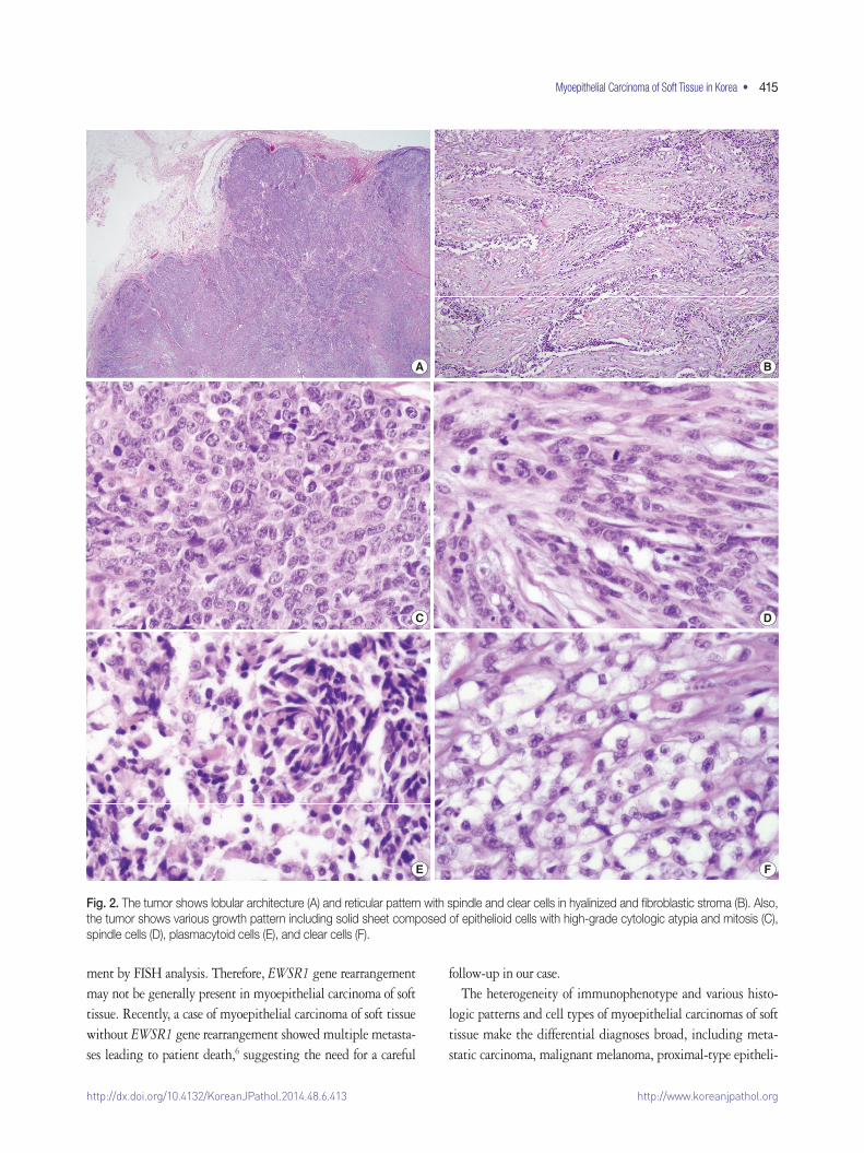
The solid mass was lobulated and the cut surface of the mass was gray tan with multiple necrotic foci (Fig. 1A). The tumor was confined to the subcutaneous soft tissue without dermal in-vasion. Although the tumor showed a well-circumscribed lobu-lating border, microscopic tumor infiltration into the adjacent soft tissue was observed (Fig. 1B). The tumor presented with various histologic growth patterns including solid sheet, tra-becular, reticular patterns, and short fascicle with myxoid and hyalinized stroma. Although tumor cells had a heterogeneous cytomorphology consisting of epithelioid cells, spindle cells, clear cells, and plasmacytoid cells, the tumor cells generally show-ed vesicular or coarse chromatin, distinct nucleoli, and thick ir-regular nuclear membrane (Fig. 2). Numerous mitoses were noted (up to 42 per 10 high-power fields). In addition, multifo-cal tumor necrosis was found. Lymphovascular invasion was also found.
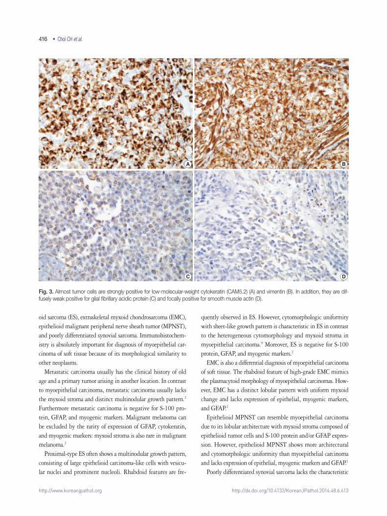
Almost all tumor cells were intensely positive for low-molec-ular-weight cytokeratin (CAM5.2), vimentin, and CD10. While the tumor cells were focally positive for cytokeratin and smooth muscle actin (SMA), they were diffusely weak positive for glial fibrillary acidic protein (GFAP). However, the tumor cells were totally negative for desmin, epithelial membrane antigen (EMA), CD15, monoclonal carcinoembryonic antigen, p63, CD34, hu-man melanoma black 45, gross cystic disease fluid protein 15, and S-100 protein (Fig. 3). Fluorescence in situ hybridization (FISH) analysis for EWSR1 gene rearrangement was performed at an outside institution, but the present case did not exhibit EWSR1 gene rearrangement. On the basis of histologic find-
The Korean Journal of Pathology 2014; 48: 413-417http://dx.doi.org/10.4132/KoreanJPathol.2014.48.6.413
▒ BRIEF CASE REPORT ▒
Corresponding AuthorYoung Chae Chu, M.D. Department of Pathology, Inha University Hospital, 27 Inhang-ro, Jung-gu, Incheon 400-711, KoreaTel: +82-32-890-3972, Fax: +82-32-890-3464, E-mail: [email protected]
Received: July 23, 2014 Revised: September 3, 2014 Accepted: September 12, 2014
Myoepithelial Carcinoma of Soft Tissue: A Case Report and Review of
the Literature
Chang Hwan Choi · Young Chae Chu · Lucia Kim · Suk Jin Choi · In Suh Park · Jee Young Han · Joon Mee Kim
Department of Pathology, Inha University Hospital, Inha University School of Medicine, Incheon, Korea
http://www.koreanjpathol.org http://dx.doi.org/10.4132/KoreanJPathol.2014.48.6.413
414 • Choi CH et al.
ings and immunohistochemical study results, a diagnosis of myo-epithelial carcinoma was rendered.
DISCUSSION
Myoepithelial carcinoma of soft tissue is morphologically identical to that of salivary gland. Although myoepithelial car-cinoma in the salivary gland is a well-known tumor, only few cases of myoepithelial carcinoma of soft tissue have ever been reported because of their rarity. Myoepithelial carcinoma of soft tissue has not been reported in Korea or China. Only one case of myoepithelial carcinoma arising in the deep soft tissue of the forearm was reported from Japan.5 The present case is the first report of myoepithelial carcinoma of soft tissue in Korea and the second one in East Asia.
To confirm the myoepithelial differentiation of the tumor, we used immunohistochemical staining with keratin, EMA, S-100 protein, GFAP, SMA, and vimentin as the immunohistochemi-cal criteria, which have been used by other authors.1,2 In our case, the tumor cells were positive for vimentin, CAM5.2, cyto-keratin, SMA, and GFAP, but negative for EMA, p63, and S-100 protein. In fact, previous studies have revealed that the positivity of these markers was variable.1,2 By comprehensive consideration of histologic features and immunohistochemical profiles of our case, the tumor is consistent with myoepithelial carcinoma.
Several pathologic criteria have been suggested for distinguish-ing myoepithelial carcinoma from benign myoepithelial tumor in soft tissue.1-5 Like their counterparts in the salivary gland, cytologic atypia, high mitotic rate, infiltrative growth into sur-
rounding soft tissues, and/or tumor necrosis have been adopted as criteria for myoepithelial carcinoma of soft tissue. Hornick and Fletcher2 revealed that the microscopically infiltrative growth without cytologic atypia and mitotic activity was not associated with recurrence or metastasis. They emphasized that the cyto-logic atypia including prominent nucleoli, vesicular or coarse chromatin, and nuclear pleomorphism was suggestive of aggres-sive behavior and tendency for metastasis. Therefore, myoepi-thelial tumor of the soft tissue with at least moderate cytologi-cal atypia should be managed as myoepithelial carcinoma. In fact, there is no definitive cutoff of mitotic rate for malignant criteria.
EWSR1 is located on chromosome band 22q12 and encodes a promoter-specific transactivator. EWSR1 gene rearrangement is associated with several neoplasms such as Ewing sarcoma, des-moplastic small round cell tumor, clear cell sarcoma, angioma-toid fibrous histiocytoma, myxoid liposarcoma, and extraskele-tal myxoid chondrosarcoma.4 Also EWSR1 gene rearrangement has been found in myoepithelial tumors including myoepithe-lial carcinoma.1,4,6-8 The EWSR1-ZNF444 fusion gene was found in a case of myoepithelial carcinoma of soft tissue with a lung metastasis.7 Furthermore, a case of myoepithelial carcinoma of soft tissue with round cell morphology was positive for EWSR1 gene rearrangement on FISH analysis.8 However, these reports do not mean that all myoepithelial carcinomas of soft tissue ex-hibit EWSR1 gene rearrangement. Gleason and Fletcher1 de-scribed two cases with EWSR1 gene rearrangement and one case without this aberration. Furthermore, EWSR1 gene rearrange-ments were observed in only 50% of myoepithelial carcinomas of soft tissue.4 Our case did not have EWSR1 gene rearrange-
Fig. 1. (A) A well-circumscribed lobulating tumor is noted in subcutaneous soft tissue with gray tan cut surface and multiple necrotic foci. (B) Microscopic infiltration into the adjacent soft tissue is found.
AB BA
http://www.koreanjpathol.orghttp://dx.doi.org/10.4132/KoreanJPathol.2014.48.6.413
Myoepithelial Carcinoma of Soft Tissue in Korea • 415
C
A
E
D
B
F
C
A
D
B
E F
Fig. 2. The tumor shows lobular architecture (A) and reticular pattern with spindle and clear cells in hyalinized and fibroblastic stroma (B). Also, the tumor shows various growth pattern including solid sheet composed of epithelioid cells with high-grade cytologic atypia and mitosis (C), spindle cells (D), plasmacytoid cells (E), and clear cells (F).
ment by FISH analysis. Therefore, EWSR1 gene rearrangement may not be generally present in myoepithelial carcinoma of soft tissue. Recently, a case of myoepithelial carcinoma of soft tissue without EWSR1 gene rearrangement showed multiple metasta-ses leading to patient death,6 suggesting the need for a careful
follow-up in our case.The heterogeneity of immunophenotype and various histo-
logic patterns and cell types of myoepithelial carcinomas of soft tissue make the differential diagnoses broad, including meta-static carcinoma, malignant melanoma, proximal-type epitheli-
http://www.koreanjpathol.org http://dx.doi.org/10.4132/KoreanJPathol.2014.48.6.413
416 • Choi CH et al.
A
C
B
D
A B
C D
Fig. 3. Almost tumor cells are strongly positive for low-molecular-weight cytokeratin (CAM5.2) (A) and vimentin (B). In addition, they are dif-fusely weak positive for glial fibrillary acidic protein (C) and focally positive for smooth muscle actin (D).
oid sarcoma (ES), extraskeletal myxoid chondrosarcoma (EMC), epithelioid malignant peripheral nerve sheath tumor (MPNST), and poorly differentiated synovial sarcoma. Immunohistochem-istry is absolutely important for diagnosis of myoepithelial car-cinoma of soft tissue because of its morphological similarity to other neoplasms.
Metastatic carcinoma usually has the clinical history of old age and a primary tumor arising in another location. In contrast to myoepithelial carcinoma, metastatic carcinoma usually lacks the myxoid stroma and distinct multinodular growth pattern.2 Furthermore metastatic carcinoma is negative for S-100 pro-tein, GFAP, and myogenic markers. Malignant melanoma can be excluded by the rarity of expression of GFAP, cytokeratin, and myogenic markers: myxoid stroma is also rare in malignant melanoma.2
Proximal-type ES often shows a multinodular growth pattern, consisting of large epithelioid carcinoma-like cells with vesicu-lar nuclei and prominent nucleoli. Rhabdoid features are fre-
quently observed in ES. However, cytomorphologic uniformity with sheet-like growth pattern is characteristic in ES in contrast to the heterogeneous cytomorphology and myxoid stroma in myoepithelial carcinoma.9 Moreover, ES is negative for S-100 protein, GFAP, and myogenic markers.2
EMC is also a differential diagnosis of myoepithelial carcinoma of soft tissue. The rhabdoid feature of high-grade EMC mimics the plasmacytoid morphology of myoepithelial carcinomas. How-ever, EMC has a distinct lobular pattern with uniform myxoid change and lacks expression of epithelial, myogenic markers, and GFAP.2
Epithelioid MPNST can resemble myoepithelial carcinoma due to its lobular architecture with myxoid stroma composed of epithelioid tumor cells and S-100 protein and/or GFAP expres-sion. However, epithelioid MPNST shows more architectural and cytomorphologic uniformity than myoepithelial carcinoma and lacks expression of epithelial, myogenic markers and GFAP.1
Poorly differentiated synovial sarcoma lacks the characteristic
http://www.koreanjpathol.orghttp://dx.doi.org/10.4132/KoreanJPathol.2014.48.6.413
Myoepithelial Carcinoma of Soft Tissue in Korea • 417
features of myoepithelial carcinoma including lobular architec-ture, epithelioid cells and expression of myogenic markers and GFAP.10
In summary, we are reporting the first case of myoepithelial carcinoma of soft tissue in Korea. To the best of our knowledge, the present case is the second report from East Asia described in the English literature. Myoepithelial carcinoma should be con-sidered in the differential diagnosis of malignant tumors that have various morphologic features of tumor cells including epi-thelioid, spindled, plasmacytoid, or clear cell with myxoid back-ground in soft tissue. Although further study is needed to con-firm the criteria for myoepithelial carcinoma of soft tissue, pa-thologists have to be conscious that myoepithelial tumors of soft tissue with cytologic atypia, mitotic activity, and tumor in-filtration suggest the malignant potential of myoepithelial tu-mor of soft tissue.
Conflicts of InterestNo potential conflict of interest relevant to this article was
reported.
AcknowledgmentsThis work was supported by Inha University Research Grant.
REFERENCES
1. Gleason BC, Fletcher CD. Myoepithelial carcinoma of soft tissue in children: an aggressive neoplasm analyzed in a series of 29 cases. Am J Surg Pathol 2007; 31: 1813-24.
2. Hornick JL, Fletcher CD. Myoepithelial tumors of soft tissue: a clin-icopathologic and immunohistochemical study of 101 cases with
evaluation of prognostic parameters. Am J Surg Pathol 2003; 27: 1183-96.
3. Lee JR, Georgi DE, Wang BY. Malignant myoepithelial tumor of soft tissue: a report of two cases of the lower extremity and a review of the literature. Ann Diagn Pathol 2007; 11: 190-8.
4. Rekhi B, Sable M, Jambhekar NA. Histopathological, immunohis-tochemical and molecular spectrum of myoepithelial tumours of soft tissues. Virchows Arch 2012; 461: 687-97.
5. Harada O, Ota H, Nakayama J. Malignant myoepithelioma (myo-epithelial carcinoma) of soft tissue. Pathol Int 2005; 55: 510-3.
6. Aparicio MA, López-Barea F, Cruz JJ, et al. Soft tissue myoepithelial carcinoma without EWSR1 gene rearrangement and poor outcome: a case report. Rev Esp Patol 2012; 45: 58-63.
7. Brandal P, Panagopoulos I, Bjerkehagen B, Heim S. t(19;22)(q13;q12) translocation leading to the novel fusion gene EWSR1-ZNF444 in soft tissue myoepithelial carcinoma. Genes Chromosomes Cancer 2009; 48: 1051-6.
8. El-Kabany M, Al-Abdulghani R, Ali AE, Francis IM, Hussein SA. Soft tissue high grade myoepithelial carcinoma with round cell mor-phology: report of a newly described entity with EWSR1 gene rear-rangement. Gulf J Oncolog 2011; (9): 73-7.
9. Guillou L, Wadden C, Coindre JM, Krausz T, Fletcher CD. “Proxi-mal-type” epithelioid sarcoma, a distinctive aggressive neoplasm showing rhabdoid features. Clinicopathologic, immunohistochemi-cal, and ultrastructural study of a series. Am J Surg Pathol 1997; 21: 130-46.
10. Olsen SH, Thomas DG, Lucas DR. Cluster analysis of immunohis-tochemical profiles in synovial sarcoma, malignant peripheral nerve sheath tumor, and Ewing sarcoma. Mod Pathol 2006; 19: 659-68.


![Ductal Carcinoma Arising from Ectopic Breast Tissue ... · PDF filetively common feature of invasive carcinoma or ductal carci-noma in situ [4,5]. However, the microcalcification of](https://img.pdfslide.net/doc/110x75/5a78f6d07f8b9a5a148e5db7/ductal-carcinoma-arising-from-ectopic-breast-tissue-common-feature-of-invasive.jpg)