Embed Size (px)
Citation preview
The Journal of Manual & Manipulative TherapyVol. 14 No. 4 (2006), 203 - 221 Myofascial Trigger Points: An Evidence-Informed Review / 203
Myofascial Trigger Points: An Evidence-Informed Review
Address all correspondence and request for reprints to:Jan DommerholtBethesda Physiocare, Inc. 7830 Old Georgetown Road, Suite C-15 Bethesda, MD 20814-2440 [email protected]
Abstract: This article provides a best evidence-informed review of the current scientific un-derstanding of myofascial trigger points with regard to their etiology, pathophysiology, and clinical implications. Evidence-informed manual therapy integrates the best available scien-tific evidence with individual clinicians’ judgments, expertise, and clinical decision-making. After a brief historical review, the clinical aspects of myofascial trigger points, the interrater reliability for identifying myofascial trigger points, and several characteristic features are discussed, including the taut band, local twitch response, and referred pain patterns. The etiology of myofascial trigger points is discussed with a detailed and comprehensive review of the most common mechanisms, including low-level muscle contractions, uneven intramus-cular pressure distribution, direct trauma, unaccustomed eccentric contractions, eccentric contractions in unconditioned muscle, and maximal or sub-maximal concentric contractions. Many current scientific studies are included and provide support for considering myofascial trigger points in the clinical decision-making process. The article concludes with a summary of frequently encountered precipitating and perpetuating mechanical, nutritional, metabolic, and psychological factors relevant for physical therapy practice. Current scientific evidence strongly supports that awareness and working knowledge of muscle dysfunction and in par-ticular myofascial trigger points should be incorporated into manual physical therapy practice consistent with the guidelines for clinical practice developed by the International Federation of Orthopaedic Manipulative Therapists. While there are still many unanswered questions in explaining the etiology of myofascial trigger points, this article provides manual therapists with an up-to-date evidence-informed review of the current scientific knowledge.
Key Words : Myofascia l Pain Syndrome, Tr igger Points , Myofascia l , Et io logy, Pathophysiology
Jan Dommerholt, PT, MPS, FAAPMCarel Bron, PTJo Franssen, PT
During the past few decades, myofascial trigger points (MTrPs) and myofascial pain syndrome (MPS) have
received much attention in the scientific and clinical literature. Researchers worldwide are investigating various aspects of MTrPs, including their specific etiol-ogy, pathophysiology, histology, referred pain patterns, and clinical applications. Guidelines developed by the International Federation of Orthopaedic Manipulative
Therapists (IFOMT) confirm the importance of muscle dysfunction for orthopedic manual therapy clinical prac-tice. The IFOMT has defined orthopedic manual therapy as “a specialized area of physiotherapy/physical therapy for the management of neuromusculoskeletal condi-tions, based on clinical reasoning, using highly specific treatment approaches including manual techniques and therapeutic exercises.” The educational standards of IFOMT require that skills will be demonstrated in—among others—“analysis and specific tests for functional status of the muscular system,” “a high level of skill in other manual and physical therapy techniques required to mobilize the articular, muscular or neural systems,” and “knowledge of various manipulative therapy approaches as practiced within physical therapy, medicine, osteopathy and chiropractic”1.
204 / The Journal of Manual & Manipulative Therapy, 2006
However, articles about muscle dysfunction in the manual therapy literature are sparse and they generally focus on muscle injury and muscle repair mechanisms2 or on muscle recruitment3. Until very recently, the current scientific knowledge and clinical implications of MTrPs were rarely included4-7. It appears that orthopedic manual therapists have not paid much attention to the patho-physiology and clinical manifestations of MTrPs. Manual therapy educational programs in the US seem to reflect this orientation and tend to place a strong emphasis on joint dysfunction, mobilizations, and manipulations with only about 10%-15% of classroom education devoted to muscle pain and muscle dysfunction.
This review of the MTrP literature is based on current best scientific evidence. The field of manual therapy has joined other medical disciplines by embrac-ing evidence-based medicine, which proposes that the results of scientific research need to be integrated into clinical practice8. Evidence-based medicine has been defined as “the conscientious, explicit, and judicious use of current best evidence in making decisions about the care of individual patients”9,10. Within the evidence-based medicine paradigm, evidence is not restricted to randomized controlled trials, systematic reviews, and meta-analyses, although this restricted view seems to be prevalent in the medical and physical therapy literature. Sackett et al9,10 emphasized that external clinical evidence can inform but not replace individual clinical expertise. Clinical expertise determines whether external clinical evidence applies to an individual patient, and if so, how it should be integrated into clinical decision-making. Pencheon11 shared this perspective and suggested that high-quality healthcare is about combining “wisdom produced by years of experience” with “evidence produced by generalizable research” in “ways with which patients are happy.” He suggested shifting from evidence-based to evidence-informed medicine, where clinical decision-making is informed by research evidence but not driven by it and always includes knowledge from experience. Evidence-informed manual therapy involves integrat-ing the best available external scientific evidence with individual clinicians’ judgments, expertise, and clini-cal decision-making12. The purpose of this article is to provide a best evidence-informed review of the current scientific understanding of MTrPs, including the etiology, pathophysiology, and clinical implications, against the background of extensive clinical experience.
Brief Historical ReviewWhile Dr. Janet Travell (1901-1997) is generally cred-
ited for bringing MTrPs to the attention of health care providers, MTrPs have been described and rediscovered for several centuries by various clinicians and researchers13,14. As far back as the 16th century, de Baillou (1538-1616), as cited by Ruhmann, described what is now known as myofascial pain syndrome (MPS)15. MPS is defined as
the “sensory, motor, and autonomic symptoms caused by MTrPs” and has become a recognized medical diag-nosis among pain specialists16,17. In 1816, British physi-cian Balfour, as cited by Stockman, described “nodular tumors and thickenings which were painful to the touch, and from which pains shot to neighboring parts”18. In 1898, the German physician Strauss discussed “small, tender and apple-sized nodules and painful, pencil-sized to little-finger-sized palpable bands”19. The first trigger point manual was published in 1931 in Germany nearly a decade before Travell became interested in MTrPs20. While these early descriptions may appear a bit archaic and unusual—for example, in clinical practice one does not encounter “apple-sized nodules” —these and other historic papers did illustrate the basic features of MTrPs quite accurately14.
In the late 1930s, Travell, who at that time was a cardiologist and medical researcher, became particularly interested in muscle pain following the publication of several articles on referred pain21. Kellgren’s descriptions of referred pain patterns of many muscles and spinal ligaments after injecting these tissues with hypertonic saline22-25 eventually moved Travell to shift her medical career from cardiology to musculoskeletal pain. During the 1940s, she published several articles on injection techniques of MTrPs26-28. In 1952, she described the myofascial genesis of pain with detailed referred pain patterns for 32 muscles29. Other clinicians also became interested in MTrPs. European physicians Lief and Chaitow developed a treatment method, which they referred to as “neuromuscular technique”30. German physician Gutstein described the characteristics of MTrPs and effective manual therapy treatments in several papers under the names of Gutstein, Gutstein-Good, and Good31-34. In Australia, Kelly produced a series of articles about fibrositis, which paralleled Travel’s writings35-38.
In the US, chiropractors Nimmo and Vannerson39 described muscular “noxious generative points,” which were thought to produce nerve impulses and eventually result in “vasoconstriction, ischaemia, hypoxia, pain, and cellular degeneration.” Later in his career, Nimmo adopted the term “trigger point” after having been introduced to Travell’s writings. Nimmo maintained that hypertonic muscles are always painful to pressure, a statement that later became known as “Nimmo’s law.” Like Travell, Nimmo described distinctive referred pain patterns and recommended releasing these dysfunctional points by applying the proper degree of manual pressure. Nimmo’s “receptor-tonus control method” continues to be popular among chiropractic physicians39,40. According to a 1993 report by the National Board of Chiropractic Economics, over 40% of chiropractors in the US frequently apply Nimmo’s techniques41. Two spin-offs of Nimmo’s work are St. John Neuromuscular Therapy (NMT) method and NMT American version, which have become particularly popular among massage therapists30.
Myofascial Trigger Points: An Evidence-Informed Review / 205
In 1966, Travell founded the North American Academy of Manipulative Medicine, together with Dr. John Mennell, who also published several articles about MTrPs42,43. Throughout her career Travell promoted integrating myofascial treatments with articular treatments16. One of her earlier papers described a technique for reduc-ing sacroiliac displacement44. However, Travell, as cited by Paris45, maintained the opinion that manipulations were the exclusive domain of physicians and she re-jected membership in the North American Academy of Manipulative Medicine by physical therapists.
In the early 1960s, Dr. David Simons was introduced to Travell and her work, which became the start of a fruitful collaboration eventually resulting in several pub-lications, including the Trigger Point Manuals, consist-ing of a 1983 first volume (upper half of the body) and a 1992 second volume (lower half of the body)46,47. The first volume has since been revised and updated and a second edition was released in 199916. The Trigger Point Manuals are the most comprehensive review of nearly 150 muscle referred-pain patterns based on Travell’s clinical observations, and they include an extensive review of the scientific basis of MTrPs. Both volumes have been translated into several foreign languages, including Russian, German, French, Italian, Japanese, and Spanish. Several other clinicians worldwide have also published their own trigger point manuals48-54.
Clinical Aspects of Myofascial Trigger Points
An MTrP is described as “a hyperirritable spot in skeletal muscle that is associated with a hypersensitive palpable nodule in a taut band”16. Myofascial trigger points are classified into active and latent trigger points16. An active MTrP is a symptom-producing MTrP and can trigger local or referred pain or other paraesthesiae. A latent MTrP does not trigger pain without being stimulated. Myofascial trigger points are the hallmark characteris-tics of MPS and feature motor, sensory, and autonomic components. Motor aspects of active and latent MTrPs may include disturbed motor function, muscle weak-ness as a result of motor inhibition, muscle stiffness, and restricted range of motion55,56. Sensory aspects may include local tenderness, referral of pain to a distant site, and peripheral and central sensitization. Peripheral sensitization can be described as a reduction in threshold and an increase in responsiveness of the peripheral ends of nociceptors, while central sensitization is an increase in the excitability of neurons within the central nervous system. Signs of peripheral and central sensitization are allodynia (pain due to a stimulus that does not normally provoke pain) and hyperalgesia (an increased response to a stimulus that is normally painful). Both active and latent MTrPs are painful on compression. Vecchiet et al57-
59 described specific sensory changes over MTrPs. They
observed significant lowering of the pain threshold over active MTrPs when measured by electrical stimulation, not only in the muscular tissue but also in the overlying cutaneous and subcutaneous tissues. In contrast, with latent MTrPs, the sensory changes did not involve the cutaneous and subcutaneous tissues57-59. Autonomic aspects of MTrPs may include, among others, vasoconstriction, vasodilatation, lacrimation, and piloerection16,60-63.
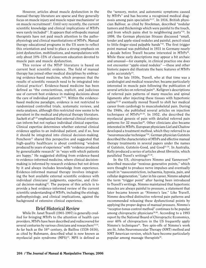
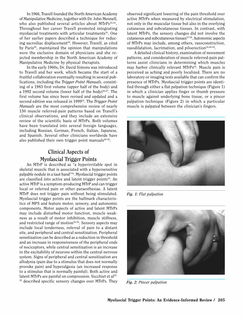
A detailed clinical history, examination of movement patterns, and consideration of muscle referred-pain pat-terns assist clinicians in determining which muscles may harbor clinically relevant MTrPs64. Muscle pain is perceived as aching and poorly localized. There are no laboratory or imaging tests available that can confirm the presence of MTrPs. Myofascial trigger points are identi-fied through either a flat palpation technique (Figure 1) in which a clinician applies finger or thumb pressure to muscle against underlying bone tissue, or a pincer palpation technique (Figure 2) in which a particular muscle is palpated between the clinician’s fingers.
Fig. 1: Flat palpation
Fig. 2: Pincer palpation
206 / The Journal of Manual & Manipulative Therapy, 2006
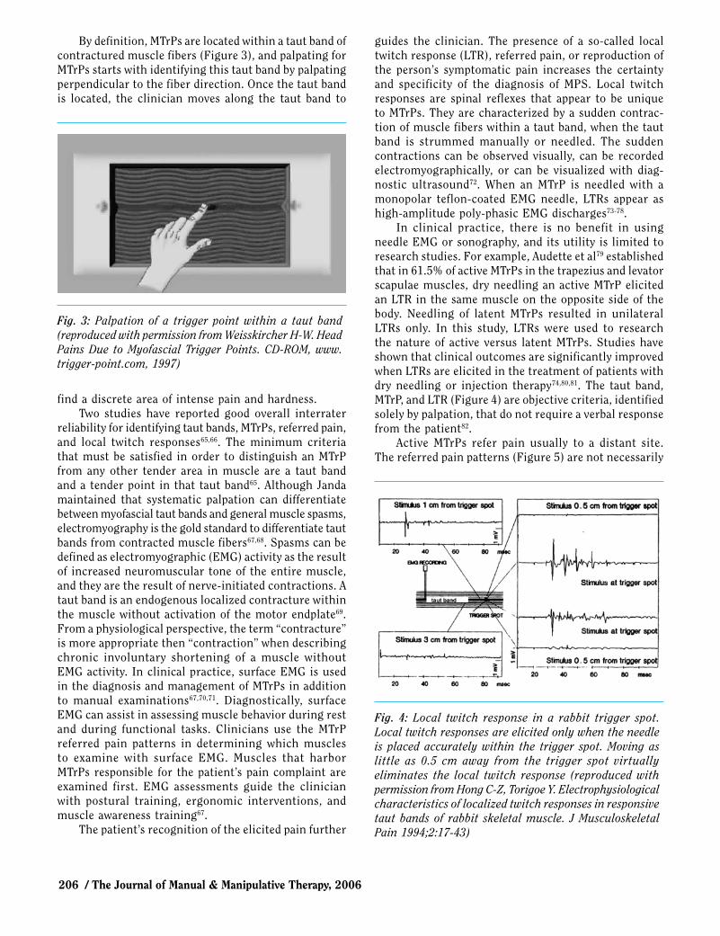
By definition, MTrPs are located within a taut band of contractured muscle fibers (Figure 3), and palpating for MTrPs starts with identifying this taut band by palpating perpendicular to the fiber direction. Once the taut band is located, the clinician moves along the taut band to
guides the clinician. The presence of a so-called local twitch response (LTR), referred pain, or reproduction of the person’s symptomatic pain increases the certainty and specificity of the diagnosis of MPS. Local twitch responses are spinal reflexes that appear to be unique to MTrPs. They are characterized by a sudden contrac-tion of muscle fibers within a taut band, when the taut band is strummed manually or needled. The sudden contractions can be observed visually, can be recorded electromyographically, or can be visualized with diag-nostic ultrasound72. When an MTrP is needled with a monopolar teflon-coated EMG needle, LTRs appear as high-amplitude poly-phasic EMG discharges73-78.
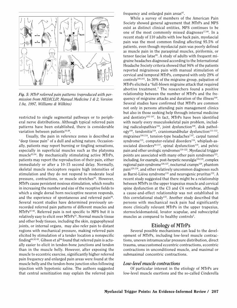
In clinical practice, there is no benefit in using needle EMG or sonography, and its utility is limited to research studies. For example, Audette et al79 established that in 61.5% of active MTrPs in the trapezius and levator scapulae muscles, dry needling an active MTrP elicited an LTR in the same muscle on the opposite side of the body. Needling of latent MTrPs resulted in unilateral LTRs only. In this study, LTRs were used to research the nature of active versus latent MTrPs. Studies have shown that clinical outcomes are significantly improved when LTRs are elicited in the treatment of patients with dry needling or injection therapy74,80,81. The taut band, MTrP, and LTR (Figure 4) are objective criteria, identified solely by palpation, that do not require a verbal response from the patient82.
Active MTrPs refer pain usually to a distant site. The referred pain patterns (Figure 5) are not necessarily
Fig. 3: Palpation of a trigger point within a taut band (reproduced with permission from Weisskircher H-W. Head Pains Due to Myofascial Trigger Points. CD-ROM, www.trigger-point.com, 1997)
find a discrete area of intense pain and hardness. Two studies have reported good overall interrater
reliability for identifying taut bands, MTrPs, referred pain, and local twitch responses65,66. The minimum criteria that must be satisfied in order to distinguish an MTrP from any other tender area in muscle are a taut band and a tender point in that taut band65. Although Janda maintained that systematic palpation can differentiate between myofascial taut bands and general muscle spasms, electromyography is the gold standard to differentiate taut bands from contracted muscle fibers67,68. Spasms can be defined as electromyographic (EMG) activity as the result of increased neuromuscular tone of the entire muscle, and they are the result of nerve-initiated contractions. A taut band is an endogenous localized contracture within the muscle without activation of the motor endplate69. From a physiological perspective, the term “contracture” is more appropriate then “contraction” when describing chronic involuntary shortening of a muscle without EMG activity. In clinical practice, surface EMG is used in the diagnosis and management of MTrPs in addition to manual examinations67,70,71. Diagnostically, surface EMG can assist in assessing muscle behavior during rest and during functional tasks. Clinicians use the MTrP referred pain patterns in determining which muscles to examine with surface EMG. Muscles that harbor MTrPs responsible for the patient’s pain complaint are examined first. EMG assessments guide the clinician with postural training, ergonomic interventions, and muscle awareness training67.
The patient’s recognition of the elicited pain further
Fig. 4: Local twitch response in a rabbit trigger spot. Local twitch responses are elicited only when the needle is placed accurately within the trigger spot. Moving as little as 0.5 cm away from the trigger spot virtually eliminates the local twitch response (reproduced with permission from Hong C-Z, Torigoe Y. Electrophysiological characteristics of localized twitch responses in responsive taut bands of rabbit skeletal muscle. J Musculoskeletal Pain 1994;2:17-43)
Myofascial Trigger Points: An Evidence-Informed Review / 207
restricted to single segmental pathways or to periph-eral nerve distributions. Although typical referred pain patterns have been established, there is considerable variation between patients16,48.
Usually, the pain in reference zones is described as “deep tissue pain” of a dull and aching nature. Occasion-ally, patients may report burning or tingling sensations, especially in superficial muscles such as the platysma muscle83,84. By mechanically stimulating active MTrPs, patients may report the reproduction of their pain, either immediately or after a 10-15 second delay. Normally, skeletal muscle nociceptors require high intensities of stimulation and they do not respond to moderate local pressure, contractions, or muscle stretches85. However, MTrPs cause persistent noxious stimulation, which results in increasing the number and size of the receptive fields to which a single dorsal horn nociceptive neuron responds, and the experience of spontaneous and referred pain86. Several recent studies have determined previously un-recorded referred pain patterns of different muscles and MTrPs87-90. Referred pain is not specific to MPS but it is relatively easy to elicit over MTrPs91. Normal muscle tissue and other body tissues, including the skin, zygapophyseal joints, or internal organs, may also refer pain to distant regions with mechanical pressure, making referred pain elicited by stimulation of a tender location a nonspecific finding84,92-95. Gibson et al96 found that referred pain is actu-ally easier to elicit in tendon-bone junctions and tendon than in the muscle belly. However, after exposing the muscle to eccentric exercise, significantly higher referred pain frequency and enlarged pain areas were found at the muscle belly and the tendon-bone junction sites following injection with hypotonic saline. The authors suggested that central sensitization may explain the referred pain
frequency and enlarged pain areas97.While a survey of members of the American Pain
Society showed general agreement that MTrPs and MPS exist as distinct clinical entities, MPS continues to be one of the most commonly missed diagnoses17,98. In a recent study of 110 adults with low back pain, myofascial pain was the most common finding affecting 95.5% of patients, even though myofascial pain was poorly defined as muscle pain in the paraspinal muscles, piriformis, or tensor fasciae latae99. A study of adults with frequent mi-graine headaches diagnosed according to the International Headache Society criteria showed that 94% of the patients reported migrainous pain with manual stimulation of cervical and temporal MTrPs, compared with only 29% of controls100,101. In 30% of the migraine group, palpation of MTrPs elicited a “full-blown migraine attack that required abortive treatment.” The researchers found a positive relationship between the number of MTrPs and the fre-quency of migraine attacks and duration of the illness100. Several studies have confirmed that MTrPs are common not only in persons attending pain management clinics but also in those seeking help through internal medicine and dentistry102-107. In fact, MTrPs have been identified with nearly every musculoskeletal pain problem, includ-ing radiculopathies104, joint dysfunction108, disk pathol-ogy109, tendonitis110, craniomandibular dysfunction111-113, migraines100,114, tension-type headaches7,87, carpal tunnel syndrome115, computer-related disorders116, whiplash-as-sociated disorders60,117, spinal dysfunction118, and pelvic pain and other urologic syndromes119-122. Myofascial trigger points are associated with many other pain syndromes123, including, for example, post-herpetic neuralgia124,125, complex regional pain syndrome126,127, nocturnal cramps128, phantom pain129,130, and other relatively uncommon diagnoses such as Barré-Liéou syndrome131 and neurogenic pruritus132. A recent study suggested that there might be a relationship between MTrPs in the upper trapezius muscle and cervical spine dysfunction at the C3 and C4 vertebrae, although a cause-and-effect relationship was not established in this correlational study133. Another study described that persons with mechanical neck pain had significantly more clinically relevant MTrPs in the upper trapezius, sternocleidomastoid, levator scapulae, and suboccipital muscles as compared to healthy controls5.
Etiology of MTrPsSeveral possible mechanisms can lead to the devel-
opment of MTrPs, including low-level muscle contrac-tions, uneven intramuscular pressure distribution, direct trauma, unaccustomed eccentric contractions, eccentric contractions in unconditioned muscle, and maximal or submaximal concentric contractions.
Low-level muscle contractionsOf particular interest in the etiology of MTrPs are
low-level muscle exertions and the so-called Cinderella
Fig. 5: MTrP referred pain patterns (reproduced with per-mission from MEDICLIP, Manual Medicine 1 & 2, Version 1.0a, 1997, Williams & Wilkins)
208 / The Journal of Manual & Manipulative Therapy, 2006
Hypothesis developed by Hägg in 1988134. The Cinderella Hypothesis postulates that occupational myalgia is caused by selective overloading of the earliest recruited and last de-recruited motor units according to the ordered recruitment principle or Henneman’s “size principle”134,135. Smaller motor units are recruited before and de-recruited after larger ones; as a result, the smaller type 1 fibers are continuously activated during prolonged motor tasks135. According to the Cinderella Hypothesis, muscular force generated at sub-maximal levels during sustained muscle contractions engages only a fraction of the motor units available without the normally occurring substitution of motor units during higher force contractions, which in turn can result in metabolically overloaded motor units, prone to loss of cellular Ca2+-homeostasis, subsequent activation of autogenic destructive processes, and muscle pain136,137. The other pillar of the Cinderella Hypothesis is the finding of an excess of ragged red fibers in myalgic patients136. Indeed, several researchers have demonstrated the presence of ragged red fibers and moth-eaten fibers in subjects with myalgia, which are indications of struc-tural damage to the cell membrane and mitochondria and a change in the distribution of mitochondria or the sarcotubular system respectively138-142.
There is growing evidence that low-level static muscle contractions or exertions can result in degeneration of muscle fibers143. Gissell144,145 has shown that low-level exertions can result in an increase of Ca2+-release in skeletal muscle cells, muscle membrane damage due to leakage of the intracellular enzyme lactate dehydrogenase, structural damage, energy depletion, and myalgia. Low-level muscle stimulation can also lead to the release of interleukin 6 (IL-6) and other cytokines146,147.
Several studies have confirmed the Cinderella Hy-pothesis and support the idea that in low-level static exertions, muscle fiber recruitment patterns tend to be stereotypical with continuous activation of smaller type 1 fibers during prolonged motor tasks148-152. As Hägg indicated, the continuous activity and metabolic overload of certain motor units does not occur in all subjects136. The Cinderella Hypothesis was recently applied to the development of MTrPs116. In a well-de-signed study, Treasters et al116 established that sustained low-level muscle contractions during continuous typing for as little as 30 minutes commonly resulted in the formation of MTrPs. They suggested that MTrPs might provide a useful explanation for muscle pain and injury that can occur from low-level static exertions116. Myo-fascial trigger points are common in office workers, musicians, dentists, and other occupational groups exposed to low-level muscle exertions153. Chen et al154 also suggested that low-level muscle exertions can lead to sensitization and development of MTrPs. Forty piano students showed significantly reduced pressure thresholds over latent MTrPs after only 20 minutes of continuous piano playing154.
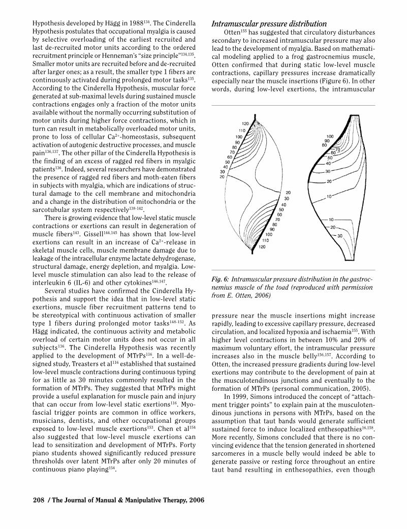
Intramuscular pressure distributionOtten155 has suggested that circulatory disturbances
secondary to increased intramuscular pressure may also lead to the development of myalgia. Based on mathemati-cal modeling applied to a frog gastrocnemius muscle, Otten confirmed that during static low-level muscle contractions, capillary pressures increase dramatically especially near the muscle insertions (Figure 6). In other words, during low-level exertions, the intramuscular
Fig. 6: Intramuscular pressure distribution in the gastroc-nemius muscle of the toad (reproduced with permission from E. Otten, 2006)
pressure near the muscle insertions might increase rapidly, leading to excessive capillary pressure, decreased circulation, and localized hypoxia and ischaemia155. With higher level contractions in between 10% and 20% of maximum voluntary effort, the intramuscular pressure increases also in the muscle belly156,157. According to Otten, the increased pressure gradients during low-level exertions may contribute to the development of pain at the musculotendinous junctions and eventually to the formation of MTrPs (personal communication, 2005).
In 1999, Simons introduced the concept of “attach-ment trigger points” to explain pain at the musculoten-dinous junctions in persons with MTrPs, based on the assumption that taut bands would generate sufficient sustained force to induce localized enthesopathies16,158. More recently, Simons concluded that there is no con-vincing evidence that the tension generated in shortened sarcomeres in a muscle belly would indeed be able to generate passive or resting force throughout an entire taut band resulting in enthesopathies, even though
Myofascial Trigger Points: An Evidence-Informed Review / 209
there may be certain muscles or conditions where this could occur (personal communication, 2005). To the contrary, force generated by individual motor units is always transmitted laterally to the muscle’s connective tissue matrix, involving at least two protein complexes containing vinculin and dystrophin, respectively159. There is also considerable evidence that the assumption that muscle fibers pass from tendon to tendon is without basis160. Trotter160 has demonstrated that skeletal muscle is comprised of in-series fibers. In other words, there is evidence that a single muscle fiber does not run from tendon to tendon. The majority of fibers are in series with inactive fibers, which makes it even more unlikely that the whole muscle length-tension properties would be dictated by the shortest contractured fibers in the muscle161.
In addition, it is important to consider the mechanical and functional differences between fast and slow motor units162,163. Slow motor units are always stiffer than fast units, although fast units can produce more force. If there were any transmission of force along the muscle fiber, as Simons initially suggested, fast fibers would be better suited to accomplish this. Yet, fast motor units have larger series of elastic elements, which would absorb most of the force displacement164,165. Fast fibers show a progressive decrease in cross-sectional area and end in a point within the muscle fascicle, making force transmission even more unlikely163. Fast fibers rely on transmitting a substantial proportion of their force to the endomysium, transverse cytoskeleton, and adja-cent muscle fibers162,163. In summary, the development of so-called “attachment trigger points” as a result of increased tension by contractured sarcomeres in MTrPs is not clear and more research is needed to explain the clinical observation that MTrPs appear to be linked to pain at the musculotendinous junction. The increased tension in the muscle belly is likely to dissipate across brief sections of the taut band on both sides of the MTrP and laterally through the transverse cytoskeleton166-168. Instead, Otten’s model of increased intramuscular pressure, decreased circulation, localized hypoxia, and ischaemia at the muscle insertions provides an alternative model for the clinically observed pain near the musculotendinous junction and osseous insertions in persons with MTrPs, even though the model does not explain why taut bands are commonly present155.
Direct traumaThere is general agreement that acute muscle over-
load can activate MTrPs, although systematic studies are lacking169. For example, people involved in whiplash injuries commonly experience prolonged muscle pain and dysfunction170-173. In a retrospective review, Schul-ler et al174 found that 80% of 1096 subjects involved in low-velocity collisions demonstrated evidence of muscle pain with myogeloses among the most common find-
ings. Although Schuller et al174 did not define these myogeloses, Simons has suggested that a myogelosis describes the same clinical entity as an MTrP175. Baker117 reported that the splenius capitis, semispinalis capitis, and sternocleidomastoid muscles developed symptomatic MTrPs in 77%, 62%, and 52% of 52 whiplash patients, respectively. In a retrospective review of 54 consecutive chronic whiplash patients, Gerwin and Dommerholt176 reported that clinically relevant MTrPs were found in every patient, with the trapezius muscle involved most often. Following treatment emphasizing the inactivation of MTrPs and restoration of normal muscle length, ap-proximately 80% of patients experienced little or no pain, even though the average time following the initiating injury was 2.5 years at the beginning of the treatment regimen. All patients had been seen previously by other physicians and physical therapists who apparently had not considered MTrPs in their thought process and clinical management176. Fernández-de-las-Peñas et al177,178 confirmed that inactivation of MTrPs should be included in the management of persons suffering from whiplash-associated disorders. In their research-based treatment protocol, the combination of cervical and thoracic spine manipulations with MTrP treatments proved superior to more conventional physical therapy consisting of massage, ultrasound, home exercises, and low-energy high-frequency pulsed electromagnetic therapy177.
Direct trauma may create a vicious cycle of events wherein damage to the sarcoplasmic reticulum or the muscle cell membrane may lead to an increase of the calcium concentration, a subsequent activation of actin and myosin, a relative shortage of adenosine triphosphate (ATP), and an impaired calcium pump, which in turn will increase the intracellular calcium concentration even more, completing the cycle. The calcium pump is responsible for returning intracellular Ca2+ to the sar-coplasmic reticulum against a concentration gradient, which requires a functional energy supply. Simons and Travell179 considered this sequence in the development of the so-called “energy crisis hypothesis” introduced in 1981. Sensory and motor system dysfunction have been shown to develop rapidly after injury and actually may persist in those who develop chronic muscle pain and in individuals who have recovered or continue to have persistent mild symptoms172,180. Scott et al181 de-termined that individuals with chronic whiplash pain develop more widespread hypersensitivity to mechanical pressure and thermal stimuli than those with chronic idiopathic neck pain. Myofascial trigger points are a likely source of ongoing peripheral nociceptive input, and they contribute to both peripheral and central sensitization, which may explain the observation of widespread allodynia and hypersensitivity60,62,63. In addi-tion to being caused by whiplash injury, acute muscle overload can occur with direct impact, lifting injuries, sports performance, etc.182.
210 / The Journal of Manual & Manipulative Therapy, 2006
Eccentric and (sub)maximal concentric contractionsMany patients report the onset of pain and activation
of MTrPs following either acute, repetitive, or chronic muscle overload183. Gerwin et al184 suggested that likely mechanisms relevant for the development of MTrPs included either unaccustomed eccentric exercise, ec-centric exercise in unconditioned muscle, or maximal or sub-maximal concentric exercise. A brief review of pertinent aspects of exercise follows, preceding linking this body of research to current MTrP research.
Eccentric exercise is associated with myalgia, muscle weakness, and destruction of muscle fibers, partially because eccentric contractions cause an irregular and uneven lengthening of muscle fibers185-187. Muscle sore-ness and pain occur because of local ultra-structural damage, the release of sensitizing algogenic substances, and the subsequent onset of peripheral and central sensitization85,188-190. Muscle damage occurs at the cyto-skeletal level and frequently involves disorganization of the A-band, streaming of the Z-band, and disruption of cytoskeletal proteins, such as titin, nebulin, and desmin, even after very short bouts of eccentric exercise186,189-194. Loss of desmin can occur within 5 minutes of eccentric loading, even in muscles that routinely contract eccen-trically during functional activities, but does not occur after isometric or concentric contractions193,195. Lieber and Fridén193 suggested that the rapid loss of desmin might indicate a type of enzymatic hydrolysis or protein phosphorylation as a likely mechanism.
One of the consequences of muscle damage is muscle weakness196-198. Furthermore, concentric and eccentric contractions are linked to contraction-induced capil-lary constrictions, impaired blood flow, hypoperfusion, ischaemia, and hypoxia, which in turn contribute to the development of more muscle damage, a local acidic milieu, and an excessive release of protons (H+), potassium (K+), calcitonin-gene-related-peptide (CGRP), bradykinin (BK), and substance P (SP), and sensitization of muscle nociceptors184,188. There are striking similarities with the chemical environment of active MTrPs established with microdialysis, suggesting an overlap between the research on eccentric exercise and MTrP research184,199. However, at this time, it is premature to conclude that there is solid evidence that eccentric and sub-maximal concentric exercise are absolute precursors to the de-velopment of MTrPs. In support of this hypothesized causal relation, Itoh et al200 demonstrated in a recent study that eccentric exercise can lead to the formation of taut and tender ropy bands in exercised muscle, and they hypothesized that eccentric exercise may indeed be a useful model for the development of MTrPs.
Eccentric and concentric exercise and MTrPs have been associated with localized hypoxia, which appears to be one of the most important precursors for the development of MTrPs201. As mentioned, hypoxia leads to the release of multiple algogenic substances. In this
context, recent research by Shah et al199 at the US Na-tional Institutes of Health is particularly relevant. Shah et al analyzed the chemical milieu of latent and active MTrPs and normal muscles. They found significantly in-creased concentrations of BK, CGRP, SP, tumor necrosis factor- (TNF- ), interleukin-1 (IL-1 ), serotonin, and norepinephrine in the immediate milieu of active MTrPs only199. These substances are well-known stimulants for various muscle nociceptors and bind to specific receptor molecules of the nerve endings, including the so-called purinergic and vanilloid receptors85,202.
Muscle nociceptors are dynamic structures whose receptors can change depending on the local tissue environment. When a muscle is damaged, it releases ATP, which stimulates purinergic receptors, which are sensitive to ATP, adenosine diphosphate, and adenosine. They bind ATP, stimulate muscle nociceptors, and cause pain. Vanilloid receptors are sensitive to heat and respond to an increase in H+-concentration, which is especially relevant under conditions with a lowered pH, such as ischaemia, inflammation, or prolonged and exhaustive muscle contractions85. Shah et al199 determined that the pH at active MTrP sites is significantly lower than at latent MTrP sites. A lowered pH can initiate and main-tain muscle pain and mechanical hyperalgesia through activation of acid-sensing ion channels203,204. Neuroplastic changes in the central nervous system facilitate me-chanical hyperalgesia even after the nociceptive input has been terminated (central sensitization)203,204. Any noxious stimulus sufficient to cause nociceptor activa-tion causes bursts of SP and CGRP to be released into the muscle, which have a significant effect on the local biochemical milieu and microcirculation by stimulating “feed-forward” neurogenic inflammation. Neurogenic inflammation can be described as a continuous cycle of increasing production of inflammatory mediators and neuropeptides and an increasing barrage of nociceptive input into wide dynamic-range neurons in the spinal cord dorsal horn184.
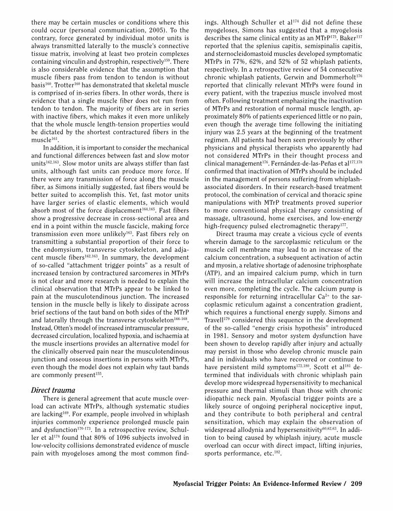
The Integrated Trigger Point HypothesisThe integrated trigger point hypothesis (Figure 7)
has evolved since its first introduction as the “energy crisis hypothesis” in 1981. It is based on a combination of electrodiagnostic and histopathological evidence179,183.
Already in 1957, Weeks and Travell205 had published a report that outlined a characteristic electrical activ-ity of an MTrP. It was not until 1993 that Hubbard et al206 confirmed that this EMG discharge consists of low-amplitude discharges in the order of 10-50 µV and intermittent high-amplitude discharges (up to 500 µV) in painful MTrPs. Initially, the electrical activity was termed “spontaneous electrical activity” (SEA) and thought to be related to dysfunctional muscle spindles206. Best available evidence now suggests that the SEA is in fact endplate noise (EPN), which is found much more
Myofascial Trigger Points: An Evidence-Informed Review / 211
commonly in the endplate zone near MTrPs than in an endplate zone outside MTrPs207-209. The electrical discharges occur with frequencies that are 10-1,000 times that of normal endplate potentials, and they have been found in humans, rabbits, and recently even in horses209,210. The discharges are most likely the result of an abnormally excessive release of acetylcholine (ACh) and indicative of dysfunctional motor endplates, contrary to the com-monly accepted notion among electromyographers that endplate noise arises from normal motor endplates183. The effectiveness of botulinum toxin in the treatment of MTrPs provides indirect evidence of the presence of excessive ACh211. Botulinum toxin (BoTox) is a neurotoxin that blocks the release of ACh from presynaptic choliner-gic nerve endings. A recent study in mice demonstrated that the administration of botulinum toxin resulted in a complete functional repair of dysfunctional endplates212. There is some early evidence that muscle stretching and hypertonicity may also enhance the excessive release of ACh213,214. Tension on the integrins in the presynaptic membrane at the motor nerve terminal is hypothesized to mechanically trigger an ACh release that does not require Ca2+ 213-215. Integrins are receptor proteins in the cell membrane involved in attaching individual cells to the extracellular matrix.
Excessive ACh affects voltage-gated sodium chan-nels of the sarcoplasmic reticulum and increases the intracellular calcium levels, which triggers sustained muscle contractures. It is conceivable that in MTrPs, myosin filaments literally get stuck in the Z-band of the sarcomere. During sarcomere contractions, titin filaments are folded into a gel-like structure at the Z-band. In MTrPs, the gel-like titin may prevent the myosin filaments from detaching. The myosin filaments may actually damage the regular motor assembly and prevent
the sarcomere from restoring its resting length216. Muscle contractures are also maintained because of the relative shortage of ATP in an MTrP, as ATP is required to break the cross-bridges between actin and myosin filaments. The question remains whether sustained contractures require an increase of oxygen availability.
At the same time, the shortened sarcomeres compro-mise the local circulation causing ischaemia. Studies of oxygen saturation levels have demonstrated severe hypoxia in MTrPs201. Hypoxia leads to the release of sensitizing substances and activates muscle nociceptors as reviewed above. The combined decreased energy supply and pos-sible increased metabolic demand would also explain the common finding of abnormal mitochondria in the nerve terminal and the previously mentioned ragged red fibers. In mice, the onset of hypoxia led to an immediate increased ACh release at the motor endplate217.
The combined high-intensity mechanical and chemi-cal stimuli may cause activation and sensitization of the peripheral nerve endings and autonomic nerves, activate second order neurons including so-called “sleep-ing” receptors, cause central sensitization, and lead to the formation of new receptive fields, referred pain, a long-lasting increase in the excitability of nociceptors, and a more generalized hyperalgesia beyond the initial nociceptive area. An expansion of a receptive field means that a dorsal horn neuron receives information from areas it has not received information from previously218. Sensitization of peripheral nerve endings can also cause pain through SP activating the neurokin-1 receptors and glutamate activating N-methyl-D-aspartate recep-tors, which opens post-synaptic channels through which Ca2+ ions can enter the dorsal horn and activate many enzymes involved in the sensitization85.
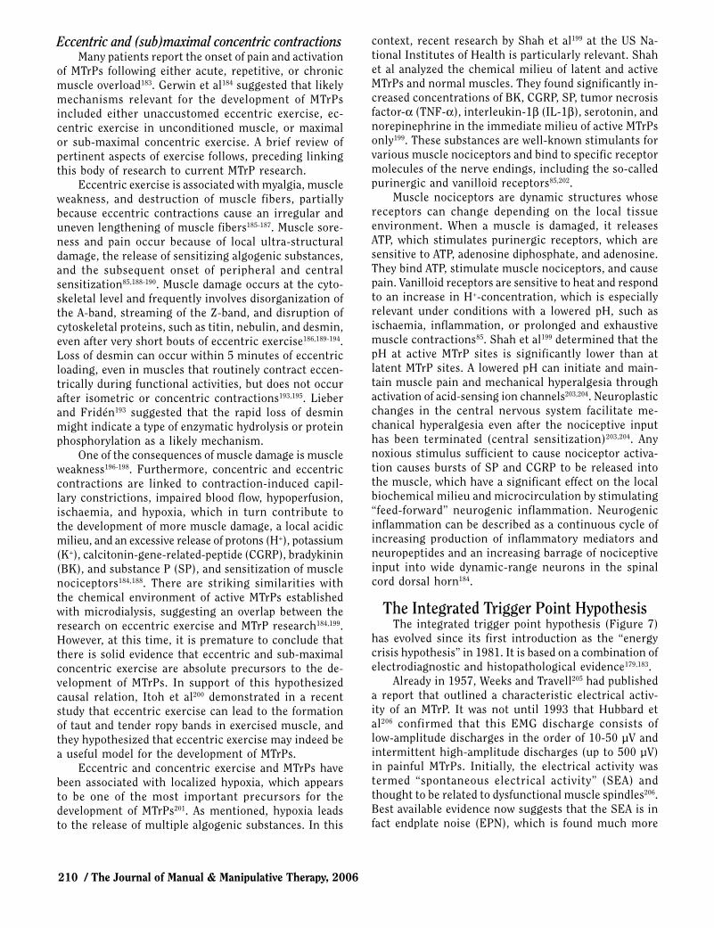
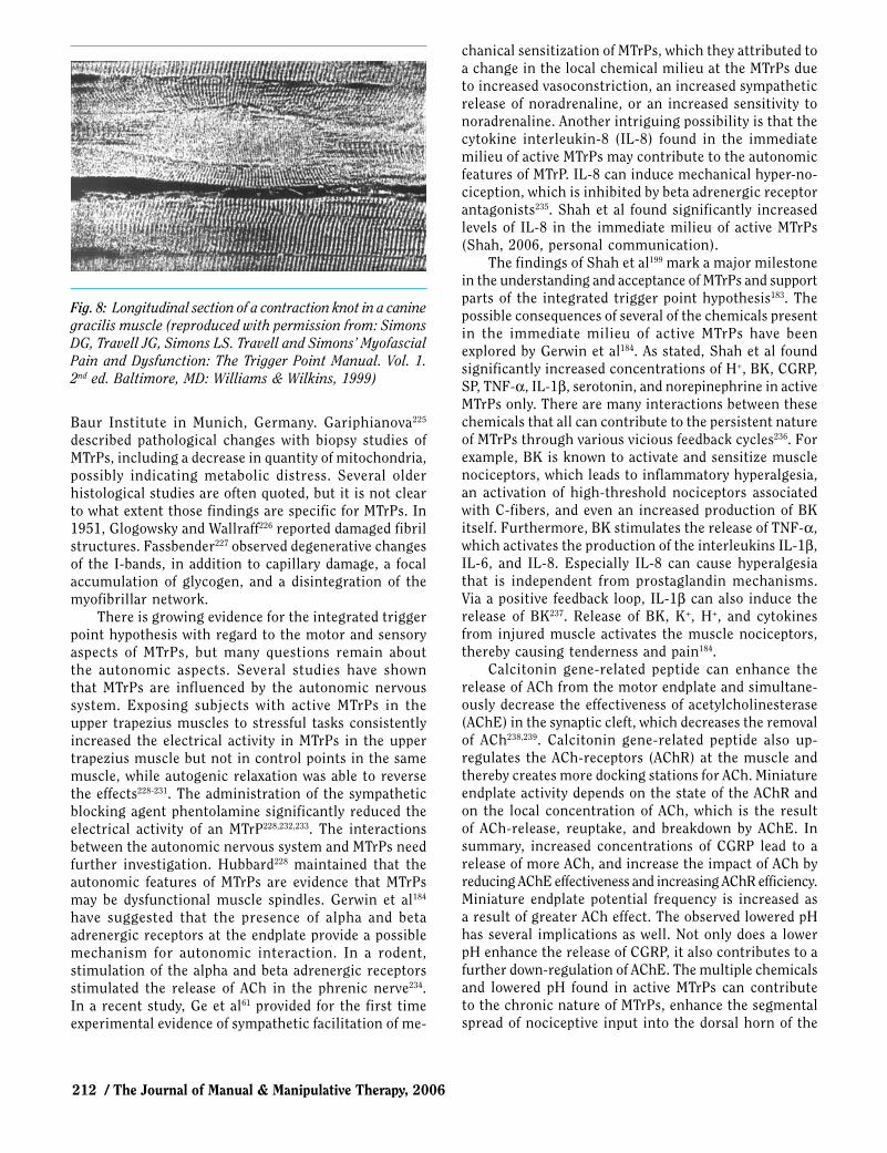
Several histological studies offer further support for the integrated trigger point hypothesis. In 1976, Simons and Stolov published the first biopsy study of MTrPs in a canine muscle and reported multiple contraction knots in various individual muscle fibers (Figure 8)219. The knots featured a combination of severely shortened sarcomeres in the center and lengthened sarcomeres outside the immediate MTrP region219.
Reitinger et al220 reported pathologic alterations of the mitochondria as well as increased width of A-bands and decreased width of I-bands in muscle sarcomeres of MTrPs in the gluteus medius muscle. Windisch et al221 determined similar alterations in a post-mortem histo-logical study of MTrPs completed within 24 hours of time of death. Mense et al222 studied the effects of electrically induced muscle contractions and a cholinesterase blocker on muscles with experimentally induced contraction knots and found evidence of localized contractions, torn fibers, and longitudinal stripes. Pongratz and Spath223, 224 dem-onstrated evidence of a contraction disk in a region of an MTrP using light microscopy. New MTrP histopathological studies are currently being conducted at the Friedrich
Fig. 7: The integrated trigger point hypothesis.Ach- acetylcholine; AchE- acetylcholinesterase; AchR- acetylcholine receptor
212 / The Journal of Manual & Manipulative Therapy, 2006
Baur Institute in Munich, Germany. Gariphianova225 described pathological changes with biopsy studies of MTrPs, including a decrease in quantity of mitochondria, possibly indicating metabolic distress. Several older histological studies are often quoted, but it is not clear to what extent those findings are specific for MTrPs. In 1951, Glogowsky and Wallraff226 reported damaged fibril structures. Fassbender227 observed degenerative changes of the I-bands, in addition to capillary damage, a focal accumulation of glycogen, and a disintegration of the myofibrillar network.
There is growing evidence for the integrated trigger point hypothesis with regard to the motor and sensory aspects of MTrPs, but many questions remain about the autonomic aspects. Several studies have shown that MTrPs are influenced by the autonomic nervous system. Exposing subjects with active MTrPs in the upper trapezius muscles to stressful tasks consistently increased the electrical activity in MTrPs in the upper trapezius muscle but not in control points in the same muscle, while autogenic relaxation was able to reverse the effects228-231. The administration of the sympathetic blocking agent phentolamine significantly reduced the electrical activity of an MTrP228,232,233. The interactions between the autonomic nervous system and MTrPs need further investigation. Hubbard228 maintained that the autonomic features of MTrPs are evidence that MTrPs may be dysfunctional muscle spindles. Gerwin et al184 have suggested that the presence of alpha and beta adrenergic receptors at the endplate provide a possible mechanism for autonomic interaction. In a rodent, stimulation of the alpha and beta adrenergic receptors stimulated the release of ACh in the phrenic nerve234. In a recent study, Ge et al61 provided for the first time experimental evidence of sympathetic facilitation of me-
chanical sensitization of MTrPs, which they attributed to a change in the local chemical milieu at the MTrPs due to increased vasoconstriction, an increased sympathetic release of noradrenaline, or an increased sensitivity to noradrenaline. Another intriguing possibility is that the cytokine interleukin-8 (IL-8) found in the immediate milieu of active MTrPs may contribute to the autonomic features of MTrP. IL-8 can induce mechanical hyper-no-ciception, which is inhibited by beta adrenergic receptor antagonists235. Shah et al found significantly increased levels of IL-8 in the immediate milieu of active MTrPs (Shah, 2006, personal communication).
The findings of Shah et al199 mark a major milestone in the understanding and acceptance of MTrPs and support parts of the integrated trigger point hypothesis183. The possible consequences of several of the chemicals present in the immediate milieu of active MTrPs have been explored by Gerwin et al184. As stated, Shah et al found significantly increased concentrations of H+, BK, CGRP, SP, TNF- , IL-1 , serotonin, and norepinephrine in active MTrPs only. There are many interactions between these chemicals that all can contribute to the persistent nature of MTrPs through various vicious feedback cycles236. For example, BK is known to activate and sensitize muscle nociceptors, which leads to inflammatory hyperalgesia, an activation of high-threshold nociceptors associated with C-fibers, and even an increased production of BK itself. Furthermore, BK stimulates the release of TNF- , which activates the production of the interleukins IL-1 , IL-6, and IL-8. Especially IL-8 can cause hyperalgesia that is independent from prostaglandin mechanisms. Via a positive feedback loop, IL-1 can also induce the release of BK237. Release of BK, K+, H+, and cytokines from injured muscle activates the muscle nociceptors, thereby causing tenderness and pain184.
Calcitonin gene-related peptide can enhance the release of ACh from the motor endplate and simultane-ously decrease the effectiveness of acetylcholinesterase (AChE) in the synaptic cleft, which decreases the removal of ACh238,239. Calcitonin gene-related peptide also up-regulates the ACh-receptors (AChR) at the muscle and thereby creates more docking stations for ACh. Miniature endplate activity depends on the state of the AChR and on the local concentration of ACh, which is the result of ACh-release, reuptake, and breakdown by AChE. In summary, increased concentrations of CGRP lead to a release of more ACh, and increase the impact of ACh by reducing AChE effectiveness and increasing AChR efficiency. Miniature endplate potential frequency is increased as a result of greater ACh effect. The observed lowered pH has several implications as well. Not only does a lower pH enhance the release of CGRP, it also contributes to a further down-regulation of AChE. The multiple chemicals and lowered pH found in active MTrPs can contribute to the chronic nature of MTrPs, enhance the segmental spread of nociceptive input into the dorsal horn of the
Fig. 8: Longitudinal section of a contraction knot in a canine gracilis muscle (reproduced with permission from: Simons DG, Travell JG, Simons LS. Travell and Simons’ Myofascial Pain and Dysfunction: The Trigger Point Manual. Vol. 1. 2nd ed. Baltimore, MD: Williams & Wilkins, 1999)
Myofascial Trigger Points: An Evidence-Informed Review / 213
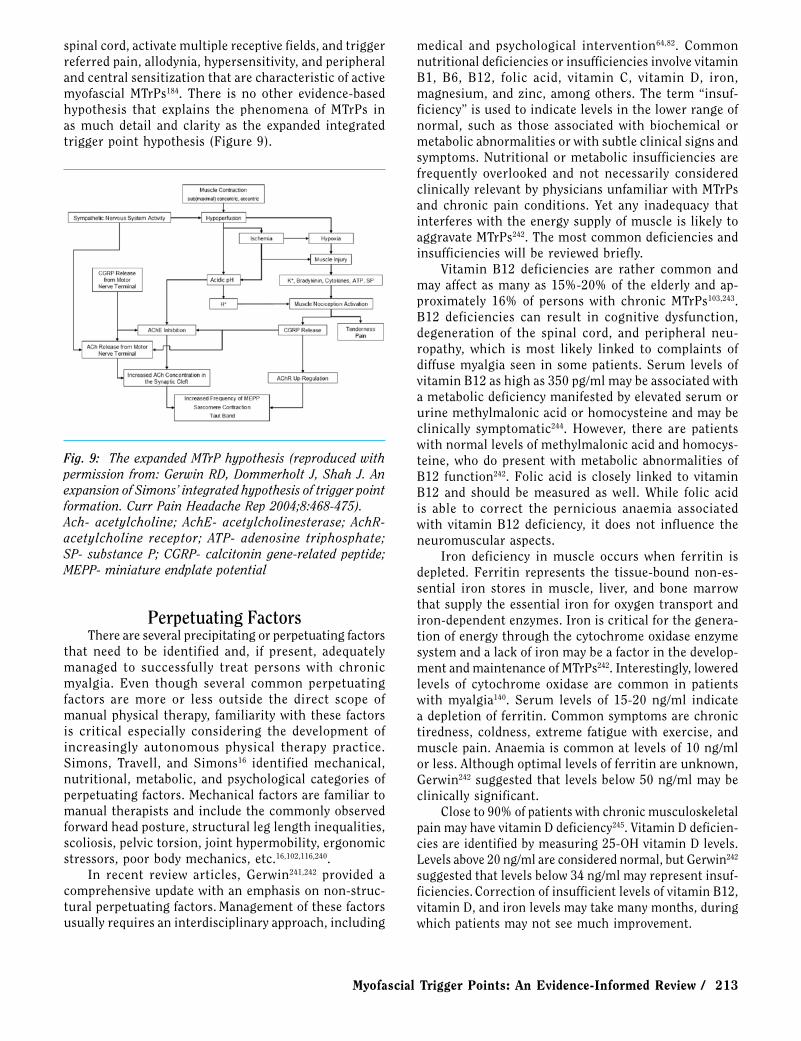
spinal cord, activate multiple receptive fields, and trigger referred pain, allodynia, hypersensitivity, and peripheral and central sensitization that are characteristic of active myofascial MTrPs184. There is no other evidence-based hypothesis that explains the phenomena of MTrPs in as much detail and clarity as the expanded integrated trigger point hypothesis (Figure 9).
medical and psychological intervention64,82. Common nutritional deficiencies or insufficiencies involve vitamin B1, B6, B12, folic acid, vitamin C, vitamin D, iron, magnesium, and zinc, among others. The term “insuf-ficiency” is used to indicate levels in the lower range of normal, such as those associated with biochemical or metabolic abnormalities or with subtle clinical signs and symptoms. Nutritional or metabolic insufficiencies are frequently overlooked and not necessarily considered clinically relevant by physicians unfamiliar with MTrPs and chronic pain conditions. Yet any inadequacy that interferes with the energy supply of muscle is likely to aggravate MTrPs242. The most common deficiencies and insufficiencies will be reviewed briefly.
Vitamin B12 deficiencies are rather common and may affect as many as 15%-20% of the elderly and ap-proximately 16% of persons with chronic MTrPs103,243. B12 deficiencies can result in cognitive dysfunction, degeneration of the spinal cord, and peripheral neu-ropathy, which is most likely linked to complaints of diffuse myalgia seen in some patients. Serum levels of vitamin B12 as high as 350 pg/ml may be associated with a metabolic deficiency manifested by elevated serum or urine methylmalonic acid or homocysteine and may be clinically symptomatic244. However, there are patients with normal levels of methylmalonic acid and homocys-teine, who do present with metabolic abnormalities of B12 function242. Folic acid is closely linked to vitamin B12 and should be measured as well. While folic acid is able to correct the pernicious anaemia associated with vitamin B12 deficiency, it does not influence the neuromuscular aspects.
Iron deficiency in muscle occurs when ferritin is depleted. Ferritin represents the tissue-bound non-es-sential iron stores in muscle, liver, and bone marrow that supply the essential iron for oxygen transport and iron-dependent enzymes. Iron is critical for the genera-tion of energy through the cytochrome oxidase enzyme system and a lack of iron may be a factor in the develop-ment and maintenance of MTrPs242. Interestingly, lowered levels of cytochrome oxidase are common in patients with myalgia140. Serum levels of 15-20 ng/ml indicate a depletion of ferritin. Common symptoms are chronic tiredness, coldness, extreme fatigue with exercise, and muscle pain. Anaemia is common at levels of 10 ng/ml or less. Although optimal levels of ferritin are unknown, Gerwin242 suggested that levels below 50 ng/ml may be clinically significant.
Close to 90% of patients with chronic musculoskeletal pain may have vitamin D deficiency245. Vitamin D deficien-cies are identified by measuring 25-OH vitamin D levels. Levels above 20 ng/ml are considered normal, but Gerwin242 suggested that levels below 34 ng/ml may represent insuf-ficiencies. Correction of insufficient levels of vitamin B12, vitamin D, and iron levels may take many months, during which patients may not see much improvement.
Fig. 9: The expanded MTrP hypothesis (reproduced with permission from: Gerwin RD, Dommerholt J, Shah J. An expansion of Simons’ integrated hypothesis of trigger point formation. Curr Pain Headache Rep 2004;8:468-475).Ach- acetylcholine; AchE- acetylcholinesterase; AchR- acetylcholine receptor; ATP- adenosine triphosphate; SP- substance P; CGRP- calcitonin gene-related peptide; MEPP- miniature endplate potential
Perpetuating FactorsThere are several precipitating or perpetuating factors
that need to be identified and, if present, adequately managed to successfully treat persons with chronic myalgia. Even though several common perpetuating factors are more or less outside the direct scope of manual physical therapy, familiarity with these factors is critical especially considering the development of increasingly autonomous physical therapy practice. Simons, Travell, and Simons16 identified mechanical, nutritional, metabolic, and psychological categories of perpetuating factors. Mechanical factors are familiar to manual therapists and include the commonly observed forward head posture, structural leg length inequalities, scoliosis, pelvic torsion, joint hypermobility, ergonomic stressors, poor body mechanics, etc.16,102,116,240.
In recent review articles, Gerwin241,242 provided a comprehensive update with an emphasis on non-struc-tural perpetuating factors. Management of these factors usually requires an interdisciplinary approach, including
214 / The Journal of Manual & Manipulative Therapy, 2006
Even when active MTrPs have been identified in a particular patient, clinicians must always consider that MTrPs may be secondary to metabolic insufficiencies or other medical diagnoses. It is questionable whether physical therapy and—as an integral part of physical therapy management—manual therapy intervention can be successful when patients have nutritional or metabolic insufficiencies or deficiencies. A close working relationship with physicians familiar with this body of literature is essential. Therapists should consider the possible interactions between arthrogenic or neurogenic dysfunction and MTrPs4,5,118,133,246,247.
Clinically, physical therapists should address all aspects of the dysfunction. There are many other con-ditions that feature muscle pain and MTrPs, including hypothyroidism, systemic lupus erythematosis, Lyme disease, babesiosis, ehrlichiosis, candida albicans infec-tions, myoadenylate deaminase deficiency, hypoglycaemia, and parasitic diseases such as fascioliasis, amoebiasis, and giardia64, 242. Therapists should be familiar with the symptoms associated with these medical diagnoses64.
Psychological stress may activate MTrPs. Electromyo-graphic activity in MTrPs has been shown to increase dramatically in response to mental and emotional stress, whereas adjacent non-trigger point muscle EMG activity remained normal229, 230. Relaxation techniques, such as autogenic relaxation, can diminish the electrical activ-ity231. In addition, many patients with persistent MTrPs are dealing with depression, anxiety, anger, and feelings of hopelessness248. Pain-related fear and avoidance can lead to the development and maintenance of chronic pain249. Sleep disturbance can also be a major factor in the perpetuation of musculoskeletal pain and must be addressed. Sleep problems may be related to pain, apnea, or to mood disorders like depression or anxiety. Manage-ment can be both pharmacologic and non-pharmacologic. Pharmacologic treatment utilizes drugs that promote normal sleep patterns and induce and maintain sleep through the night without causing daytime sedation. Non-pharmacologic treatment emphasizes sleep hygiene,
such as using the bed only for sleep and sex, and not for reading, television viewing, and eating250. Therapists must be sensitive to the impact of psychological and emotional distress and refer patients to clinical social workers or psychologists when appropriate.
The Role of Manual TherapyAlthough the various management approaches are
beyond the scope of this article, manual therapy is one of the basic treatment options and the role of orthope-dic manual physical therapists cannot be overempha-sized82,158. Myofascial trigger points are treated with manual techniques, spray and stretch, dry needling, or injection therapy. Dry needling is within the scope of physical therapy practice in many countries including Canada, Spain, Ireland, South Africa, Australia, the Netherlands, and Switzerland. In the United States, the physical therapy boards of eight states have ruled that physical therapists can engage in the practice of dry needling: New Hampshire, Maryland, Virginia, South Carolina, Georgia, Kentucky, New Mexico, and Colorado80. A promising new development used in the diagnosis and treatment of MTrPs involves shockwave therapy, but as of yet, there are no controlled studies substantiating its use251,252.
SummaryAlthough MTrPs are a common cause of pain and
dysfunction in persons with musculoskeletal injuries and diagnoses, the importance of MTrPs is not obvious from reviewing the orthopedic manual therapy litera-ture. Current scientific evidence strongly supports that awareness and a working knowledge of muscle dysfunc-tion; in particular, MTrPs should be incorporated into manual physical therapy practice consistent with the IFOMT guidelines for clinical practice. While there are still many unanswered questions with regard to explain-ing the etiology of MTrPs, this article provides manual therapists with an up-to-date evidence-informed review of the current scientific knowledge.
REFERENCES 1. IFOMT. Available at: http://www.ifomt.org/ifomt/about/standards.
Accessed November 15, 2006.2. Huijbregts PA. Muscle injury, regeneration, and repair. J Manual
Manipulative Ther 2001;9:9-16.3. Urquhart DM, Hodges PW, Allen TJ, Story IH. Abdominal muscle
recruitment during a range of voluntary exercises. Man Ther 2005;10:144-153.
4. Fernández-de-las-Peñas C, Alonso-Blanco C, Alguacil-Diego IM, Miangolarra-Page JC. Myofascial trigger points and postero-an-
terior joint hypomobility in the mid-cervical spine in subjects presenting with mechanical neck pain: A pilot study. J Manual Manipulative Ther 2006;14:88-94.
5. Fernández-de-las-Peñas C, Alonso-Blanco C, Miangolarra JC. Myofascial trigger points in subjects presenting with mechanical neck pain: A blinded, controlled study Man Ther (In press).
6. Lew PC, Lewis J, Story I. Inter-therapist reliability in locating latent myofascial trigger points using palpation. Man Ther 1997;2:87-90.
Myofascial Trigger Points: An Evidence-Informed Review / 215
7. Fernández-de-las-Peñas C, Alonso-Blanco C, Cuadrado ML, Pareja JA. Myofascial trigger points in the suboccipital muscles in episodic tension-type headache. Man Ther 2006;11:225-230.
8. Moore A, Petty N. Evidence-based practice: Getting a grip and finding a balance. Man Ther 2001;6:195-196.
9. Sackett DL, Rosenberg WM. The need for evidence-based medi-cine. J R Soc Med 1995;88:620-624.
10. Sackett DL, Rosenberg WM, Gray JA, Haynes RB, Richardson WS. Evidence-based medicine: What it is and what it isn’t. BMJ 1996;312:71-72.
11. Pencheon D. What’s next for evidence-based medicine? Evidence-Based Healthcare Public Health 2005;9:319-321.
12. Cicerone KD. Evidence-based practice and the limits of rational rehabilitation. Arch Phys Med Rehabil 2005;86:1073-1074.
13. Baldry PE. Acupuncture, Trigger Points and Musculoskeletal Pain. Edinburgh, UK: Churchill Livingstone, 2005.
14. Simons DG. Muscle pain syndromes. Part 1. Am J Phys Med 1975;54:289-311.
15. Ruhmann W. The earliest book on rheumatism. Br J Rheumatism 1940;11:140-162.
16. Simons DG, Travell JG, Simons LS. Travell and Simons’ Myo-fascial Pain and Dysfunction: The Trigger Point Manual. Vol. 1. 2nd ed. Baltimore, MD: Williams & Wilkins, 1999.
17. Harden RN, Bruehl SP, Gass S, Niemiec C, Barbick B. Signs and symptoms of the myofascial pain syndrome: A national survey of pain management providers. Clin J Pain 2000;16:64-72.
18. Stockman R. The causes, pathology, and treatment of chronic rheumatism. Edinburgh Med J 1904;15:107-116.
19. Strauss H. ber die sogenannten‚ Rheumatische Muskelschwiele‘ [German; With regard to the so-called myogelosis]. Klin Wo-chenschr 1898;35:89-91,121-123.
20. Lange M. Die Muskelhärten (Myogelosen) [German; The Muscle Hardenings (Myogeloses)]. Munich, Germany: J.F. Lehmann‘s Verlag, 1931.
21. Travell J. Office Hours: Day and Night. The Autobiography of Janet Travell, MD. New York, NY: World Publishing, 1968.
22. Kellgren JH. Deep pain sensibility. Lancet 1949;1:943-949.23. Kellgren JH. Observations on referred pain arising from muscle.
Clin Sci 1938;3:175-190.24. Kellgren JH. A preliminary account of referred pains arising
from muscle. British Med J 1938;1:325-327.25. Simons DG. Cardiology and myofascial trigger points: Janet G.
Travell’s contribution. Tex Heart Inst J 2003;30(1):3-7.26. Travell J. Basis for the multiple uses of local block of somatic
trigger areas (procaine infiltration and ethyl chloride spray). Miss Valley Med 1949;71:13-22.
27. Travell J, Bobb AL. Mechanism of relief of pain in sprains by local injection techniques. Fed Proc 1947;6:378.
28. Travell JG, Rinzler S, Herman M. Pain and disability of the shoulder and arm: Treatment by intramuscular infiltration with procaine hydrochloride. JAMA 1942;120:417-422.
29. Travell JG, Rinzler SH. The myofascial genesis of pain. Postgrad Med 1952;11:452-434.
30. Chaitow L, DeLany J. Neuromuscular techniques in orthopedics. Techniques in Orthopedics 2003;18(1):74-86.
31. Good MG. Five hundred cases of myalgia in the British army. Ann Rheum Dis 1942;3:118-138.
32. Good MG. The role of skeletal muscle in the pathogenesis of diseases. Acta Medica Scand 1950;138:285-292.
33. Gutstein M. Common rheumatism and physiotherapy. Br J Phys Med 1940;3:46-50.
34. Gutstein M. Diagnosis and treatment of muscular rheumatism. Br J Phys Med 1938;1:302-321.
35. Kelly M. The nature of fibrositis. I. The myalgic lesion and its secondary effects: A reflex theory. Ann Rheum Dis 1945;5:1-7.
36. Kelly M. The nature of fibrositis. II. A study of the causation of the myalgic lesion (rheumatic, traumatic, infective). Ann Rheum Dis 1946;5:69-77.
37. Kelly M. The relief of facial pain by procaine (Novocaine) injec-tions. J Am Geriatr Soc 1963;11:586-96.
38. Kelly M. The treatment of fibrositis and allied disorders by local anesthesia. Med J Aust 1941;1:294-298.
39. Schneider M, Cohen J, Laws S. The Collected Writings of Nimmo & Vannerson: Pioneers of Chiropractic Trigger Point Therapy. Pittsburgh, PA: Schneider, 2001.
40. Cohen JH, Gibbons RW. Raymond L. Nimmo and the evolution of trigger point therapy, 1929-1986. J Manipulative Physiol Ther 1998;21:167-172.
41. National Board of Chiropractic Examiners. Chiropractic Treat-ment Procedures. Greeley, CO: NBCE, 1993.
42. Mennell J. Spray-stretch for the relief of pain from muscle spasm and myofascial trigger points. J Am Podiatry Assoc 1976;66:873-876.
43. Mennell J. Myofascial trigger points as a cause of headaches. J Manipulative Physiol Ther, 1989;12:308-313.
44. Travell W, Travell JG. Technic for reduction and ambulatory treatment of sacroiliac displacement. Arch Phys Ther 1942;23:222-232.
45. Paris SV. In the best interests of the patient. Phys Ther 2006;86:1541-1553.
46. Travell JG, Simons DG. Myofascial Pain and Dysfunction: The Trigger Point Manual. Vol. 2. Baltimore, MD: Williams & Wilkins, 1992.
47. Travell JG, Simons DG. Myofascial Pain and Dysfunction: The Trigger Point Manual. Vol. 1. Baltimore, MD: Williams & Wilkins, 1983.
48. Dejung B, Gröbli C, Colla F, Weissmann R. Triggerpunkttherapie [German; Trigger Point Therapy]. Bern, Switzerland: Hans Huber, 2003.
49. Ferguson LW, Gerwin R. Clinical Mastery in the Treatment of Myofascial Pain. Philadelphia, PA: Lippincott Williams & Wilkins, 2005.
50. Kostopoulos D, Rizopoulos K. The Manual of Trigger Point and Myofascial Therapy. Thorofare, NJ: Slack, 2001.
51. Prateepavanich P. Myofascial Pain Syndrome: A Common Problem in Clinical Practice. Bangkok, Thailand: Ammarind, 1999.
52. Rachlin ES, Rachlin IS. Myofascial Pain and Fibromyalgia: Trigger Point Management. St. Louis, MO: Mosby, 2002.
53. Cardinal S. Points Détente et Acupuncture: Approche Neuro-physiologique [French; Trigger Points and Acupuncture: Neu-rophysiological Approach]. Montreal, Canada: Centre Collégial de Développement de Matériel Didactique, 2004.
54. Jonckheere PDM. Spieren en Dysfuncties, Trigger punten, Ba-sisprincipes van de Myofasciale Therapie [Dutch; Muscles and Dysfunctions, Basic Principles of Myofascial Therapy]. Brussels, Belgium: Satas, 1993.
55. Lucas KR, Polus BI, Rich PS. Latent myofascial trigger points: Their effect on muscle activation and movement efficiency. J Bodywork Mov Ther 2004;8:160-166.
56. Weissmann RD. Überlegungen zur Biomechanik in der Myo-faszialen Triggerpunkttherapie [German; Considerations with regard to the biomechanics related to myofascial trigger point
216 / The Journal of Manual & Manipulative Therapy, 2006
therapy]. Physiotherapie 2000;35(10):13-21.57. Vecchiet L, Giamberardino MA, De Bigontina P. Comparative
sensory evaluation of parietal tissues in painful and nonpainful areas in fibromyalgia and myofascial pain syndrome. In: Gebhart GF, Hammond DL, Jensen TS, eds. Proceedings of the 7th World Congress on Pain (Progress in Pain Research and Management). Seattle, WA: IASP Press, 1994:177-185.
58. Vecchiet L, Giamberardino MA, Dragani L. Latent myofascial trigger points: Changes in muscular and subcutaneous pain thresholds at trigger point and target level. J Manual Medicine 1990;5:151-154.
59. Vecchiet L, Pizzigallo E, Iezzi S, Affaitati G, Vecchiet J, Giambe-rardino MA. Differentiation of sensitivity in different tissues and its clinical significance. J Musculoskeletal Pain 1998;6:33-45.
60. Dommerholt J. Persistent myalgia following whiplash. Curr Pain Headache Rep 2005;9:326-330.
61. Ge HY, Fernández-de-las-Peñas C, Arendt-Nielsen L. Sympathetic facilitation of hyperalgesia evoked from myofascial tender and trigger points in patients with unilateral shoulder pain. Clin Neurophysiol 2006;117:1545-1550.
62. Lidbeck J. Central hyperexcitability in chronic musculoskeletal pain: A conceptual breakthrough with multiple clinical implica-tions. Pain Res Manag 2002;7(2):81-92.
63. Munglani R. Neurobiological mechanisms underlying chronic whiplash associated pain: The peripheral maintenance of central sensitization. J Musculoskeletal Pain 2000;8:169-178.
64. Dommerholt J, Issa T. Differential diagnosis: Myofascial pain. In: Chaitow L, ed. Fibromyalgia Syndrome: A Practitioner’s Guide to Treatment. Edinburgh, UK: Churchill Livingstone, 2003:149-177.
65. Gerwin RD, Shannon S, Hong CZ, Hubbard D, Gevirtz R. Inter-rater reliability in myofascial trigger point examination. Pain 1997;69(1-2):65-73.
66. Sciotti VM, Mittak VL, DiMarco L, Ford LM, Plezbert J, Santipadri E, Wigglesworth J, Ball K. Clinical precision of myofascial trigger point location in the trapezius muscle. Pain 2001;93(3):259-266.
67. Franssen JLM. Handboek Oppervlakte Elektromyografie [Dutch; Manual Surface Electromyography]. Utrecht, The Netherlands: De Tijdstroom, 1995.
68. Janda V. Muscle spasm: A proposed procedure for differential diagnosis. J Manual Med 1991;6:136-139.
69. Mense S. Pathophysiologic basis of muscle pain syndromes. In: Fischer AA, ed. Myofascial Pain: Update in Diagnosis and Treat-ment. Philadelphia, PA: W.B. Saunders Company, 1997:23-53.
70. Headly BJ. Evaluation and treatment of myofascial pain syndrome utilizing biofeedback. In: Cram JR, ed. Clinical Electromyogra-phy for Surface Recordings. Nevada City, NV: Clinical Resources, 1990:235-254.
71. Headly BJ. Chronic pain management. In: O’Sullivan SB, Schmitz TS, eds. Physical Rehabilitation: Assessment and Treatment. Philadelphia, PA: F.A. Davis Company, 1994:577-600.
72. Gerwin RD, Duranleau D. Ultrasound identification of the myofascial trigger point. Muscle Nerve 1997;20:767-768.
73. Hong CZ. Persistence of local twitch response with loss of conduction to and from the spinal cord. Arch Phys Med Rehabil 1994;75:12-16.
74. Hong C-Z, Torigoe Y. Electrophysiological characteristics of localized twitch responses in responsive taut bands of rabbit skeletal muscle. J Musculoskeletal Pain 1994;2:17-43.
75. Hong C-Z, Yu J. Spontaneous electrical activity of rabbit trig-
ger spot after transection of spinal cord and peripheral nerve. J Musculoskeletal Pain 1998;6(4):45-58.
76. Fricton JR, Auvinen MD, Dykstra D, Schiffman E. Myofascial pain syndrome: Electromyographic changes associated with local twitch response. Arch Phys Med Rehabil 1985;66:314-317.
77. Simons DG, Dexter JR. Comparison of local twitch responses elicited by palpation and needling of myofascial trigger points. J Musculoskeletal Pain 1995;3:49-61.
78. Wang F, Audette J. Electrophysiological characteristics of the local twitch response with active myofascial pain of neck compared with a control group with latent trigger points. Am J Phys Med Rehabil 2000;79:203.
79. Audette JF, Wang F, Smith H. Bilateral activation of motor unit potentials with unilateral needle stimulation of active myofascial trigger points. Am J Phys Med Rehabil 2004;83:368-374, quiz 375-377,389.
80. Dommerholt J. Dry needling in orthopedic physical therapy practice. Orthop Phys Ther Pract 2004;16(3):15-20.
81. Hong C-Z. Lidocaine injection versus dry needling to myofascial trigger point: The importance of the local twitch response. Am J Phys Med Rehabil 1994;73:256-263.
82. Gerwin RD, Dommerholt J. Treatment of myofascial pain syn-dromes. In: Boswell MV, Cole BE, eds. Weiner’s Pain Manage-ment: A Practical Guide for Clinicians. Boca Raton, FL: CRC Press, 2006:477-492.
83. Vecchiet L, Dragani L, De Bigontina P, Obletter G, Giamberardino MA. Experimental referred pain and hyperalgesia from muscles in humans. In: Vecchiet L, et al, eds. New Trends in Referred Pain and Hyperalgesia. Amsterdam, The Netherlands: Elsevier Science, 1993:239-249.
84. Vecchiet L, Giamberardino MA. Referred pain: Clinical significance, pathophysiology and treatment. In: Fischer AA, ed. Myofascial Pain: Update in Diagnosis and Treatment. Philadelphia, PA: W.B. Saunders Company, 1997:119-136.
85. Mense S. The pathogenesis of muscle pain. Curr Pain Headache Rep 2003;7:419-425.
86. Mense S. Referral of muscle pain: New aspects. Amer Pain Soc J 1994;3:1-9.
87. Fernández-de-las-Peñas CF, Cuadrado ML, Gerwin RD, Pareja JA. Referred pain from the trochlear region in tension-type headache: A myofascial trigger point from the superior oblique muscle. Headache 2005;45:731-737.
88. Fernández-de-las-Peñas C, Cuadrado ML, Gerwin RD, Pareja JA. Myofascial disorders in the trochlear region in unilateral migraine: A possible initiating or perpetuating factor. Clin J Pain 2006;22:548-553.
89. Hwang M, Kang YK, Kim DH. Referred pain pattern of the pronator quadratus muscle. Pain 2005;116:238-242.
90. Hwang M, Kang YK, Shin JY, Kim DH. Referred pain pattern of the abductor pollicis longus muscle. Am J Phys Med Rehabil 2005;84:593-597.
91. Hong CZ, Kuan TS, Chen JT, Chen SM. Referred pain elicited by palpation and by needling of myofascial trigger points: A comparison. Arch Phys Med Rehabil 1997;78:957-960.
92. Dwyer A, Aprill C, Bogduk N. Cervical zygapophyseal joint pain patterns. I: A study in normal volunteers. Spine 1990;15:453-457.
93. Giamberardino MA, Vecchiet L. Visceral pain, referred hyperal-gesia and outcome: New concepts. Eur J Anaesthesiol (Suppl) 1995;10:61-66.
94. Scudds RA, Landry M, Birmingham T, Buchan J, Griffin K. The
Myofascial Trigger Points: An Evidence-Informed Review / 217
frequency of referred signs from muscle pressure in normal healthy subjects (abstract). J Musculoskeletal Pain 1995;3 (Suppl. 1):99.
95. Torebjörk HE, Ochoa JL, Schady W. Referred pain from intra-neural stimulation of muscle fascicles in the median nerve. Pain 1984;18:145-156.
96. Gibson W, Arendt-Nielsen L, Graven-Nielsen T. Referred pain and hyperalgesia in human tendon and muscle belly tissue. Pain 2006;120(1-2):113-123.
97. Gibson W, Arendt-Nielsen L, Graven-Nielsen T. Delayed onset muscle soreness at tendon-bone junction and muscle tissue is associated with facilitated referred pain. Exp Brain Res 2006 (In press).
98. Hendler NH, Kozikowski JG. Overlooked physical diagnoses in chronic pain patients involved in litigation. Psychosomatics 1993;34:494-501.
99. Weiner DK, Sakamoto S, Perera S, Breuer P. Chronic low back pain in older adults: Prevalence, reliability, and validity of physi-cal examination findings. J Am Geriatr Soc 2006;54:11-20.
100. Calandre EP, Hidalgo J, Garcia-Leiva JM, Rico-Villademoros F. Trigger point evaluation in migraine patients: An indication of peripheral sensitization linked to migraine predisposition? Eur J Neurol 2006;13:244-249.
101. Headache Classification Subcommittee of the International Headache Society: The international classification of headache disorders. Cephalalgia 2004;24(Suppl 1):9-160.
102. Fricton JR, Kroening R, Haley D, Siegert R. Myofascial pain syn-drome of the head and neck: A review of clinical characteristics of 164 patients. Oral Surg Oral Med Oral Pathol 1985;60:615-623.
103. Gerwin R. A study of 96 subjects examined both for fibromy-algia and myofascial pain (abstract). J Musculoskeletal Pain 1995;3(Suppl 1):121.
104. Rosomoff HL, Fishbain DA, Goldberg N, Rosomoff RS. Myofascial findings with patients with chronic intractable benign pain of the back and neck. Pain Management 1989;3:114-118.
105. Skootsky SA, Jaeger B, Oye RK. Prevalence of myofascial pain in general internal medicine practice. West J Med 1989;151:157-160.
106. Chaiamnuay P, Darmawan J, Muirden KD, Assawatanabodee P. Epidemiology of rheumatic disease in rural Thailand: A WHO-ILAR COPCORD study. Community Oriented Programme for the Control of Rheumatic Disease. J Rheumatol 1998;25:1382-1387.
107. Graff-Radford B. Myofascial trigger points: Their importance and diagnosis in the dental office. J Dent Assoc S Afr 1984;39:249-253.
108. Bajaj P, Bajaj P, Graven-Nielsen T, Arendt-Nielsen L. Trigger points in patients with lower limb osteoarthritis. J Musculosk-eletal Pain 2001;9(3):17-33.
109. Hsueh TC, Yu S, Kuan TS, Hong C-Z. Association of active myofascial trigger points and cervical disc lesions. J Formosa Med Assoc 1998;97(3):174-80.
110. Wang C-F, Chen M, Lin M-T, Kuan T-S, Hong C-Z. Teres minor tendinitis manifested with chronic myofascial pain syndrome in the scapular muscles: A case report. J Musculoskeletal Pain 2006;14(1):39-43.
111. Fricton JR. Etiology and management of masticatory myofascial pain. J Musculoskeletal Pain 1999;7(1/2):143-160.
112. Teachey WS. Otolaryngic myofascial pain syndromes. Curr Pain Headache Rep 2004;8:457-462.
113. Dommerholt J. El sindrome de dolor miofascial en la region craneomandibular. [Spanish; Myofascial pain syndrome in the craniomandibular region]. In: Padrós Serrat E, ed. Bases diagnosticas, terapeuticas y posturales del functionalismo craniofacial. Madrid, Spain: Ripano, 2006:564-581.
114. Hesse J, Mogelvang B, Simonsen H. Acupuncture versus meto-prolol in migraine prophylaxis: A randomized trial of trigger point inactivation. J Intern Med 1994;235:451-456.
115. Skubick DL, Clasby R, Donaldson CC, Marshall WM. Carpal tunnel syndrome as an expression of muscular dysfunction in the neck. J Occupational Rehab 1993;3:31-43.
116. Treaster D, Marras WS, Burr D, Sheedy JE, Hart D. Myofascial trigger point development from visual and postural stressors during computer work. J Electromyogr Kinesiol 2006;16:115-124.
117. Baker BA. The muscle trigger: Evidence of overload injury. J Neurol Orthop Med Surg 1986;7:35-44.
118. Fruth SJ. Differential diagnosis and treatment in a patient with posterior upper thoracic pain. Phys Ther 2006;86:254-268.
119. Doggweiler-Wiygul R. Urologic myofascial pain syndromes. Curr Pain Headache Rep 2004;8:445-451.
120. Jarrell J. Myofascial dysfunction in the pelvis. Curr Pain Head-ache Rep 2004;8:452-456.
121. Jarrell JF, Vilos GA, Allaire C, Burgess S, Fortin C, Gerwin R, Lapensee L, Lea RH, Leyland NA, Martyn P, Shenassa H, Taenzer P, Abu-Rafea B. Consensus guidelines for the management of chronic pelvic pain. J Obstet Gynaecol Can 2005;27:869-887.
122. Weiss JM. Pelvic floor myofascial trigger points: Manual therapy for interstitial cystitis and the urgency-frequency syndrome. J Urol 2001;166:2226-2231.
123. Dommerholt J. Muscle pain syndromes. In: Cantu RI, Grodin AJ, eds. Myofascial Manipulation. Gaithersburg, MD: Aspen, 2001:93-140.
124. Weiner DK, Schmader KE. Postherpetic pain: More than sensory neuralgia? Pain Med 2006;7:243-249; discussion 250.
125. Chen SM, Chen JT, Kuan TS, Hong C-Z. Myofascial trigger points in intercostal muscles secondary to herpes zoster infection of the intercostal nerve. Arch Phys Med Rehabil 1998;79:336-338.
126. Dommerholt J. Complex regional pain syndrome. Part 1: His-tory, diagnostic criteria and etiology. J Bodywork Mov Ther 2004;8:167-177.
127. Rashiq S, Galer BS. Proximal myofascial dysfunction in complex regional pain syndrome: A retrospective prevalence study. Clin J Pain,1999;15:151-153.
128. Prateepavanich P, Kupniratsaikul VC. The relationship between myofascial trigger points of gastrocnemius muscle and nocturnal calf cramps. J Med Assoc Thailand 1999;82:451-459.
129. Kern KU, Martin C, Scheicher S, Muller H. Auslosung von Phan-tomschmerzen und -sensationen durch Muskuläre Stumpftrig-gerpunkte nach Beinamputationen [German; Referred pain from amputation stump trigger points into the phantom limb]. Schmerz 2006;20:300-306.
130. Kern U, Martin C, Scheicher S, M ller H. Does botulinum toxin A make prosthesis use easier for amputees? J Rehabil Med 2004;36:238-239.
131. Longbottom J. A case report of postulated “Barré Liéou syn-drome.” Acupunct Med 2005;23:34-38.
132. Stellon A. Neurogenic pruritus: An unrecognized problem? A retrospective case series of treatment by acupuncture. Acupunct Med 2002;20:186-190.
133. Fernández-de-las-Peñas C, Fernández-Carnero J, Miangolarra-
218 / The Journal of Manual & Manipulative Therapy, 2006
Page JC. Musculoskeletal disorders in mechanical neck pain: Myofascial trigger points versus cervical joint dysfunction. J Musculoskeletal Pain 2005;13(1):27-35.
134. Hägg GM. Ny förklaringsmodell för muskelskador vid statisk belastning i skuldra och nacke [Swedish; New explanation for muscle damage as a result of static loads in the neck and shoulder]. Arbete Människa Miljö 1988;4:260-262.
135. Henneman E, Somjen G, Carpenter DO. Excitability and in-hibitability of motoneurons of different sizes. J Neurophysiol 1965;28:599-620.
136. Hägg GM. The Cinderella Hypothesis. In: Johansson H, et al, eds. Chronic Work-Related Myalgia. Gävle, Sweden: Gävle University Press, 2003:127-132.
137. Armstrong RB. Initial events in exercise-induced muscular injury. Med Sci Sports Exerc 1990;22:429-435.
138. Hägg GM. Human muscle fibre abnormalities related to oc-cupational load. Eur J Appl Physiol 2000;83(2-3):159-165.
139. Kadi F, Hagg G, Hakansson R, Holmner S, Butler-Browne GS, Thornell LE. Structural changes in male trapezius muscle with work-related myalgia. Acta Neuropathol (Berl) 1998;95:352-360.
140. Kadi F, Waling K, Ahlgren C, Sundelin G, Holmner S, Butler-Browne GS, Thornell LE. Pathological mechanisms implicated in localized female trapezius myalgia. Pain 1998;78:191-196.
141. Larsson B, Bjork J, Kadi F, Lindman R, Gerdle B. Blood supply and oxidative metabolism in muscle biopsies of female cleaners with and without myalgia. Clin J Pain 2004;20:440-446.
142. Henriksson KG, Bengtsson A, Lindman R, Thornell LE. Morpho-logical changes in muscle in fibromyalgia and chronic shoulder myalgia. In: Værøy H, Merskey H, eds. Progress in Fibromyalgia and Myofascial Pain. Amsterdam, The Netherlands: Elsevier, 1993:61-73.
143. Lexell J, Jarvis J, Downham D, Salmons S. Stimulation-induced damage in rabbit fast-twitch skeletal muscles: A quantitative morphological study of the influence of pattern and frequency. Cell Tissue Res 1993;273:357-362.
144. Gissel H. Ca2+ accumulation and cell damage in skeletal muscle during low frequency stimulation. Eur J Appl Physiol 2000;83(2-3):175-180.
145. Gissel H, Clausen T. Excitation-induced Ca(2+) influx in rat soleus and EDL muscle: Mechanisms and effects on cellular integrity. Am J Physiol Regul Integr Comp Physiol 2000;279:R917-924.
146. Febbraio MA, Pedersen BK. Contraction-induced myokine production and release: Is skeletal muscle an endocrine organ? Exerc Sport Sci Rev 2005;33(3):114-119.
147. Pedersen BK, Febbraio M. Muscle-derived interleukin-6: A pos-sible link between skeletal muscle, adipose tissue, liver, and brain. Brain Behav Immun 2005;19:371-376.
148. Forsman M, Birch L, Zhang Q, Kadefors R. Motor unit recruit-ment in the trapezius muscle with special reference to coarse arm movements. J Electromyogr Kinesiol 2001;11:207-216.
149. Forsman M, Kadefors R, Zhang Q, Birch L, Palmerud G. Motor-unit recruitment in the trapezius muscle during arm movements and in VDU precision work. Int J Ind Ergon 1999;24:619-630.
150. Forsman M, Taoda K, Thorn S, Zhang Q. Motor-unit recruitment during long-term isometric and wrist motion contractions: A study concerning muscular pain development in computer operators. Int J Ind Ergon 2002;30:237-250.
151. Zennaro D, Laubli T, Krebs D, Klipstein A, Krueger H. Continu-ous, intermitted and sporadic motor unit activity in the trape-
zius muscle during prolonged computer work. J Electromyogr Kinesiol 2003;13:113-124.
152. Zennaro D, Laubli T, Krebs D, Krueger H, Klipstein A. Trapezius muscle motor unit activity in symptomatic participants during finger tapping using properly and improperly adjusted desks. Hum Factors 2004;46:252-66.
153. Andersen JH, Kærgaard A, Rasmussen K. Myofascial pain in different occupational groups with monotonous repetitive work (abstract). J Musculoskeletal Pain 1995;3(Suppl 1):57.
154. Chen S-M, Chen J-T, Kuan T-S, Hong J, Hong C-Z. Decrease in pressure pain thresholds of latent myofascial trigger points in the middle finger extensors immediately after continuous piano practice. J Musculoskeletal Pain 2000;8(3):83-92.
155. Otten E. Concepts and models of functional architecture in skeletal muscle. Exerc Sport Sci Rev 1988;16:89-137.
156. Sjogaard G, Lundberg U, Kadefors R. The role of muscle activity and mental load in the development of pain and degenerative processes at the muscle cell level during computer work. Eur J Appl Physiol 2000;83(2-3):99-105.
157. Sjogaard G, Sogaard K. Muscle injury in repetitive motion disorders. Clin Orthop 1998;351:21-31.
158. Simons DG. Understanding effective treatments of myofascial trigger points. J Bodywork Mov Ther 2002;6:81-88.
159. Proske U, Morgan DL. Stiffness of cat soleus muscle and tendon during activation of part of muscle. J Neurophysiol 1984;52:459-468.
160. Trotter JA. Functional morphology of force transmission in skeletal muscle: A brief review. Acta Anat (Basel) 1993;146(4):205-222.
161. Monti RJ, Roy RR, Hodgson JA, Edgerton VR. Transmission of forces within mammalian skeletal muscles. J Biomech 1999;32:371-380.
162. Bodine SC, Roy RR, Eldred E, Edgerton VR. Maximal force as a function of anatomical features of motor units in the cat tibialis anterior. J Neurophysiol 1987;57:1730-1745.
163. Ounjian M, Roy RR, Eldred E, Garfinkel A, Payne JR, Armstrong A, Toga AW, Edgerton VR. Physiological and developmental implications of motor unit anatomy. J Neurobiol 1991;22:547-559.
164. Petit J, Filippi GM, Emonet-Denand F, Hunt CC, Laporte Y. Changes in muscle stiffness produced by motor units of dif-ferent types in peroneus longus muscle of cat. J Neurophysiol 1990;63:190-197.
165. Petit J, Filippi GM, Gioux M, Hunt CC, Laporte Y. Effects of tetanic contraction of motor units of similar type on the initial stiffness to ramp stretch of the cat peroneus longus muscle. J Neurophysiol 1990;64:1724-1732.
166. Altringham JD, Bottinelli R. The descending limb of the sarco-mere length-force relation in single muscle fibres of the frog. J Muscle Res Cell Motil 1985;6:585-600.
167. Street SF. Lateral transmission of tension in frog myofibers: A myofibrillar network and transverse cytoskeletal connections are possible transmitters. J Cell Physiol 1983;114:346-364.
168. Denoth J, Stussi E, Csucs G, Danuser G. Single muscle fiber contraction is dictated by inter-sarcomere dynamics. J Theor Biol 2002;216(1):101-122.
169. Dommerholt J, Royson MW, Whyte-Ferguson L. Neck pain and dysfunction following whiplash. In: Whyte-Ferguson L, Gerwin RD, eds. Clinical Mastery of Myofascial Pain Syndrome. Balti-more, MD: Lippincott, Williams & Wilkins, 2005:57-89.
170. Jull GA. Deep cervical flexor muscle dysfunction in whiplash. J
Myofascial Trigger Points: An Evidence-Informed Review / 219
Musculoskeletal Pain 2000;8(1/2):143-154.171. Kumar S, Narayan Y, Amell T. Analysis of low velocity frontal
impacts. Clin Biomech 2003;18:694-703.172. Sterling M, Jull G, Vicenzino B, Kenardy J, Darnell R. Develop-
ment of motor system dysfunction following whiplash injury. Pain 2003;103(1-2):65-73.
173. Sterling M, Jull G, Vicenzino B, Kenardy J, Darnell R. Physical and psychological factors predict outcome following whiplash injury. Pain 2005;114(1-2):141-148.
174. Schuller E, Eisenmenger W, Beier G. Whiplash injury in low speed car accidents. J Musculoskeletal Pain 2000;8(1/2):55-67.
175. Simons DG. Triggerpunkte und Myogelose [German; Trigger points and myogeloses]. Manuelle Medizin 1997;35:290-294.
176. Gerwin RD, Dommerholt J. Myofascial trigger points in chronic cervical whiplash syndrome. J Musculoskeletal Pain 1998;6(Suppl. 2):28.
177. Fernández-de-las-Peñas C, Fernández-Carnero J, Palomeque-del-Cerro L, Miangolarra-Page JC. Manipulative treatment vs. conventional physiotherapy treatment in whiplash injury: A randomized controlled trial. J Whiplash Rel Disord 2004;3(2):73-90.
178. Fernández-de-las-Peñas C, Palomeque-del-Cerro L, Fernández-Carnero J. Manual treatment of post-whiplash injury. J Bodywork Mov Ther 2005;9:109-119.
179. Simons DG, Travell J, Myofascial trigger points: A possible explanation. Pain 1981;10:106-109.
180. Sterling M, Jull G, Vicenzino B, Kenardy J. Sensory hypersen-sitivity occurs soon after whiplash injury and is associated with poor recovery. Pain 2003;104:509-517.
181. Scott D, Jull G, Sterling M. Widespread sensory hypersensitiv-ity is a feature of chronic whiplash-associated disorder but not chronic idiopathic neck pain. Clin J Pain 2005;21:175-181.
182. Vecchiet L, Vecchiet J, Bellomo R, Giamberardino MA. Muscle pain from physical exercise. J Musculoskeletal Pain 1999;7(1/2):43-53.
183. Simons DG. Review of enigmatic MTrPs as a common cause of enigmatic musculoskeletal pain and dysfunction. J Electromyogr Kinesiol 2004;14:95-107.
184. Gerwin RD, Dommerholt J, Shah J. An expansion of Simons’ integrated hypothesis of trigger point formation. Curr Pain Headache Rep 2004;8:468-475.
185. Newham DJ, Jones DA, Clarkson PM. Repeated high-force ec-centric exercise: Effects on muscle pain and damage. J Appl Physiol 1987;63:1381-1386.
186. Fridén J, Lieber RL. Segmental muscle fiber lesions after repeti-tive eccentric contractions. Cell Tissue Res 1998;293:165-171.
187. Stauber WT, Clarkson PM, Fritz VK, Evans WJ. Extracellular matrix disruption and pain after eccentric muscle action. J Appl Physiol 1990;69:868-874.
188. Graven-Nielsen T, Arendt-Nielsen L. Induction and assessment of muscle pain, referred pain, and muscular hyperalgesia. Curr Pain Headache Rep 2003;7:443-451.
189. Lieber RL, Shah S, Fridén J. Cytoskeletal disruption after ec-centric contraction-induced muscle injury. Clin Orthop 2002;403:S90-S99.
190. Lieber RL, Thornell LE, Fridén J. Muscle cytoskeletal disruption occurs within the first 15 min of cyclic eccentric contraction. J Appl Physiol 1996;80:278-284.
191. Barash IA, Peters D, Fridén J, Lutz GJ, Lieber RL. Desmin cytoskeletal modifications after a bout of eccentric exercise in the rat. Am J Physiol Regul Integr Comp Physiol 2002;283:
R958-R963.192. Thompson JL, Balog EM, Fitts RH, Riley DA. Five myofibrillar
lesion types in eccentrically challenged, unloaded rat adductor longus muscle: A test model. Anat Rec 1999;254:39-52.
193. Lieber RL, Fridén J. Mechanisms of muscle injury gleaned from animal models. Am J Phys Med Rehabil 2002;81(11 Suppl):S70-S79.
194. Peters D, Barash IA, Burdi M, Yuan PS, Mathew L, Fridén J, Lieber RL. Asynchronous functional, cellular and transcriptional changes after a bout of eccentric exercise in the rat. J Physiol 2003;553(Pt 3):947-957.
195. Bowers EJ, Morgan DL, Proske U. Damage to the human quad-riceps muscle from eccentric exercise and the training effect. J Sports Sci 2004;22(11-12):1005-1014.
196. Byrne C, Twist C, Eston R. Neuromuscular function after exercise-induced muscle damage: Theoretical and applied implications. Sports Med 2004;34(1):49-69.
197. Hamlin MJ, Quigley BM. Quadriceps concentric and eccentric exercise. 2: Differences in muscle strength, fatigue and EMG activity in eccentrically-exercised sore and non-sore muscles. J Sci Med Sport 2001;4(1):104-115.
198. Pearce AJ, Sacco P, Byrnes ML, Thickbroom GW, Mastaglia FL. The effects of eccentric exercise on neuromuscular function of the biceps brachii. J Sci Med Sport 1998;1(4):236-244.
199. Shah JP, Phillips TM, Danoff JV, Gerber LH. An in-vivo micro-analytical technique for measuring the local biochemical milieu of human skeletal muscle. J Appl Physiol 2005;99:1980-1987.
200. Itoh K, Okada K, Kawakita K. A proposed experimental model of myofascial trigger points in human muscle after slow eccentric exercise. Acupunct Med 2004;22(1):2-12; discussion 12-13.
201. Brückle W, Sückfull M, Fleckenstein W, Weiss C, Müller W. Gewebe-pO2-Messung in der verspannten Rückenmuskulatur (m. erector spinae) [German; Tissue pO2 in hypertonic back muscles]. Z. Rheumatol 1990;49:208-216.
202. McCleskey EW, Gold MS. Ion channels of nociception. Annu Rev Physiol 1999;61:835-856.
203. Sluka KA, Kalra A, Moore SA. Unilateral intramuscular injections of acidic saline produce a bilateral, long-lasting hyperalgesia. Muscle Nerve 2001;24:37-46.
204. Sluka KA, Price MP, Breese NM, Stucky CL, Wemmie JA, Welsh MJ. Chronic hyperalgesia induced by repeated acid injections in muscle is abolished by the loss of ASIC3, but not ASIC1. Pain 2003;106:229-239.
205. Weeks VD. Travell J. How to give painless injections. In: AMA Scientific Exhibits. New York, NY: Grune & Stratton, 1957:318-322.
206. Hubbard DR, Berkoff GM. Myofascial trigger points show spon-taneous needle EMG activity. Spine 1993;18:1803-1807.
207. Couppé C, Midttun A, Hilden J, Jørgensen U, Oxholm P, Fuglsang-Frederiksen A. Spontaneous needle electromyographic activity in myofascial trigger points in the infraspinatus muscle: A blinded assessment. J Musculoskeletal Pain 2001;9(3):7-17.
208. Simons DG. Do endplate noise and spikes arise from normal motor endplates? Am J Phys Med Rehabil 2001;80:134-140.
209. Simons DG, Hong C-Z, Simons LS. Endplate potentials are common to midfiber myofascial trigger points. Am J Phys Med Rehabil 2002;81:212-222.
210. Macgregor J, Graf von Schweinitz D. Needle electromyographic activity of myofascial trigger points and control sites in equine cleidobrachialis muscle: An observational study. Acupunct Med 2006;24(2):61-70.
220 / The Journal of Manual & Manipulative Therapy, 2006
211. Mense S. Neurobiological basis for the use of botulinum toxin in pain therapy. J Neurol 2004;251(Suppl. 1):I1-7.
212. De Paiva A, Meunier FA, Molgo J, Aoki KR, Dolly JO. Functional repair of motor endplates after botulinum neurotoxin type A poisoning: Biphasic switch of synaptic activity between nerve sprouts and their parent terminals. Proc Natl Acad Sci USA 1999;96:3200-3205.
213. Chen BM, Grinnell AD. Kinetics, Ca2+ dependence, and biophysi-cal properties of integrin-mediated mechanical modulation of transmitter release from frog motor nerve terminals. J Neurosci 1997;17:904-916.
214. Grinnell AD, Chen BM, Kashani A, Lin J, Suzuki K, Kidokoro Y. The role of integrins in the modulation of neurotransmitter release from motor nerve terminals by stretch and hypertonicity. J Neurocytol 2003;32(5-8):489-503.
215. Kashani AH, Chen BM, Grinnell AD. Hypertonic enhancement of transmitter release from frog motor nerve terminals: Ca2+ independence and role of integrins. J Physiol 2001;530(Pt 2):243-252.
216. Wang K, Yu L. Emerging concepts of muscle contraction and clinical implications for myofascial pain syndrome (abstract). In: Focus on Pain. Mesa, AZ: Janet G. Travell, MD Seminar Series, 2000.
217. Bukharaeva EA, Salakhutdinov RI, Vyskocil F, Nikolsky EE. Spontaneous quantal and non-quantal release of acetylcho-line at mouse endplate during onset of hypoxia. Physiol Res 2005;54:251-255.
218. Hoheisel U, Mense S, Simons D, Yu X-M. Appearance of new receptive fields in rat dorsal horn neurons following noxious stimulation of skeletal muscle: A model for referral of muscle pain? Neurosci Lett 1993;153:9-12.
219. Simons DG, Stolov WC. Microscopic features and transient contraction of palpable bands in canine muscle. Am J Phys Med 1976;55:65-88.
220. Reitinger A, Radner H, Tilscher H, Hanna M, Windisch A, Feigl W. Morphologische Untersuchung an Triggerpunkten [German; Morphological investigation of trigger points]. Manuelle Medizin 1996;34:256-262.
221. Windisch A, Reitinger A, Traxler H, Radner H, Neumayer C, Feigl W, Firbas W. Morphology and histochemistry of myogelosis. Clin Anat 1999;12:266-271.
222. Mense S, Simons DG, Hoheisel U, Quenzer B. Lesions of rat skeletal muscle after local block of acetylcholinesterase and neu-romuscular stimulation. J Appl Physiol 2003;94:2494-2501.
223. Pongratz D. Neuere Ergebnisse zur Pathogenese Myofaszi-aler Schmerzsyndrom [German; New findings with regard to the etiology of myofascial pain syndrome]. Nervenheilkunde 2002;21(1):35-37.
224. Pongratz DE, Späth M. Morphologic aspects of muscle pain syndromes. In: Fischer AA, ed. Myofascial Pain: Update in Diagnosis and Treatment. Philadelphia, PA: W.B. Saunders Company, 1997:55-68.
225. Gariphianova MB. The ultrastructure of myogenic trigger points in patients with contracture of mimetic muscles (abstract). J Musculoskeletal Pain 1995;3(Suppl 1):23.
226. Glogowsky C, Wallraff J. Ein Beitrag zur Klinik und Histologie der Muskelhärten (Myogelosen) [German; A contribution on clinical aspects and histology of myogeloses]. Z Orthop 1951;80:237-268.
227. Fassbender HG. Morphologie und Pathogenese des Weich-teilrheumatismus [German; Morphology and etiology of soft
tissue rheumatism]. Z Rheumaforsch 1973;32:355-374.228. Hubbard DR. Chronic and recurrent muscle pain: Pathophysi-
ology and treatment, and review of pharmacologic studies. J Musculoskeletal Pain 1996;4:123-143.
229. Lewis C, Gevirtz R, Hubbard D, Berkoff G. Needle trigger point and surface frontal EMG measurements of psychophysiological responses in tension-type headache patients. Biofeedback & Self-Regulation 1994;3:274-275.
230. McNulty WH, Gevirtz RN, Hubbard DR, Berkoff GM. Needle electromyographic evaluation of trigger point response to a psychological stressor. Psychophysiology 1994;31:313-316.
231. Banks SL, Jacobs DW, Gevirtz R, Hubbard DR. Effects of auto-genic relaxation training on electromyographic activity in active myofascial trigger points. J Musculoskeletal Pain 1998;6(4):23-32.
232. Chen JT, Chen SM, Kuan TS, Chung KC, Hong C-Z. Phentolamine effect on the spontaneous electrical activity of active loci in a myofascial trigger spot of rabbit skeletal muscle. Arch Phys Med Rehabil 1998;79:790-794.
233. Chen SM, Chen JT, Kuan TS, Hong C-Z. Effect of neuromuscular blocking agent on the spontaneous activity of active loci in a myofascial trigger spot of rabbit skeletal muscle. J Musculosk-eletal Pain 1998;6(Suppl. 2):25.
234. Bowman WC, Marshall IG, Gibb AJ, Harborne AJ. Feedback control of transmitter release at the neuromuscular junction. Trends Pharmacol Sci 1988;9(1):16-20.
235. Cunha FQ, Lorenzetti BB, Poole S, Ferreira SH. Interleukin-8 as a mediator of sympathetic pain. Br J Pharmacol 1991;104:765-767.
236. Verri WA, Jr., Cunha TM, Parada CA, Poole S, Cunha FQ, Fer-reira SH. Hypernociceptive role of cytokines and chemokines: Targets for analgesic drug development? Pharmacol Ther 2006 (In press).
237. Poole S, de Queiroz Cunha F, Ferreira SH. Hyperalgesia from subcutaneous cytokines. In: Watkins LR, Maier SF, eds. Cytokines and Pain. Basel, Switzerland: Birkhaueser, 1999:59-87.
238. Fernandez HL, Hodges-Savola CA. Physiological regulation of G4 AChE in fast-twitch muscle: Effects of exercise and CGRP. J Appl Physiol 1996;80:357-362.
239. Hodges-Savola CA, Fernandez HL. A role for calcitonin gene-related peptide in the regulation of rat skeletal muscle G4 acetylcholinesterase. Neurosci Lett 1995;190(2):117-20.
240. Fernandez-de-las-Penas C, Alonso-Blanco C, Cuadrado ML, Gerwin RD, Pareja JA. Trigger points in the suboccipital muscles and forward head posture in tension-type headache. Headache 2006;46:454-460.
241. Gerwin RD. Factores que promueven la persistencia de mialgia en el síndrome de dolor miofascial y en la fibromyalgia [Span-ish; Factors that promote the continued existence of myalgia in myofascial pain syndrome and fibromyalgia]. Fisioterapia 2005;27(2):76-86.
242. Gerwin RD. A review of myofascial pain and fibromyalgia: Factors that promote their persistence. Acupunct Med 2005;23(3):121-134.
243. Andres E, Loukili NH, Noel E, Kaltenbach G, Abdelgheni MB, Perrin AE, Noblet-Dick M, Maloisel F, Schlienger JL, Blickle JF. Vitamin B12 (cobalamin) deficiency in elderly patients. CMAJ 2004;171:251-259.
244. Pruthi RK, Tefferi A. Pernicious anemia revisited. Mayo Clin Proc 1994;69:144-150.
245. Plotnikoff GA, Quigley JM. Prevalence of severe hypovitaminosis
Myofascial Trigger Points: An Evidence-Informed Review / 221
D in patients with persistent, nonspecific musculoskeletal pain. Mayo Clin Proc 2003;78:1463-1470.
246. Bogduk N, Simons DG. Neck pain: Joint pain or trigger points. In: Værøy H, Merskey H, eds. Progress in Fibromyalgia and Myofascial Pain. Amsterdam, The Netherlands: Elsevier, 1993:267-273.
247. Padamsee M, Mehta N, White GE. Trigger point injection: A neglected modality in the treatment of TMJ dysfunction. J Pedod 1987;12(1):72-92.
248. Linton SJ. A review of psychological risk factors in back and neck pain. Spine 2000;25:1148-1156.
249. Vlaeyen JW, Linton SJ. Fear-avoidance and its consequences in chronic musculoskeletal pain: A state of the art. Pain 2000;85:317-332.
250. Menefee LA, Cohen MJ, Anderson WR, Doghramji K, Frank ED, Lee H. Sleep disturbance and nonmalignant chronic pain: A comprehensive review of the literature. Pain Med 2000;1:156-172.
251. Bauermeister W. Diagnose und Therapie des Myofaszialen Triggerpunkt Syndroms durch Lokalisierung und Stimulation sensibilisierter Nozizeptoren mit fokussierten elektrohydraulische Stosswellen [German; Diagnosis and therapy of myofascial trigger point symptoms by localization and stimulation of sensitized nociceptors with focused ultrasound shockwaves]. Medizinisch-Orthopädische Technik 2005;5:65-74.
252. Müller-Ehrenberg H, Licht G. Diagnosis and therapy of myofascial pain syndrome with focused shock waves (ESWT). Medizinisch-Orthopädische Technik 2005;5:1-6.
![THE LINK BETWEEN TINNITUS AND MYOFASCIAL TRIGGER … Link between Tinnitus and Myofascial Trigger Points.pdfSomatosensory [Proprioceptive] System cause tinnitus 5. HOW to treat the](https://img.pdfslide.net/doc/110x75/5e3b4da7432a6c74a3606023/the-link-between-tinnitus-and-myofascial-trigger-link-between-tinnitus-and-myofascial.jpg)

![Current Trends in Medicine - Somato Publications · myofascial pain [28,29]. Myofascial pain is associated with myofascial trigger points (MTPs), muscles in sustained contraction](https://img.pdfslide.net/doc/110x75/5e43a3f992ffb312756e8245/current-trends-in-medicine-somato-publications-myofascial-pain-2829-myofascial.jpg)