Embed Size (px)
Citation preview
J. Neurol. 219, 107--116 (1978) Journal of
Neurology © by Springer-Verlag 1978
Myotonia Induced with Clofibrate in Rats*
Hubert Kwiecifiski
Department of Neurology (Head: Prof. Dr. I. Hausmanowa-Petrusewicz), Medical Academy, ul. Oczki 6, 02-007 Warsaw, Poland
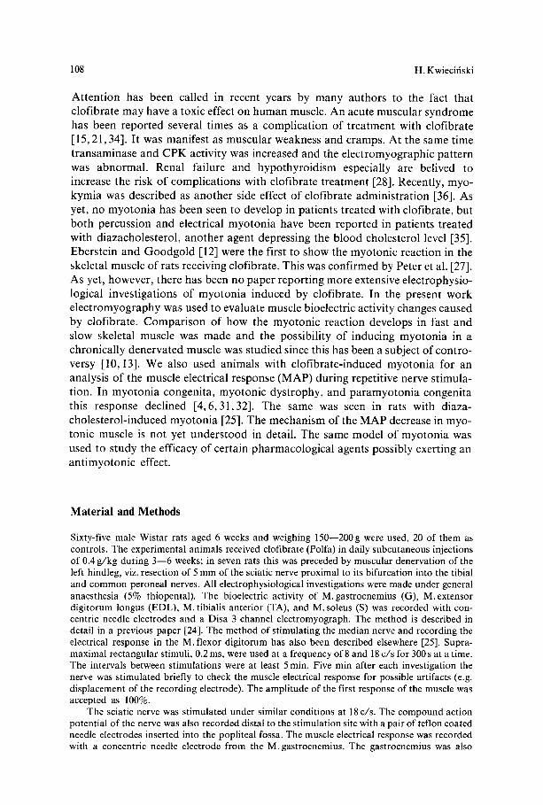
Summary. Myotonia was induced in rats with clofibrate given in daily sub- cutaneous injections of 0.4 g/kg. The first myotonic discharges were recorded electromyographically from the extensor digitorum longus, tibialis anterior, and gastrocnemius muscles after 4 days on clofibrate, but from the soleus not until after 11 days. Clofibrate induced myotonic activity in chronically de- nervated muscle also. During repetitive nerve stimulation the electrical response of the muscle was declining in all myotonic rats. It did so also when repetitive stimulation was applied directly to the muscle, which would seem to suggest a myotonic defect as the cause. Several drugs were tested and diphenylhydantoin proved to inhibit myotonia most effectively. Animals on an extended clofibrate schedule (12 weeks) had ECG abnormalities resem- bling those seen in patients with myotonic dystrophy.
Key words: Clofibrate - Experimental myotonia - Slow and fast muscle - Denervation - Repetitive stimulation - Antimyotonic drugs.
Zusammenfassung. Myotonie wurde in Ratten durch Verabreichung von Clofibrat in t~iglichen subkutanen Injektionsdosen von 0,4 g/kg erzeugt. Die ersten Entladungen wurden elektromyographisch in den Extensor-digitorum- longus-, Tibialis-anterior- und Gastrocnemius-Muskeln 4Tage nach Beginn der Clofibrat-Verabreichung registriert. Im Soleus erschienen ~ihnliche Ent- ladungen erst nach 11 Tagen. Die mit Clofibrat induzierte myotonische T~itig- keit entsteht auch in einem chronisch denervierten Muskel. Eine zunehmende Verringerung der elektrischen Antwort des Muskels (MAP) im Verlauf repeti- tiver Reizung wurde in allen Ratten mit Myotonie festgestellt. Eine ~ihnliche Verringerung der Muskel-Antwortamplitude tritt im Verlauf direkter repeti- tiver Stimulation ein, was auf einen myotonischen Defekt als Ursache hin- weisen mag. Unter mehreren untersuchten Arzneimitteln wies Diphenyl- hydantoin die wirksamsten antimyotonischen Eigenschaften auf. Ratten, denen Clofibrat l~inger verabreicht wurde (12 Wochen), wiesen EKG-St6run- gen auf, die denjenigen der Dystrophia-myotonica-Kranken ~ihnlich waren.
* This study was done under the agreement with NIH. Bethesda, Md., U.S.A., No. 05-002-N and was supported by a grant from the Muscular Dystrophy Association of America
0340-5354/78/0219/0107/$02.00
108 H. Kwiecifiski
A t t en t ion has been cal led in recent years by many au thors to the fact that c lof ibra te may have a toxic effect on human muscle. An acute muscular syndrome has been repor ted several t imes as a compl ica t ion of t rea tment with c lof ibrate [15, 21,34]. It was manifes t as muscu la r weakness and cramps. At the same t ime t r ansaminase and C P K act ivi ty was increased and the e lec t romyograph ic pa t t e rn was abnorma l . Renal failure and hypo thyro id i sm especial ly are belived to increase the risk o f compl ica t ions with c lof ibrate t rea tment [28]. Recently, myo- kymia was descr ibed as ano the r side effect of c lof ibrate admin i s t r a t ion [36]. As yet, no m y o t o n i a has been seen to develop in pat ients t rea ted with clof ibrate , but bo th percuss ion and electr ical m y o t o n i a have been repor ted in pat ients t rea ted with d iazacholes tero l , ano the r agent depressing the b lood cholesterol level [35]. Ebers te in and G o o d g o t d [12] were the first to show the myoton ic reac t ion in the skeletal muscle of rats receiving clof ibrate . This was conf i rmed by Peter et al. [27]. As yet, however , there has been no p a p e r repor t ing more extensive e lec t rophys io- logical invest igat ions o f m y o t o n i a induced by clofibrate . In the present work e l ec t romyography was used to evaluate muscle bioelectr ic act ivi ty changes caused by clof ibrate . C o m p a r i s o n o f how the myoton ic react ion develops in fast and slow skeletal muscle was made and the poss ibi l i ty o f inducing m y o t o n i a in a chronica l ly dene rva ted muscle was s tudied since this has been a subject of cont ro- versy [10, 13]. We also used animals with c lof ibra te- induced myo ton ia for an analysis o f the muscle electr ical response ( M A P ) dur ing repeti t ive nerve s t imula- t ion. In myo ton ia congeni ta , myoton ic dys t rophy , and p a r a m y o t o n i a congeni ta this response decl ined [4 ,6 ,31 ,32] . The same was seen in rats with d iaza- choles te ro l - induced m y o t o n i a [25]. The mechanism of the M A P decrease in myo- tonic muscle is not yet unde r s tood in detail . The same model o f m y o t o n i a was used to s tudy the efficacy of cer tain pha rmaco log ica l agents poss ib ly exert ing an an t imyo ton ic effect.
Material and Methods
Sixty-five male Wistar rats aged 6 weeks and weighing 150--200g were used, 20 of them as controls. The experimental animals received clofibrate (Polfa) in daily subcutaneous injections of 0.4 g/kg during 3--6 weeks; in seven rats this was preceded by muscular denervation of the left hindleg, viz. resection of 5 mm of the sciatic nerve proximal to its bifurcation into the tibial and common peroneal nerves. All electrophysiological investigations were made under general anaesthesia (5% thiopental). The bioelectric activity of M. gastrocnemius (G), M. extensor digitorum longus (EDL), M. tibialis anterior (TA), and M. soleus (S) was recorded with con- centric needle electrodes and a Disa 3 channel electromyograph. The method is described in detail in a previous paper [24]. The method of stimulating the median nerve and recording the electrical response in the M.flexor digitorum has also been described elsewhere [25]. Supra- maximal rectangular stimuli, 0.2 ms, were used at a frequency of 8 and 18 c/s for 300 s at a time. The intervals between stimulations were at least 5 min. Five min after each investigation the nerve was stimulated briefly to check the muscle electrical response for possible artifacts (e.g. displacement of the recording electrode). The amplitude of the first response of the muscle was accepted as 100%.
The sciatic nerve was stimulated under similar conditions at 18 c/s. The compound action potential of the nerve was also recorded distal to the stimulation site with a pair of teflon coated needle electrodes inserted into the popliteal fossa. The muscle electrical response was recorded with a concentric needle electrode from the M. gastrocnemius. The gastrocnemius was also

Myotonia Induced with Clofibrate in Rats 109
stimulated directly; the frequency was 18 c/s, and the MAP was recorded. The method was similar to that described by McQuillen and Johns [23]. For a statistical analysis of the mean values Student's t test was used.
Results
Myotonic Activity Induced by Clofibrate
The first myotonic discharges were recorded from G, TA, and EDL after 4 days of subcutaneous administration of clofibrate in doses of 0.4g/kg. In S they appeared only after 11 days. In all, 168 myotonic trains were recorded from the four muscles of rats receiving clofibrate for 3--6 weeks. They appeared usually in response to the insertion or slight displacement of the electrode, also after light tapping of the muscle, and only rarely spontaneously. They were usually fusi- form in shape. Extinction of myotonic activity was gradual, through reduction of the amplitude and frequency of the component potentials (Fig. 1). The electrical activity was associated by the characteristic dive bomber sound. This kind of repetitive muscle activity is considered typical of congenital myotonic syndromes in contrast to pseudomyotonia [29]. Table 1 shows the characteristics of the amplitude and frequency of myotonic trains in different muscles. The longest trains were seen in EDL (mean 1.74+ 0.42 s) and the shortest in S (mean 0.98 _+ 0.28 s). In G and TA they lasted 1.35 + 0.36s and 1.31 + 0.31 s respectively. A single intraperitoneal 0.4-g/kg dose of clofibrate caused no myotonic dis- charges, but a treble dose (1.2 g/kg) did within 5--10 min. As already mentioned, seven animals received clofibrate after denervation of the left hindleg. After 2 weeks of clofibrate administration E M G examinations were performed. A1-
sooopv
L 2ooms
5ooopv mmm- L oo°.
m 2o.~k,v
L2OOms
Fig. 1. Myotonic trains recorded from different muscles of a rat kept 3 weeks on clofibrate
110 H. Kwiecifiski
Table 1. Correlation between the amplitude and frequency characteristics of myotonic trains in different muscles. Changing pattern (increasing-decreasing) / \ ; decreasing pattern\ ; constant pattern - -
Amplitude /X \ /~ \ Others Total \ \ - _
Frequency
EDL 18 9 6 3 4 40
TA 15 11 4 4 5 39
G 19 11 8 6 7 51
S 12 12 5 4 5 38
5000,uV
' !'. I f I,L,L,I I,L~,L,L,L l II,L l LL,L,L ~_~oo~ ~ii,,,:::i;;:;ii::~!!;!!!!!!!!!!!!!i~.!!!!!t!~.s.,~
:!,4 ¢ 4:! 4 !,!t:! :! :! :!,! :! :! ! ¢ l i t . . . . . . . . . . . . . i t l i i i l l
t-144444444JJJJJJJJJJTlll/ / / / / i
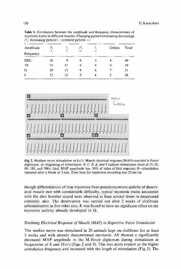
Fig. 2. Medium nerve stimulation at 8 c/s. Muscle electrical response (MAP) recorded in flexor digitorum. A--beginning of stimulation: B, C, D, E, and F indicate stimulation times of 15, 30, 60, 180, and 300s; final MAP amplitude has 50% of value of first response; R--stimulation repeated after a break of 5 min. Time base for transverse recording was 25 ms/cm
though d i f ferent ia t ion o f true m y o t o n i a f rom pseudomyo ton i c act ivi ty of denerv- a ted muscle met with cons iderable difficulty, typical myoton ic t ra ins associa ted with the dive b o m b e r sound were observed at least several t imes in denerva ted ext remi ty also. The denerva t ion was carr ied out after 2 weeks of c lof ibrate admin i s t r a t ion in five o ther rats. I t was found to have no significant effect on the myo ton ic act ivi ty a l ready deve loped in G.
Declining Electrical Response of Muscle (MAP) to Repetitive Nerve Stimulation
The median nerve was s t imula ted in 20 animals kept on c lof ibra te for at least 3 weeks and with a l ready demons t r a t ed myoton ia . Al l showed a s ignif icant ly decreased M A P ampl i tude in the M. f lexor d ig i to rum dur ing s t imula t ion at frequencies of 8 and 18 c / s (Figs. 2 and 3). This was more evident at the higher s t imula t ion f requency and increased with the length o f s t imula t ion (Fig. 3). The
Myotonia Induced with Clofibrate in Rats 111
• J + , , '"i i i J~ii~ililliiliiiilillllltllttllllflllt|tlll|llllllltNIIIllttlltlli~t:Hu I1111111111111111 IIIII111111111111111111111III111111111111111111111t1111111111111111111III11 5000~V
t, t , k k k k k+k+k+k.~,,kk, k=k L t , k+kk k t , k2ooms
...... * ................. , . . . . . . . . . . . . . . . . . . + . i . : . . ........... +,+., .................................... 1111111111111111111n111 II11111111111| I I l ! l l l l l l l l l l l l l l l l l l l l l l l l l l l l l l l l l l l ~ e ~ e ~ l l ~ r ~ l l l l l l l l l l l e ! e l ! ! l l l l l l l
++k+k+k k+k.,+++k L_,_L k.. k.k.k, k k k k k k k k k k k k k k
. . . . . . . . . . . . . . . . . . . . . . . . . . . . . . . . . . . . . . . . . . . . . . . . . . . . . . . . . . . . . . . . . . . +"fllllllllllllllllllllllllllllllllllllll
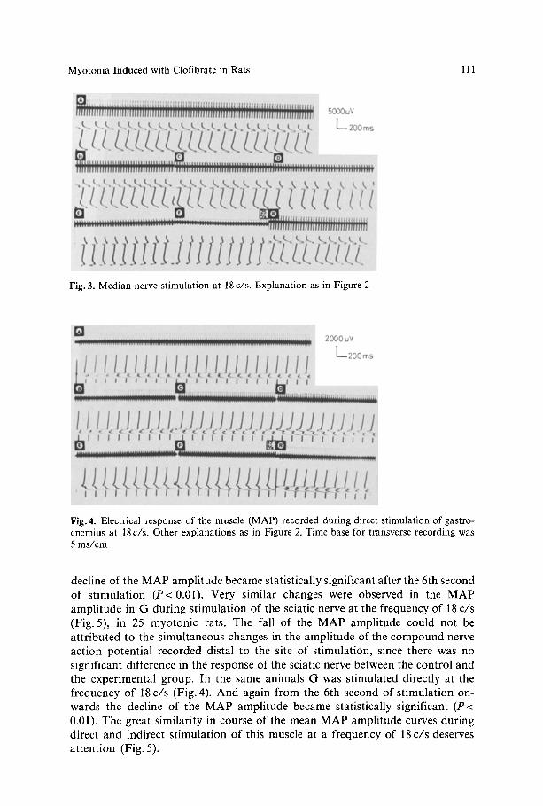
t ] ]]1 ]] ]1 u. u Fig. 3. Median nerve stimulation at 18 c/s. Explanation as in Figure 2
::::::::::::::::::::::::::::::::::::::::::::::::::::::::::::: ....................... ................ .mm 2000uV
L t / / ~ l ] !_ t ] ! / / ] ] ] ] ] ] ] i / ] ] L~2oOms
t ! / ]+1 ,!/~//,~/+].///] J ]./.JJJj~//+] / / , , + I I I i I I I I I I I I I I I I I I i I i
+.m ............................ .ell . . . . . . . . . . . . . . . . . . ~ . . . . . . .
Fig. 4. Electrical response o f the muscle (MAP) recorded dur ing direct s t imula t ion o f gas t ro- cnemius at 18 c / s . Other exp lana t ions as in Figure 2. T ime base for t ransverse recording was 5 m s / c m
decline of the MAP amplitude became statistically significant after the 6th second of stimulation (P< 0.01). Very similar changes were observed in the MAP amplitude in G during stimulation of the sciatic nerve at the frequency of 18 c/s (Fig. 5), in 25 myotonic rats. The fall of the MAP amplitude could not be attributed to the simultaneous changes in the amplitude of the compound nerve action potential recorded distal to the site of stimulation, since there was no significant difference in the response of the sciatic nerve between the control and the experimental group. In the same animals G was stimulated directly at the frequency of 18 c/s (Fig. 4). And again from the 6th second of stimulation on- wards the decline of the MAP amplitude became statistically significant (P< 0.01). The great similarity in course of the mean MAP amplitude curves during direct and indirect stimulation of this muscle at a frequency of 18c/s deserves attention (Fig. 5).
112 H. Kwiecifiski
1 O0 ~,;. a,. J '~':",'". . . . . . . . CONTROL 90
~0 J \\ "'::~"'~.,, - - MYOTONIA
Go \ \ . . . . . . . . . . . . . . .
4O
3O
2 o ~ 10 B
1530 (~0 1:~0 180 2~<)[sec] 3'00"
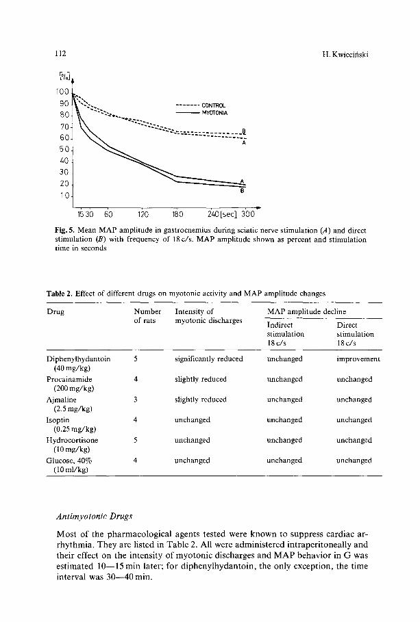
Fig. 5. Mean MAP amplitude in gastrocnemius during sciatic nerve stimulation (.4) and direct stimulation (B) with frequency of 18 c/s. MAP amplitude shown as percent and stimulation time in seconds
Table 2. Effect of different drugs on myotonic activity and MAP amplitude changes
Drug Number Intensity of MAP amplitude decline of rats myotonic discharges Indirect Direct
stimulation stimulation 18 c/s 18 c/s
Diphenylhydantoin 5 significantly reduced unchanged improvement (40 mg/kg)
Procainamide 4 slightly reduced unchanged unchanged (200 mg/kg)
Ajmaline 3 slightly reduced unchanged unchanged (2.5 mg/kg)
Isoptin 4 unchanged unchanged unchanged (0.25 mg/kg)
Hydrocortisone 5 unchanged unchanged unchanged (10 mg/kg)
Glucose, 40% 4 unchanged unchanged unchanged ( 10 ml/kg)
Antimyotonic Drugs
Most of the pha rmaco log i ca l agents tested were known to suppress card iac ar- rhy thmia . They are l isted in Table 2. Al l were admin i s te red in t raper i tonea l ly and their effect on the intensi ty o f myo ton ic discharges and M A P behavior in G was es t imated 10--15 min later; for d ipheny lhydan to in , the only except ion, the t ime interval was 30 - -40 min.
Myotonia Induced with Clofibrate in Rats 113
C O N T R O L
lOOmm/sec i l i J I T T I - T ] T F I I I !,j,L...LJ__.I_I I 1 I :1 I ; I I I
I T + i I i I I t I I I I I 1
1 1 t I I
" ! ' " t - - ~ I I t 1 I I I I I . . . . [_[ &_L i l l l
.... ' " I i Ill i III I I11 I
.... i..~._l~l I I I I I: 1 I , i , ~ I I I I I / I I 1 I
i i . . 21 . . . . . . . . . I
M Y O T O N I
1 ]T- ITT-1--1 ...... , L t l t l l t 1
I 1 I I _ l i l ] I 111121_ / . I IlL I t,. R I t l . F T'II T-I-'t" ] I-P"
! I l I t l I t
}-f-I I I I I I I ,1 t I lil l
I I | t I 1 IA:I I
l l l i I 1 1
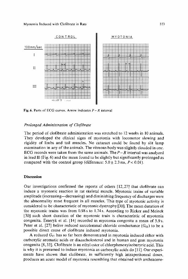
Fig. 6. Parts of ECG curves. Arrow indicates P - R interval
I I ]11 t
Prolonged Administration of Clofibrate
The period o f clofibrate administrat ion was stretched to 12 weeks in 10 animals. They developed the clinical signs o f myotonia with locomotor slowing and rigidity o f limbs and tail muscles. No cataract could be found by slit lamp examinat ion in any of the animals. The vitreous body was slightly clouded in one. E C G records were taken f rom the same animals. The P - R interval was analyzed in lead II (Fig. 6) and the mean found to be slightly but significantly prolonged as compared with the control group (difference: 5 . 0 _ 2.3 ms, P < 0.01).
D i s c u s s i o n
Our investigations c o n f r m e d the reports of others [12,27] that clofibrate can induce a myotonic reaction in rat skeletal muscle. Myotonic trains of variable ampli tude ( increasing--decreasing) and diminishing frequency of discharges were the abnormal i ty most frequent in all muscles. This type o f myotonic activity is considered to be characteristic of myotonic dys t rophy [30]. The mean durat ion o f the myotonic trains was f rom 0.98 s to 1.74 s. According to Ricker and Meinck [30] such short durat ion of the myotonic train is characteristic of myoton ia congenita. Emeryk et al. [14] recorded in myotonia congenita a mean of 5.9 s. Peter et al. [27] belive reduced sarcolemmal chloride conductance (Ga) to be a possible direct cause o f clofibrate induced myotonia .
A reduced G a has so far been demonstra ted in myotonia induced either with carboxylic aromat ic acids or diazacholesterol and in human and goat myoton ia congeni ta [8, 33]. Clofibrate is an ethyl ester of chlorphenoxyisobutyr ic acid. This is why it is presumed to induce myoton ia as carboxylic acids do [11]. Our experi- ments have shown that clofibrate, in sufficiently high intraperitoneal doses, produces an acute model o f myotonia resembling that obtained with anthracene-
114 H. Kwiecifiski
9-COOH [24]. The clofibrate induced myotonic activity took longest to show (at least 11 days) in the soleus with myotonic trains being fewest in it and relatively brief (mean: 0.98 s). In diazacholesterol induced myotonia the myotonic reaction has also been reported to be retarded in the soleus [24]. Why this slow muscle is relatively resistant to myotonia is not entirely clear. Recent investigations [22] have shown no significant difference in Ga between the slow and the fast skeletal muscle of rats. Other reports [26], however, have shown nearly 80% of the total resting Gc~ of muscle to be connected with the system of T tubules, much more developed in fast muscle, in which therefore Gc~ may be presumed to play a particularly important role. This would explain why the myotonic reaction devel- ops more quickly in it. Clofibrate induced myotonia seems to develop independ- ently of efferent neural influences. Clofibrate induced typical myotonic trains in a chronically denervated muscle, as also nerve section, did not abolish a myotonic reaction already developed by use of the drug. The development of true myotonia in denervated muscle is a much discussed problem. Brumlik et al. [7] recorded myotonic volleys from completely denervated muscles of patients. Mro~ek et al. [24] and Eberstein et al. [13] have described diazacholesterol induced myotonia in a denervated muscle. Caccia et al. [9, 10] disagree and belive diazacholesterol induces myotonia only in innervated muscles. According to Iyer et al. [18, 19] 2,4-D does not cause myotonia in a chronically denervated muscle. They belive that myotonia requires a diminuation of Ga relative to potassium conductance (GK), which is said to be impossible in a denervated muscle because of its lowered GK [18]. This explanation seems hardly acceptable in the light of recent findings of raised GK in denervated muscle [22]. It is worth stressing that Gc~ is reduced both in myotonic and in denervated muscle, and this reduction is cause enough for repetitive electrical activity [5]. Reduction of Gcl in a muscle after nerve section is a slow process and may take over 6 weeks [3]. Theoretically, chemical agents inducing myotonia, administrated in the course of the denervation pro- cess, can also lower Gc~ and thereby give rise to myotonic discharges in the denervated muscle. Adrian and Marschall [2] considered that lowered Gc~ was not alone responsible for the membrane instability of myotonic muscle. Ab- normal sodium conductance is probably another factor and may be shown by future investigations to be a still more characteristic feature of myotonia. We may expect voltage clamp studies to clarify in considerable detail the mechanism leading to myotonic discharges and ultimately settle the question whether or not myotonia can develop in a denervated muscle.
During repetitive nerve stimulation the electrical response of the muscle (MAP) diminished in all myotonic rats. The decline was continuous and more evident at the higher stimulation frequency. Why, under similar conditions, it was transient in diazacholesterol induced myotonia [25] remains obscure. Both kinds of MAP decline, the continuous and the transient, were seen by Aminoff et al. [4] in patients with myotonic disorders. It may hence be concluded to be directly connected with the myotonic defect of the muscle fiber, as is suggested by experi- ments with direct stimulations of the gastrocnemius, in which the decline of the MAP amplitude was very similar to that during nerve stimulation (Fig. 4).
Similar results were obtained by Brown [6] in a patient with myotonia con- genita. Some authors [4] belive that MAP can be regarded as the sum of action
Myotonia Induced with Clofibrate in Rats 115
potentials recorded f rom many muscle fibers. I f this is so, the decline of its amplitude in myoton ia can be explained by the lowered Gc~. Repetitive stimula- tion is known to reduce the action potential amplitude, depress the resting potential, and increase ear ly 'af ter-potent ia l in normal muscle [17]. The after- potential is part icularly marked when the muscle is stimulated in a chloride free medium [20], which is a situation very much like that in myotonic muscle, in which the decreased Gcl is the immediate cause o f after-depolarization [1].
On compar ison of several pharmacological agents, d iphenylhydantoin was found to give the best ant imyotonic effect. This agrees with the views of others [16]. In addit ion to inhibiting myoton ia it also lessened the decline o f the M A P ampli tude during direct st imulation of the muscle.
E C G records demonst ra ted abnormal atriventricular conduct ion in rats with clofibrate induced myotonia . The findings were similar in most patients with myotonic dys t rophy [16]. The electromyographic pattern o f the myotonic trains investigated, together with the E C G abnormalities, may seem to suggest a considerable similarity of clofibrate induced myotonia to myotonic dystrophy.
Nevertheless, in the light of our results clofibrate induced myotonia cannot be regarded as an animal model either o f myotonic dyst rophy or myotonia con- genita.
Fur ther studies of clofibrate induced myotonia , including electrophysiological parameters of muscle cell membrane by intracellular recording, seem to be necessary and we are going to carry out such studies.
Our investigations suggest that clofibrate is contraindicated for patients with myotonic disorders as being a myoton ia inducing agent.
Acknowledgement. The author wishes to thank Professor I. Hausmanowa-Petrusewicz for her interest, many helpful suggestions and constructive criticism.
References
1. Adrian, R. H., Bryant, S. H.: On the repetitive discharge in myotonic muscle. J. Physiol. (Lond.) 240, 505--515 (1974)
2. Adrian, R. H., Marshall, M. W.: Action potentials reconstructed in normal and myotonic muscle fibres. J. Physiol. (Lond.) 258, 125--143 (1976)
3. Albuquerque, E. X., Mclsaac, R. J.: Fast and slow mammalian muscles after denervation. Exp. Neurol. 26, 183--202 (1970)
4. Aminoff, M. J., Layter, R. B., Satya-Murti, S., Faden, A. J.: The declining electrical response of muscle to repetitive nerve stimulation in myotonia. Neurology (Minneap.) 27, 812--816 (1977)
5. Barchi, R. L.: Myotonia. An evaluation of the chloride hypothesis. Arch. Neurol. (Chic.) 32, 175--180 (1975)
6. Brown, J. C.: Muscle weakness after rest in myotonic disorders: an electrophysiological study. J. Neurol. Neurosurg. Psychiat. 37, 1336--1342 (1974)
7. Brumlik, J., Cuetter, A. C.: Denervation myotonia: a subclinical electromyographic finding. Electromyography 9, 297--310 (1969)
8. Bryant, S. H.: The electrophysiology of myotonia with review of congenital myotonia of goats. In: New Developments in electromyography and clinical neurophysiology (J. E. Desmedt, ed.), Vol. 1, p. 420. Basel: Karger 1973
9. Caccia, M. R.: Denervation and diazacholesterol myotonia: a study of the isometric twitch of the rat. Exp. Neurol. 56, 628--633 (1977)
116 H. Kwiecifiski
10. Caccia, M. R., Boiardi, A., Andreussi, L., Cornelio, F.: Nerve supply and experimental myo- tonia in rats. J. Neurol. Sci. 24, 145--150 (1975)
11. Dromgoole, S. H., Campion, D. S., Peter, J. B.: Myotonia induced by clofibrate and sodium chlorphenoxyisobutyrate. Biochem. Med. 14,238--240.(1975)
12. Eberstein, A., Goodgold, J.: Myotonia induced with clofibrate. Electromyogr. clin. Neuro- physiol. 13, 141 (1973)
13. Eberstein, A., Goodgold, J., Johnston, R.: Myotonia induced in denervated muscles. Exp. Neurol. 51,266--271 (1976)
14. Emeryk, B., Hausmanowa-Petrusewicz, I., Nowak, T.: Spontaneous volleys of bizarre high frequency potentials in neuromuscular diseases. Electromyogr. clin. Neurophysiol. 14, 339--354 (1974)
15. Geltner, D., Chaco, M., Shapiro, M.: Reversible myopathy induced by clofibrate. Postgrad. med. J. 51, 184--185 (1975)
16. Griggs, R. C., Davis, R. J., Anderson, D. C., Dove, J. T.: Cardiac conduction in myotonic dystrophy. Amer. J. Med. 59, 37--42 (1975)
17. Hanson, J. R.: The effects of repetitive stimulation on the action potential and the twitch of rat muscle. Acta physiol, scand. 90, 387--400 (1974)
18. Iyer, V., Ranish, N. A., Fenichel, G. M.: The effect of denervation on subsequent in vitro induction of myotonia. Neurology (Minneap.) 27, 669--671 (1977)
19. Iyer, V., Whiting, M., Fenichel, G. M.: Neural influence on experimental myotonia. Neuro- logy (Minneap.) 27, 73--76 (1977)
20. Kirsch, G. E., Nichols, R. A., Nakajima, S.: Delayed rectification in the transverse tubules. J. gen. Physiol. 70, 1--21 (1977)
21. Kra, S. J.: Muscle syndrome with clofibrate usage. Conn. Med. 1, 348--349 (1974) 22. Lorkovi6, H., Tomanek, R. J.: Potassium and chloride conductances in normal and de-
nervated rat muscles. Amer. J. Physiol. 232, C109--C114 (1977) 23. McQuillen, M. P., Johns, R. J.: The nature of the defect in the Eaton-Lambert syndrome.
Neurology (Minneap.) 17, 527--536 (1967) 24. Mro~'ek, K., Kwiecifiski, H., Kamifiska, A.: Experimental myotonia. Myotonic activity of
the fast and slow muscles. Acta physiol, pol. 25, 321--327 (1974) 25. Mroiek, K., Kwiecifiski, H.: Neuromuscular failure in myotonic rats. Europ. Neurol. 13,
47--54 (1975) 26. Palade, P. T., Barchi, R. L.: Characteristics of the chloride conductance in muscle fibers of
the rat diaphragm. J. gen. Physiol. 69,325--342 (1977) 27. Peter, J. B., Campion, D. S., Dromgoole, S. H., Nagatomo, T., Andiman, R. M.: Similar-
ities and differences between human myotonia and drug-induced myotonia in rats. In: Recent Advances in Myology (W. G. Bradley, ed.), p. 434. Amsterdam: Excerpta Medica 1975
28. Pierides, A. M., Alvarez-Vole, F., Kerr, D. N. S., Skillen, A. W.: Clofibrate-induced muscle damage in patients with chronical renal failure. Lancet 1975 II, 1279--1282
29. Richardson, A. T., Barwick, D. D.: Clinical electromyography. In: Disorders of voluntary muscle (J. N. Walton, ed.), p. 1003. London: Churchill Livingstone 1974
30. Ricker, K., Meinck, H. M.: Vergleich myotoner Entladungen bei Myotonia congenita und Dystrophia myotonica. Z. Neurol. 201, 62--72 (1972)
31. Ricker, K., Meinck, H. M., Stumpf, H.: Neurophysiologische Untersuchungen t~ber das Stadium passagerer Lahmung bei Myotonia congenita und Dystrophia myotonica. Z. Neurol. 204, 135--148 (1973)
32. Rowifiska-Marcifiska, K., Mro~ek, K.: Block of evoked muscle activity in myotonia. Electro- myogr, clin. Neurophysiol. 16, 483---490 (1976)
33. Rtidel, R., Senges, J.: Experimental myotonia in mammalian skeletal muscle: changes in membrane properties. Pflt~gers Arch. ges. Physiol. 331,324--334 (1972)
34. Rumpf, K. W., Albers, R., Scheler, F.: Clofibrate-induced myopathy syndrome. Lancet 1976 I, 249--250
35. Somers, J. E., Winer, N.: Reversible myopathy and myotonia following administration of a hypocholesterolemic agent. Neurology (Minneap.) 16,761--765 (1966)
36. Ter/ivainen, H., MS.kitie, J.: Myokymia, unusual side-effect of clofibrate. Lancet 1976 II, 1289
Received April 14, 1978