Embed Size (px)
Citation preview
University of KentuckyUKnowledge
Theses and Dissertations--Nutritional Sciences Nutritional Sciences
2012
DIET, BACTERIA AND INFLAMMATION:THE INTESTINAL MUCOSA ANDMETABOLIC SYNDROMENadeem K. MohammedUniversity of Kentucky, [email protected]
This Doctoral Dissertation is brought to you for free and open access by the Nutritional Sciences at UKnowledge. It has been accepted for inclusion inTheses and Dissertations--Nutritional Sciences by an authorized administrator of UKnowledge. For more information, please [email protected].
Recommended CitationMohammed, Nadeem K., "DIET, BACTERIA AND INFLAMMATION: THE INTESTINAL MUCOSA AND METABOLICSYNDROME" (2012). Theses and Dissertations--Nutritional Sciences. Paper 4.http://uknowledge.uky.edu/nutrisci_etds/4
STUDENT AGREEMENT:
I represent that my thesis or dissertation and abstract are my original work. Proper attribution has beengiven to all outside sources. I understand that I am solely responsible for obtaining any needed copyrightpermissions. I have obtained and attached hereto needed written permission statements(s) from theowner(s) of each third-party copyrighted matter to be included in my work, allowing electronicdistribution (if such use is not permitted by the fair use doctrine).
I hereby grant to The University of Kentucky and its agents the non-exclusive license to archive and makeaccessible my work in whole or in part in all forms of media, now or hereafter known. I agree that thedocument mentioned above may be made available immediately for worldwide access unless apreapproved embargo applies.
I retain all other ownership rights to the copyright of my work. I also retain the right to use in futureworks (such as articles or books) all or part of my work. I understand that I am free to register thecopyright to my work.
REVIEW, APPROVAL AND ACCEPTANCE
The document mentioned above has been reviewed and accepted by the student’s advisor, on behalf ofthe advisory committee, and by the Director of Graduate Studies (DGS), on behalf of the program; weverify that this is the final, approved version of the student’s dissertation including all changes requiredby the advisory committee. The undersigned agree to abide by the statements above.
Nadeem K. Mohammed, Student
Dr. Erik Eckhardt, Major Professor
Dr. Howard Glauert, Director of Graduate Studies
TITLE PAGE
TITLE PAGE
DIET, BACTERIA AND INFLAMMATION: THE INTESTINAL MUCOSA AND METABOLIC SYNDROME
______________________________________________________________
THESIS ______________________________________________________________
A thesis is submitted in partial fulfillment of the
requirements for the degree of Doctorate of Philosophy in the College of Nutritional Sciences at the University of Kentucky
By
Nadeem K. Mohammed
Lexington, Kentucky
Co-Directors: Dr. Erik Eckhardt, Professor of Nutritional Sciences and Dr. Deneys van der Westhuyzen, Professor of Nutritional
Sciences
Lexington, Kentucky
2012
Copyright© Nadeem K. Mohammed
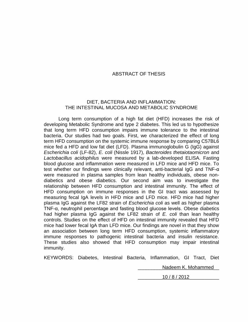
ABSTRACT OF THESIS
DIET, BACTERIA AND INFLAMMATION: THE INTESTINAL MUCOSA AND METABOLIC SYNDROME
Long term consumption of a high fat diet (HFD) increases the risk of
developing Metabolic Syndrome and type 2 diabetes. This led us to hypothesize that long term HFD consumption impairs immune tolerance to the intestinal bacteria. Our studies had two goals. First, we characterized the effect of long term HFD consumption on the systemic immune response by comparing C57BL6 mice fed a HFD and low fat diet (LFD). Plasma immunoglobulin G (IgG) against Escherichia coli (LF-82), E. coli (Nissle 1917), Bacteroides thetaiotaomicron and Lactobacillus acidophilus were measured by a lab-developed ELISA. Fasting blood glucose and inflammation were measured in LFD mice and HFD mice. To test whether our findings were clinically relevant, anti-bacterial IgG and TNF-α were measured in plasma samples from lean healthy individuals, obese non-diabetics and obese diabetics. Our second aim was to investigate the relationship between HFD consumption and intestinal immunity. The effect of HFD consumption on immune responses in the GI tract was assessed by measuring fecal IgA levels in HFD mice and LFD mice. HFD mice had higher plasma IgG against the LF82 strain of Escherichia coli as well as higher plasma TNF-α, neutrophil percentage and fasting blood glucose levels. Obese diabetics had higher plasma IgG against the LF82 strain of E. coli than lean healthy controls. Studies on the effect of HFD on intestinal immunity revealed that HFD mice had lower fecal IgA than LFD mice. Our findings are novel in that they show an association between long term HFD consumption, systemic inflammatory immune responses to pathogenic intestinal bacteria and insulin resistance. These studies also showed that HFD consumption may impair intestinal immunity.
KEYWORDS: Diabetes, Intestinal Bacteria, Inflammation, GI Tract, Diet
_________Nadeem K. Mohammed__
_________10 / 8 / 2012___________
DIET, BACTERIA AND INFLAMMATION:
THE INTESTINAL MUCOSA AND METABOLIC SYNDROME
By Nadeem K. Mohammed
__________________Dr. Erik Eckhardt_ _ Co-Director of Thesis
______ Dr. Deneys van der Westhuyzen _ Co-Director of Thesis
________________ Dr. Howard Glauert__
Director of Graduate Studies
_________________10 / 4 / 2012_______ Date

iii
ACKNOWLEDGMENTS
Earning a Doctorate in Philosophy is an achievement that few individuals
ever have the opportunity to attain. I would like to first of all thank Dr. Erik
Eckhardt for giving me the opportunity to be a member of his laboratory. My
experience working for Dr. Eckhardt exposed me to a multitude of scientific
disciplines including nutritional sciences, immunology, microbiology and
gastroenterology. I would also like to thank my dissertation committee, Dr.
Deneys van der Westhuyzen, Dr. Nancy Webb and Dr. Jerold Woodward for
providing me feedback and advice on my scientific work. I owe gratitude to
members of the following labs in the Graduate Center for Nutritional Sciences:
Dr. Vicki King’s lab, Dr. Shuxia Wang’s lab and Dr van der Westhuyzen’s lab for
loaning me reagents and equipment that was needed. I would also like to thank
John Cranfil prepared reagents and media for me in a prompt and efficient
manner. There are two labs from the Department of Microbiology and
Immunology which provided me with assistance: Dr. Woodward’s lab for
ELISPOT analysis and Dr. Charlotte Kaetzel and Dr. Maria Bruno for providing
our lab with pIgR knockout mice as well as breeding pairs for us to conduct a
crucial feeding study. I would like to extend my gratitude to Dr. Geza Bruckner
who was extremely helpful as my academic advisor. The members of my lab
Jianing Li, Dr. Lihua Tang and Yu Wang were helpful colleagues who
demonstrated techniques to me and gave me useful advice, in addition to being
wonderful friends. Finally I would like to acknowledge the Graduate Center for
Nutritional Sciences for all their support and for nominating me for the Kentucky
Opportunity Fellowship as well as the support of the NIH COBRE GRANT#
5P2ORRO21954.
iv
TABLE OF CONTENTS
Table of Contents
ACKNOWLEDGMENTS ............................................................................ iii
LIST OF TABLES ..................................................................................... vii
LIST OF FIGURES .................................................................................. viii
CHAPTER 1 INTRODUCTION .................................................................. 1
Metabolic syndrome: a global public health threat. ................................. 1
Introduction to the immune system. ........................................................ 3
Inflammation is associated with Metabolic Syndrome. ........................... 6
High fat diet consumption and inflammation. .......................................... 7
Linking intestinal bacteria, dietary fat and inflammation. ...................... 10
The GI tract as the source of inflammation. .......................................... 12
The intestinal bacteria are a source of inflammatory molecules. .......... 13
Regulation and confinement of the intestinal bacteria. ......................... 14
Adaptations of intestinal immunity. ....................................................... 16
The adaptive immune response to the intestinal bacteria. .................... 18
pIgR knockout mice: defining the role of active IgA secretion. ............. 21
Immune responses to intestinal bacteria are localized. ........................ 22
Mesenteric lymph nodes are crucial for oral tolerance. ........................ 23
Proposed hypothesis. ........................................................................... 24
Experimental approach. ........................................................................ 26
CHAPTER 2 THE EFFECT OF HIGH FAT DIET ON SYSTEMIC IMMUNITY. ......................................................................................................... 27
INTRODUCTION. ................................................................................. 27
METHODS............................................................................................ 28
PRELIMINARY ANIMAL STUDIES ................................................... 28
Long term feeding study on C57BL6 mice. ....................................... 32
HUMAN STUDIES. ............................................................................ 36
RESULTS ............................................................................................. 41
Preliminary mouse studies. ............................................................... 41
LONG TERM FEEDING STUDY IN C57BL6 MICE. .......................... 43
HUMAN STUDIES ............................................................................. 50
v
DISCUSSION ....................................................................................... 53
PRELIMINARY MOUSE STUDIES: HFD mice have higher plasma IgG against invasive intestinal bacteria. ....................................................... 53
HFD mice have higher anti-bacterial IgG .......................................... 55
Results from long term feeding study in C57BL6 mice ...................... 56
Long term HFD consumption leads to increased plasma TNF-α in mice. ............................................................................................................ 57
Plasma IgG against invasive intestinal bacteria is increased in HFD mice. ............................................................................................................ 58
Neutrophil percentage is increased in HFD mice. ............................. 59
Long term HFD consumption promotes intestinal inflammation. ....... 60
HUMAN STUDIES ............................................................................. 62
CHAPTER 3 HIGH FAT DIET CONSUMPTION AND INTESTINAL IMMUNITY. ......................................................................................................... 67
INTRODUCTION .................................................................................. 67
METHODS............................................................................................ 69
Determining the effect of long term HFD consumption on fecal IgA .. 69
Fecal IgA extraction ........................................................................... 69
Total fecal IgA measurements ........................................................... 69
Quantifying total fecal output and food intake. .................................. 70
Effect of HFD consumption on plasma IgA. ....................................... 71
To evaluate whether impaired IgA secretion increases the risk of developing metabolic syndrome. .................................................................. 71
Processing of samples for analysis. .................................................. 72
Identification of mice. ......................................................................... 72
Body weight and body fat analyses ................................................... 73
Plasma cytokine analysis .................................................................. 73
Hematological analysis. ..................................................................... 73
Oral glucose tolerance test (OGTT) .................................................. 74
Real time PCR analysis ..................................................................... 74
Statistical analyses. ........................................................................... 74
ANALYSIS OF HUMAN PLASMA IgA. ................................................. 75
RESULTS ............................................................................................. 75
HFD mice have reduced fecal IgA. .................................................... 75
HFD consumption reduces fecal output. ........................................... 76
Diet does not alter food intake. .......................................................... 76
vi
No effect of HFD consumption on plasma IgA. ................................. 78
To assess whether impaired IgA secretion into the lumen of the GI tract is a predisposing factor for development of metabolic syndrome. ........ 79
DISCUSSION ....................................................................................... 93
Long term HFD consumption decreases fecal IgA. ........................... 93
The effect of long term HFD consumption on plasma IgA ................. 94
Absence of pIgR does not exacerbate inflammation. ........................ 94
Reduced plasma IgA in obese diabetics. .......................................... 98
CHAPTER 4 DISCUSSION ................................................................... 100
EVALUATION OF PROPOSED HYPOTHESIS. ................................. 100
Significance of studies. ....................................................................... 103
Technical contributions. ...................................................................... 104
EVALUATION OF THESE STUDIES. ................................................ 105
EXPERIMENTAL DESIGN. ................................................................ 108
Alternative reasons for increased plasma anti-E. coli LF82 IgG. ........ 110
FUTURE DIRECTION OF STUDIES. ................................................. 110
Concluding statement: ........................................................................ 114
REFERENCES ...................................................................................... 116
VITA ....................................................................................................... 128
vii
LIST OF TABLES
Table 2.1 Details of Rodent Diets. ........................................................ 39
Table 2.2 Human plasma sample details. ............................................. 39
Table 2.3 List of primer sequences used for genotyping ...................... 40
Table 2.4 List of real time PCR primer sequences ............................... 40
viii
LIST OF FIGURES
Figure 1.1 IgA secretion ........................................................................... 20 Figure 1. 2 Experimental Model ............................................................... 25 Figure 2.1 Anti-E. coli LF82 IgG .............................................................. 42 Figure 2.2 Antibacterial IgG ..................................................................... 42 Figure 2.3 Comparison of LFD mice and HFD mice during a long time feeding study ........................................................................................... 46 Figure 2.4 The effect of long term HFD consumption on plasma IgG against different intestinal bacteria in C57BL6 mice. ............................... 47 Figure 2.5 Hematological analysis of LFD mice and HFD mice .............. 48 Figure 2.6 RT-PCR analysis of the ileums and colons of LFD mice and HFD mice. ................................................................................................ 49 Figure 2.7 Western Blot Analysis of plasma from human test subjects ... 50 Figure 2.8 Plasma IgG against different intestinal bacteria human subjects
........................................................................................................................... 51 Figure 2.9 Plasma TNF-α in obese human subjects ................................ 53 Figure 3.1 Effect of diet on fecal IgA. ....................................................... 77 Figure 3.2 The effect of HFD consumption on plasma IgA in C57BL6 mice.
........................................................................................................................... 78 Figure 3.3 Plasma IgA in pIgR KO mice and wild type C57BL6 controls. 79 Figure 3.4 Bodyweight and body fat analyses 82 Figure 3.5 Plasma cytokine levels. .......................................................... 83 Figure 3.6 Plasma cytokine levels. .......................................................... 84 Figure 3.7 Plasma adipokines .................................................................. 85 Figure 3.8 Anti-E coli LF82 IgG in plasma ............................................... 86 Figure 3.9 Hematological analysis ........................................................... 88 Figure 3.10 Intestinal inflammatory markers ............................................ 89 Figure 3.11 Oral glucose tolerance tests (OGTTs) .................................. 91 Figure 3.12 Human plasma IgA. .............................................................. 92 Figure 4.1 A proposed model based on our experimental findings of how the intestinal bacteria can promote inflammation and insulin resistance. ............................................................................................................... 115
1
CHAPTER 1 INTRODUCTION
Metabolic syndrome: a global public health threat.
The drastic increase in the prevalence of type 2 diabetes is a worldwide
problem, with an estimated six percent of the world’s adults categorized as
diabetic. The main impact of type 2 diabetes in the past has been in the
developed world. However this is rapidly changing as a result of the increased
affluence of developing nations. By the year 2025 it is projected that 300 million
individuals will be diagnosed with diabetes worldwide with most new cases being
from developing countries [1]. The diagnostic criteria for type 2 diabetes is a
fasting plasma glucose level >126 mg dl-1 [2]. Untreated diabetes may lead to a
variety of debilitating complications. These include coronary artery disease,
peripheral vascular disease, blindness and amputations [3]. Therefore it is no
surprise that the rise in prevalence of type 2 diabetes has been accompanied by
an increased financial burden via increased medical expenditure as well as lost
productivity [4]. As a consequence, investigators are interested in fully
characterizing the underlying causes of diabetes to develop new strategies to
prevent the development of this condition. The dramatic surge in the number of
diabetics has been attributed to the increase in the number of individuals who are
overweight and obese. According to the guidelines set by the World Health
Organization a body mass index (BMI) between twenty five and thirty qualifies an
adult as being overweight. Obesity is defined as a BMI of thirty or higher [5].
There has been a dramatic increase in the prevalence of obesity in all segments
of American society in the past four decades [6]. It is estimated that a third of the
2
adults in the United States are obese [7]. Furthermore, seven in ten American
adults can be categorized as being overweight and obese [8]. The increase in the
prevalence of overweight and obese individuals has been accompanied by the
increased prevalence of type 2 diabetes. As a result, excess body weight is an
established risk factor for developing type 2 diabetes [9]. While lifestyle choices
are a major cause for the rise in the number of diabetics it is worth noting that
not all diabetics are obese. Additionally not all obese individuals develop
diabetes, since a third of adults are obese but the prevalence of type 2 diabetes
in the total population is 8.3% [10]. There are other risk factors for developing
diabetes. These include genetic defects of beta-cell function and genetic defects
in insulin action [11].
Increased body weight results when the intake of calories exceeds the
expenditure of calories. Surplus dietary calories are converted into triglyceride
and stored. There are two major forces that have contributed to increased
average bodyweight. The first is an increase in dietary intake as larger portions of
food can be obtained at a cheaper cost. The second factor is a more sedentary
lifestyle due to automation and limited physical activity. High caloric intake and
sedentary lifestyle can predispose an individual to developing a variety of
metabolic abnormalities including elevated triglyceride levels, reduced high
density lipoprotein cholesterol levels and increased blood pressure values that
may occur simultaneously with increased body weight. This is referred to as
metabolic syndrome.
3
Metabolic syndrome (MetS), also referred to as metabolic syndrome X,
syndrome X or insulin resistance syndrome, refers to a combination of risk
factors that increases the risk of developing type 2 diabetes and cardiovascular
disease [12]. It has been estimated that metabolic syndrome affects almost one
quarter of US adults [13]. The risk factors for metabolic syndrome utilized by the
National Cholesterol Education Program (NCEP) are any three of the following
five risk factors: a waist circumference ≥ 102 centimeters (cm) in men or 88 cm in
women, triglyceride levels ≥150mg/dL, high density lipoprotein cholesterol levels
(≤50 in women, ≤40 in men), fasting blood glucose ≥100mg/dL) and systolic BP
≥130mmHg and diastolic BP ≥85mmHg [14]. The International Diabetes
Federation (IDF) criteria is waist circumference values ≥ 94 centimeters (cm) in
men, ≥ 80 cm in women and any two of the other aforementioned risk factors.
Introduction to the immune system.
Since these studies involve characterization of immune responses, it is
essential first of all to introduce some key immunology concepts. The immune
system exists to protect the host from infection by potentially harmful
microorganisms. The human body is under the constant threat of infection by
microorganisms. Microorganisms such as bacteria, viruses, fungi and parasites
are ubiquitous. Although the majority of microorganisms are harmless, some
species have the potential to cause disease and death. Furthermore, there is
also the threat of aberrant cellular replication which may lead to tumor
development. To eliminate these threats the body has devised elaborate and
diverse mechanisms that are highly specialized.
4
The entrance of microorganisms into the tissues and systemic circulation
is termed infection. There are generalized mechanisms which protect against
infection. The skin acts as a physical barrier which prevents infection. Chemical
mechanisms also act against a broad range of infectious microbes. For example,
lysozyme cleaves bacterial cell wall components. Lysozyme is present in nasal
secretions, tears, on skin and in other bodily secretions [15]. A variety of immune
cells eliminate infectious microorganisms. These include natural killer (NK) cells,
macrophages and neutrophils [16-18]. These components of the host response
are the first lines of defense against the establishment of infection. Inflammation
also eliminates certain infections and it involves leukocytes and plasma proteins.
The generalized mechanisms of immunity that eliminate microbes are collectively
referred to as innate immunity.
There are receptors which recognize molecular structures resulting from
the presence of pathogens referred to as pattern recognition receptors (PRRs).
There are membrane bound PRRs and cytoplasmic PRRs. The membrane
bound PRRs include the C-type lectin receptors and the toll like receptors
(TLRs). The C-type lectin receptors (CLRs) are present on dendritic cells and
detect carbohydrate structures on pathogens [19]. CLRs play a vital role in the
elimination of pathogens [20]. This is because detection by CLRs results in
endocytosis and degradation of pathogens [21]. This facilitates the presentation
of antigens for detection by the B and T lymphocytes. Another group of PRRs are
the toll like receptors (TLRs). TLRs detect microbial compounds present on the
surface of microbes such as lipopolysaccharide (LPS), peptidoglycan, flagellin
5
and lipotechoic acid. They also detect the genetic material of foreign
microorganisms such as single stranded RNA, double stranded RNA as well as
CpG-containing DNA. Detection of molecular structures found on
microorganisms by TLRs triggers signaling cascades that culminate in NF-κB
translocation into the nucleus and subsequent expression of genes encoding
inflammatory products [22, 23].
There are also cytosolic sensors to detect microbial products. These
include the NOD-like receptors (NLRs) [24-26]. NLRs act by eliciting the
production of inflammatory cytokines, antimicrobial peptides and interferon-β
[27]. Alternatively, NLR activation results in procaspase-1 activation and the
production of the inflammatory cytokines interleukin-1β (IL-1β) and IL-18 [28, 29]
However, many pathogenic microorganisms have devised mechanisms
that have enabled them to avoid the mechanisms of innate immunity. This has
resulted in the development of elaborate and specific mechanisms of host
immunity. These specific mechanisms of immunity which combat infections by
microorganisms are characterized as the adaptive immune response. In
addition to pathogen-specific effector mechanisms, another hallmark of the
adaptive response is immunological memory. Subsequent infection by the same
pathogen does result in disease since the immune system can mount a more
rapid response. This prevents the reoccurrence of disease. The key components
of the adaptive immune response are cells called lymphocytes that mature from
lymphoid progenitors. Lymphocytes are categorized based on the location of
6
their maturation and their effector functions. There are two kinds of lymphocytes:
B lymphocytes and T lymphocytes.
B lymphocytes refer to lymphocytes which mature in the bone marrow and
possess immunoglobulin receptors [30]. They detect antigens in soluble form via
immunoglobulin receptors. They respond to infection by secreting antibodies.
The binding of antibodies to antigens lead to their destruction via a variety of
specialized immune pathways and cells. The next group of lymphocytes is the T-
lymphocytes. They possess T-cell receptors and mature in the thymus. The T-
lymphocytes can further be subdivided on the basis of surface markers and
effector mechanisms [31]. Those that express the CD4-surface glycoprotein and
respond to exogenous peptides are the CD4 T lymphocytes. Exogenous peptides
are derived from phagocytosed antigens, e.g., extracellular protozoan parasites
and bacteria. Those that bear the CD8 glycoprotein and respond to endogenous
peptides are the CD8 T lymphocytes. Endogenous peptides are derived from
proteins synthesized within the cell cytosol. These include the proteins
synthesized in the cytosol by viruses or intracellular bacteria that have infected
host cells.
Inflammation is associated with Metabolic Syndrome.
Some of the hallmarks of metabolic syndrome including central obesity,
elevated blood glucose and hypertension have been shown to be associated with
inflammation. Inflammation is a complex host defense mechanism that results
from both internal and external stimuli [32]. Bacterial infection is a potent trigger
of inflammation. Under normal circumstances the processes involved in
7
inflammation are self-limiting. However disease may arise if inflammation
becomes continuous and chronic. A variety of disease states are associated with
chronic inflammation. These include rheumatoid arthritis, Crohn’s Disease and
systemic lupus erythematosus [33-35]. Chronic unwanted inflammation is also
associated with impaired glucose tolerance [36]. It has been reported that when
tumor necrosis factor alpha (TNF-α), an inflammatory marker is administered to
cultured cells insulin action is impaired [37]. Furthermore, enhanced insulin
sensitivity has been observed in obese mice deficient in functional TNF-α
receptors [38]. These findings suggested that the insulin signaling pathway may
be altered by TNF-α. Insulin is produced by the beta cells of the pancreas and it
is essential to metabolic pathways of carbohydrate and fat [39]. Insulin interacts
with cell surface receptors in adipose and muscle tissue. This results in
autophosphorylation and phosphorylation of the insulin receptor substrate (IRS)
family, initiating the insulin signaling pathway which culminates with glucose
uptake. TNF-α and fatty acids have been shown to inhibit the phosphorylation of
IRS-1, which in turn impairs insulin action [40, 41]. As a result deciphering the
underlying causes of chronic systemic inflammation has become an important
priority.
High fat diet consumption and inflammation.
Consumption of high levels of dietary fat are associated with the
development of the obesity, insulin resistance, elevated blood pressure and
dyslipidemia. Therefore, high fat diet consumption increases the risk of
developing metabolic syndrome [42]. There has been an appreciable increase in
8
the fat content of food. The most extreme cases can be found at some fast food
restaurants where a single serving contains more than twice the recommended
daily intake of saturated fat.
Dietary fat itself has been implicated as the culprit that promotes
inflammation. This is not surprising given the association between high fat diet
consumption and inflammatory disorders. Furthermore, various studies have
implicated high fat intake to the development of insulin resistance. Saturated fats
and monounsaturated fatty acids have been shown to be detrimental to insulin
signaling, while omega-3 fatty acids and polyunsaturated fatty acids do not
negatively affect insulin signaling [43, 44]. Furthermore, the inhibition of insulin
signaling has been attributed to increased free fatty acid levels [36]. As a
consequence, it was imperative to investigate the role of fatty acids in promoting
inflammation. Free fatty acids activated toll like receptor 4 signaling in adipocytes
and macrophages [45]. Furthermore, myristic acid, palmitic acid and oleic acid
induced interlukin-6 (IL-6) messenger RNA expression in the RAW264.7
macrophage cell line.
However, the role of fatty acids in promoting inflammation is controversial.
It seems counterintuitive that the host would mount inflammatory immune
responses against fatty acids. This is because fatty acids are integral in cell
membrane structure and serve a variety of physiologic roles. Furthermore, a
subsequent study showed that the bovine serum albumin (BSA) that had been
complexed with fatty acids in the previous study were contaminated by
lipopolysaccharide (LPS), the ligand of the toll like receptor 4 (TLR 4) [46]. Fatty
9
acids complexed with BSA elicted TLR dependent signaling. However, fatty acids
by themselves were unable to elicit TLR signaling. The findings of this study cast
serious doubt on the role of dietary fat as the causative factor for inflammation.
Adipose tissue inflammation and insulin resistance.
Adipose tissue was initially thought to be an inert triglyceride storage
depot. However, the association between obesity and conditions such as type 2
diabetes and cardiovascular disease prompted researchers to take a closer look
at adipose tissue. It was observed that use of BMI values by itself was not an
accurate means of predicting the risk of developing type 2 diabetes [47]. Visceral
adipose tissue accumulation is associated with an increased risk of insulin
resistance, whereas the subcutaneous adipose tissue depot is not associated
with an increased risk of developing insulin resistance [48-51]. A variety of
mechanisms have been proposed to explain how visceral adipose tissue
accumulation results in the development of insulin resistance.
One proposed mechanism by which visceral fat promotes insulin
resistance is the secretion of adipokines which impair insulin signaling in the liver
and muscle tissues [52]. Excess lipid accumulation also promotes the
development of insulin resistance. Saturated fatty acids may increase the
biosynthesis of ceramide which may precede insulin resistance [53]. The
conversion of triacylglycerols to diacylglyerols by adipose triglyceride lipase
(ATGL) may also result in intracellular diacylgyercerol accumulation. ATGL
activity has been shown to promote the development of insulin resistance [54].
10
Macrophage accumulation in visceral adipose tissue is another proposed
mechanism by which adipose tissue may promote insulin resistance. The
macrophages that accumulate in the adipose tissue release inflammatory
cytokines, which can impair insulin sensitivity. It has been estimated that in lean
mice and humans the percentage of macrophages is less than ten percent. This
increases to forty percent in obese individuals [55]. Additionally, it was shown
that macrophages present in the adipose tissue secrete a variety of inflammatory
markers including TNF-α, iNOS and IL-6 [56-62]. It should also be noted that the
adaptive immune response is involved in the inflammation that results from long
term HFD consumption. CD4-T lymphocytes are present in the adipose tissue
inflammation observed in mice fed a HFD [63]. Evidence suggests that the T
lymphocytes may contribute to inflammation in the visceral adipose tissue prior to
the recruitment of macrophages. Considering all these experimental findings, it is
not surprising that inflammation associated with high fat diet intake was thought
to be the result of excess adipose tissue accumulation.
Linking intestinal bacteria, dietary fat and inflammation.
Germ free mice (GF mice) were crucial in linking the intestinal bacteria to
the obesity, inflammation and insulin resistance. Germ free mice do not harbor
microorganisms [64]. This is because they are reared in sterile isolators which
prevent them from being colonized by bacteria, viruses and eukaryotic parasites.
The intestinal bacteria have been linked to the development of obesity [65].
Three groups of adult B6 male mice were analyzed; GF mice, mice colonized by
intestinal bacteria from birth to adulthood (conventionally raised, CONV-R) and
11
GF mice colonized with bacteria from the cecum (conventionalized, CONV-D).
GF mice had significantly lower body fat percentage compared to CONV-R and
CONV-D mice. Furthermore, the epididymal fat pads of GF mice weighed less
than CONV-R and CONV-D mice. These findings were intriguing since the GF
mice consumed more food and had lower metabolic rates than the CONV-R and
CONV-D mice. These findings were confirmed in a subsequent study when it
was observed that GF mice are less prone to becoming obese after long term
consumption of a high-fat, high-carbohydrate Western Diet [66]. These studies
indicated that the intestinal bacteria may play a role in the development of
obesity.
However, it was a study by Rabot et al that showed the full extent of the
effects of the intestinal bacteria. GF C57BL/6J mice and conventionally raised
(conv) C57BL/6J mice were fed a high fat diet (60% of energy from fat). The GF
mice had improved metabolic parameters compared to conventionally raised
(conv) controls [67]. These included lower fasting blood glucose levels compared
to the conventional controls on the high fat diet (conv/HF) as well as lower
plasma insulin concentrations and showed improved response to an oral glucose
challenge. GF/HF mice also had significantly lower plasma TNF-α, serum
amyloid A (SAA), leptin and IFN-γ levels. Collectively these studies implicated
the intestinal bacteria in the development of obesity and accumulation of adipose
tissue. Furthermore, they showed that the intestinal bacteria play a role in the
development of systemic inflammation and insulin resistance. However, these
studies failed to identify the point of origin of the systemic inflammation.
12
The growing body of evidence linking the intestinal bacteria to the
development of obesity and inflammation suggests that inflammation is not solely
due to adipose tissue depots. Studies had also shown that the gut microbiota
differed between obese and lean mice, as well as lean and obese human
subjects [68]. These findings suggested that the GI tract may be the source of
inflammation. This is not an unreasonable proposal. There are situations where
intestinal inflammation occurs, giving rise to a variety of debilitating conditions,
including Crohn’s Disease and Ulcerative Colitis [69].
The GI tract as the source of inflammation.
A study performed by Ding et al was the first to investigate whether high
fat diet consumption in combination with the intestinal bacteria can result in pro-
inflammatory changes within the GI tract [70]. Conventional specific pathogen
free (SPF) C57BL/6 mice and GF mice were fed a HFD (45% kcal from fat) or a
LFD (10% kcal from fat). Their findings were in accordance to previous studies
using GF mice. GF mice on a HFD gained less weight than CONV controls, were
more responsive to insulin and had lower levels of inflammatory plasma
cytokines. TNF-α levels in the ileum of CONV/HFD was significantly increased
compared to CONV/LFD mice whereas there was no increase in ileal TNF-α
levels in the GF/HFD mice. This provides evidence that the GI tract is the point of
origin of inflammation. The GI tract contains large numbers of immune cells
which regulate the intestinal bacteria in a non-inflammatory manner.
Inflammation of the GI tract can result in severe gastroenterological disorders,
collectively referred to as inflammatory bowel disease (IBD). A variety of factors
13
are associated with IBD. These include certain pathogenic bacteria as well as
defects in the immune system. For example certain strains of E. coli are
associated with Crohn’s Disease [71, 72]. With regards to the immune system,
defective sensors for molecular structures located on pathogenic microbes result
in chronic inflammation [73, 74]. These include mutations in constituents of the
inflammasomes [75]. Inflammasomes by definition are protein complexes
capable of eliciting immune responses to a variety of stimuli including bacteria
[76]. NLRP6 deficiency leads to reduced IL-18 and outgrowth of the
Bacteroidetes. This promoted the development of colitis (inflammation of the
colon).
The intestinal bacteria are a source of inflammatory molecules.
An estimated one hundred trillion (1014) microorganisms reside in the
gastrointestinal tract [77]. To put things in perspective the number of
microorganisms in the GI tract is so immense that intestinal microbes outnumber
the total number of cells in the host by a factor of ten. The overwhelming majority
of microorganisms that inhabit the GI tract are bacteria. Smaller numbers of
archea and eukarya are also present [78]. Characterization of the intestinal
bacteria remains challenging. There is considerable variability in the bacterial
composition between different individuals. Furthermore, most of the intestinal
bacterial species are anaerobic and difficult to culture using conventional
methods. Advances in molecular techniques have facilitated characterization of
the intestinal bacteria. Sequencing of the 16S ribosomal RNA gene (rRNA) led
14
researchers to identify the dominant intestinal bacterial divisions as the
Bacteroidetes and the Firmicutes [79].
Pyrosequencing, which involves the detection of chemiluminescent signals
subsequent to DNA synthesis has led researchers to identify in excess of 5,000
bacterial taxa [80]. The majority of the intestinal bacterial species belong to the
phyla Firmicutes, Bacteroidetes, Actinobacteria, Proteobacteria and
Verrucomicrobia [81]. The large and diverse bacterial population of the
gastrointestinal (GI) tract is a rich source of inflammatory molecules.
Lipopolysaccharide (LPS) is present in the outer cell membranes of the gram
negative intestinal bacteria [82]. LPS is a potent inducer of inflammation,
triggering the release of pro-inflammatory cytokines such as TNF-alpha, IL-6, and
IL-1 [83]. It should be noted that gram positive bacteria residing in the GI tract
can also trigger inflammation. Bacterial DNA from gram positive bacteria induces
inflammation [84]. Furthermore, long term HFD consumption is associated with
increased plasma LPS. Long term HFD consumption increases LPS containing
bacteria residing in the GI tract [85]. It was observed that LPS infusion results in
insulin resistance, thereby establishing a crucial link between the contents of the
GI tract, inflammation and insulin resistance.
Regulation and confinement of the intestinal bacteria.
Considering the sheer quantity of inflammatory molecules within the GI
tract, it is not surprising that a variety of mechanisms exist to prevent intestinal
bacteria from entering the systemic circulation. The intestinal epithelial cells
(IECs) form a physical barrier referred to as the intestinal epithelial-cell barrier
15
[86]. The IECs also possess a brush border, which is an actin rich microvilli
covered surface on the apical surface of the IECs. This prevents the intestinal
bacteria from adhering to the surface, thereby minimizing bacterial invasion of
the epithelial layer. Furthermore, the proteins occludin and claudin form tight
physical seals at the apical epithelial surface [87, 88]. However, it should be
noted that these so called tight junctions are not fully impermeable since the GI
tract has to absorb a variety of nutrients and ions. Goblet cells also play a role in
preventing the intestinal bacteria from adhering to the epithelial layer. Goblet
cells are differentiated epithelial cells which secrete mucus. The secretion of
mucus forms a viscous impermeable glyocalyx which prevents bacterial adhesion
to the epithelium of the GI tract [89].
The IECs do not only function as a physical barrier. They also act by
secreting a variety of host defense peptides that kill bacteria. These include
defensins and cathelicidins which have broad spectrum antibiotic activity [90, 91].
Defensins and cathelicidins are cationic proteins. These antimicrobial proteins
function by forming pores in bacterial cells walls. IECs are not the only cells that
secrete these antimicrobial peptides. Paneth cells, which are specialized
epithelial cells, secrete antimicrobial peptides [92]. In additions to defensins and
cathelicidins other anti-microbial compounds have been identified including
phospholipases, lysozyme, and Reg III-gamma [93-97].
Intestinal epithelial serum amyloid A (SAA) also plays a role in regulating
the intestinal bacteria population. SAA regulates Gram-negative bacteria present
in the lumen of the GI tract, including E. coli [98]. SAA binds to the outer
16
membrane protein A (OmpA) of E. coli, which leads to its elimination by
neutrophils [99]. SAA also promotes homeostasis with the intestinal bacteria,
since SAA deficient mice are more susceptible to dextran sodium sulfate (DSS)-
induced colitis [100] . The production of such a diverse array of antimicrobial
compounds emphasizes the importance of regulating the immense population of
bacteria residing within the lumen of the GI tract.
Adaptations of intestinal immunity.
It has been previously stated that the immense antigenic load of the GI
tract is a vast reservoir of inflammatory molecules. As a consequence a number
of mechanisms exist to prevent the huge bacterial population of the lumen of the
GI tract from entering the systemic circulation. Entrance of the immense
antigenic load into the systemic circulation has the potential to result in
catastrophic consequences including systemic shock and sepsis. Furthermore,
the GI tract is also exposed to a variety of pathogens that are ingested by the
host. The large surface area of the GI tract (approximately 300m2) makes it
especially susceptible to infection. In order to prevent bacteria from the lumen of
the GI tract from undergoing uncontrolled proliferation and entering the
circulation, the GI tract contains a vast and diverse population of immune cells.
The GI tract contains B and T lymphocytes, dendritic cells as well as the largest
reservoir of macrophages (Mψ) in the body [101]. The immune cells are located
in specialized structures including the Peyer’s Patches as well as in isolated
lymphoid follicles.
17
However, a paradox exists. On one hand, the presence of immune
defenses is required to prevent the intestinal bacteria from entering the systemic
circulation. However, it is crucial that the intestinal bacteria be regulated without
inflammation. Failure to engage bacteria without inflammation can have
catastrophic consequences that are usually observed in patients with
inflammatory bowel diseases such as ulcerative colitis (UC) and Crohn’s Disease
(CD) [102]. Furthermore it is essential that the immune system tolerate the
intestinal bacteria since some bacterial species perform vitamin K and biotin
synthesis [103-105]. Additionally, the intestinal bacteria may play a role in the
overall health of the digestive system [106]. Germ-free animals are more
susceptible to infection [107].
Although intestinal macrophages are abundant, they possess a variety of
modifications that render them inert against the bacteria in the lumen of the GI
tract. These include a lack of receptors that may activate inflammatory pathways
such as CD14, complement receptors (CR) and the triggering receptor
expressed on myeloid cells (TREM-1) [108, 109]. It was initially proposed that the
intestinal macrophages did not express toll like receptors (TLRs) [110]. However,
further analysis revealed that intestinal macrophages do in fact express toll like
receptors [111]. The reason for these macrophages existing in an anergic state is
a lack of downstream signaling molecules including MyD88, IRAK and TRAF6
[112-115]. The presence of molecules that inhibit TLR pathways such as IRAK-M
also play a role in rendering intestinal macrophages hyporesponsive [116].
Impaired TLR signaling prevents the transcription factor NF-κB from translocating
18
into the nucleus. This prevents the expression of inflammatory genes and
inflammatory cytokines.
Another mechanism of intestinal immune tolerance is the inhibition of
inflammatory cytokine production by antigen presenting cells (APCs). APCs are
cells which express major histocompatibility complex (MHC) molecules which
bind and present antigenic peptides for recognition by the T lymphocytes [117].
Dendritic cells (DCs) are specialized APCs which can induce cell mediated
immune responses by the T lymphocytes [118]. However, the DCs are also
capable of inducing tolerance to certain antigens, including those of the intestinal
bacteria. The intestinal epithelial cells (IECs) secrete certain factors which limit
inflammatory cytokine production by DCs. The most prominent of these IEC
derived factors are thymic stromal lymphopoietin (TSLP), transforming growth
factor-β (TGF-β), Interleukin-10 (IL-10) and prostaglandin E2 [119-122]. These
IEC derived factors create a microenvironment that promotes the tolerogenic
phenotype of dendritic cells.
The adaptive immune response to the intestinal bacteria.
In addition to the aforementioned generalized mechanisms, specialized
immune responses occur to prevent the intestinal bacteria from entering the
systemic circulation. Since the 1960’s, researchers have observed that the vast
majority of immunoglobulins in the external secretions belong to the
immunoglobulin A (IgA) isotype [123]. IgA exists in a monomeric form or a
polymeric form. The predominant form of secreted polymeric IgA (pIgA) is
dimeric IgA, which is comprised of two IgA subunits bridged by the J chain
19
polypeptide [124]. Plasma cells present in the lamina propria secrete dimeric IgA
[125].
The polymeric immunoglobulin receptor (pIgR) is responsible for
transepithelial transport of polymeric IgA and IgM [126]. pIgR binds and
internalizes polymeric immunoglobulin at the basolateral surface of the intestinal
epithelial cells. The bound immunoglobulin is shuttled through the cell to the
apical surface where it is secreted. It is secreted with the extracellular ligand-
binding fragment of pIgR as secretory IgA (sIgA) [127]. Adherence and invasion
of the intestinal epithelium by bacteria is minimized by secretory IgA (sIgA) and
secretory IgM (sIgM) [128-130]. IgA is the predominant immunoglobulin subclass
produced in the GI tract [131]. The IgA subclass accounts for four fifths of the
immunoglobulins produced in the duodenum and jejunum. IgA comprises ninety
percent of the immunoglobulins produced in the colon.
In addition to its established role in preventing the bacteria present in the
lumen of the GI tract from entering the systemic circulation, sIgA is essential to
preventing inflammatory responses. sIgA minimizes inflammatory immune
responses against the commensal bacteria by downregulating the expression of
the inflammatory cytokines TNF-α, IL-6, Cox-2 and IFN-γ [132]. The presence of
intestinal bacteria stimulates the production of intestinal IgA [133, 134]. Although
the vast majority of IgA is produced at the intestinal mucosa, there is also IgA
present in the plasma. Plasma IgA production is independent of intestinal IgA
production [135]. Plasma IgA is primarily monomeric and is produced by B
20
lymphocytes resident in the bone marrow [136]. Small quantities of monomeric
serum IgA enter intestinal secretions by diffusion [137].
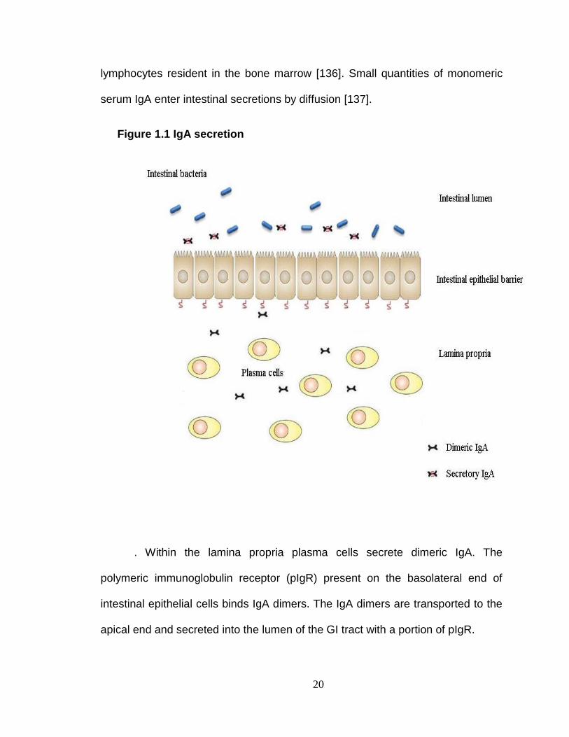
Figure 1.1 IgA secretion
. Within the lamina propria plasma cells secrete dimeric IgA. The
polymeric immunoglobulin receptor (pIgR) present on the basolateral end of
intestinal epithelial cells binds IgA dimers. The IgA dimers are transported to the
apical end and secreted into the lumen of the GI tract with a portion of pIgR.
21
pIgR knockout mice: defining the role of active IgA secretion.
Dimeric IgA and pentameric IgM exported by the polymeric
immunoglobulin receptor are not the only immunoglobulins present in the
intestinal muocsa. Serum IgA and IgG also enter intestinal mucosal secretions by
paracellular diffusion. Mice deficient in the polymeric immunoglobulin receptor
(pIgR KO mice) were generated to characterize the role of active immunoglobulin
secretion by insertion of a targeting vector into the third exon of the polymeric Ig
receptor locus (PIGR). Immunofluorescence staining of small intestine sections
revealed that pIgR KO mice have significantly reduced IgA at the epithelial
surface compared to wild type controls. However, pIgR KO mice have increased
interstitial IgA indicating that the lack of IgA at the epithelium is due to impaired
active transport, not to a defect in IgA synthesis. Comparison of serum, whole
saliva, small intestinal secretions and fecal extracts revealed a number of major
differences between pIgR KO mice with wild type controls. pIgR KO mice had
elevated serum IgA. This was the direct result of impaired IgA secretion since
Western Blot analysis revealed that the majority of this IgA was polymeric IgA.
pIgR KO mice had increased serum anti-E. coli IgG. However, there was no
difference in anti-Lactobacillus IgG between pIgR KO mice and wild type
controls. Therefore, reduced active secretion of IgA by pIgR resulted plasma IgG
being produced selectively against E. coli. IgG was higher in the small intestinal
secretions of pIgR KO mice than in the wild type controls. Fecal IgG was also
significantly higher in pIgR KO mice than in the wild type controls. This shows
22
that impairment of the mucosal barrier results in the bulk transport of IgG into the
mucosa.
Studies have been performed using pIgR KO mice to determine the role of
active immunoglobulin secretion in protection against disease. pIgR KO mice are
more susceptible to Mycobacterium bovis bacillus Calmette-Guérin (BCG)
infections than wild-type mice [138]. pIgR KO mice had higher bacterial loads in
their lungs compared to the wild-type controls. The capacity of pIgR KO mice to
produce IFN-γ and TNF-α was also significantly reduced compared to wild type
controls. pIgR KO mice are also more susceptible to nasal colonization by
Streptococcus pneumonia [139] . Wild type C57BL/6 mice had significantly less
serum IgG against intestinal bacterial antigens than pIgR KO mice. pIgR KO
mice also had increased numbers of bacteria in their mesenteric lymph nodes
than wild type controls. These findings provide evidence that active IgA secretion
is essential in the containment of bacteria to the lumen of the GI tract [140].
Immune responses to intestinal bacteria are localized.
As previously stated, immune responses are mounted against the
intestinal bacteria. These immune responses involve the generation of IgA
exclusively within the GI tract [141]. A study by Konrad et al characterized
systemic immune responses against select intestinal bacterial antigens [142].
ELISA assays revealed that the serum immunoglobulin G (IgG) response to
these antigens was negligible in C3H/HeJ mice. Furthermore, there were no
specific CD4+ T-cell responses to bacterial antigens. However, although these
mice did not have systemic immune responses against bacterial protein antigens
23
they produced intestinal IgA to the same antigens. This provides strong evidence
of a very tight compartmentalization of immunity.
Mesenteric lymph nodes are crucial for oral tolerance.
There is an abundance of antigens in the GI tract. These include bacterial
antigens as well as numerous food antigens. The oral administration of antigens
significantly impairs systemic immune responses to these same antigens if they
are administered intravenously. This phenomenon is referred to as oral tolerance
[141]. Mesenteric lymphadenectomy (the removal of mesenteric lymph nodes)
abolishes oral tolerance [143]. This provides evidence that the mesenteric lymph
nodes (MLNs) are the sites of induction of tolerance to harmless antigens
present in the GI tact. The MLNs act as barriers, preventing the dendritic cells
carrying commensal bacteria from gaining access to the systemic circulation
[144]. The inability of dendritic cells carrying commensal bacteria to enter the
systemic circulation is a major contributing factor in preventing systemic immune
responses to the intestinal bacteria.
The MLNs are situated along the route of chylomicron transport [145].
Chylomicrons are lipoprotein particles secreted by the enterocytes subsequent to
consuming a meal containing triglycerides [146]. Triacylglyceride (TAG) is the
major constituent of chylomicrons. In addition to TAGs chylomicrons contain
phospholipids, cholesterol as well as proteins [147]. Chylomicrons are comprised
of a hydrophobic core containing TAG and cholesterol esters. The hydrophilic
surface is a phospholipid monolayer in addition to cholesterol and proteins. It is
therefore possible that the long term consumption of high levels of dietary fat can
24
affect the functioning of the mesenteric lymph nodes. Atrophy of the mesenteric
lymph nodes has been observed in obese mice along with a reduction in the
numbers of regulatory T lymphocytes. Long term consumption of a high fat diet
is associated with apoptosis of the regulatory T lymphocytes within the MLNs
[148].
The regulatory T cells (Tregs) downregulate immune responses,
preventing uncontrolled immune responses [149]. A reduction in Tregs due to
long term consumption of a HFD may result in a switch from tightly regulated
immune responses to the intestinal bacteria to uncontrolled systemic
inflammation. These findings were exciting since they demonstrated that the
implications of long term HFD consumption may also extend to impairment of
immune tolerance. If this occurs then a switch from the tightly controlled non-
inflammatory responses against the intestinal bacteria to systemic inflammatory
responses against the intestinal bacteria could result. Loss of tolerance to the
intestinal bacteria being linked to metabolic syndrome was a novel concept that
we sought to investigate in a series of studies.
Proposed hypothesis.
Based on our preliminary findings and the published literature we propose
that long term consumption of a high fat diet impairs the immune tolerance to the
intestinal bacteria resulting in increased systemic inflammation. This systemic
inflammation may become chronic and lead to impaired insulin signaling and the
development of type 2 diabetes.
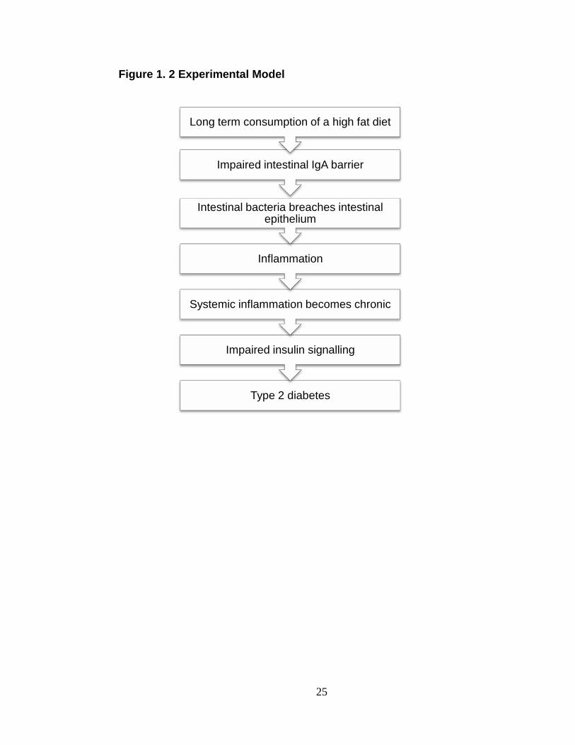
25
Type 2 diabetes
Impaired insulin signalling
Systemic inflammation becomes chronic
Inflammation
Intestinal bacteria breaches intestinal epithelium
Impaired intestinal IgA barrier
Long term consumption of a high fat diet
Figure 1. 2 Experimental Model
26
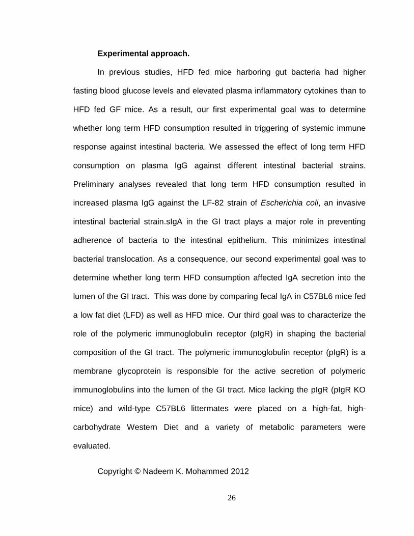
Experimental approach.
In previous studies, HFD fed mice harboring gut bacteria had higher
fasting blood glucose levels and elevated plasma inflammatory cytokines than to
HFD fed GF mice. As a result, our first experimental goal was to determine
whether long term HFD consumption resulted in triggering of systemic immune
response against intestinal bacteria. We assessed the effect of long term HFD
consumption on plasma IgG against different intestinal bacterial strains.
Preliminary analyses revealed that long term HFD consumption resulted in
increased plasma IgG against the LF-82 strain of Escherichia coli, an invasive
intestinal bacterial strain.sIgA in the GI tract plays a major role in preventing
adherence of bacteria to the intestinal epithelium. This minimizes intestinal
bacterial translocation. As a consequence, our second experimental goal was to
determine whether long term HFD consumption affected IgA secretion into the
lumen of the GI tract. This was done by comparing fecal IgA in C57BL6 mice fed
a low fat diet (LFD) as well as HFD mice. Our third goal was to characterize the
role of the polymeric immunoglobulin receptor (pIgR) in shaping the bacterial
composition of the GI tract. The polymeric immunoglobulin receptor (pIgR) is a
membrane glycoprotein is responsible for the active secretion of polymeric
immunoglobulins into the lumen of the GI tract. Mice lacking the pIgR (pIgR KO
mice) and wild-type C57BL6 littermates were placed on a high-fat, high-
carbohydrate Western Diet and a variety of metabolic parameters were
evaluated.
Copyright © Nadeem K. Mohammed 2012
27
CHAPTER 2 THE EFFECT OF HIGH FAT DIET ON SYSTEMIC
IMMUNITY.
INTRODUCTION.
Studies showing improved metabolic parameters in GF mice prompted an
investigation as to whether the bacteria residing in the GI tract are responsible for
the development of inflammation and insulin resistance. As previously
mentioned, immune responses against the bacteria in the GI tract are generated
at the intestinal mucosal surface. Theoretically the systemic immune cells are
oblivious to the presence of intestinal bacteria. Serum immunoglobulin G (IgG)
and CD4+ T-cell specific responses against intestinal bacterial antigens are
negligible.
To gain a more comprehensive understanding of the effects of long term
HFD consumption, pilot studies were performed using the plasma from BALBc
mice fed a high fat diet (Research Diets Inc. D12492 Rodent Diet with 60% kCal
fat) and mice fed a low fat diet (Open Source Diets D12450B Rodent Diet with 10
kcal% fat) for 10 weeks. The pilot analyses involved measuring plasma IgG
against the LF82 strain of E. coli, an intestinal bacterial strain. Plasma IgG was
evaluated by Western Blot analysis, as well as by ELISA analysis. After
performing the preliminary studies in mice, a 10 week feeding study was
performed on C57BL6 mice fed a HFD and a LFD. Plasma IgG against protein
extracts from E. coli LF82, E. coli Nissle 1917 (EcN), Bacteroides
thetaiotaomicron (B. thetaiotaomicron) and Lactobacilus acidophilus (L.
acidophilus) were measured by a lab-developed ELISA. Plasma TNF-α, blood
28
cell composition and fasting blood glucose levels were measured in order to
determine whether a link existed between plasma IgG against intestinal bacteria,
systemic inflammation and elevated fasting blood glucose.
To determine whether the findings in the animal studies were clinically
relevant, plasma samples from human subjects were also analyzed. Plasma IgG
against E. coli LF82, E. coli Nissle 1917 (EcN), Bacteroides thetaiotaomicron (B.
thetaiotaomicron) and Lactobacilus acidophilus (L. acidophilus) was measured in
lean healthy controls as well as obese non-diabetics and obese diabetics.
Plasma TNF-α was also measured in human plasma samples to determine
whether a relationship existed between plasma IgG against intestinal bacteria
and inflammation. The findings of this study can be found in the journal
Metabolism: Clinical and Experimental. The title of the paper is Elevated IgG
levels against specific bacterial antigens in obese patients with diabetes
and in mice with diet-induced obesity and glucose intolerance. The article is
available online and it is currently still in press. Permission to reproduce this data
was obtained.
METHODS.
PRELIMINARY ANIMAL STUDIES
The preliminary studies involved the analysis of plasma from eight BALB/c
mice. Four of the mice had been placed on a high fat diet (Research Diets Inc.
D12492 Rodent Diet with 60% kCal fat) and the other four had been placed on a
low fat diet (Open Source Diets D12450B Rodent Diet with 10 kcal% fat) for ten
29
weeks. Western blots were performed to determine whether long term HFD
consumption was associated with increased plasma IgG against the intestinal
bacteria.
Bacterial cultures.
The LF82 strain of the gut microbe Eschericha coli was cultured overnight
in Luria-Bertani Medium (LB Medium) at 36.9°C in an Isotemp Incubator (Fisher
Scientific).
Preparation of bacterial proteins.
In order to obtain bacterial proteins, approximately 5 ml of bacterial
cultures were centrifuged at 10,000 rpm for ten minutes. The supernatant was
discarded and the bacterial pellet obtained was re-suspended in two hundred
and fifty microliters (250µl) 4X SDS and seven hundred and fifty microliters
(750µl) water. The re-suspended bacterial pellet was then boiled for ten minutes.
Western Blot Analysis of anti-E. coli LF82 IgG.
The E. coli proteins were separated on NuPAGE® 4-12% Bis Tris Gels and
then transferred by an Invitrogen iBlot® Gel Transfer Device to PVDF
membranes. The membranes were blocked with non-animal protein (NAP)
blocking solution diluted 1:2 with 1X femto-TBST. The PVDF membranes were
cut into individual strips. Each membrane strip was incubated overnight in
plasma from an individual mouse that had been diluted 1:10 in the blocking
solution. The membrane strips were then washed with 1X Tris-Buffered Saline
Tween-20 (TBST). Anti-Mouse IgG (Fc specific)-Peroxidase antibody was diluted
30
1:5000 to detect the presence of anti-E.coli IgG. The membrane strips were then
washed again with TBST and then simultaneously exposed to ECL Western
Blotting Substrate. The western blots were then visualized on the Kodak Image
Station 440.
Western Blot Analysis of plasma anti-bacterial IgG.
The intestinal bacteria are poorly characterized. To determine whether
long term HFD consumption resulted in increased systemic immune responses to
the intestinal bacteria, the contents of the cecum were isolated. The contents of
the cecums were removed from each of the LFD mice, pooled and added to
750µl of water and 250µl 4X SDS loading buffer. The mixture was boiled for
approximately ten minutes. The mixture was then centrifuged at 10,000 rpm for
ten minutes. The pellet was discarded while the supernatant containing the cecal
antigens was kept for analysis. The cecal antigens were separated on NuPAGE®
4-12% Bis Tris Gels and then transferred by an Invitrogen iBlot® Gel Transfer
Device to PVDF membranes.
Each membrane was cut into four individual strips. Each individual
membrane strip was placed in non-animal protein (NAP) blocking solution diluted
1:2 with 1X femto-TBST. Each membrane strip was incubated overnight in
plasma from an individual mouse that had been diluted ten times in the blocking
solution. The membrane strips were then washed with Tris-Buffered Saline
Tween-20 (TBST) [1X]. Anti-Mouse IgG (Fc specific)-Peroxidase antibody was
diluted 1:5000 to detect the presence of anti-E.coli IgG. The membrane strips
were then washed again with TBST and then simultaneously exposed to ECL
31
Western Blotting Substrate. The western blots were then visualized on the Kodak
Image Station 440.
Development of a novel ELISA technique to quantify anti-bacterial
IgG.
We wanted to assess whether long term HFD consumption resulted in
quantitative differences in IgG against bacterial proteins. To do this we
developed a novel enzyme-linked immunosorbent assay (ELISA) to measure IgG
against the bacteria used in these studies: E. coli LF82, E. coli Nissle 1917,
Bacteroides thetaiotaomicron and Lactobacilus acidophilus. Soluble proteins
from these bacteria were extracted using B-PER Bacterial Protein Extraction
Reagents (ThermoSCIENTIFIC). Bacteria cultures were centrifuged at 5000 × g
for 10 minutes.
The supernatant was discarded and the remaining pellet was weighed. B-
PER Reagent was added at a ratio of 4ml/gram of cell pellet. Lysozyme and
DNase I were added at a ratio of 2μL/mL of B-PER Reagent in order to obtain a
higher concentration of soluble proteins. The suspension was incubated at room
temperature for 15 minutes and then centrifuged at 15,000 × g for 5 minutes. The
supernatant containing soluble bacterial proteins was used for preparing the
ELISA plates. Bacterial proteins were quantified using the BCA™ Protein Assay
(Thermo Scientific) and dissolved in a carbonate coating buffer of pH 9.6 to give
a final concentration of 100µg/ml. 96 well flat-bottom ELISA plates (BD-Falcon)
were coated with 10μg of bacterial protein per well and incubating the plate
overnight at 4ºC. Details of how the ELISAs were performed are given below.
32
Long term feeding study on C57BL6 mice.
Twelve six week old male C57BL6 mice were ordered at 5 weeks of age
from Jackson Laboratories. The mice were housed three per cage and
maintained in a 12hour light/dark cycle. We allowed the mice to become
acclimated to their environment. We then marked them for identification using
tail markings. We measured the body weight and fasting blood glucose values
prior to switching these mice from regular chow diets to special diets.
The special diets of the mice were open formula, open source purified
ingredients. The mice were divided into two equal groups (n = 6). One group was
fed a high fat diet (Rodent Diets with 60 kcal% Fat D12492 from Research Diets
Incorporated). The other group was fed a low fat diet (Rodent Diets with 10
kcal% Fat D12450B from Research Diets Incorporated). Details of these diets
are listed in table 2.1. The mice were placed on these diets at six weeks of age
and euthanized after 10 weeks on the special diets.
Measurement of body weights and fasting blood glucose
The mice were weighed weekly to monitor changes in bodyweights. To
assess fasting glucose levels, mice were fasted for 4 hours. Blood was collected
from the tail tips of the mice. Blood glucose levels were assessed using the
TRUEtrack® glucose meter (Home Diagnostics Inc.).
33
Measurement of plasma anti-bacterial IgG.
The ELISA developed by this laboratory was used to measure plasma IgG
against intestinal bacteria. 96 well flat-bottom ELISA plates (BD-Falcon) were
coated with 10μg of bacterial protein per well. Plates were incubated overnight at
4ºC. The wells were washed five times with 1X Tris-Buffered Saline Tween-20
(TBST). Each washing step was performed for duration of five minutes. The
plates were then blocked for one hour at room temperature by adding 250µl of
NAP Blocking reagent diluted 1:2 in 1X TBST to each well. 100µl of plasma
diluted 1:100 in blocking solution was then added to each well and incubated for
2 hours at room temperature. The plates were then washed five times with 1X
Tris-Buffered Saline Tween-20 (TBST). Bound IgG was detected by adding
100µl/well of alkaline phosphatase-conjugated anti-mouse IgG (Fc specific) from
Sigma-Aldrich diluted 1:5000 in blocking buffer for 1 hour. The plates were then
washed five times with 1X Tris-Buffered Saline Tween-20 (TBST). 50µl/well of p-
nitrophenyl phosphate (pNPP) from Sigma-Aldrich) was added, and the color
reaction was stopped with 2M sulfuric acid. Absorbance values were read at
optical density 405nm (A405) in a Bio-Rad microplate reader.
Processing of samples.
Mice were humanely euthanized using carbon dioxide followed by cervical
dislocation. Blood was immediately collected by cardiac puncture and placed into
EDTA containing eppendorf tubes that had been kept on ice. The majority of
blood obtained was immediately centrifuged at 8,000 rpm for 10 minutes. The
34
plasma was collected and stored at -82°C. The remaining whole blood fractions
were used to characterize the blood cell types present by hematological analysis.
Plasma TNF-α measurements.
To assess the levels of circulating TNF-α we utilized the MILLIPLEX®
Mouse Cytokine Kit (from Millipore Catalog # MPXMYCTO-70K). The data was
read in a Bio-Plex® 200 system (BIO-RAD).
Hematological analysis.
Subsequent to euthanasia approximately 50µl of whole blood was
collected from the mice by cardiac puncture. The tubes containing the whole
blood was briefly rocked. The whole blood was then analyzed using a
HEMAVET® 950 FS Multispecies Hematology Systems (Drew Scientific Inc.).
This is a device which gives comprehensive hematology profiles using 20 µL of
whole blood from different animals including mice. The hematology profiles given
are for both leukocytes and erythrocytes.
The leukocyte parameters given are white blood cell count, absolute
number and percentage of neutrophils, absolute number and percentage of
lymphocyte, absolute number and percentage of monocyte, absolute number and
percentage of eosinophil, and absolute number and percentage of basophil. The
erythrocyte parameters given are red blood cell count, hemoglobin, hematocrit,
mean cell volume, mean corpuscular hemoglobin concentration and red cell
distribution width. Additionally thrombocyte parameters including platelet counts
and mean platelet volume are given.
35
Measuring plasma IgG against intestinal bacteria.
Cultures of E. coli Nissle 1917, Bacteroides thetaiotaomicron and
Lactobacilus acidophilus were kindly provided by Dr. Charlotte Kaetzel
(Department of Microbiology, Immunology and Molecular Genetics, University of
Kentucky). These bacteria had been obtained from the American Type Culture
Collection (ATCC). Proteins were extracted from E. coli Nissle 1917, Bacteroides
thetaiotaomicron and Lactobacilus acidophilus as well as the LF82 strain of E.
coli. The ELISA developed by this laboratory was used to measure plasma IgG
against these intestinal bacteria. Ninety six well flat-bottom ELISA plates (BD-
Falcon) were coated overnight with 100µl of the bacterial protein extracts.
Plasma from the LFD mice and the HFD mice was diluted 1:100 to quantify IgG
against E. coli LF82, E. coli Nissle 1917, Bacteroides thetaiotaomicron and
Lactobacilus acidophilus.
To determine if inflammation originates in the GI tract.
RNA was extracted from ileum and colon using using the E.Z.N.A® Total
RNA Kit. We used the Thermo Scientific NanoDrop 2000 spectrophotometer
(Thermo Scientific) to measure RNA concentration. The qScript™ cDNA
Synthesis Kit was used to generate cDNA. We analyzed the intestinal samples
for expression of inflammatory genes. The genes we examined were Chemokine
(C-C motif) ligand 5 (CCL5 also known as RANTES), NOD-like receptor family
pyrin domain containing 6 (NLRP6), IL-18 and thymic stromal lymphopoietin
(TSLP) expression using a BIORAD CFX96™ Real Time PCR detection system.
Gene expression was normalized to Glyceraldehyde 3-phosphate
36
dehydrogenase (GAPDH). The reason for examining NLRP6, IL-8 and CCL5 was
due to their association with inflammatory responses against intestinal bacteria.
TSLP was examined since TSLP secreted by the intestinal epithelial cells
promote tolerance to the intestinal bacteria. The primer sequences used for RT-
PCR analysis of these genes are listed in table 2.4.
HUMAN STUDIES.
Human plasma samples from a Centers of Biomedical Research
Excellence (COBRE) pilot study “The influence of SAA and CETP activity on
HDL remodeling during active weight loss” were kindly provided by Dr. Anisa
Jahangiri. All of the plasma samples obtained from this study were from obese
patients (BMI ≥ 30). One group of the obese patients had not been diagnosed
with type 2 diabetics (obese non-diabetics). The other group of obese patients
had been diagnosed with type 2 diabetes (fasting plasma glucose ≥126 mg/dl).
Plasma samples from non-diabetic individuals with BMI values in the healthy
range (BMI ≥ 20 and ≤25) were obtained commercially from Biospecialty Corp.
(Colmar, PA, USA) for experimental controls. The study was approved by the
Institutional Review Board (IRB) at the University of Kentucky. Plasma samples
were stored at -82°C and not subjected to freeze thaw cycles. Details of the
human plasma donors are given in table 2.2.
37
Western Blot Analysis of human plasma.
Proteins were then extracted from Escherichia coli and separated on
NuPAGE® 4-12% Bis Tris Gels as previously described. After the proteins were
transferred to PVDF membranes, the membranes were placed in non-animal
protein (NAP) blocking solution diluted 1:2 with 1X femto-TBST. The PVDF
membranes were sliced into individual strips and incubated with plasma samples
from ten healthy control individuals as well as six obese non-diabetics and six
obese diabetics.
The plasma samples were diluted 1:400 in the blocking solution and
incubated overnight at 4°C. The strips were then washed 5 times in 1X TBST.
The membrane strips were incubated in anti-human IgG (Fc specific)-Peroxidase
antibody diluted 1:5000 in the blocking solution for one hour. The membrane
strips were washed five times in 1X TBST. ECL Western Blotting Substrate was
simultaneously added to each of the membrane strips and imaging was
performed on the Kodak Image Station 440.
Measurement of anti-bacterial IgG.
IgG against E. coli Nissle 1917, Bacteroides thetaiotaomicron and
Lactobacilus acidophilus and E. coli LF82 was measured in plasma samples from
the lean healthy controls, obese non-diabetics and obese diabetics by lab
developed ELISA. Human plasma samples were diluted 1:400 in blocking
solution.
38
Bound IgG was detected by adding 100µl/well of alkaline phosphatase-
conjugated anti-human IgG (Fc specific) from Sigma-Aldrich diluted 1:5000 in
blocking buffer for 1 hour. 50µl/well of p-nitrophenyl phosphate (pNPP) from
Sigma-Aldrich) was added, and the color reaction was stopped with 2M sulfuric
acid. Absorbance values were read at optical density 405nm (A405) in a Bio-Rad
microplate reader.
Measurement of TNF-α
Plasma TNF-α was measured in the obese non-diabetics and the obese-
diabetics using the Human TNF-alpha High Sensitivity ELISA from eBioscience
(BMS223HS; sensitivity 0.13 pg/mL). 50 μl of human plasma was added in
duplicate. The human plasma samples were diluted in 50 μl of sample diluent.
Statistical analyses.
Results were expressed as mean ± S.E.M and were analyzed with
GraphPad Prism v5.04. Groups were compared with unpaired Student’s t-tests
and ANOVA and Bonferroni’s post-hoc analysis. Statistical significance was
assumed when p<0.05.
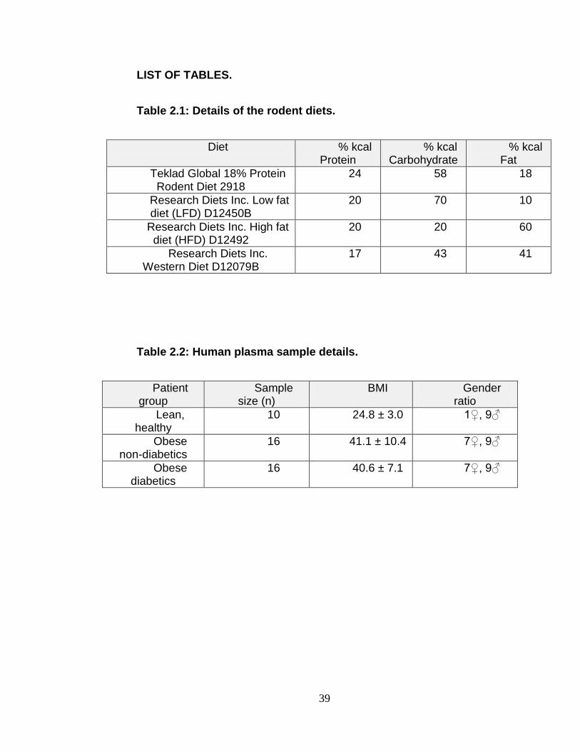
39
LIST OF TABLES.
Table 2.1: Details of the rodent diets.
Diet % kcal Protein
% kcal Carbohydrate
% kcal Fat
Teklad Global 18% Protein Rodent Diet 2918
24 58 18
Research Diets Inc. Low fat diet (LFD) D12450B
20 70 10
Research Diets Inc. High fat diet (HFD) D12492
20 20 60
Research Diets Inc. Western Diet D12079B
17 43 41
Table 2.2: Human plasma sample details.
Patient group
Sample size (n)
BMI Gender ratio
Lean, healthy
10 24.8 ± 3.0
1♀, 9♂
Obese non-diabetics
16 41.1 ± 10.4
7♀, 9♂
Obese diabetics
16 40.6 ± 7.1
7♀, 9♂
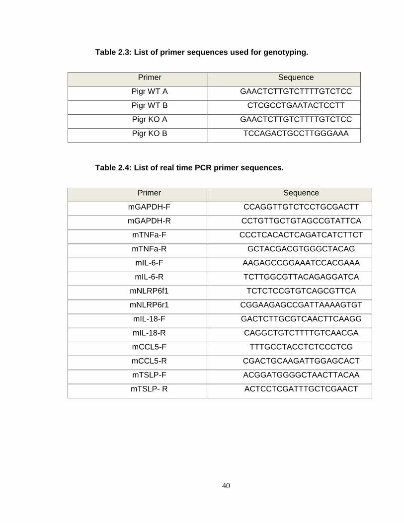
40
Table 2.3: List of primer sequences used for genotyping.
Primer Sequence
Pigr WT A GAACTCTTGTCTTTTGTCTCC
Pigr WT B CTCGCCTGAATACTCCTT
Pigr KO A GAACTCTTGTCTTTTGTCTCC
Pigr KO B TCCAGACTGCCTTGGGAAA
Table 2.4: List of real time PCR primer sequences.
Primer Sequence
mGAPDH-F CCAGGTTGTCTCCTGCGACTT
mGAPDH-R CCTGTTGCTGTAGCCGTATTCA
mTNFa-F CCCTCACACTCAGATCATCTTCT
mTNFa-R GCTACGACGTGGGCTACAG
mIL-6-F AAGAGCCGGAAATCCACGAAA
mIL-6-R TCTTGGCGTTACAGAGGATCA
mNLRP6f1 TCTCTCCGTGTCAGCGTTCA
mNLRP6r1 CGGAAGAGCCGATTAAAAGTGT
mIL-18-F GACTCTTGCGTCAACTTCAAGG
mIL-18-R CAGGCTGTCTTTTGTCAACGA
mCCL5-F TTTGCCTACCTCTCCCTCG
mCCL5-R CGACTGCAAGATTGGAGCACT
mTSLP-F ACGGATGGGGCTAACTTACAA
mTSLP- R ACTCCTCGATTTGCTCGAACT
41
RESULTS
Preliminary mouse studies.
HFD mice have higher plasma IgG against invasive intestinal
bacteria.
To investigate whether a high fat diet is associated with systemic immune
responses against the intestinal bacteria, we performed Western Blots to
determine whether there is an association between long term (10 weeks)
HFD consumption and immunoglobulin G (IgG) against invasive intestinal
bacteria. Our data showed that the plasma of the mice on the HFD had
higher levels of IgG against antigens from invasive intestinal bacteria than
the mice on the LFD (Figure 2.1).
HFD mice have higher anti-bacterial IgG.
Western blot analysis was then performed to compare plasma IgG against
the antigens from the cecums of LFD mice. Plasma IgG against the
antigens present in the cecum were low in each of the LFD mice (left
panel Figure 2.2). Plasma IgG against the antigens present in the cecum
was high in each of the individual HFD mice.
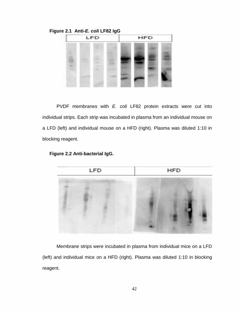
42
Figure 2.1 Anti-E. coli LF82 IgG
PVDF membranes with E. coli LF82 protein extracts were cut into
individual strips. Each strip was incubated in plasma from an individual mouse on
a LFD (left) and individual mouse on a HFD (right). Plasma was diluted 1:10 in
blocking reagent.
Figure 2.2 Anti-bacterial IgG.
Membrane strips were incubated in plasma from individual mice on a LFD
(left) and individual mice on a HFD (right). Plasma was diluted 1:10 in blocking
reagent.
43
LONG TERM FEEDING STUDY IN C57BL6 MICE.
Increased weight gain and fasting blood glucose in HFD mice.
As expected, HFD fed mice subsequently gained significantly more weight
over the duration of the study than the mice on the LFD. Six weeks after
commencing the special diets we observed a statistically significant difference
between the bodyweights of LFD and HFD mice (Figure 2.3 A). Fasting blood
glucose was measured using the TRUEtrack® glucose meter. After 10 weeks on
the special diets, HFD mice had significantly higher fasting blood glucose levels
(Figure 2.3 B, p<0.05).
Long term HFD consumption leads to increased plasma TNF-α in
mice.
Plasma obtained from mice by cardiac puncture after the 10 week feeding
regimen was analyzed for total TNF-α by the MILLIPLEX® Mouse Cytokine Kit.
HFD mice had higher plasma TNF-α than LFD mice. The difference between the
plasma TNF-α in the HFD mice and the LFD mice was significant (Figure 2.3 C,
p<0.05).
Plasma IgG against invasive intestinal bacteria is increased in HFD
mice.
HFD mice had significantly higher plasma IgG against protein extracts
from the LF82 strain of E. coli (Figure 2.4 A). However statistical analysis
revealed that there were no significant differences between LFD and HFD mice
44
in plasma IgG against E. coli Nissle 1917, Bacteroides thetaiotaomicron and
Lactobacilus acidophilus (Figures 2.4 B, 2.4 C and 2.4 D).
Neutrophil percentage is increased in HFD mice.
The HEMAVET® 950 FS Multispecies Hematology System was used in
order to measure the inflammatory immune cell subsets. It was observed that the
HFD mice had a higher percentage of neutrophils compared to LFD mice.
Statistical analysis revealed that the difference between both groups was
significant (Figure 2.5 A, p < 0.05). Lymphocyte percentage in the HFD mice was
significantly lower than in the LFD mice (Figure 2.5 B, p < 0.05). No differences
were observed between the LFD mice and the HFD mice in the percentages of
the monocytes, eosinophils and basophils.
Long term HFD consumption promotes intestinal inflammation.
Real-time quantitative PCR was performed in order to determine whether
inflammation was present in the GI tract of HFD mice. A variety of pro-
inflammatory genes were analyzed. These included interleukin-18 (IL-18),
chemokine (C-C motif) ligand 5 (CCL5) and the NOD-like receptor family pyrin
domain containing 6 (NLRP6). These genes were chosen since they encode
constituents associated with specialized inflammatory complexes against
intestinal bacteria referred to as inflammosomes. The ileum and colon sections
were of special interest since they harbor the highest densities of intestinal
bacteria. There were no differences in IL-18 expression in the colon (Figure 2.6
A). However a trend towards increased CCL5 expression in the ileum was
observed (Figure 2.6 B, p =0.19). There was also a trend towards increased
45
NLRP6 expression in the ileum of HFD mice (Figure 2.6 C, p = 0.1154). Thymic
stromal lymphopoietin (TSLP) expression was measured to determine whether
tolerance to the intestinal bacteria was impaired by long term HFD consumption.
TSLP is secreted by the intestinal epithelial cells (IECs) to prevent the dendritic
cells from promoting inflammatory responses. A trend towards decreased TSLP
expression in the colon of HFD mice was observed (Figure 2.6 D, p = 0.1857).
Collectively the trend towards increased inflammation and decreased tolerance
provides evidence that the systemic inflammation observed in HFD mice
originates in the GI tract.
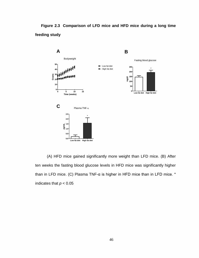
46
Figure 2.3 Comparison of LFD mice and HFD mice during a long time
feeding study
Time (weeks)
Gra
ms
0 5 10 150
10
20
30
40
50Low fat diet
High fat diet
Bodyweight
mg
/dl
Low fat diet High fat diet0
50
100
150
200
250*
Fasting blood glucose
pg
/mL
Low fat diet High fat diet0.0
0.5
1.0
1.5
2.0
2.5*
Plasma TNF-
A B
C
(A) HFD mice gained significantly more weight than LFD mice. (B) After
ten weeks the fasting blood glucose levels in HFD mice was significantly higher
than in LFD mice. (C) Plasma TNF-α is higher in HFD mice than in LFD mice. *
indicates that p < 0.05
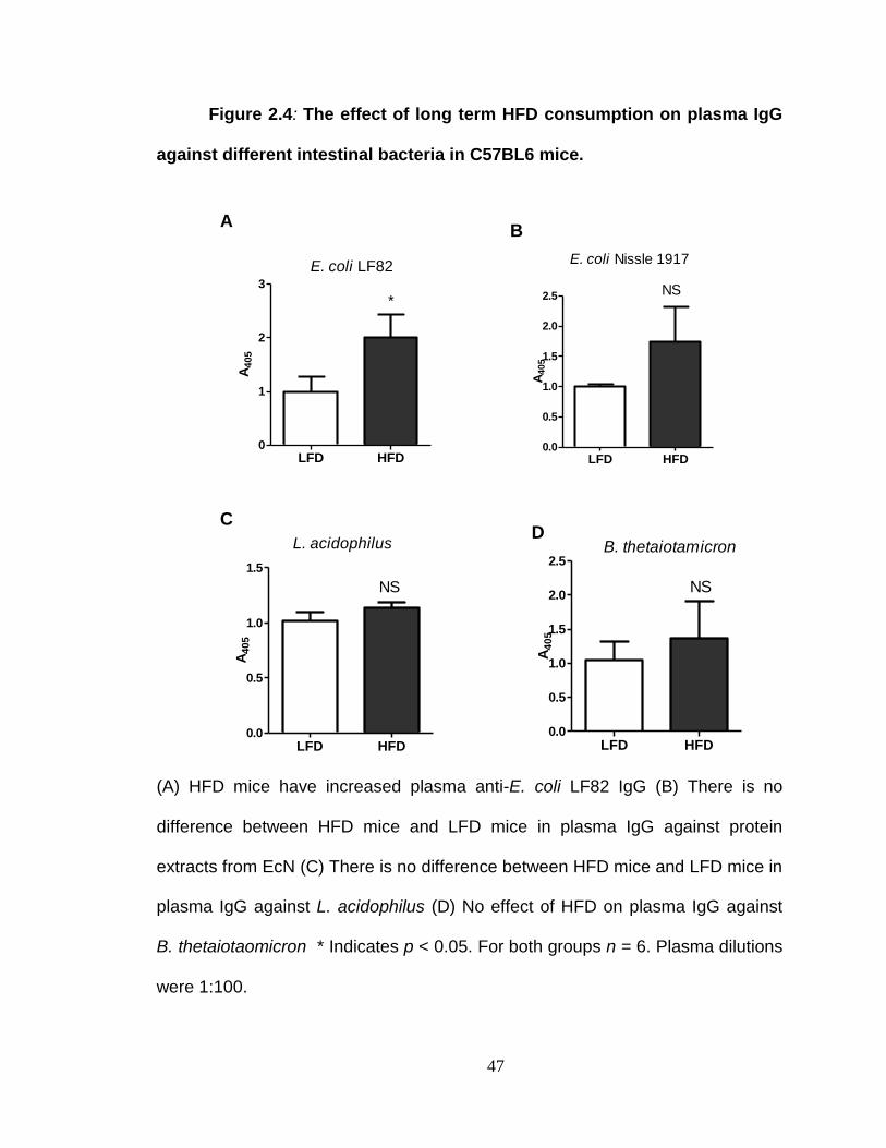
47
Figure 2.4: The effect of long term HFD consumption on plasma IgG
against different intestinal bacteria in C57BL6 mice.
A405
LFD HFD0
1
2
3
*
E. coli LF82
A4
05
LFD HFD0.0
0.5
1.0
1.5
2.0
2.5 NS
E. coli Nissle 1917
A405
LFD HFD0.0
0.5
1.0
1.5
NS
L. acidophilusA
40
5
LFD HFD0.0
0.5
1.0
1.5
2.0
2.5
NS
B. thetaiotamicron
AB
CD
(A) HFD mice have increased plasma anti-E. coli LF82 IgG (B) There is no
difference between HFD mice and LFD mice in plasma IgG against protein
extracts from EcN (C) There is no difference between HFD mice and LFD mice in
plasma IgG against L. acidophilus (D) No effect of HFD on plasma IgG against
B. thetaiotaomicron * Indicates p < 0.05. For both groups n = 6. Plasma dilutions
were 1:100.
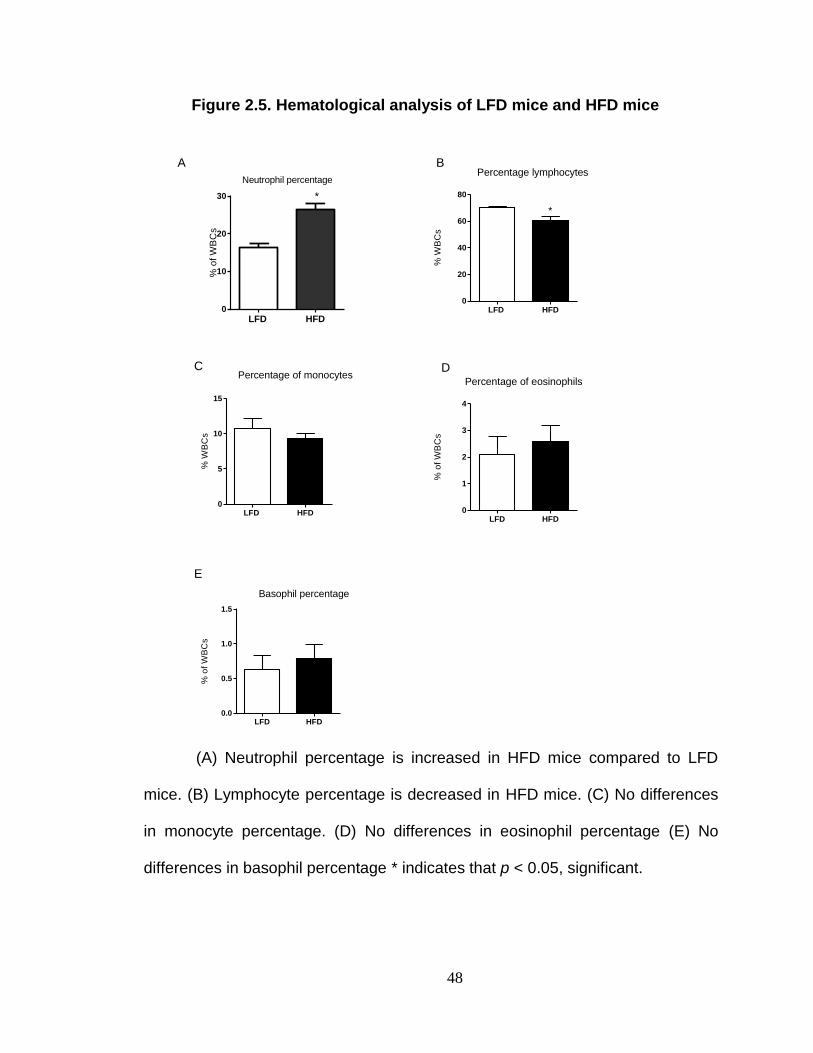
48
Figure 2.5. Hematological analysis of LFD mice and HFD mice
(A) Neutrophil percentage is increased in HFD mice compared to LFD
mice. (B) Lymphocyte percentage is decreased in HFD mice. (C) No differences
in monocyte percentage. (D) No differences in eosinophil percentage (E) No
differences in basophil percentage * indicates that p < 0.05, significant.
Neutrophil percentage
% o
f W
BC
s
LFD HFD0
10
20
30 *
% W
BC
s
LFD HFD0
20
40
60
80
Percentage lymphocytes
*%
WB
Cs
LFD HFD0
5
10
15
Percentage of monocytes
% o
f W
BC
s
LFD HFD0
1
2
3
4
Percentage of eosinophils
% o
f W
BC
s
LFD HFD0.0
0.5
1.0
1.5
Basophil percentage
A B
C D
E
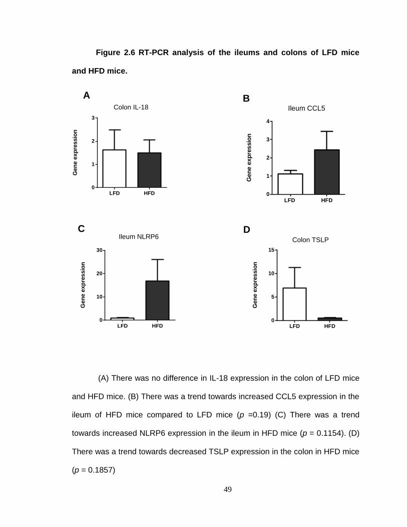
49
Figure 2.6 RT-PCR analysis of the ileums and colons of LFD mice
and HFD mice.
(A) There was no difference in IL-18 expression in the colon of LFD mice
and HFD mice. (B) There was a trend towards increased CCL5 expression in the
ileum of HFD mice compared to LFD mice (p =0.19) (C) There was a trend
towards increased NLRP6 expression in the ileum in HFD mice (p = 0.1154). (D)
There was a trend towards decreased TSLP expression in the colon in HFD mice
(p = 0.1857)
Gen
e e
xp
ressio
n
LFD HFD0
1
2
3
Colon IL-18
Gen
e e
xp
ressio
n
LFD HFD0
1
2
3
4
Ileum CCL5
Gen
e e
xp
res
sio
n
LFD HFD0
10
20
30
Ileum NLRP6
Gen
e e
xp
ressio
n
LFD HFD0
5
10
15
Colon TSLP
A B
C D
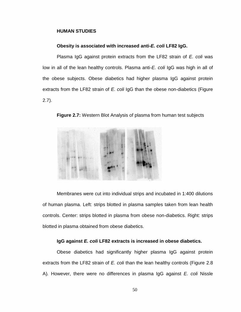
50
HUMAN STUDIES
Obesity is associated with increased anti-E. coli LF82 IgG.
Plasma IgG against protein extracts from the LF82 strain of E. coli was
low in all of the lean healthy controls. Plasma anti-E. coli IgG was high in all of
the obese subjects. Obese diabetics had higher plasma IgG against protein
extracts from the LF82 strain of E. coli IgG than the obese non-diabetics (Figure
2.7).
Figure 2.7: Western Blot Analysis of plasma from human test subjects
Membranes were cut into individual strips and incubated in 1:400 dilutions
of human plasma. Left: strips blotted in plasma samples taken from lean health
controls. Center: strips blotted in plasma from obese non-diabetics. Right: strips
blotted in plasma obtained from obese diabetics.
IgG against E. coli LF82 extracts is increased in obese diabetics.
Obese diabetics had significantly higher plasma IgG against protein
extracts from the LF82 strain of E. coli than the lean healthy controls (Figure 2.8
A). However, there were no differences in plasma IgG against E. coli Nissle
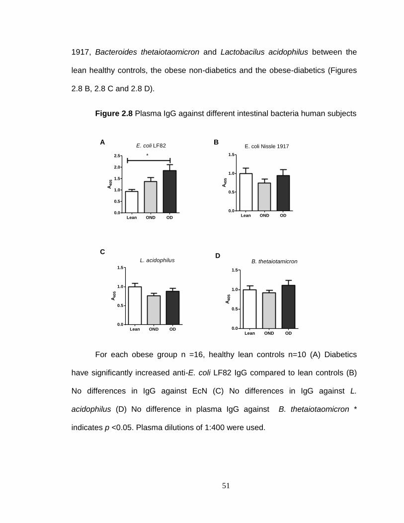
51
1917, Bacteroides thetaiotaomicron and Lactobacilus acidophilus between the
lean healthy controls, the obese non-diabetics and the obese-diabetics (Figures
2.8 B, 2.8 C and 2.8 D).
Figure 2.8 Plasma IgG against different intestinal bacteria human subjects
For each obese group n =16, healthy lean controls n=10 (A) Diabetics
have significantly increased anti-E. coli LF82 IgG compared to lean controls (B)
No differences in IgG against EcN (C) No differences in IgG against L.
acidophilus (D) No difference in plasma IgG against B. thetaiotaomicron *
indicates p <0.05. Plasma dilutions of 1:400 were used.
A4
05
Lean OND OD0.0
0.5
1.0
1.5
2.0
2.5 *
E. coli LF82
A4
05
Lean OND OD0.0
0.5
1.0
1.5
E. coli Nissle 1917
A40
5
Lean OND OD0.0
0.5
1.0
1.5
L. acidophilus
A4
05
Lean OND OD0.0
0.5
1.0
1.5
B. thetaiotamicron
A B
CD
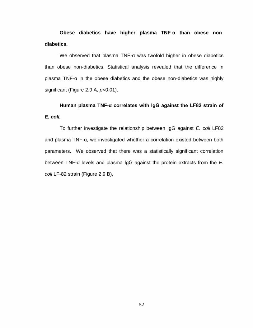
52
Obese diabetics have higher plasma TNF-α than obese non-
diabetics.
We observed that plasma TNF-α was twofold higher in obese diabetics
than obese non-diabetics. Statistical analysis revealed that the difference in
plasma TNF-α in the obese diabetics and the obese non-diabetics was highly
significant (Figure 2.9 A, p<0.01).
Human plasma TNF-α correlates with IgG against the LF82 strain of
E. coli.
To further investigate the relationship between IgG against E. coli LF82
and plasma TNF-α, we investigated whether a correlation existed between both
parameters. We observed that there was a statistically significant correlation
between TNF-α levels and plasma IgG against the protein extracts from the E.
coli LF-82 strain (Figure 2.9 B).
53
Figure 2.9 Analysis of plasma TNF-α in obese human subjects
TN
F-
(p
g/m
l)
OND OD0
1
2
3
4 **
TNF- in obese patients
An
ti-E
. co
li L
F-8
2 Ig
G (
A 4
50)
0 50 100 150 2000.0
0.2
0.4
0.6
R2 = 0.1968
P = 0.0181
Correlation analysis
A B
For each group n =16 (A) Obese diabetics had significantly higher plasma TNF-
α than the obese non-diabetics (B) There is a statistically significant correlation
between TNF-α and plasma anti-E. coli LF82 IgG. ** indicates p < 0.001. Plasma
dilution 1:2.
DISCUSSION
PRELIMINARY MOUSE STUDIES: HFD mice have higher plasma IgG
against invasive intestinal bacteria.
These studies were commenced in March 2010. By that time there was
accumulating evidence that the intestinal bacteria in conjunction with long term
consumption of a high fat diet played a role in the development of systemic
inflammation and insulin resistance. Our research group hypothesized that long
term consumption of a high fat diet might cause a loss of systemic immune
54
ignorance to the intestinal bacteria. We postulated that long term HFD
consumption would result in increased systemic immune responses to the
intestinal bacteria. To do this we decided to look at systemic immune responses
against bacteria found exclusively in the GI tract.
The intestinal bacteria are poorly characterized. We chose to measure
plasma IgG against the LF82 strain of E. coli which was isolated from an ileal
lesion of a Crohn’s Disease patient [150]. E. coli LF82 is designated as an
adherent invasive E. coli strain (AIEC strain). Invasive strains are characterized
by their ability to enter epithelial cells and subsequently survive and replicate
[151, 152]. E. coli LF82 invades a number of epithelial cell lines including Caco-2,
Intestine-407, and HCT-8 cells [153]. E. coli LF82 induces altered cytoskeletal
arrangements in epithelial cells which leads to its uptake by endocytic vacuoles.
This bacterial strain is more prevalent in the lesions of Crohn’s disease patients
than in control patients without inflammatory bowel disease [154]. It should be
emphasized that this bacterial strain is not present in the GI tract of mice.
However, since it is an adherent and invasive strain, there may be antigens that
are shared between this bacterial strain and uncharacterized invasive bacterial
strains.
Our pilot western blot analysis of the plasma of LFD and HFD mice
revealed drastic differences between the two groups. The plasma from HFD mice
had higher levels of IgG against invasive intestinal bacteria than the mice on the
LFD. Plasma IgG against invasive intestinal bacteria very low in all four individual
LFD mice (Figure 3). The systemic immune system is oblivious to the presence
55
of the immense bacterial load of the GI tract. This is essential since the intestinal
bacteria play important roles in vitamin synthesis and contribute to the overall
health of the host individual.
The higher levels of plasma IgG against invasive intestinal bacteria in HFD
mice suggested that these mice had lost systemic ‘ignorance’ to the bacteria in
the GI tract. IgG is produced in response to the presence of antigens in the
systemic circulation. This may lead to triggering of the systemic immune
response against these intestinal bacterial pathogens, resulting in increased IgG
the amount of antigenic material present in the systemic circulation. Therefore
the higher plasma IgG against intestinal bacteria in HFD mice is indicative of a
loss of systemic immune ignorance to invasive intestinal bacteria.
HFD mice have higher anti-bacterial IgG
As it has been previously mentioned, the GI tract contains at least 500
bacterial species. Although our finding that higher levels of anti- E. coli IgG in the
plasma of HFD mice suggested that these mice had lost systemic ‘ignorance’ to
the bacteria in the GI tract, we needed to establish whether there were higher
levels of IgG against other bacterial species in the plasma of HFD mice.
However, this aim was complicated by two major factors. The first was the
poor characterization of the composition of the intestinal bacteria. Additionally,
most intestinal bacterial species are anaerobic. This makes the culturing of
intestinal bacteria challenging. To overcome these problems cecal extracts were
prepared. The cecum is a pouch shaped structure situated between the ileum
56
and the colon. It is situated in a region of the GI tract where bacterial population
density is extremely high. It is therefore a reservoir of intestinal bacteria.
HFD mice had higher levels of plasma IgG against the antigenic contents
of the cecum compared to the LFD control group (Figure 2.2). This further
confirmed that HFD consumption is associated with a loss of systemic immune
ignorance to the intestinal bacteria. This is because the systemic immune system
is theoretically supposed to be oblivious to the contents of the cecum.
Furthermore these findings strengthened our hypothesis since IgG against the
intestinal bacteria of mice was measured. On the other hand, the LFD controls
had low levels of IgG against the cecal contents. This may be due to the fact that
these mice have not experienced any significant translocation of the intestinal
bacteria into the systemic circulation. As a result, the levels of IgG against the
antigenic components of the cecum are significantly much lower in the LFD
controls than the HFD fed mice.
Results from long term feeding study in C57BL6 mice
The preliminary studies resulted in the observation that mice fed a high fat
diet (Rodent Diets with 60 kcal% Fat D12492 from Research Diets Incorporated)
for 10 weeks had elevated plasma anti-E. coli LF82 IgG. Although these findings
were novel, they offered only a narrow range of interpretation. This is because
the only parameter that had been examined thus far was plasma anti-E. coli
LF82 IgG in relation to high fat diet consumption. To gain greater insight into the
effects of long term HFD consumption on the systemic immune system a long
term (ten week) feeding study was performed using C57BL6 mice.
57
Two parameters were used to define the end point of the study (i.e. the
point at which the mice would be euthanized). These were body weight and
fasting blood glucose. As expected, C57BL6 mice fed a HFD (60% calories from
fat) gained significantly more weight than the mice fed a LFD (10% calories from
fat), with bodyweights diverging significantly six weeks into the study (Figure 2.3
A). However, weight gain by itself is not an accurate indicator of the development
of elevated fasting blood glucose. The mice were euthanized once a significant
difference in fasting blood glucose levels was observed. Based on the proposed
hypothesis a significant elevation in fasting blood glucose would be observed
subsequent to systemic inflammation and bacterial translocation.
After 10 weeks the fasting blood glucose levels of the HFD mice were
higher than the LFD mice (Figure 2.3 B). Statistical analysis revealed that these
differences were significant (p<0.05) and it was at this point the study was
terminated. The mice were euthanized and we were able to collect samples to
evaluate a wide range of parameters.
Long term HFD consumption leads to increased plasma TNF-α in
mice.
HFD mice had elevated plasma IgG against invasive intestinal bacteria.
Previous studies have linked the IgG response to certain bacterial species to
disease severity [155]. This is because cross-linking of FcγRs leads to
inflammation [156, 157]. However, plasma IgG levels alone cannot be used to
determine whether there is increased systemic inflammation. TNF-α is a widely
used marker of inflammation. Since the proposed hypothesis states that the
58
immune responses to the intestinal bacteria lead to systemic inflammation, TNF-
α was measured in LFD mice and HFD mice. Statistical analysis revealed that
the plasma TNF-α in the HFD mice was higher than in the LFD mice (Figure 2.3
C; p <0.05). These findings strengthened the hypothesis that long term HFD
consumption leads to systemic inflammatory immune responses being mounted
against the bacterial antigens present in the lumen of the GI tract.
A possible explanation for the increased plasma TNF-α could be the
increase in intestinal bacteria crossing the intestinal epithelial barrier. This could
lead to immune responses being mounted by the B lymphocytes. The B
lymphocytes produce IgG which bind to the antigens present on the surface of
the intestinal bacteria. It is possible that the increase in systemic IgG against
intestinal bacteria results in increased FcγR crosslinking. This in turn leads to the
release of TNF-α.
Plasma IgG against invasive intestinal bacteria is increased in HFD
mice.
HFD mice had higher plasma IgG against invasive intestinal bacteria than
LFD mice (Figure 2.4 A). However, there were no significant differences in
plasma IgG against protein extracts from E. coli Nissle 1917, Bacteroides
thetaiotaomicron and Lactobacilus acidophilus in HFD and LFD mice (Figures 2.4
B, 2.4 C and 2.4 D). As it has been previously stated the systemic immune cells
are naïve to the presence of the intestinal bacteria. This feeding study resulted in
increased IgG against invasive intestinal bacteria in the HFD mice. These results
further support the proposal that long term HFD consumption is associated with
59
increased IgG against antigens that are usually confined to the lumen of the GI
tract. IgG is produced as a consequence of the B lymphocytes detecting the
presence of a particular antigen. Thus the elevated IgG against invasive
intestinal bacteria in HFD mice was possibly due to the fact that that the B
lymphocytes were no longer oblivious to these antigens. A possible explanation
for this is that long term HFD consumption promotes entry of the bacteria present
in the lumen of the GI tract into the systemic circulation. This triggers systemic
immune responses including IgG production.
Neutrophil percentage is increased in HFD mice.
A variety of immune cells participate in inflammatory reactions. Since the
sample number for the mouse studies (n = 6 for each group) was relatively small,
further evidence was needed that long term HFD consumption results in
inflammatory immune responses. The HEMAVET® 950 FS Multispecies
Hematology Systems is useful in characterizing the various blood cell types
present. After the ten week feeding study was completed, and the mice were
euthanized we characterized the various immune cells present in whole blood
fractions. The neutrophil percentage in the blood of the HFD mice was
significantly increased compared to the LFD mice (Figure 2.5 A, p <0.0001). The
increase in neutrophil percentage in HFD mice provides further evidence that
long term HFD consumption leads to increased systemic inflammatory immune
responses against the intestinal bacteria. Neutrophils are present in the initial
phase of inflammation [158]. They ingest infectious microbes and eliminate them.
The intracellular destruction of microbes by neutrophils is mediated by their
60
cytoplasmic granules via superoxide production as well as the proteases
elastase and cathepsin G [159].
A possible explanation for the increase in neutrophil percentage may be
the increased plasma IgG against invasive intestinal bacteria in the HFD mice.
Since FcγRs are expressed on the surface of neutrophils, there may be
increased cross-linking of FcγRs with the Fc domains of the IgG against the
intestinal bacterial antigens [160]. No significant differences were noted in
monocyte percentage, basophil percentage or eosinophil percentage in the blood
of HFD mice and LFD mice (Figures 2.5 C, 2.5 D and 2.5 E). This was not
surprising since basophils are usually produced in response to infections by
helminthes [161]. They are also usually associated with allergens and are
associated with allergic reactions. Eosinophil percentage did not differ between
the LFD and HFD mice. Eosinophils protect against parasitic infections [162]. Our
discovery that long term consumption of a high fat diet specifically increases the
percentage of neutrophils was significant. The increase in neutrophil percentage
was observed along with increased plasma IgG against invasive intestinal
bacteria, providing further evidence that the systemic immune responses against
intestinal bacteria are inflammatory.
Long term HFD consumption promotes intestinal inflammation.
Real time PCR (RT-PCR) analysis was performed in order to determine
whether the systemic inflammation we observed originated in the GI tract. It has
been traditionally thought that the higher levels of inflammatory cytokines
associated with high fat diet consumption originate in the adipose tissue [163].
61
However, there is a growing body of evidence which suggests that inflammation
resulting from long term HFD consumption may originate in the gastrointestinal
(GI) tract [65]. For example, it has been reported that long term HFD
consumption may alter the bacterial composition of the GI tract [68]. Furthermore
Ding et al observed that conventionally raised (CONV) mice consuming a high fat
diet experienced intestinal inflammation [70]. By contrast GF mice fed a high fat
diet did not exhibit intestinal inflammation.
Therefore it was crucial to determine whether the HFD mice experienced
intestinal inflammation. The ileum and colon sections of the GI tract were
analyzed for intestinal inflammation. This is because these regions of the GI tract
harbor the largest bacterial populations. Different pro-inflammatory markers were
measured. These were the NOD-like receptor family pyrin domain containing 6
(NLRP6), Chemokine (C-C motif) ligand 5 (CCL5 also known as RANTES) and
IL-18. RT-PCR revealed a trend towards increased NLRP6 expression in the
ileum HFD mice (LFD 1.000 ± 0.2285, HFD 16.90 ± 9.220, p = 0.1154). NLRP6 is
thought to play a role in inflammation via the activation of NF-κB [164]. Although
the difference between the LFD mice and the HFD mice was not statistically
significant, this was probably due to the relatively small sample size (where n =
6) for each group. Although intestinal IL-18 did not differ between LFD and HFD
mice, there was a trend towards increased CCL5 in the ileum of the HFD mice
(LFD 1.136 ± 0.1660, HFD 2.444 ± 1.001, p = 0.1900). CCL5 plays a role in
inflammation by recruiting macrophages [165]. Thus the trend towards increased
62
CCL5 expression in the GI tract supports the idea that the systemic inflammation
observed originates from the GI tract.
Thymic stromal lymphopoietin (TSLP) expression was also measured.
TSLP is constitutively secreted by the intestinal epithelial cells (IECs). The IECs
secrete TSLP which prevents the dendritic cells (DCs) from promoting
inflammatory immune responses. The DCs sample antigens and present them to
T lymphocytes. RT-PCR showed a trend towards reduction of TSLP expression
in the colon of HFD mice compared to the LFD controls (LFD 6.867 ± 4.469, HFD
0.5141 ± 0.1141, p = 0.1857). Since TSLP is crucial to preventing intestinal
inflammation, the trend towards decreased TSLP expression provides further
evidence that the systemic inflammation may have originated in the GI tract.
HUMAN STUDIES
We decided to investigate whether long term consumption of a high fat
diet is associated with increased plasma IgG against invasive intestinal bacteria
in humans. We made the assumption that humans who had consumed a high fat
diet over a long time period would have body mass index values in the
overweight and obese range (≥ 25). When we performed Western Blot analysis
we observed what appeared to be striking differences between the three groups.
Plasma IgG against protein extracts from the LF82 strain of E. coli was lowest in
the lean healthy control group. Plasma anti-E. coli LF82 IgG was higher in the
plasma of the obese individuals. Notably there appeared to be a difference in
plasma anti-E. coli LF82 IgG between the non-diabetic individuals and diabetic
63
patients. The obese diabetics had higher plasma anti-E. coli LF82 IgG than their
non-diabetic counterparts.
These findings provided further evidence that long term HFD consumption
is associated with a loss of systemic immune ignorance to the intestinal bacteria.
As it has been previously stated, the LF82 strain of E. coli is only known to reside
in the GI tract. The low levels of plasma anti-E. coli LF82 IgG in the lean healthy
controls was not surprising. The systemic immune system is ‘ignorant’ to the
existence of the immense bacterial load of the GI tract. This is important for
minimizing potentially harmful inflammatory immune responses against the
immense reservoir of antigenic material in the GI tract. However, what was
particularly striking was the difference in anti-E. coli LF82 IgG in obese non-
diabetics and obese diabetics. Anti-E. coli LF82 IgG was higher in obese
diabetics than in obese non-diabetics. This finding suggested that the systemic
immune responses against intestinal bacterial pathogens may be linked to insulin
resistance and the development of type 2 diabetes.
IgG against invasive intestinal bacteria is increased in obese
diabetics.
Quantifying plasma IgG against the intestinal bacteria was crucial in
determining whether the apparent differences observed in our Western Blot
analyses had statistical significance. Furthermore, in order to determine whether
any quantitative relationships existed between IgG against the intestinal bacteria
and inflammatory cytokine levels we needed a reliable quantitative assay. As a
consequence, we developed our own laboratory made ELISA by coating 96 well
64
flat-bottom ELISA plates with soluble protein extracts from E. coli Nissle 1917,
Bacteroides thetaiotaomicron and Lactobacilus acidophilus and E. coli LF82
dissolved in a carbonate coating buffer. The difference between the plasma anti-
E. coli LF82 IgG in lean controls and obese diabetics was significant (p =
0.0107). The lean controls had the lowest levels of plasma IgG against invasive
intestinal bacteria. The low plasma anti- E. coli LF82 IgG in lean healthy controls
is probably due to the fact that the immune system these individuals maintain a
state of ignorance to the intestinal bacteria.
What was particularly interesting was that while obese diabetics had
higher plasma anti- E. coli LF82 IgG than the lean controls, there was no
difference in plasma anti-E. coli LF82 in obese non-diabetic individuals compared
to the lean healthy controls. The obese non-diabetics had body mass index
values that were statistically equivalent to the obese diabetic patients. This led us
to hypothesize that plasma IgG against the intestinal bacteria may be related to
the transition from a non-diabetic state to a diabetic state.
Immunoglobulin G is the major immunoglobulin subclass produced in
response to the presence of antigens in the systemic circulation. The IgG
response to certain bacterial species has also been correlated with disease
severity [166]. IgG is comprised of two identical heavy chains and two identical
light chains, arranged in a Y shaped configuration. The IgG molecule is
comprised of an antigen binding fragment (Fab) domain and a crystalizeable
fragment (Fc) domain. Receptors for the Fc domain of IgG (FcγRs) are located
on the surface of immune cells. Inflammation results from the cross-linking of
65
FcγRs. It is possible that the increased IgG against intestinal bacteria could have
resulted in increased FcγR cross-linking. This in turn could have led to increased
systemic inflammation. Uncontrolled systemic inflammation can adversely affect
insulin signaling pathways, leading to insulin resistance and the development of
type 2 diabetes.
Human plasma TNF-α correlates with IgG against the LF82 strain of
E. coli.
Statistical analysis was performed to determine whether there was a
correlation between anti-E. coli LF82 IgG and TNF-α. This was done to establish
a more definitive link between the immune responses against the intestinal
bacteria and inflammatory markers. The sample size in the animal study (n=6 for
each group) was insufficient to perform a correlation analysis.
The correlation analysis was performed using the human plasma
samples. This is because the sample size for human subjects was larger (n=16
for each group, 32 in total). There were two groups of obese subjects: obese
non-diabetics and obese diabetics. Both groups had mean BMI values that were
statistically equivalent (obese non-diabetics mean BMI 41.1 ± 10.4 versus obese
diabetics mean BMI 40.6 ± 7.1). As a consequence, a discrepancy in adiposity
could be ruled out. It was observed that there was a statistical correlation
between anti-E. coli LF82 IgG and plasma TNF-α in the human subjects. The
correlation between anti-E. coli LF82 IgG and TNF-α is further evidence that the
systemic immune responses to the intestinal bacterial antigens are associated
with inflammation. It is possible that the increase in IgG against intestinal
66
antigens promotes cross-linking of FcγRs, which in turn causes the release of
pro-inflammatory markers such as TNF-α [167].
The correlation between anti-E. coli LF82 IgG and plasma TNF-α linked
inflammation to the immune responses against intestinal antigens. If systemic
inflammation was linked to the development of type 2 diabetes, then theoretically
the obese diabetics would have higher levels of plasma TNF-α. To determine
whether this was the case, TNF-α in the human plasma samples was measured
by ELISA. It was observed that there was a difference in plasma TNF-α between
both groups of obese subjects. TNF-α in the obese diabetics was significantly
higher than in the obese non-diabetics. This finding provides further evidence
that the inflammation resulting from intestinal bacterial antigens may lead to the
development of type 2 diabetes. It is possible that insulin signaling could have
been adversely affected by the presence of high levels of plasma TNF-α.
Copyright © Nadeem K. Mohammed 2012
67
CHAPTER 3 HIGH FAT DIET CONSUMPTION AND INTESTINAL IMMUNITY.
INTRODUCTION
Immunoglobulin A (IgA) actively secreted into the GI tract minimizes the
adherence of bacteria to the intestinal epithelium. In our initial studies we
observed an association between long term consumption of a HFD and elevated
plasma anti-E. coli LF82 IgG. As a consequence, we investigated whether long
term HFD consumption impairs IgA secretion into the lumen of the GI tract. To do
this we measured fecal IgA in mice fed a LFD (10% calories from fat) and mice
fed a HFD (60% calories from fat). Plasma IgA levels were measured in LFD and
HFD mice. IgA is the second most abundant immunoglobulin in the plasma.
Plasma IgA neutralizes antigens and suppresses inflammatory effects of
antibody dependent cellular toxicity [168]. Furthermore, small quantities of
plasma IgA enter the intestinal mucosal secretions by paracellular diffusion
[131].
A goal of this study was to assess whether the absence of the active
secretion of IgA into the lumen of the GI tract by the pIgR is a predisposing factor
for development of metabolic syndrome. The polymeric immunoglobulin receptor
(pIgR) actively secretes polymeric immunoglobulins into the lumen of the GI tract.
An estimated 3 grams of IgA are secreted daily. Since this involves considerable
energy expenditure, we reasoned that this specialized secretory mechanism
plays a role in preventing adherence of the commensal bacteria to the intestinal
epithelial cells.
68
. Our previous studies had shown that long term HFD consumption was
associated with increased IgG against adherent invasive intestinal bacteria. We
postulated that perhaps mice lacking pIgR (pIgR KO mice) might be more
susceptible to intestinal epithelial cell adherence and invasion. This could result
in inflammatory immune responses being mounted against the resident gut
bacteria. To determine whether this was the case, pIgR knockout mice and
C57BL6 wild type littermate controls were placed on a Western Diet (Rodent
Diets with 41 kcal% Fat, 43 kcal% Carbohydrate and 17 kcal% Protein). A
Western Diet feeding regimen was chosen since the Western Diet is
representative of the diets that are associated with metabolic disorders in
humans.
The human plasma samples were also analyzed by total IgA ELISA. This
was done in order to determine whether plasma IgA levels differed between the
lean healthy controls, obese non-diabetics and obese diabetics. It was observed
that plasma IgA differed between the three groups of human subjects. Lean
controls had the highest levels of plasma IgA. Obese non-diabetics had
significantly reduced plasma IgA compared to the lean controls. Obese diabetics
have significantly reduced plasma IgA compared to both obese non-diabetics
and lean controls.
69
METHODS
Determining the effect of long term HFD consumption on fecal IgA
C57BL6 mice aged 6 weeks were divided into two equal groups (n = 6).
One group was fed a high fat diet (Rodent Diets with 60 kcal% Fat D12492 from
Research Diets Incorporated). The other group was fed a low fat diet (Rodent
Diets with 10 kcal% Fat D12450B from Research Diets Incorporated). Details of
these diets are listed in table 2.1. The mice were placed on the special diets for
10 weeks.
Fecal IgA extraction
Fecal IgA was extracted utilizing a protocol previously employed by
Ferguson et al [169]. Fecal IgA extraction buffer was prepared utilizing
phosphate-buffered saline (PBS, pH 7.4), 0.5% Tween® 20 and complete, EDTA-
free protease inhibitor cocktail. The extraction buffer was added to each tube at a
ratio of 1 ml of buffer to 0.10 g of feces. The samples were manually
homogenized and the resulting mixtures were vortexed. The resulting fecal
suspensions were centrifuged at 14,000 rpm for 20 minutes and the supernatants
were collected for further use.
Total fecal IgA measurements
Total fecal IgA was quantified using an ELISA for Mouse IgA from
MABTECH. 96 well flat-bottom ELISA plates (BD-Falcon) were coated overnight
with monoclonal anti-IgA antibody diluted 2µg/ml in PBS (pH 7.4). Blocking was
performed for one hour at room temperature using incubation buffer (PBS with
70
0.05% Tween 20 and 0.1% BSA). Subsequent to blocking fecal IgA extracts
were diluted 100X in incubation buffer and added in triplicate. Total IgA was
detected using anti-IgA-ALP diluted 1:500 in incubation buffer. P-nitrophenyl
phosphate was then added and the optical density was measured at 405nm
using a Bio-Rad microplate reader. IgA concentration was divided by the mass of
fecal pellets to give the quantity of IgA present per gram of feces (IgA/g feces).
Quantifying total fecal output and food intake.
To gain an accurate estimate of total fecal IgA production per day total
daily fecal output was measured in a separate group of C57BL6 mice. C57BL6
mice were divided into two groups. One group was fed a high fat diet (Rodent
Diets with 60 kcal% Fat D12492 from Research Diets Incorporated). The other
group was fed a low fat diet (Rodent Diets with 10 kcal% Fat D12450B from
Research Diets Incorporated).
Total fecal output: Mice were placed in a cage with clean bedding, with
free access to food and water. Fecal pellets were collected at intervals of 24
hours and weighed. It should be noted that metabolic cages were available from
the COCVD PHYSIOLOGIC RESEARCH CORES at the University of Kentucky.
However, the Eckhardt Laboratory did not have the required protocol approvals
to measure fecal output in mice using these facilities.
Quantifying food intake: Mice were given free access to food and water.
Food was weighed at 24 hour intervals. The weight of food was subtracted from
the previous weight to give total consumption. This was then divided by the
71
number of mice in the cage to give an estimate of the amount of food consumed
by each mouse.
Effect of HFD consumption on plasma IgA.
The effect of HFD consumption on plasma IgA was assessed in HFD mice
and LFD mice. Plasma IgA was measured using the ELISA for Mouse IgA. 96
well flat-bottom ELISA plates (BD-Falcon) were coated overnight with
monoclonal anti-IgA antibody diluted 2µg/ml in PBS (pH 7.4). Blocking was
performed for one hour at room temperature using incubation buffer (PBS with
0.05% Tween 20 containing 0.1% BSA). Subsequent to blocking plasma diluted
100X in incubation buffer was added in triplicate. Total IgA was detected using
anti-IgA-ALP diluted 1:500 in incubation buffer. P-nitrophenyl phosphate was
then added and the optical density was measured at 405nm using a Bio-Rad
microplate reader.
To evaluate whether impaired IgA secretion increases the risk of
developing metabolic syndrome.
Mice: Six pIgR knockout mice (pIgR KO mice) on a C57BL/6 background
were kindly donated by Dr. Charlotte Kaetzel (Department of Microbiology,
Immunology and Molecular Genetics, University of Kentucky). pIgR KO mice are
unable to actively secrete polymeric IgA into the lumen of the GI tract. This is
because the third exon of the PIGR locus has been disrupted by the use of a
targeting vector [170]. For controls we utilized wild type C57BL/6 littermates
(n=7). Mice were housed three per cage and maintained in a 12h light / dark
cycle. We allowed the mice to become acclimated to their new environment.
72
When the mice were obtained they were being fed the Teklad Global 18%
Protein Rodent Diet (hereafter referred to as regular chow diet). Both groups of
mice were maintained on the regular chow diet for one week after receiving
them. The reason for doing this was to measure a variety of baseline metabolic
parameters. The mice were then switched to a high fat, high carbohydrate
Western Diet (Rodent Diets with 41 kcal% Fat, 43 kcal% Carbohydrate, 17 kcal%
Protein). Details of the diets are listed in table 2.1. The mice were fed the
Western Diet for 16 weeks.
Processing of samples for analysis.
Mice were euthanized using carbon dioxide. Blood was immediately
collected by cardiac puncture and placed into EDTA containing eppendorf tubes
that had been kept on ice. The majority of blood obtained was immediately
centrifuged at 8,000 rpm for 10 minutes. The plasma was collected and stored at
-82°C. The remaining blood (whole blood fraction) was used for hematological
analysis. After mice were euthanized the ileums and colons were harvested.
Intestinal mucosal scrapings were collected and frozen immediately in liquid
nitrogen.
Identification of mice.
After the mice were obtained they were implanted with IPTT-300
transponders (Vendor BMDS) for identification. After the mice were euthanized,
they were identified and tail snips were collected from individual mice. DNA was
extracted from the tail snips using the Qiagen DNeasy Blood and Tissue kit
(QIAGEN Catalog# 69504). Polymerase chain reaction (PCR) was then
73
performed using a Perkin Elmer GeneAmp PCR System 9600. The primer
sequences to detect the Pigr mutant gene (Pigr KO) and the wild type Pigr gene
(Pigr WT) are listed in table 2.3.
Body weight and body fat analyses
pIgR KO mice and the C57BL/6 controls were weighed prior to
commencing the Western Diet feeding regimen. Once the feeding study was
started the mice were weighed weekly. EchoMRI™ analysis was performed in
order to determine whether there were differences in body fat percentage.
Plasma cytokine analysis
To assess the levels of circulating inflammatory cytokines we utilized
MILLIPLEX® Mouse Cytokine Kits (from Millipore). The first kit (Catalog #
MPXMYCTO-70K) quantified the cytokines IL-1b, IL-6, IL-10 and TNF-α, IL-12,
IL-7, IL-5 and IL-13 prior to commencing the Western Diet, as well as two and
sixteen weeks after commencing the Western Diet feeding regimen. The second
kit used (Catalog # MADPK-71K) measured insulin, resistin, leptin, PAI-1 and
TNF-α eight and sixteen weeks after commencing the Western Diet feeding
regimen.
Hematological analysis.
50µl of whole blood fractions collected from the mice after euthanasia was
used. The tubes containing the whole blood was placed on a rocker for 2
minutes. Samples were then analyzed using a HEMAVET® 950 FS Multispecies
Hematology Systems (Drew Scientific Inc.).
74
Oral glucose tolerance test (OGTT)
To assess oral glucose tolerance, mice were fasted for 6 hours. Blood was
collected from the tail tips of mice and blood glucose levels were recorded using
the TrueTrack glucose meter (Home Diagnostics Inc). The mice were gavaged
with a dose of 2.0g/kg glucose. Blood glucose measurements were recorded at
15, 30, 60, 90 and 120 minutes post-gavage. We performed the OGTT prior to
commencing the Western Diet feeding and two weeks, ten weeks and sixteen
weeks after commencing the Western Diet feeding regimen.
Real time PCR analysis
We compared the levels of inflammatory markers in the intestinal tissue of
pIgR KO mice and C57BL6 controls. After mice were euthanized the small
intestine and colon were harvested. RNA was extracted from intestinal tissues by
the E.Z.N.A® Total RNA Kit. The RNA was converted into cDNA using the
qScript™ cDNA Synthesis Kit. We analyzed the samples for expression of TNF-
α and IL-6 using a BIORAD CFX96™ Real Time PCR detection system.
Statistical analyses.
Results were expressed as mean ± S.E.M and were analyzed with
GraphPad Prism v5.04. Groups were compared with unpaired Student’s t-tests
and ANOVA and Bonferroni’s post-hoc analysis. Statistical significance was
assumed when p<0.05. Area under the curve analyses were performed using
SigmaPlot.
75
ANALYSIS OF HUMAN PLASMA IgA.
We also measured plasma IgA in humans. Details of the human plasma
donors are listed in table 2.2. Total plasma IgA in the humans was assessed with
a Human IgA ELISA kit from MABTECH (3860-1AD-6). 96 well flat-bottom
ELISA plates (BD-Falcon) were coated overnight with MT57 antibody diluted
2µg/ml in PBS (pH 7.4). Blocking was performed with incubation buffer and
human plasma was diluted 400X in incubation buffer and added in triplicate.
Total IgA present in human plasma was detected by adding a 1000 fold dilution
of MT20-ALP. P-nitrophenyl phosphate was then added and the optical density
was measured at 405nm using a Bio-Rad microplate reader.
RESULTS
HFD mice have reduced fecal IgA.
The effect of diet on secretion of IgA into the lumen of the GI tract was
studied in C57BL6 mice fed a HFD and a LFD. During the first three weeks of the
study statistical analysis revealed that there were no differences between the
fecal IgA levels in the LFD mice or the HFD mice. However, by the fourth week of
the study fecal IgA levels between LFD and HFD mice had diverged significantly.
The LFD mice had significantly higher fecal IgA than the HFD mice. Higher fecal
IgA in the LFD mice compared to the HFD mice was observed throughout the
duration of the feeding study (Figure 3.1 A). Analysis of the fecal IgA levels in
the LFD mice and HFD mice revealed that there was an increase in fecal IgA
over time in the LFD mice. On the other hand, the levels of fecal IgA in the HFD
mice remained relatively unchanged. Thus the differences noted between fecal
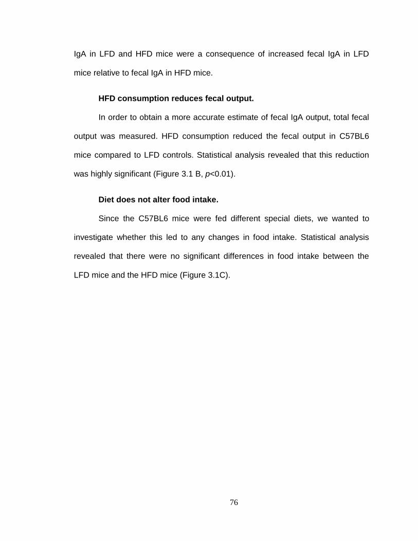
76
IgA in LFD and HFD mice were a consequence of increased fecal IgA in LFD
mice relative to fecal IgA in HFD mice.
HFD consumption reduces fecal output.
In order to obtain a more accurate estimate of fecal IgA output, total fecal
output was measured. HFD consumption reduced the fecal output in C57BL6
mice compared to LFD controls. Statistical analysis revealed that this reduction
was highly significant (Figure 3.1 B, p<0.01).
Diet does not alter food intake.
Since the C57BL6 mice were fed different special diets, we wanted to
investigate whether this led to any changes in food intake. Statistical analysis
revealed that there were no significant differences in food intake between the
LFD mice and the HFD mice (Figure 3.1C).
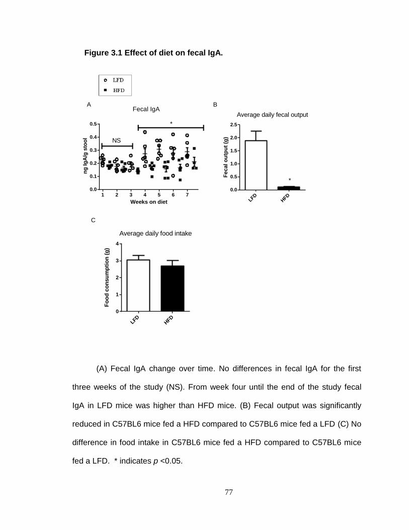
77
Figure 3.1 Effect of diet on fecal IgA.
(A) Fecal IgA change over time. No differences in fecal IgA for the first
three weeks of the study (NS). From week four until the end of the study fecal
IgA in LFD mice was higher than HFD mice. (B) Fecal output was significantly
reduced in C57BL6 mice fed a HFD compared to C57BL6 mice fed a LFD (C) No
difference in food intake in C57BL6 mice fed a HFD compared to C57BL6 mice
fed a LFD. * indicates p <0.05.
Weeks on diet
ng
Ig
A/g
sto
ol
1 2 3 4 5 6 70.0
0.1
0.2
0.3
0.4
0.5
NS
*
Fecal IgAAverage daily fecal output
Fe
cal
ou
tpu
t (g
)
LFDHFD
0.0
0.5
1.0
1.5
2.0
2.5
*
Average daily food intake
Fo
od
co
nsu
mp
tio
n (
g)
LFDHFD
0
1
2
3
4
A B
C
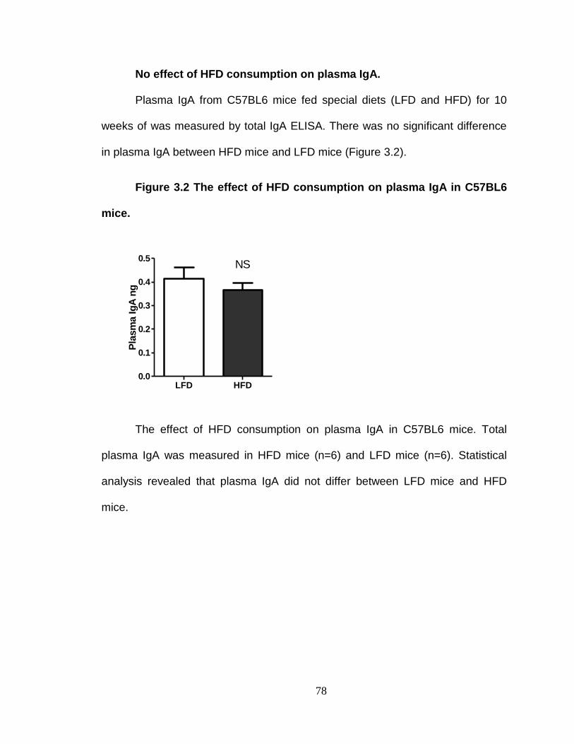
78
No effect of HFD consumption on plasma IgA.
Plasma IgA from C57BL6 mice fed special diets (LFD and HFD) for 10
weeks of was measured by total IgA ELISA. There was no significant difference
in plasma IgA between HFD mice and LFD mice (Figure 3.2).
Figure 3.2 The effect of HFD consumption on plasma IgA in C57BL6
mice.
Pla
sm
a Ig
A n
g
LFD HFD0.0
0.1
0.2
0.3
0.4
0.5NS
The effect of HFD consumption on plasma IgA in C57BL6 mice. Total
plasma IgA was measured in HFD mice (n=6) and LFD mice (n=6). Statistical
analysis revealed that plasma IgA did not differ between LFD mice and HFD
mice.
79
To assess whether impaired IgA secretion into the lumen of the GI
tract is a predisposing factor for development of metabolic syndrome.
pIgR KO mice have higher plasma IgA.
To investigate whether long term consumption of a high-fat, high-
carbohydrate Western Diet would affect plasma IgA in pIgR KO mice a total IgA
ELISA was performed. Plasma IgA in the pIgR KO mice was higher than in the
wild type C57BL6 controls. Statistical analysis revealed that this difference was
highly significant (Figure 3.3; p < 0.01).
Figure 3.3 Plasma IgA in pIgR KO mice and wild type C57BL6
controls.
A4
05
C57BL6 pIgR KO0.0
0.1
0.2
0.3
0.4
**
Plasma IgA
The pIgR KO mice had higher plasma IgA compared to the C57BL6 WT
controls. ** indicates p < 0.001. Plasma dilution 1:100.
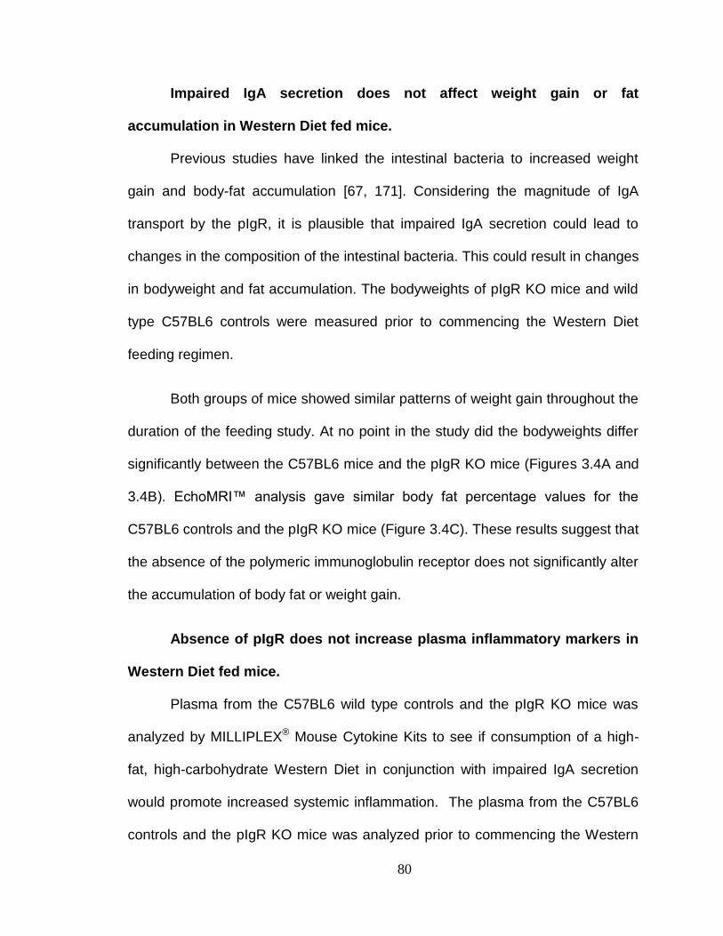
80
Impaired IgA secretion does not affect weight gain or fat
accumulation in Western Diet fed mice.
Previous studies have linked the intestinal bacteria to increased weight
gain and body-fat accumulation [67, 171]. Considering the magnitude of IgA
transport by the pIgR, it is plausible that impaired IgA secretion could lead to
changes in the composition of the intestinal bacteria. This could result in changes
in bodyweight and fat accumulation. The bodyweights of pIgR KO mice and wild
type C57BL6 controls were measured prior to commencing the Western Diet
feeding regimen.
Both groups of mice showed similar patterns of weight gain throughout the
duration of the feeding study. At no point in the study did the bodyweights differ
significantly between the C57BL6 mice and the pIgR KO mice (Figures 3.4A and
3.4B). EchoMRI™ analysis gave similar body fat percentage values for the
C57BL6 controls and the pIgR KO mice (Figure 3.4C). These results suggest that
the absence of the polymeric immunoglobulin receptor does not significantly alter
the accumulation of body fat or weight gain.
Absence of pIgR does not increase plasma inflammatory markers in
Western Diet fed mice.
Plasma from the C57BL6 wild type controls and the pIgR KO mice was
analyzed by MILLIPLEX® Mouse Cytokine Kits to see if consumption of a high-
fat, high-carbohydrate Western Diet in conjunction with impaired IgA secretion
would promote increased systemic inflammation. The plasma from the C57BL6
controls and the pIgR KO mice was analyzed prior to commencing the Western
81
Diet as well as two and sixteen weeks after commencing the Western Diet
feeding regimen. No differences were observed between the C57BL6 controls
and the pIgR KO mice in plasma IL-1b, IL-6, IL-10 and TNF-α at any time point
(Figures 3.5 A, 3.5 B, 3.5 C and 3.5 D). No differences were observed in plasma
IL-13, IL-12, IL-5 and IL-7 (Figures 3.6 A, 3.6 B, 3.6 C and 3.6 D). These results
indicate that there are no baseline differences in plasma inflammatory cytokines
between C57BL6 controls and pIgR KO mice prior or subsequent to consuming a
high-fat, high-carbohydrate Western Diet.
Plasma adipokine levels were also analyzed subsequent to commencing
the Western Diet feeding regimen. Notably, the pIgR KO had reduced plasma
PAI-1 compared to the C57BL6 controls. Statistical analysis revealed that this
difference was significant (Figure 3.7 B). There were no differences between the
C57BL6 controls and the pIgR KO mice in plasma insulin or resistin (Figures 3.7
A and 3.7 C). Plasma IL-6 and TNF-α were below the detectable range in the
C57BL6 controls and the pIgR KO mice. These results provide evidence that the
absence of the polymeric immunoglobulin receptor does not promote increased
systemic inflammation either in the presence or absence of high-fat diet
consumption. It should be noted that plasma adipokine levels were not measured
in these mice prior to commencing the Western Die and as a result there is no
baseline date for the plasma adipokine values.
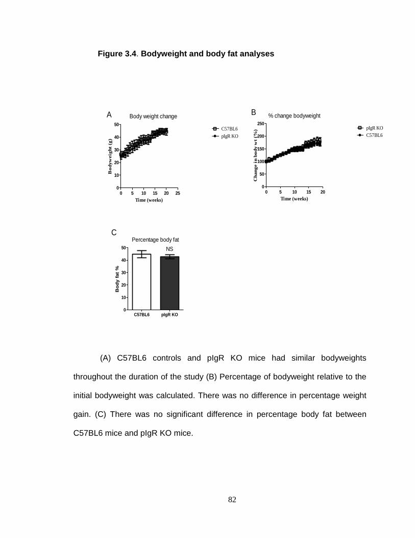
82
Figure 3.4. Bodyweight and body fat analyses
Body weight change
Time (weeks)
Bo
dyw
eig
ht
(g)
0 5 10 15 20 250
10
20
30
40
50C57BL6
pIgR KO
% change bodyweight
Time (weeks)
Ch
an
ge i
n b
od
y w
t (%
)
0 5 10 15 200
50
100
150
200
250pIgR KO
C57BL6
Percentage body fat
Bo
dy f
at
%
C57BL6 pIgR KO0
10
20
30
40
50 NS
A B
C
(A) C57BL6 controls and pIgR KO mice had similar bodyweights
throughout the duration of the study (B) Percentage of bodyweight relative to the
initial bodyweight was calculated. There was no difference in percentage weight
gain. (C) There was no significant difference in percentage body fat between
C57BL6 mice and pIgR KO mice.
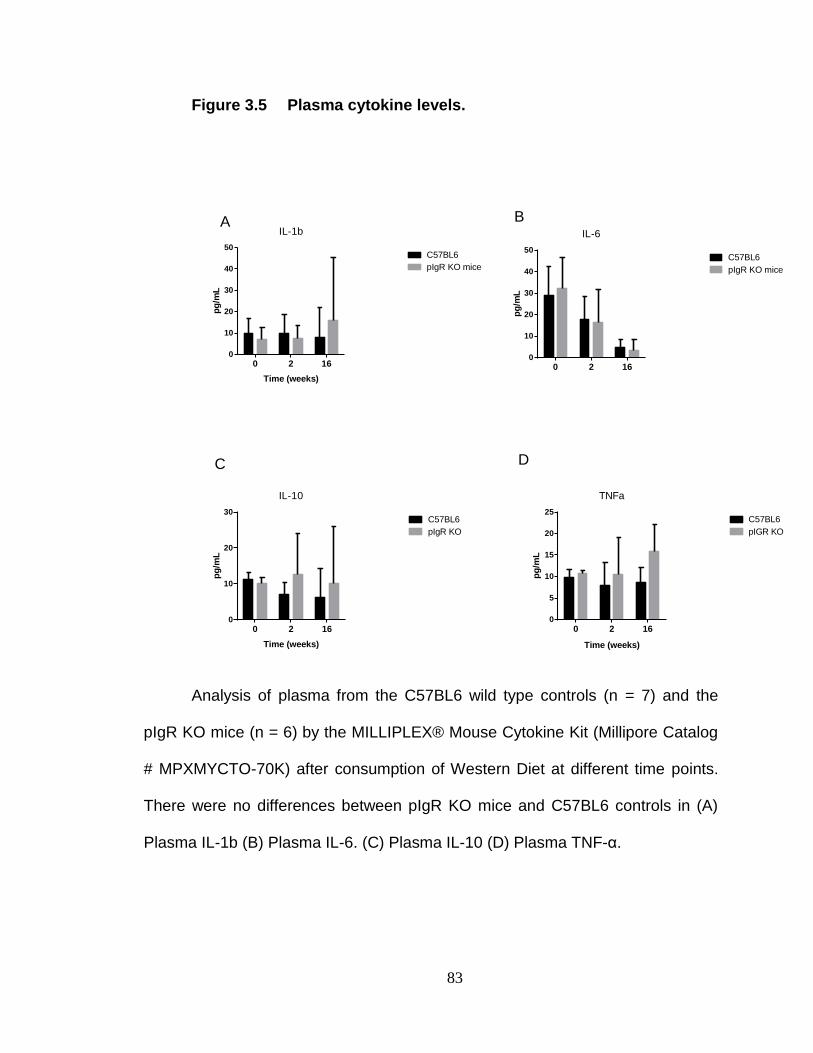
83
Figure 3.5 Plasma cytokine levels.
Analysis of plasma from the C57BL6 wild type controls (n = 7) and the
pIgR KO mice (n = 6) by the MILLIPLEX® Mouse Cytokine Kit (Millipore Catalog
# MPXMYCTO-70K) after consumption of Western Diet at different time points.
There were no differences between pIgR KO mice and C57BL6 controls in (A)
Plasma IL-1b (B) Plasma IL-6. (C) Plasma IL-10 (D) Plasma TNF-α.
IL-1b
Time (weeks)
pg
/mL
0 2 160
10
20
30
40
50C57BL6
pIgR KO mice
IL-6
pg
/mL
0 2 160
10
20
30
40
50C57BL6
pIgR KO mice
IL-10
Time (weeks)
pg
/mL
0 2 160
10
20
30C57BL6
pIgR KO
TNFa
Time (weeks)
pg
/mL
0 2 160
5
10
15
20
25C57BL6
pIGR KO
A B
C D
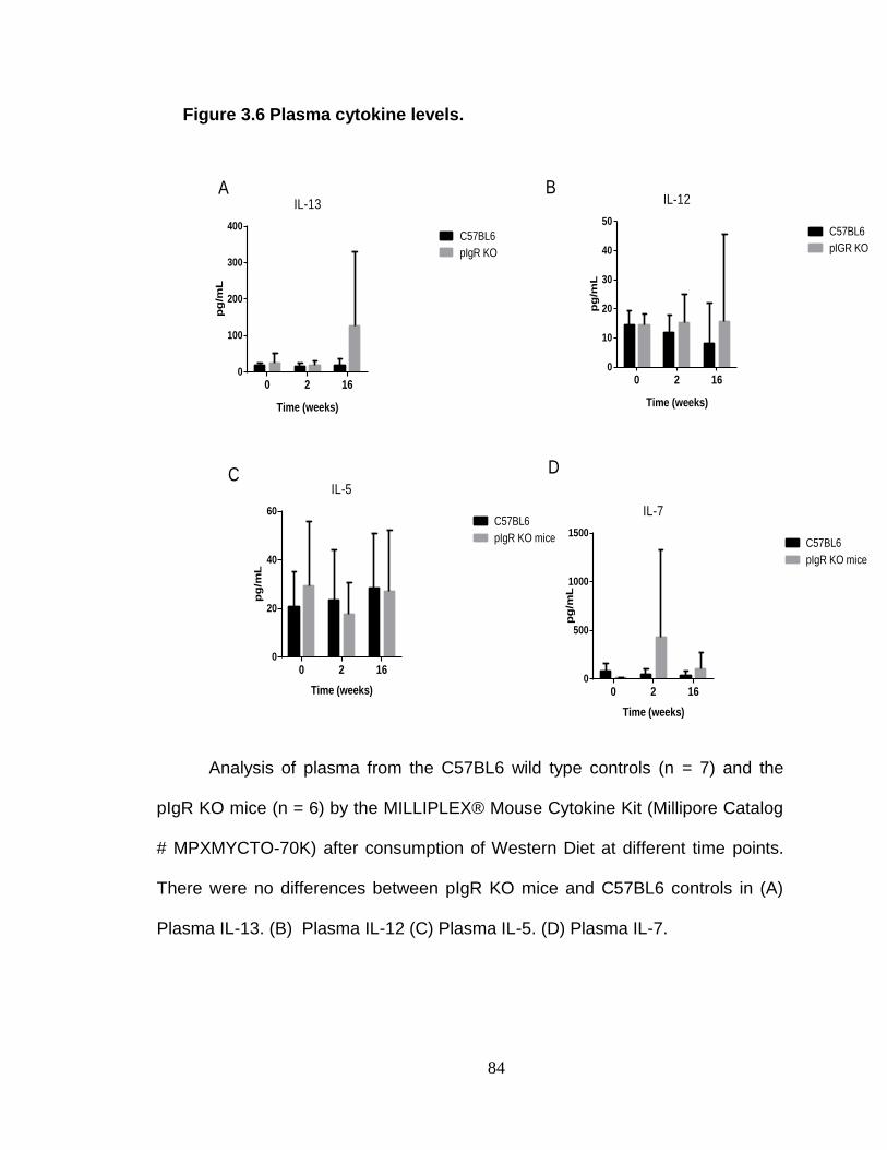
84
Figure 3.6 Plasma cytokine levels.
Analysis of plasma from the C57BL6 wild type controls (n = 7) and the
pIgR KO mice (n = 6) by the MILLIPLEX® Mouse Cytokine Kit (Millipore Catalog
# MPXMYCTO-70K) after consumption of Western Diet at different time points.
There were no differences between pIgR KO mice and C57BL6 controls in (A)
Plasma IL-13. (B) Plasma IL-12 (C) Plasma IL-5. (D) Plasma IL-7.
IL-5
Time (weeks)
pg
/mL
0 2 160
20
40
60C57BL6
pIgR KO mice
IL-7
Time (weeks)
pg
/mL
0 2 160
500
1000
1500C57BL6
pIgR KO mice
IL-13
Time (weeks)
pg
/mL
0 2 160
100
200
300
400C57BL6
pIgR KO
A B
C D
IL-12
Time (weeks)
pg
/mL
0 2 160
10
20
30
40
50C57BL6
pIGR KO
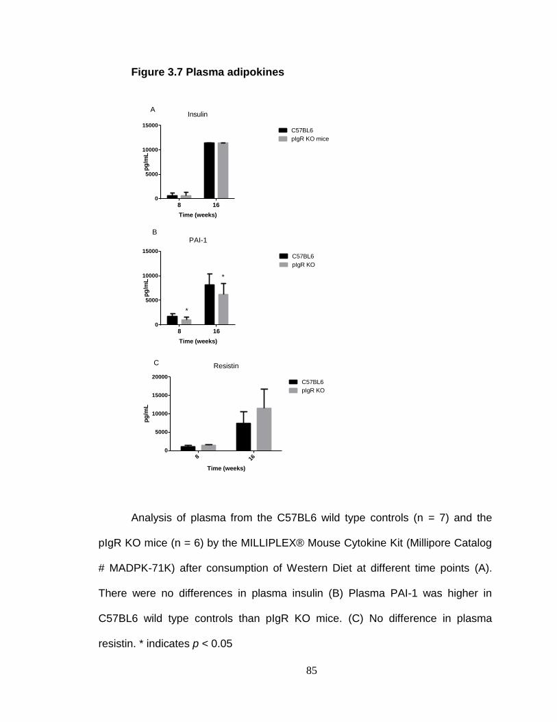
85
Figure 3.7 Plasma adipokines
Analysis of plasma from the C57BL6 wild type controls (n = 7) and the
pIgR KO mice (n = 6) by the MILLIPLEX® Mouse Cytokine Kit (Millipore Catalog
# MADPK-71K) after consumption of Western Diet at different time points (A).
There were no differences in plasma insulin (B) Plasma PAI-1 was higher in
C57BL6 wild type controls than pIgR KO mice. (C) No difference in plasma
resistin. * indicates p < 0.05
Insulin
Time (weeks)
pg
/mL
8 160
5000
10000
15000C57BL6
pIgR KO mice
PAI-1
Time (weeks)
pg
/mL
8 160
5000
10000
15000C57BL6
pIgR KO
*
*
Resistin
Time (weeks)
pg
/mL
8 16
0
5000
10000
15000
20000C57BL6
pIgR KO
A
B
C
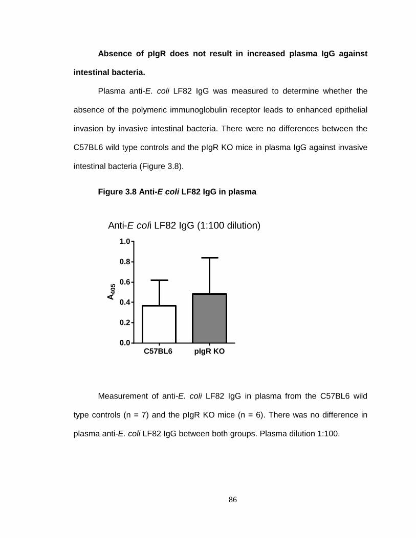
86
Absence of pIgR does not result in increased plasma IgG against
intestinal bacteria.
Plasma anti-E. coli LF82 IgG was measured to determine whether the
absence of the polymeric immunoglobulin receptor leads to enhanced epithelial
invasion by invasive intestinal bacteria. There were no differences between the
C57BL6 wild type controls and the pIgR KO mice in plasma IgG against invasive
intestinal bacteria (Figure 3.8).
Figure 3.8 Anti-E coli LF82 IgG in plasma
Measurement of anti-E. coli LF82 IgG in plasma from the C57BL6 wild
type controls (n = 7) and the pIgR KO mice (n = 6). There was no difference in
plasma anti-E. coli LF82 IgG between both groups. Plasma dilution 1:100.
Anti-E coli LF82 IgG (1:100 dilution)
A4
05
C57BL6 pIgR KO0.0
0.2
0.4
0.6
0.8
1.0
87
Absence of pIgR does not alter the percentages of immune cell
subsets after long term Western Diet consumption.
In order to determine whether the absence of the polymeric
immunoglobulin receptor leads to increased inflammation after consuming a
high-fat Western Diet, inflammatory immune cells were measured by the
HEMAVET® 950 FS Multispecies Hematology System. Hematological analysis of
whole blood revealed that there were no differences between the C57BL6 wild
type controls and the pIgR KO mice in neutrophil percentage, basophil
percentage, monocyte percentage and eosinophil percentage (Figures 3.9 A, 3.9
B, 3.9 C, and 3.9 D).
Absence of pIgR does not promote increased intestinal inflammation
after long term Western Diet consumption.
Real time PCR (RT-PCR) was performed in order to investigate whether
the absence of pIgR in conjunction with long term consumption of a high-fat
Western Diet can promote intestinal inflammation. TNF-α and IL-6 expression
was measured in the ileum and the colon. These regions of the GI tract were
selected for analysis since they harbor the largest bacterial populations. After
sixteen weeks of consuming the Western Diet, no differences were observed
between the C57BL6 controls and the pIgR KO mice in IL-6 expression in the
ileum, and colon (Figures 3.10 A and 3.10 B). There were no significant
differences in TNF-α expression in the ileum and colon (Figures 3.10 C and 3.10
D). These findings suggest that impaired IgA secretion by pIgR does not lead to
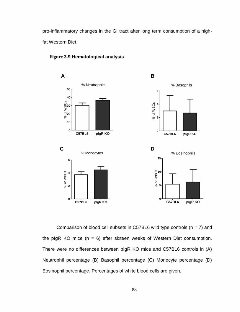
88
pro-inflammatory changes in the GI tract after long term consumption of a high-
fat Western Diet.
Figure 3.9 Hematological analysis
% o
f W
BC
s
C57BL6 pIgR KO0
10
20
30
40
50
% Neutrophils
% o
f W
BC
s
C57BL6 pIgR KO0
2
4
6
% Monocytes%
of
WB
Cs
C57BL6 pIgR KO0
5
10
15
% Eosinophils%
of
WB
Cs
C57BL6 pIgR KO0
2
4
6
% Basophils
A B
C D
Comparison of blood cell subsets in C57BL6 wild type controls (n = 7) and
the pIgR KO mice (n = 6) after sixteen weeks of Western Diet consumption.
There were no differences between pIgR KO mice and C57BL6 controls in (A)
Neutrophil percentage (B) Basophil percentage (C) Monocyte percentage (D)
Eosinophil percentage. Percentages of white blood cells are given.
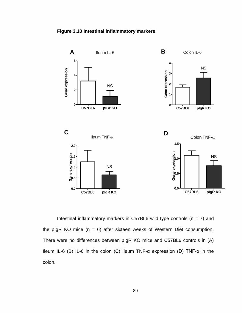
89
Figure 3.10 Intestinal inflammatory markers
Gen
e e
xp
ressio
n
C57BL6 pIGr KO0
2
4
6
NS
Ileum IL-6
Gen
e e
xp
ressio
n
C57BL6 pIgR KO0
1
2
3
4
NS
Colon IL-6
Gen
e e
xp
ressio
n
C57BL6 pIgR KO0.0
0.5
1.0
1.5
2.0
NS
Ileum TNF-
Gen
e e
xp
ressio
n
C57BL6 pIgR KO0.0
0.5
1.0
1.5
NS
Colon TNF-
A B
C D
Intestinal inflammatory markers in C57BL6 wild type controls (n = 7) and
the pIgR KO mice (n = 6) after sixteen weeks of Western Diet consumption.
There were no differences between pIgR KO mice and C57BL6 controls in (A)
Ileum IL-6 (B) IL-6 in the colon (C) Ileum TNF-α expression (D) TNF-α in the
colon.
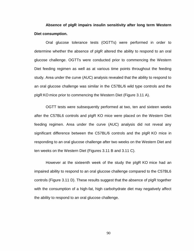
90
Absence of pIgR impairs insulin sensitivity after long term Western
Diet consumption.
Oral glucose tolerance tests (OGTTs) were performed in order to
determine whether the absence of pIgR altered the ability to respond to an oral
glucose challenge. OGTTs were conducted prior to commencing the Western
Diet feeding regimen as well as at various time points throughout the feeding
study. Area under the curve (AUC) analysis revealed that the ability to respond to
an oral glucose challenge was similar in the C57BL/6 wild type controls and the
pIgR KO mice prior to commencing the Western Diet (Figure 3.11 A).
OGTT tests were subsequently performed at two, ten and sixteen weeks
after the C57BL6 controls and pIgR KO mice were placed on the Western Diet
feeding regimen. Area under the curve (AUC) analysis did not reveal any
significant difference between the C57BL/6 controls and the pIgR KO mice in
responding to an oral glucose challenge after two weeks on the Western Diet and
ten weeks on the Western Diet (Figures 3.11 B and 3.11 C).
However at the sixteenth week of the study the pIgR KO mice had an
impaired ability to respond to an oral glucose challenge compared to the C57BL6
controls (Figure 3.11 D). These results suggest that the absence of pIgR together
with the consumption of a high-fat, high carbohydrate diet may negatively affect
the ability to respond to an oral glucose challenge.
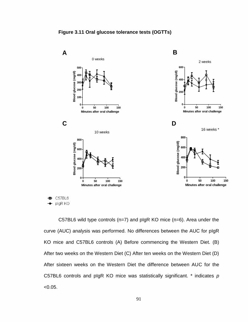
91
Figure 3.11 Oral glucose tolerance tests (OGTTs)
Minutes after oral challenge
Blo
od
glu
co
se (
mg
/dl)
0 50 100 1500
100
200
300
400
500
0 weeks
Minutes after oral challenge
Blo
od
glu
co
se (
mg
/dl)
0 50 100 1500
200
400
600
2 weeks
Minutes after oral challenge
Blo
od
glu
co
se (
mg
/dl)
0 50 100 1500
200
400
600
800
10 weeks
Minutes after oral challenge
Blo
od
glu
co
se (
mg
/dl)
0 50 100 1500
200
400
600
800
16 weeks *
A B
C D
C57BL6 wild type controls (n=7) and pIgR KO mice (n=6). Area under the
curve (AUC) analysis was performed. No differences between the AUC for pIgR
KO mice and C57BL6 controls (A) Before commencing the Western Diet. (B)
After two weeks on the Western Diet (C) After ten weeks on the Western Diet (D)
After sixteen weeks on the Western Diet the difference between AUC for the
C57BL6 controls and pIgR KO mice was statistically significant. * indicates p
<0.05.
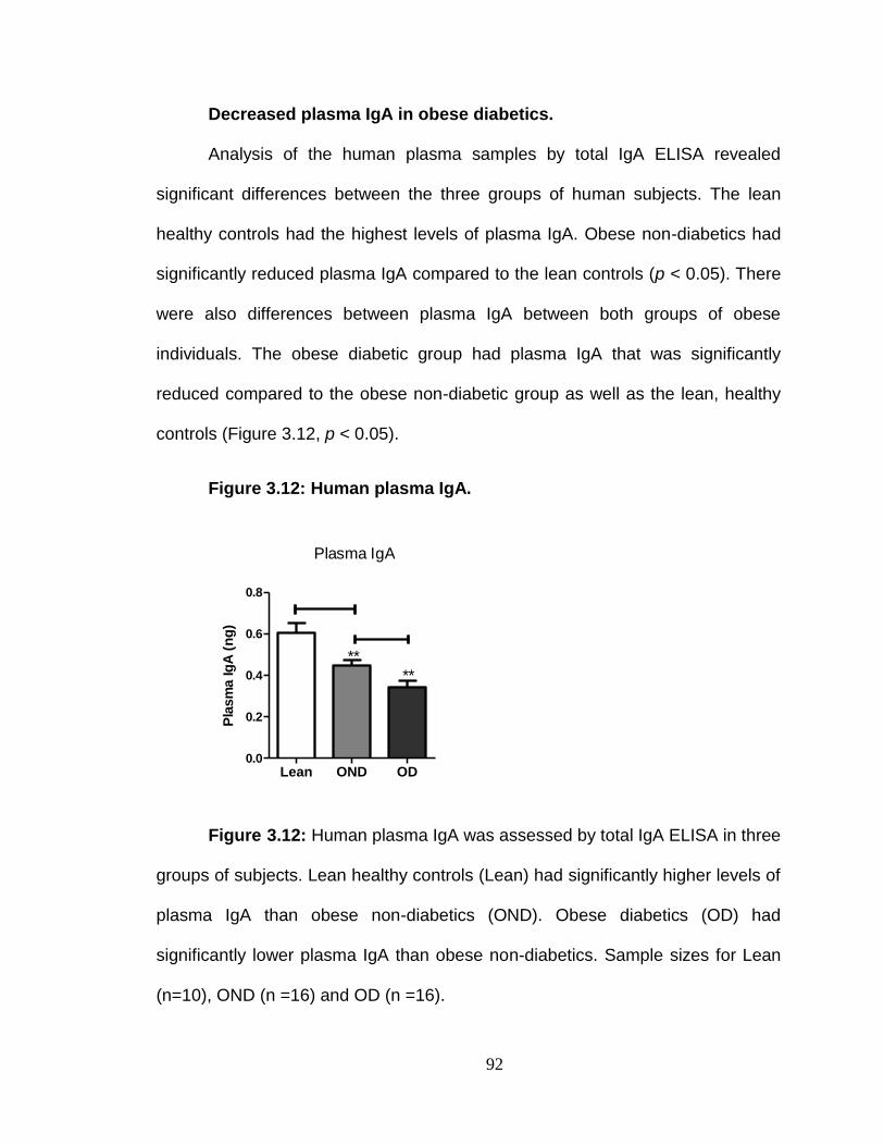
92
Decreased plasma IgA in obese diabetics.
Analysis of the human plasma samples by total IgA ELISA revealed
significant differences between the three groups of human subjects. The lean
healthy controls had the highest levels of plasma IgA. Obese non-diabetics had
significantly reduced plasma IgA compared to the lean controls (p < 0.05). There
were also differences between plasma IgA between both groups of obese
individuals. The obese diabetic group had plasma IgA that was significantly
reduced compared to the obese non-diabetic group as well as the lean, healthy
controls (Figure 3.12, p < 0.05).
Figure 3.12: Human plasma IgA.
Pla
sm
a Ig
A (
ng
)
Lean OND OD0.0
0.2
0.4
0.6
0.8
**
**
Plasma IgA
Figure 3.12: Human plasma IgA was assessed by total IgA ELISA in three
groups of subjects. Lean healthy controls (Lean) had significantly higher levels of
plasma IgA than obese non-diabetics (OND). Obese diabetics (OD) had
significantly lower plasma IgA than obese non-diabetics. Sample sizes for Lean
(n=10), OND (n =16) and OD (n =16).
93
DISCUSSION
Long term HFD consumption decreases fecal IgA.
Immunoglobulin A (IgA) secreted into the lumen of the GI tract minimizes
adherence of intestinal bacteria to the intestinal epithelium. Since long term HFD
consumption is linked to elevated plasma IgG against invasive intestinal bacteria,
it is possible that IgA secretion into the lumen of the GI tract could be impaired.
To determine whether this was the case, fecal IgA levels were measured in
C57BL6 mice fed a HFD and a LFD diet on a weekly basis. It was expected that
prior to commencing the feeding study that the fecal IgA levels would be similar.
With time, fecal IgA levels in the HFD mice would decrease relative to the LFD
controls. This study is not the first to investigate dietary intervention on fecal IgA
production. The consumption of probiotic containing foods may improve overall
health [172]. These improved indicators of health have been correlated with
increased fecal sIgA levels.
Prior to switching the mice to the special diets, both groups of mice had
similar fecal IgA levels. However, by the fourth week of the feeding study there
was a significant difference between the LFD mice and HFD mice. After four
weeks HFD mice had significantly lower fecal IgA levels compared to LFD
controls (Figure 3.1). However, fecal IgA/g feces did not decrease in the HFD
mice. Instead what was observed was an increase in LFD mice relative to the
HFD mice. When the mice were purchased they were five weeks old. The
feeding study was commenced when they were six weeks old.
94
Secretory IgA production increases with age [173]. It is possible that in this
feeding study an age effect was observed. B-1 cells play a major role in IgA
production in the gut [174]. Furthermore, B-1 cells are adversely affected by
consumption of high levels of dietary fat [175]. Therefore it is possible the
differences between fecal IgA in LFD and HFD mice are a consequence of
dietary fat inhibiting development of a normal immune repertoire in the HFD
mice.
The effect of long term HFD consumption on plasma IgA
Long term HFD consumption did not appear to have an effect on plasma
IgA in mice. After the ten week feeding study, both groups of mice had similar
levels of plasma IgA (Figure 3.2). Considering the relatively small sample size in
the study (n=6 for each group), it is likely that further studies should be performed
using larger sample sizes.
Absence of pIgR does not exacerbate inflammation.
A major component of our study was to investigate whether the absence
of active IgA secretion into the lumen of the GI tract by the polymeric
immunoglobulin receptor (pIgR) is a predisposing factor for development of
metabolic syndrome. As it has been previously mentioned, pIgR actively secretes
polymeric immunoglobulins into the lumen of the GI tract. The quantity of IgA
secreted into the lumen of the GI tract by pIgR is immense (an estimated three
grams per day) [168, 176]. What is even more intriguing is that the pIgR
traversing the IECs are never recycled. pIgR not bound to IgA is also secreted
into the lumen of the GI tract as secretory component (SC). This raises the
95
question as to whether such an elaborate transport mechanism requiring a
considerable input of energy serves a crucial purpose. We observed that long
term HFD consumption led to increased plasma IgG against an invasive
intestinal bacterial strain.
In addition to our experimental findings, we were intrigued by the study
performed by Gewirtz et al. That study utilized mice deficient in the Toll-like
receptor 5 (TLR5) [171]. TLR5 binds flagellin, a constituent of intestinal bacteria
[177]. TLR5 KO mice developed features of the metabolic syndrome. These
include increased weight gain, adiposity and insulin resistance. Since TLR5 KO
mice are unable to respond to flagellin containing bacteria, it is probable that that
these mice have an altered intestinal bacterial population from wild type control
mice.
Since pIgR KO mice lack a specialized method of transporting pIgA, it was
reasonable to hypothesize that a significant reduction in the amount of stable
secretory IgA could result in a drastically altered intestinal bacterial composition.
It was expected that a combination of an intestinal bacterial population enriched
in pathogenic strains and long term consumption of a high-fat, high-carbohydrate
Western Diet would lead to metabolic dysregulation as well as increased
systemic inflammation.
A wide range of parameters were evaluated prior to commencing the
Western Diet feeding regimen. These included body weight and body fat, plasma
inflammatory cytokines and the ability to respond to an oral glucose challenge.
96
Considering the vast quantity of IgA secreted into the lumen of the GI tract, we
reasoned that pIgR KO mice would be more susceptible to epithelial invasion by
invasive intestinal bacteria. Instead it was observed that plasma IgG levels
against invasive intestinal bacteria was similar in both pIgR KO mice and the
C57BL6 littermate controls.
No significant differences were observed in these parameters between the
pIgR KO mice and the C57BL6 littermate wild type controls prior to commencing
the Western Diet feeding regimen. The mice used were relatively young (age six
weeks). It is likely that during the nursing stage both groups of mice received
ample amounts of IgA from the breast milk thereby negating any potential
differences in intestinal bacteria due to the deficiency in pIgR. Despite the
obvious difference in IgA secreting capacity, the lack of differences in metabolic
parameters prior to consuming the Western Diet feeding regimen was not
unexpected.
Body weight, body fat percentage, plasma inflammatory cytokines,
immune cell subsets, intestinal inflammation and oral glucose tolerance were
evaluated prior to commencing the Western Diet feeding regimen, as well as at
various points subsequent to commencing the Western Diet. There were no
differences in plasma inflammatory cytokines between pIgR KO mice and
C57BL6 wild type controls either prior to commencing the Western Diet or at any
point during the feeding study. With the exception of PAI-1, plasma adipokine
levels did not differ at 8 or 16 weeks after commencing the Western Diet feeding
regimen.
97
There were no baseline differences in the ability to respond to an oral
glucose challenge between the pIgR KO mice and C57BL6 wild type controls.
Two and ten weeks after commencing Western Diet feeding there were no
differences in responding to an oral glucose challenge between pIgR KO mice
and C57BL6 wild type controls. However after sixteen weeks on the Western Diet
area under the curve (AUC) analysis revealed that pIgR KO mice had impaired
responses to an oral glucose challenge compared to C57BL6 controls.
After performing euthanasia on both groups of mice, intestinal
inflammation was measured. There were no differences in any of these
parameters between pIgR KO mice and C57BL6 controls. This was surprising
given the fact that a large quantity of IgA is secreted into the lumen of the GI tract
by pIgR. However, there are a few possible explanations as to why no
differences in intestinal inflammation were observed between the C57BL6 and
the pIgR KO mice. First of all, this study lasted for a shorter duration than the
study using TLR5 KO mice. Whereas this study was performed for a total of 16
weeks, the study performed by Gewirtz et al was a twenty week feeding study. It
is possible that the effects of pIgR deficiency had not yet become apparent.
Another possible reason no differences were observed between both
groups of mice is because pIgR KO mice are not completely deficient in intestinal
IgA. A study performed by Johansen et al, compared fecal IgA levels in pIgR KO
mice and wild type controls [170]. Fecal IgA was measured in pIgR KO mice and
C57BL/6 controls by ELISA. Statistical analysis revealed that fecal IgA in pIgR
KO mice was less than in the C57BL/6 controls. However, IgA was still
98
detectable in the feces of pIgR KO mice despite their inability to actively secrete
IgA. This is because plasma IgA diffuses into the intestinal mucosa by
paracellular diffusion. Furthermore, pIgR KO mice have significantly elevated
fecal IgG which may confer protection against the intestinal bacteria.
We observed impaired responses to an oral glucose challenge at the end
of the study in the pIgR KO mice compared to the wild type C57BL6 mice.
However this did not correspond to an increase in plasma and intestinal
inflammatory cytokines. It is possible that the MILLIPLEX analysis was not the
best method of evaluating systemic inflammation. Furthermore, in the intestinal
tissue only a limited number of inflammatory markers were examined. Perhaps, a
wider range of inflammatory markers need to be examined. The study of long
term HFD consumption on pIgR KO mice is inconclusive and should be repeated
with pIgR KO mice housed separately from wild-type controls.
Reduced plasma IgA in obese diabetics.
Analysis of the human plasma samples revealed differences between the
three groups of human subjects. The lean controls had the highest levels of
plasma IgA. Obese non-diabetics had significantly reduced plasma IgA
compared to the lean controls. Obese diabetics had significantly reduced plasma
IgA compared to both obese non-diabetics and lean controls (Figure 3.12).
Relatively little is known about the precise role of plasma IgA. This has been
attributed to difficulties in purification of plasma IgA. Plasma IgA can neutralize
antigens. However, it has a poor ability to promote opsonization. There are a
number of effector functions possessed by plasma IgA. These include
99
enhancement of antibacterial effect of lactoperoxidase and suppression of
inflammatory effects of immune lysis, NK cell activity and antibody dependent
cellular toxicity.
Reduced serum IgA is more prevalent in individuals who have lost
tolerance to normal food antigens [178]. Serum IgA neutralizes normally
‘harmless’ antigenic material such as food antigens and autoantigens. Serum IgA
clears these antigens from the systemic circulation in a non-inflammatory
manner. It is has been suggested that IgA clearance of antigens prevents
inappropriate responses to these antigens [179]. The higher prevalence of
autoimmune disorders in IgA deficient individuals has been attributed
inappropriate immune destruction of these antigens [180-182]. Thus the
significant decrease in plasma IgA in obese-diabetics may have led to
inflammatory immune responses that in turn led to insulin resistance.
However, further analysis is needed on plasma IgA levels with regards to
type 2 diabetes. A previous study by Gill et al noted that type 2 diabetics had
higher plasma IgA levels than healthy controls [183]. This was attributed to the
fact that some of the diabetic individuals in that study had acute or chronic
bacterial infections. Furthermore, subclinical infections could not be ruled out in
the diabetics who did not have bacterial infections. A possible reason for the
reduced plasma IgA in obese diabetics is a defect in IgA synthesis.
Copyright © Nadeem K. Mohammed 2012
100
CHAPTER 4 DISCUSSION
EVALUATION OF PROPOSED HYPOTHESIS.
Our preliminary findings led us to propose that long term consumption of a
high fat diet could impair intestinal IgA production, facilitating adherence and
invasion of the intestinal epithelium by the commensal bacteria. Invasion of the
intestinal epithelium by the commensal bacteria could trigger inflammatory
responses, which in turn could impair insulin signaling, culminating in the
development of type 2 diabetes.
Decreased IgA production by long term HFD consumption: Over the
course of a long term (10 week) feeding study C57BL6 mice fed a low fat diet
(10% kCal from fat) experienced an increase in fecal IgA production relative to
mice fed a high fat diet (60% kCal from fat). These findings provided evidence
that consumption of high levels of dietary fat may impair the mucosal IgA barrier.
Revision to proposed model-Invasion of the intestinal epithelium by
invasive bacteria: Initially we proposed that long term consumption of a high fat
diet could result in the commensal bacteria being able to breach the intestinal
epithelium and elicit inflammatory responses. However, a major limitation of our
preliminary studies was the exclusive use of E. coli LF82, an invasive intestinal
bacterial strain. To address this concern non-invasive intestinal bacteria were
analyzed. IgG against E. coli Nissle 1917, Bacteroides thetaiotaomicron and
Lactobacilus acidophilus was measured in the plasma of HFD and LFD mice.
Plasma IgG against these bacterial strains was measured using the lab based
101
ELISA. There were no significant differences in IgG against these bacteria in LFD
mice or HFD mice (Figures 2.4 B, 2.4 C and 2.4 D).
The studies cited earlier using GF mice suggested that the presence of
intestinal bacteria is required for the negative effects of long term HFD
consumption. However, none of those studies examined the role of specific
intestinal bacterial species. The findings of our studies provide evidence that
invasive intestinal bacterial strains are associated with inflammation and insulin
resistance.
The other intestinal bacterial strains examined are beneficial to the host.
E. coli Nissle 1917 (EcN) is a widely studied probiotic. Probiotics are viable
microbes which may confer health benefits when consumed [184]. The ability of
EcN to confer health benefits has been known for almost a century since the First
World War when it was isolated from a lone soldier who did not succumb to
enterocolitis. Investigations on the effects of EcN supplementation show
promising results for treating colitis [185, 186]. An interesting coincidence is that
EcN has been utilized to counteract the effects of the LF82 strain of E. coli [187].
Reports have also provided evidence that Bacteroides thetaiotaomicron may
prevent rotavirus infections [188]. It has been reported that Lactobacilus
acidophilus has the ability to diminish inflammation caused by Helicobacter pylori
[189]. By contrast, the LF-82 strain of E. coli has been described as being
invasive in nature [151]. E. coli LF82 was isolated from an ileal lesion of a
Crohn’s Disease patient [150]. E. coli LF82 is designated as an adherent invasive
102
E. coli strain (AIEC strain). Invasive strains are characterized by their ability to
enter epithelial cells and subsequently survive and replicate.
Immune responses against invasive intestinal bacteria are associated with
inflammation. In these studies a statistical correlation between anti-E. coli LF82
IgG and TNF-α was observed. The correlation analysis was performed using the
plasma samples from the obese patients. Both groups had equivalent mean BMI
values, ruling out differences in adiposity as a TNF-α source. The correlation
between anti-E. coli LF82 IgG and TNF-α provided evidence that immune
responses to the intestinal bacterial antigens are associated with inflammation. It
is possible that the increase in IgG against intestinal antigens promoted cross-
linking of FcγRs, which in turn caused the release of TNF-α.
Inflammatory responses resulting from invasion of the intestinal epithelium
impairs insulin signaling. Elevated plasma anti-E. coli LF82 IgG in the HFD mice
provided evidence that long term consumption of high levels of dietary fat is
associated with invasion of the intestinal epithelium by invasive bacteria.
Increased plasma TNF-α and neutrophil percentage in the blood of HFD mice
suggests that a consequence of epithelial penetration by invasive bacteria is
inflammation. Inflammation is associated with a variety of disease states.
One of these conditions is type 2 diabetes. The increase in fasting blood
glucose levels in HFD mice compared to LFD mice was significant (Figure 5B,
p<0.05). It is possible that elevated anti-E. coli LF82 IgG against intestinal
bacteria led to increased cross-linking of FcγRs with the Fc domains. This in turn
103
may have led to increased TNF-α secretion as well as increased neutrophils in
the blood. The chronic inflammation could have in turn impaired insulin signaling
pathways, leading to the development of insulin resistance.
Significance of studies.
These studies have significant clinical relevance. Although there is
widespread awareness about the risk factors for developing type 2 diabetes, the
prevalence of obesity and metabolic syndrome has continued to rise. This has
been costly as treatment and the loss of productivity due to various disabling
complications of type 2 diabetes continue to burden the healthcare sector. The
research into novel mechanisms of treating type 2 diabetes is in itself a tacit
admission that it is impossible to expect the tens of millions of overweight
individuals to alter their lifestyle radically enough to maintain a healthy
bodyweight.
Apart from individuals with Crohn’s Disease the LF82 strain of E. coli is
rare. It is unlikely that a large percentage of individuals harbor this particular
strain. However, E. coli LF82 may possess antigens that are shared by other
intestinal bacterial species. The intestinal bacteria remain poorly characterized.
There is a possibility that invasive intestinal bacterial strains that have not yet
been identified reside in the GI tract. Long term HFD consumption may enhance
the capacity of invasive bacterial strains to cross the IEC barrier. This can elicit
inflammation leading to insulin resistance.
104
Furthermore, our findings may explain why some obese individuals
develop type 2 diabetes whereas others do not. Individuals harboring invasive
intestinal bacterial strains may be more prone to chronic inflammation and
impaired insulin signaling. To more comprehensively define the role of the
intestinal bacteria in promoting systemic inflammation, further studies will need to
be performed. IgG against protein extracts from invasive intestinal bacterial
strains can be measured in diabetics and non-diabetics. Based on our findings
we expect that diabetics would have higher IgG against invasive intestinal
bacterial strains. Comparison of IgG in a larger group of obese non-diabetics and
obese-diabetics would give a better indication of whether long term HFD
consumption enhances invasion of the IEC barrier by invasive intestinal bacterial
strains. The clinical implications of these studies could be novel methods of
treating type 2 diabetes. It is possible that specific anti-microbial compounds
could be developed to selectively eliminate invasive intestinal bacterial strains
residing in obese individuals. This could spare these individuals from developing
type 2 diabetes. It would also reduce the financial burden to the healthcare sector
significantly.
Technical contributions.
These studies resulted in technical contributions for assessing immune
responses against the intestinal bacteria. ELISA kits to assay systemic immune
responses against intestinal bacterial antigens were difficult to obtain. To address
the lack of commercial ELISAs we developed our own lab based ELISA. This lab
based ELISA is a reliable, reproducible assay for assaying the humoral immune
105
response against antigens from intestinal bacterial strains. To the best of our
knowledge, no one had developed the methods of Western Blotting to analyze
the levels of plasma IgG against intestinal bacterial proteins. Researchers hoping
to assess the systemic immune response against intestinal bacterial strains in a
timely manner can use the information provided in this publication.
EVALUATION OF THESE STUDIES.
Strengths: These studies were the first to investigate the association
between specific intestinal bacteria and type 2 diabetes. Previous studies had
linked the presence of intestinal bacteria to obesity, inflammation and insulin
resistance. However, a key flaw of those studies was that they did not examine
which particular bacteria resulted in the development of inflammation and insulin
resistance. The findings of previous studies implicated that bacteria residing in
the GI tract promoted the development of inflammation. Although there is
extensive evidence supporting this proposal, it should be kept in mind that there
are hundreds of bacterial species residing in the GI tract. Given the enormous
diversity of the intestinal bacteria, it cannot be assumed that all intestinal bacteria
are equally capable of promoting systemic inflammation and insulin resistance.
As the results from our studies indicate, not all intestinal bacteria possess the
same ability to cross the intestinal epithelial cell barrier.
These studies provided a more definitive link between the intestinal
bacteria, inflammation and insulin resistance. A critical component lacking in the
previous studies in GF mice was showing that the inflammation observed as a
consequence of HFD consumption was the result of intestinal bacteria. Although
106
the studies comparing GF mice to conventionally raised mice measured a variety
of parameters, none of these studies performed immunological assays that
showed immune responses to the intestinal bacteria. This study quantified
plasma IgG against intestinal bacteria in addition to quantifying plasma TNF-α
and fasting blood glucose values, linking all three parameters.
Our studies characterized the effect of HFD consumption on the localized
immune responses at the mucosal surface of the GI tract. Although previous
studies had linked the intestinal bacteria to systemic inflammation and insulin
resistance, none of these studies investigated whether this was due to
impairment of the localized adaptive intestinal mucosal responses. It should be
noted that the effect of impairment of the intestinal immunity on shaping the
composition of the gut microbiota was examined by Gewirtz et al using TLR5
deficient mice. However, their study looked at the deficiency of innate immune
mechanisms in the GI tract. Our studies examined the effect of HFD consumption
on the adaptive immune response to the intestinal bacteria. This is an important
topic to address given the immense load of IgA secreted into the GI tract as well
as the documented protective effects of IgA.
Weaknesses: A major critique of this study is the extensive use of the
LF82 strain of E. coli. as it has been previously stated, this strain of bacteria is
recognized as an adherent invasive bacterial strain. Therefore it is not an ideal
strain to use as an experimental model. This experimental model deficiency was
addressed by assessing the effect of HFD consumption on the systemic immune
response to the non-pathogenic bacteria E. coli Nissle 1917, Bacteroides
107
thetaiotaomicron and Lactobacilus acidophilus. Despite the fact that HFD
consumption did not appear to alter the systemic immune response to these
bacteria, Western Blot analysis showed that HFD mice had higher plasma IgG
against the antigens present in the cecum of control mice. Higher plasma IgG
against the antigens from the cecum in HFD mice strengthened our hypothesis
that HFD consumption may lead to translocation of intestinal bacteria into the
systemic circulation. However, at this point, the only bacterial strain to which the
systemic immune response has shown to be increased as a result of HFD
consumption the LF82 strain of E. coli.
The findings of these studies point to the possibility that individuals
harboring invasive intestinal bacterial strains are at increased risk of developing
systemic inflammatory immune responses that may increase their risk of
developing type 2 diabetes. However, these studies did not determine whether
the composition of the gut microbes changed during the HFD feeding regimen.
As a consequence, it is not known if HFD consumption increases the proportion
of invasive intestinal bacteria.
There were flaws in the assessment of HFD consumption on the localized
immune responses at the mucosal surface of the GI tract. Although fecal IgA was
quantified, no other immunoglobulin classes were measured. It is known that IgM
plays a role in preventing the intestinal bacteria from adhering to the intestinal
epithelium. Fecal IgM was not measured. Thus it is not conclusive as to whether
HFD consumption impairs mucosal immunity. Furthermore, a number of
mechanisms prevent the commensal bacteria in the lumen of the GI tract from
108
adhering to the intestinal epithelial cells. These include mucus production by
goblet cells as well as the production of defensins, cathelicidins, phospholipases,
lysozyme, and Reg III-gamma. These parameters were not examined in LFD
mice and HFD mice.
EXPERIMENTAL DESIGN.
Lab developed ELISA: A key difference between the lab developed
ELISA and the Western Blot analysis was that the Western Blot showed
differences between the three groups of human subjects. Western Blot analysis
indicated that plasma anti-E. coli LF82 IgG was highest in obese diabetics,
followed by obese non-diabetics with the lowest plasma anti-E. coli LF82 IgG
found in the lean healthy controls. On the other hand, ELISA analysis showed
that the obese diabetics had significantly higher plasma anti-E. coli LF82 IgG
than lean controls with no significant differences between the obese non-
diabetics and lean healthy controls. A possible explanation for this discrepancy is
that the ELISA was prepared by extracting bacterial proteins using Bacterial
Protein Extraction Reagent (B-PER). This reagent specifically extracts soluble
proteins. It is possible that during the process of soluble protein extraction, non-
soluble proteins that possessed epitopes for immune recognition were lost.
Furthermore, the lab-developed ELISA only consisted of soluble bacterial
proteins. Non-protein compounds such as lipopolysaccharide (LPS) can also
elicit immune reactions. LPS is a constituent of the outer cell membrane of gram
negative bacteria including E. coli LF82. It is one of the more potent inducers of
inflammation and results in the release of the pro-inflammatory cytokines TNF-α,
109
IL-6, and IL-1 [190-192]. However, despite these limitations we chose to perform
the ELISAs since it was the most readily available method to give us quantitative
measurements. There was concern that the lab developed ELISA optimization
was not performed accurately. These do not negate our results since the
Western Blots show distinct differences between LFD mice and HFD mice.
Technical flaws of pIgR KO mouse study: The magnitude of active IgA
secretion into the lumen of the GI tract suggests that there is a rationale behind
this specialized mechanism of active IgA transport. Furthermore, pIgR KO mice
have significantly increased serum IgG against intestinal bacterial antigens than
C57BL6 wild type controls. There were a number of design flaws in these
studies. The mice were not housed according to genotype. As a consequence, it
was possible that differences in the gut microbe composition of both groups of
mice were minimized by coprophagy. Consumption of the fecal pellets produced
by the wild type C57BL6 mice could result in harmless commensal bacteria being
repopulated in the GI tract of the pIgR KO mice. These bacteria could in turn out-
compete any invasive strains, thereby preventing the anticipated outgrowth of
invasive bacterial strains in the pIgR KO mice. Co-housing of pIgR KO mice with
wild type C57BL6 controls also resulted in an additional source of IgA for the
pIgR KO mice. Small quantities of IgA reach the lumen of the GI tract in pIgR KO
mice via paracellular diffusion. The co-housed pIgR KO mice were provided with
an additional source of IgA in the form of fecal pellets. In addition to the IgA
reaching the lumen of the GI tract via paracellular diffusion, the pIgR KO mice
also obtained IgA by coprophagy. This in turn could have compensated for the
110
lack of active IgA transport by the pIgR. These technical shortcomings should be
addressed in future studies of the effect of HFD consumption on pIgR KO mice.
Alternative reasons for increased plasma anti-E. coli LF82 IgG.
A question that was not addressed in these studies is whether live
invasive bacteria invade the epithelium. The assays performed in these studies
involved quantification of plasma IgG against E. coli LF82, E. coli Nissle 1917,
Bacteroides thetaiotaomicron and Lactobacilus acidophilus. However, there were
no direct methods used to determine whether live bacteria had been able to
invade the intestinal epithelium. Given the huge bacterial population in the GI
tract it is inevitable that some leakage across the epithelium takes place. The
mesenteric lymph nodes act as a barrier between the GI tract and the systemic
circulation. Dendritic cells actively sample bacteria from the lumen of the GI tract.
The MLNs prevent the dendritic cells carrying commensal bacteria from gaining
access to the systemic circulation. HFD consumption results in atrophy of the
mesenteric lymph nodes and a reduction in the numbers of regulatory T
lymphocytes. It is possible therefore that increased systemic immune responses
resulted from defects in the immune repertoire, not increased IEC invasion by the
intestinal bacteria.
FUTURE DIRECTION OF STUDIES.
Resolving technical issues of these studies: These studies linked the
presence of systemic immune responses against intestinal bacteria to the
development of type 2 diabetes. However there are a number of technical issues
that need to be resolved. The lab based ELISA assay needs to be optimized by
111
preparing serial dilutions and plotting standard curves in order to determine the
dynamic or linear portion of the standard curves. This would make quantification
of plasma IgG more accurate.
Another technical issue was the oral glucose tolerance tests performed in
the Western Diet feeding study using the pIgR KO mice. The OGTTs were
performed by administering an oral gavage of 2.0g/kg glucose. The blood
glucose levels were beyond the detectable limit of the TrueTrack glucose meter
at various time points subsequent to the oral gavage. To address this issue, an
oral gavage of either 1.5g/kg glucose 1.0g/kg glucose needs to be administered
to the mice.
The effect of HFD consumption on the systemic immune response:
Although there is now considerable evidence that the bacteria residing within the
GI tract may promote systemic inflammation and insulin resistance, there are still
some issues that need to be addressed. The first is to provide evidence that
intestinal bacteria other than the LF82 strain of E. coli are associated with
systemic inflammation and insulin resistance. This method may rely on preparing
extracts from the cecums of control mice and performing Western Blots to assess
IgG levels in the plasma of HFD mice and LFD mice. If HFD consumption is
associated with increased plasma IgG against antigens from the cecum, then
proteomic analysis could be performed to decipher what antigens are eliciting the
systemic immune responses. This would be advantageous because identification
of antigens triggering a systemic immune response would aid in identifying
specific bacteria capable of penetrating the mucosal layer. Furthermore, these
112
antigens can be used for developing ELISAs for further quantitative assays of
HFD consumption and systemic IgG responses against bacteria resident in the
gut.
It is also necessary to determine when the intestinal bacteria associated
with systemic inflammatory immune responses are present. On one hand it is
possible that some individuals harbor invasive intestinal strains that make them
more prone to developing systemic inflammatory immune responses. On the
other hand, HFD consumption may alter the composition of the intestinal bacteria
resulting in an outgrowth of intestinal bacteria that may promote inflammation. To
assess the relationship between HFD consumption and the intestinal bacteria a
time course analysis could be performed. Fecal pellets can be collected from
mice prior to commencing a HFD feeding regimen. The bacteria present in the
feces can be characterized by 16SrRNA sequencing. Fecal pellets can be
collected at various time points during feeding study and the bacteria present can
be sequenced. These can then be referenced to LFD controls. This would
indicate whether HFD consumption alters the intestinal bacteria over time.
Our studies did not detect the presence of intestinal bacteria in the
systemic circulation. Instead, we measured systemic immune responses against
intestinal bacterial antigens by Western Blots and ELISAs that measured serum
immunoglobulin G (IgG) responses to bacterial antigens. Trying to detect the
presence of intestinal bacteria would have provided evidence that adherent
bacteria breach the intestinal epithelium.
113
HFD consumption and mucosal immunity: To further characterize the
effect of HFD consumption on the immune responses at the mucosal surface of
the GI tract, fecal IgM will have to be measured in LFD mice and HFD mice. This
would further strengthen the argument that mucosal immunity is impaired by HFD
consumption. Additionally, intestinal sections can be immunostained for IgA, IgM
and IgG in HFD mice and LFD mice. Comparisons of these IgG classes at the
epithelium and the lamina propria may give a more comprehensive indication as
to how HFD consumption affects immunity at the mucosal surface.
One proposed experiment was to characterize the role of IgA responses in
the GI tract using IgA knockout mice. The proposed study involved measuring
and comparing body weight, adiposity, plasma inflammatory cytokines, intestinal
inflammation and insulin resistance in mice lacking the gene for IgA production
(IgA KO mice) and C57BL6 wild type controls. These parameters were supposed
to be measured prior to commencing a high-fat, high-carbohydrate Western Diet
and at different time points during the feeding study.
IgA knockout mice are unable to synthesize IgA. This is because these
mice lack the gene for the switch and constant regions [179]. IgA is undetectable
in the serum and gastrointestinal secretions of IgA KO mice. Since mucosal IgA
is important in restricting bacteria to the lumen of the GI tract, it is possible that
the mucosal barrier of IgA KO mice may be impaired in comparison to wild type
controls. It should be noted that IgA KO mice have significantly increased IgM
and IgG levels in intestinal secretions. Elevations of IgM and IgG in the intestinal
secretions of IgA KO mice may be compensatory mechanisms for the lack of IgA.
114
Therefore future evaluation of the effect of HFD consumption should not
focus on one specific aspect of mucosal immunity. A more comprehensive array
of parameters should be evaluated. Since the commensal bacteria residing in the
GI tract do not cause problems in most individuals, millions of years of co-
evolution could have resulted in a number of redundant mechanisms to restrict
bacteria to the GI tract lumen. As a result future studies should not only focus
exclusively on IgA, but IgM as well as the production of mucus and anti-microbial
peptides.
Concluding statement:
The results of these studies show that long term HFD consumption in mice
is associated with increased plasma IgG against invasive intestinal bacterial
strains. This is a revision from our proposed experimental model. HFD mice had
higher levels of IgG against invasive intestinal bacteria than LFD mice. However,
there were no significant differences in plasma IgG against the non-invasive
intestinal bacterial strains E. coli Nissle 1917, Bacteroides thetaiotaomicron and
Lactobacilus acidophilus in HFD and LFD mice. These findings are clinically
relevant since obese-diabetics had significantly higher plasma IgG against the
invasive intestinal bacteria than lean healthy controls. Furthermore, a correlation
exists between TNF-α and IgG against E. coli LF82. Increased anti-E. coli LF 82
IgG and plasma TNF-α in obese-diabetics established a link between systemic
immune responses against invasive intestinal bacterial strains, systemic
inflammation and insulin resistance. The effect of long term HFD consumption on
the secretory immune response in the intestinal mucosa was also investigated. It
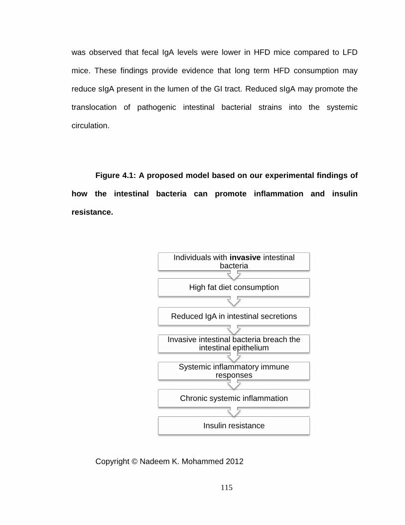
115
Insulin resistance
Chronic systemic inflammation
Systemic inflammatory immune responses
Invasive intestinal bacteria breach the intestinal epithelium
Reduced IgA in intestinal secretions
High fat diet consumption
Individuals with invasive intestinal bacteria
was observed that fecal IgA levels were lower in HFD mice compared to LFD
mice. These findings provide evidence that long term HFD consumption may
reduce sIgA present in the lumen of the GI tract. Reduced sIgA may promote the
translocation of pathogenic intestinal bacterial strains into the systemic
circulation.
Figure 4.1: A proposed model based on our experimental findings of
how the intestinal bacteria can promote inflammation and insulin
resistance.
Copyright © Nadeem K. Mohammed 2012
116
REFERENCES
1. King, H., R.E. Aubert, and W.H. Herman, Global burden of diabetes, 1995-2025: prevalence, numerical estimates, and projections. Diabetes Care, 1998. 21(9): p. 1414-31.
2. Alberti, K.G. and P.Z. Zimmet, Definition, diagnosis and classification of diabetes mellitus and its complications. Part 1: diagnosis and classification of diabetes mellitus provisional report of a WHO consultation. Diabet Med, 1998. 15(7): p. 539-53.
3. Ismail-Beigi, F., Glycemic Management of Type 2 Diabetes Mellitus. New England Journal of Medicine, 2012. 366(14): p. 1319-1327.
4. Brandle, M., et al., The direct medical cost of type 2 diabetes. Diabetes Care, 2003. 26(8): p. 2300-4.
5. Balkon, N., C. Balkon, and B.S. Zitkus, Overweight and obesity: pharmacotherapeutic considerations. J Am Acad Nurse Pract, 2011. 23(2): p. 61-6.
6. Flegal, K.M., et al., Overweight and obesity in the United States: prevalence and trends, 1960-1994. Int J Obes Relat Metab Disord, 1998. 22(1): p. 39-47.
7. Baskin, M.L., et al., Prevalence of obesity in the United States. Obes Rev, 2005. 6(1): p. 5-7.
8. Flegal, K.M., et al., Prevalence and trends in obesity among US adults, 1999-2008. JAMA, 2010. 303(3): p. 235-41.
9. Ismail-Beigi, F., Clinical practice. Glycemic management of type 2 diabetes mellitus. N Engl J Med, 2012. 366(14): p. 1319-27.
10. Shaw, J.E., R.A. Sicree, and P.Z. Zimmet, Global estimates of the prevalence of diabetes for 2010 and 2030. Diabetes Res Clin Pract, 2010. 87(1): p. 4-14.
11. Stern, M.P., et al., Does obesity explain excess prevalence of diabetes among Mexican Americans? Results of the San Antonio Heart Study. Diabetologia, 1983. 24(4): p. 272-7.
12. Isomaa, B., et al., Cardiovascular morbidity and mortality associated with the metabolic syndrome. Diabetes Care, 2001. 24(4): p. 683-9.
13. Ford, E.S., W.H. Giles, and W.H. Dietz, Prevalence of the metabolic syndrome among US adults: findings from the third National Health and Nutrition Examination Survey. JAMA, 2002. 287(3): p. 356-9.
14. Executive Summary of The Third Report of The National Cholesterol Education Program (NCEP) Expert Panel on Detection, Evaluation, And Treatment of High Blood Cholesterol In Adults (Adult Treatment Panel III). JAMA, 2001. 285(19): p. 2486-97.
15. Swaminathan, R., et al., Lysozyme: a model protein for amyloid research. Adv Protein Chem Struct Biol, 2011. 84: p. 63-111.
16. D'Andrea, A., et al., Production of natural killer cell stimulatory factor (interleukin 12) by peripheral blood mononuclear cells. J Exp Med, 1992. 176(5): p. 1387-98.
117
17. Nathan, C.F. and J.B. Hibbs, Jr., Role of nitric oxide synthesis in macrophage antimicrobial activity. Curr Opin Immunol, 1991. 3(1): p. 65-70.
18. Yamashiro, S., et al., Phenotypic and functional change of cytokine-activated neutrophils: inflammatory neutrophils are heterogeneous and enhance adaptive immune responses. J Leukoc Biol, 2001. 69(5): p. 698-704.
19. Geijtenbeek, T.B. and S.I. Gringhuis, Signalling through C-type lectin receptors: shaping immune responses. Nat Rev Immunol, 2009. 9(7): p. 465-79.
20. Drickamer, K., C-type lectin-like domains. Curr Opin Struct Biol, 1999. 9(5): p. 585-90.
21. van Kooyk, Y. and G.A. Rabinovich, Protein-glycan interactions in the control of innate and adaptive immune responses. Nat Immunol, 2008. 9(6): p. 593-601.
22. Janeway, C.A., Jr. and R. Medzhitov, Innate immune recognition. Annu Rev Immunol, 2002. 20: p. 197-216.
23. Akira, S. and K. Takeda, Toll-like receptor signalling. Nat Rev Immunol, 2004. 4(7): p. 499-511.
24. Girardin, S.E., et al., Nod1 detects a unique muropeptide from gram-negative bacterial peptidoglycan. Science, 2003. 300(5625): p. 1584-7.
25. Inohara, N., et al., Host recognition of bacterial muramyl dipeptide mediated through NOD2. Implications for Crohn's disease. J Biol Chem, 2003. 278(8): p. 5509-12.
26. Chamaillard, M., et al., An essential role for NOD1 in host recognition of bacterial peptidoglycan containing diaminopimelic acid. Nat Immunol, 2003. 4(7): p. 702-7.
27. Kanneganti, T.D., M. Lamkanfi, and G. Nunez, Intracellular NOD-like receptors in host defense and disease. Immunity, 2007. 27(4): p. 549-59.
28. Martinon, F., K. Burns, and J. Tschopp, The inflammasome: a molecular platform triggering activation of inflammatory caspases and processing of proIL-beta. Mol Cell, 2002. 10(2): p. 417-26.
29. Latz, E., The inflammasomes: mechanisms of activation and function. Curr Opin Immunol, 2010. 22(1): p. 28-33.
30. LeBien, T.W. and T.F. Tedder, B lymphocytes: how they develop and function. Blood, 2008. 112(5): p. 1570-80.
31. Kuo, C.T. and J.M. Leiden, Transcriptional regulation of T lymphocyte development and function. Annu Rev Immunol, 1999. 17: p. 149-87.
32. Levy, J.H., The human inflammatory response. J Cardiovasc Pharmacol, 1996. 27 Suppl 1: p. S31-7.
33. Vinay, D.S. and B.S. Kwon, Targeting TNF superfamily members for therapeutic intervention in rheumatoid arthritis. Cytokine, 2012. 57(3): p. 305-12.
34. Vilela, E.G., et al., Evaluation of inflammatory activity in Crohn's disease and ulcerative colitis. World J Gastroenterol, 2012. 18(9): p. 872-81.
118
35. Ohl, K. and K. Tenbrock, Inflammatory cytokines in systemic lupus erythematosus. J Biomed Biotechnol, 2011. 2011: p. 432595.
36. Wellen, K.E. and G.S. Hotamisligil, Inflammation, stress, and diabetes. J Clin Invest, 2005. 115(5): p. 1111-9.
37. Hotamisligil, G.S., N.S. Shargill, and B.M. Spiegelman, Adipose expression of tumor necrosis factor-alpha: direct role in obesity-linked insulin resistance. Science, 1993. 259(5091): p. 87-91.
38. Uysal, K.T., et al., Protection from obesity-induced insulin resistance in mice lacking TNF-alpha function. Nature, 1997. 389(6651): p. 610-4.
39. Porte, D., Jr., Banting lecture 1990. Beta-cells in type II diabetes mellitus. Diabetes, 1991. 40(2): p. 166-80.
40. Hotamisligil, G.S., et al., IRS-1-mediated inhibition of insulin receptor tyrosine kinase activity in TNF-alpha- and obesity-induced insulin resistance. Science, 1996. 271(5249): p. 665-8.
41. Aguirre, V., et al., The c-Jun NH(2)-terminal kinase promotes insulin resistance during association with insulin receptor substrate-1 and phosphorylation of Ser(307). J Biol Chem, 2000. 275(12): p. 9047-54.
42. Yang, Z.H., et al., Diet high in fat and sucrose induces rapid onset of obesity-related metabolic syndrome partly through rapid response of genes involved in lipogenesis, insulin signalling and inflammation in mice. Diabetol Metab Syndr, 2012. 4(1): p. 32.
43. Marshall, J.A., D.H. Bessesen, and R.F. Hamman, High saturated fat and low starch and fibre are associated with hyperinsulinaemia in a non-diabetic population: the San Luis Valley Diabetes Study. Diabetologia, 1997. 40(4): p. 430-8.
44. Fasching, P., et al., Metabolic effects of fish-oil supplementation in patients with impaired glucose tolerance. Diabetes, 1991. 40(5): p. 583-9.
45. Shi, H., et al., TLR4 links innate immunity and fatty acid-induced insulin resistance. J Clin Invest, 2006. 116(11): p. 3015-25.
46. Erridge, C. and N.J. Samani, Saturated fatty acids do not directly stimulate Toll-like receptor signaling. Arterioscler Thromb Vasc Biol, 2009. 29(11): p. 1944-9.
47. Kannel, W.B., Lipids, diabetes, and coronary heart disease: insights from the Framingham Study. Am Heart J, 1985. 110(5): p. 1100-7.
48. Wajchenberg, B.L., Subcutaneous and visceral adipose tissue: their relation to the metabolic syndrome. Endocr Rev, 2000. 21(6): p. 697-738.
49. Fujioka, S., et al., Contribution of intra-abdominal fat accumulation to the impairment of glucose and lipid metabolism in human obesity. Metabolism, 1987. 36(1): p. 54-9.
50. Despres, J.P., et al., Role of deep abdominal fat in the association between regional adipose tissue distribution and glucose tolerance in obese women. Diabetes, 1989. 38(3): p. 304-9.
51. Pouliot, M.C., et al., Visceral obesity in men. Associations with glucose tolerance, plasma insulin, and lipoprotein levels. Diabetes, 1992. 41(7): p. 826-34.
119
52. Hardy, O.T., et al., Body mass index-independent inflammation in omental adipose tissue associated with insulin resistance in morbid obesity. Surg Obes Relat Dis, 2011. 7(1): p. 60-7.
53. Holland, W.L., et al., Lipid-induced insulin resistance mediated by the proinflammatory receptor TLR4 requires saturated fatty acid-induced ceramide biosynthesis in mice. J Clin Invest, 2011. 121(5): p. 1858-70.
54. Badin, P.M., et al., Altered skeletal muscle lipase expression and activity contribute to insulin resistance in humans. Diabetes, 2011. 60(6): p. 1734-42.
55. Weisberg, S.P., et al., Obesity is associated with macrophage accumulation in adipose tissue. J Clin Invest, 2003. 112(12): p. 1796-808.
56. Fried, S.K., D.A. Bunkin, and A.S. Greenberg, Omental and subcutaneous adipose tissues of obese subjects release interleukin-6: depot difference and regulation by glucocorticoid. J Clin Endocrinol Metab, 1998. 83(3): p. 847-50.
57. Vgontzas, A.N., et al., Elevation of plasma cytokines in disorders of excessive daytime sleepiness: role of sleep disturbance and obesity. J Clin Endocrinol Metab, 1997. 82(5): p. 1313-6.
58. Perreault, M. and A. Marette, Targeted disruption of inducible nitric oxide synthase protects against obesity-linked insulin resistance in muscle. Nat Med, 2001. 7(10): p. 1138-43.
59. Samad, F., et al., Elevated expression of transforming growth factor-beta in adipose tissue from obese mice. Mol Med, 1997. 3(1): p. 37-48.
60. Visser, M., et al., Elevated C-reactive protein levels in overweight and obese adults. JAMA, 1999. 282(22): p. 2131-5.
61. Weyer, C., et al., Humoral markers of inflammation and endothelial dysfunction in relation to adiposity and in vivo insulin action in Pima Indians. Atherosclerosis, 2002. 161(1): p. 233-42.
62. Sartipy, P. and D.J. Loskutoff, Monocyte chemoattractant protein 1 in obesity and insulin resistance. Proc Natl Acad Sci U S A, 2003. 100(12): p. 7265-70.
63. Kintscher, U., et al., T-lymphocyte infiltration in visceral adipose tissue: a primary event in adipose tissue inflammation and the development of obesity-mediated insulin resistance. Arterioscler Thromb Vasc Biol, 2008. 28(7): p. 1304-10.
64. Wiseman, R.F., Gnotobiotics and Germ-Free Animal. Bioscience, 1965. 15(3): p. 187-&.
65. Backhed, F., et al., The gut microbiota as an environmental factor that regulates fat storage. Proc Natl Acad Sci U S A, 2004. 101(44): p. 15718-23.
66. Backhed, F., et al., Mechanisms underlying the resistance to diet-induced obesity in germ-free mice. Proc Natl Acad Sci U S A, 2007. 104(3): p. 979-84.
67. Rabot, S., et al., Germ-free C57BL/6J mice are resistant to high-fat-diet-induced insulin resistance and have altered cholesterol metabolism. FASEB J, 2010.
120
68. Turnbaugh, P.J., et al., An obesity-associated gut microbiome with increased capacity for energy harvest. Nature, 2006. 444(7122): p. 1027-31.
69. Podolsky, D.K., Inflammatory bowel disease (1). N Engl J Med, 1991. 325(13): p. 928-37.
70. Ding, S., et al., High-fat diet: bacteria interactions promote intestinal inflammation which precedes and correlates with obesity and insulin resistance in mouse. PLoS One, 2010. 5(8): p. e12191.
71. Barnich, N., et al., CEACAM6 acts as a receptor for adherent-invasive E. coli, supporting ileal mucosa colonization in Crohn disease. J Clin Invest, 2007. 117(6): p. 1566-74.
72. Sellon, R.K., et al., Resident enteric bacteria are necessary for development of spontaneous colitis and immune system activation in interleukin-10-deficient mice. Infect Immun, 1998. 66(11): p. 5224-31.
73. Hugot, J.P., et al., Association of NOD2 leucine-rich repeat variants with susceptibility to Crohn's disease. Nature, 2001. 411(6837): p. 599-603.
74. Ogura, Y., et al., A frameshift mutation in NOD2 associated with susceptibility to Crohn's disease. Nature, 2001. 411(6837): p. 603-6.
75. Elinav, E., et al., NLRP6 inflammasome regulates colonic microbial ecology and risk for colitis. Cell, 2011. 145(5): p. 745-57.
76. Schroder, K. and J. Tschopp, The inflammasomes. Cell, 2010. 140(6): p. 821-32.
77. Steinhoff, U., Who controls the crowd? New findings and old questions about the intestinal microflora. Immunol Lett, 2005. 99(1): p. 12-6.
78. Finegold, S.M., H.R. Attebery, and V.L. Sutter, Effect of diet on human fecal flora: comparison of Japanese and American diets. Am J Clin Nutr, 1974. 27(12): p. 1456-69.
79. Eckburg, P.B., et al., Diversity of the human intestinal microbial flora. Science, 2005. 308(5728): p. 1635-8.
80. O'Day, K., Gut reaction: pyrosequencing provides the poop on distal gut bacteria. PLoS Biol, 2008. 6(11): p. e295.
81. Hakansson, A. and G. Molin, Gut microbiota and inflammation. Nutrients, 2011. 3(6): p. 637-82.
82. Kasai, N. and A. Nowotny, Endotoxic glycolipid from a heptoseless mutant of Salmonella minnesota. J Bacteriol, 1967. 94(6): p. 1824-36.
83. Gangloff, S.C., et al., Lipopolysaccharide structure influences the macrophage response via CD14-independent and CD14-dependent pathways. Clin Infect Dis, 1999. 28(3): p. 491-6.
84. Gonzalez-Navajas, J.M., et al., Presence of bacterial-DNA in cirrhosis identifies a subgroup of patients with marked inflammatory response not related to endotoxin. J Hepatol, 2008. 48(1): p. 61-7.
85. Cani, P.D., et al., Metabolic endotoxemia initiates obesity and insulin resistance. Diabetes, 2007. 56(7): p. 1761-72.
86. Artis, D., Epithelial-cell recognition of commensal bacteria and maintenance of immune homeostasis in the gut. Nat Rev Immunol, 2008. 8(6): p. 411-20.
121
87. Furuse, M., et al., Occludin: a novel integral membrane protein localizing at tight junctions. J Cell Biol, 1993. 123(6 Pt 2): p. 1777-88.
88. Furuse, M., et al., Claudin-1 and -2: novel integral membrane proteins localizing at tight junctions with no sequence similarity to occludin. J Cell Biol, 1998. 141(7): p. 1539-50.
89. McAuley, J.L., et al., MUC1 cell surface mucin is a critical element of the mucosal barrier to infection. J Clin Invest, 2007. 117(8): p. 2313-24.
90. Ganz, T., Defensins: antimicrobial peptides of innate immunity. Nat Rev Immunol, 2003. 3(9): p. 710-20.
91. Agerberth, B. and G.H. Gudmundsson, Host antimicrobial defence peptides in human disease. Curr Top Microbiol Immunol, 2006. 306: p. 67-90.
92. Ouellette, A.J., Paneth cells and innate immunity in the crypt microenvironment. Gastroenterology, 1997. 113(5): p. 1779-84.
93. Weiss, J., et al., Conversion of pig pancreas phospholipase A2 by protein engineering into enzyme active against Escherichia coli treated with the bactericidal/permeability-increasing protein. J Biol Chem, 1991. 266(7): p. 4162-7.
94. Laine, V.J., D.S. Grass, and T.J. Nevalainen, Resistance of transgenic mice expressing human group II phospholipase A2 to Escherichia coli infection. Infect Immun, 2000. 68(1): p. 87-92.
95. Syder, A.J., et al., The impact of parietal cells on Helicobacter pylori tropism and host pathology: an analysis using gnotobiotic normal and transgenic mice. Proc Natl Acad Sci U S A, 2003. 100(6): p. 3467-72.
96. Ogawa, H., et al., Increased expression of HIP/PAP and regenerating gene III in human inflammatory bowel disease and a murine bacterial reconstitution model. Inflamm Bowel Dis, 2003. 9(3): p. 162-70.
97. Pellegrini, A., et al., Identification and isolation of a bactericidal domain in chicken egg white lysozyme. J Appl Microbiol, 1997. 82(3): p. 372-8.
98. Shah, C., R. Hari-Dass, and J.G. Raynes, Serum amyloid A is an innate immune opsonin for Gram-negative bacteria. Blood, 2006. 108(5): p. 1751-7.
99. Hari-Dass, R., et al., Serum amyloid A protein binds to outer membrane protein A of gram-negative bacteria. J Biol Chem, 2005. 280(19): p. 18562-7.
100. Eckhardt, E.R., et al., Intestinal epithelial serum amyloid A modulates bacterial growth in vitro and pro-inflammatory responses in mouse experimental colitis. BMC Gastroenterol, 2010. 10: p. 133.
101. Weber, B., L. Saurer, and C. Mueller, Intestinal macrophages: differentiation and involvement in intestinal immunopathologies. Semin Immunopathol, 2009. 31(2): p. 171-84.
102. Shanahan, F., The host-microbe interface within the gut. Best Pract Res Clin Gastroenterol, 2002. 16(6): p. 915-31.
103. Frick, P.G., G. Riedler, and H. Brogli, Dose response and minimal daily requirement for vitamin K in man. J Appl Physiol, 1967. 23(3): p. 387-9.
122
104. Hill, M.J., Intestinal flora and endogenous vitamin synthesis. Eur J Cancer Prev, 1997. 6 Suppl 1: p. S43-5.
105. Bowman, B.B. and I.H. Rosenberg, Biotin absorption by distal rat intestine. J Nutr, 1987. 117(12): p. 2121-6.
106. MacDonald, T.T. and P.B. Carter, Requirement for a bacterial flora before mice generate cells capable of mediating the delayed hypersensitivity reaction to sheep red blood cells. J Immunol, 1979. 122(6): p. 2624-9.
107. Karita, M., et al., Establishment of a small animal model for human Helicobacter pylori infection using germ-free mouse. Am J Gastroenterol, 1994. 89(2): p. 208-13.
108. Smythies, L.E., et al., Human intestinal macrophages display profound inflammatory anergy despite avid phagocytic and bacteriocidal activity. J Clin Invest, 2005. 115(1): p. 66-75.
109. Smith, P.D., et al., Isolation and purification of CD14-negative mucosal macrophages from normal human small intestine. J Immunol Methods, 1997. 202(1): p. 1-11.
110. Naik, S., et al., Absence of Toll-like receptor 4 explains endotoxin hyporesponsiveness in human intestinal epithelium. J Pediatr Gastroenterol Nutr, 2001. 32(4): p. 449-53.
111. Otte, J.M., E. Cario, and D.K. Podolsky, Mechanisms of cross hyporesponsiveness to Toll-like receptor bacterial ligands in intestinal epithelial cells. Gastroenterology, 2004. 126(4): p. 1054-70.
112. Smith, P.D., et al., Intestinal macrophages and response to microbial encroachment. Mucosal Immunol, 2011. 4(1): p. 31-42.
113. Jacinto, R., et al., Lipopolysaccharide- and lipoteichoic acid-induced tolerance and cross-tolerance: distinct alterations in IL-1 receptor-associated kinase. J Immunol, 2002. 168(12): p. 6136-41.
114. Sato, S., et al., A variety of microbial components induce tolerance to lipopolysaccharide by differentially affecting MyD88-dependent and -independent pathways. Int Immunol, 2002. 14(7): p. 783-91.
115. Medvedev, A.E., et al., Dysregulation of LPS-induced Toll-like receptor 4-MyD88 complex formation and IL-1 receptor-associated kinase 1 activation in endotoxin-tolerant cells. J Immunol, 2002. 169(9): p. 5209-16.
116. Kobayashi, K., et al., IRAK-M is a negative regulator of Toll-like receptor signaling. Cell, 2002. 110(2): p. 191-202.
117. van Vugt, E., et al., Antigen presenting capacity of peritoneal macrophages and dendritic cells. Adv Exp Med Biol, 1993. 329: p. 129-34.
118. Banchereau, J. and R.M. Steinman, Dendritic cells and the control of immunity. Nature, 1998. 392(6673): p. 245-52.
119. Bilsborough, J., et al., Mucosal CD8alpha+ DC, with a plasmacytoid phenotype, induce differentiation and support function of T cells with regulatory properties. Immunology, 2003. 108(4): p. 481-92.
120. Dignass, A.U. and D.K. Podolsky, Cytokine modulation of intestinal epithelial cell restitution: central role of transforming growth factor beta. Gastroenterology, 1993. 105(5): p. 1323-32.
123
121. Newberry, R.D., et al., Spontaneous and continuous cyclooxygenase-2-dependent prostaglandin E2 production by stromal cells in the murine small intestine lamina propria: directing the tone of the intestinal immune response. J Immunol, 2001. 166(7): p. 4465-72.
122. Harizi, H., et al., Dendritic cells issued in vitro from bone marrow produce PGE(2) that contributes to the immunomodulation induced by antigen-presenting cells. Cell Immunol, 2001. 209(1): p. 19-28.
123. Hanson, L.A. and P. Brandtzaeg, The discovery of secretory IgA and the mucosal immune system. Immunol Today, 1993. 14(8): p. 416-7.
124. Mestecky, J., J. Zikan, and W.T. Butler, Immunoglobulin M and secretory immunoglobulin A: presence of a common polypeptide chain different from light chains. Science, 1971. 171(3976): p. 1163-5.
125. Fagarasan, S., et al., In situ class switching and differentiation to IgA-producing cells in the gut lamina propria. Nature, 2001. 413(6856): p. 639-43.
126. Mostov, K.E., Transepithelial transport of immunoglobulins. Annu Rev Immunol, 1994. 12: p. 63-84.
127. Brandtzaeg, P., Mucosal and glandular distribution of immunoglobulin components: differential localization of free and bound SC in secretory epithelial cells. J Immunol, 1974. 112(4): p. 1553-9.
128. Stokes, C.R., J.F. Soothill, and M.W. Turner, Immune exclusion is a function of IgA. Nature, 1975. 255(5511): p. 745-6.
129. Brandtzaeg, P., Human secretory immunoglobulin M. An immunochemical and immunohistochemical study. Immunology, 1975. 29(3): p. 559-70.
130. Brown, W.R., Y. Isobe, and P.K. Nakane, Studies on translocation of immunoglobulins across intestinal epithelium. II. Immunoelectron-microscopic localization of immunoglobulins and secretory component in human intestinal mucosa. Gastroenterology, 1976. 71(6): p. 985-95.
131. Brandtzaeg, P., et al., The B-cell system of human mucosae and exocrine glands. Immunol Rev, 1999. 171: p. 45-87.
132. Boullier, S., et al., Secretory IgA-mediated neutralization of Shigella flexneri prevents intestinal tissue destruction by down-regulating inflammatory circuits. J Immunol, 2009. 183(9): p. 5879-85.
133. Benveniste, J., G. Lespinats, and J. Salomon, Serum and secretory IgA in axenic and holoxenic mice. J Immunol, 1971. 107(6): p. 1656-62.
134. Benveniste, J., et al., Immunoglobulins in intact, immunized, and contaminated axenic mice: study of serum IgA. J Immunol, 1971. 107(6): p. 1647-55.
135. Macpherson, A.J., M.B. Geuking, and K.D. McCoy, Immune responses that adapt the intestinal mucosa to commensal intestinal bacteria. Immunology, 2005. 115(2): p. 153-62.
136. Delacroix, D.L., et al., IgA subclasses in various secretions and in serum. Immunology, 1982. 47(2): p. 383-5.
137. Delacroix, D.L., et al., Selective transport of polymeric immunoglobulin A in bile. Quantitative relationships of monomeric and polymeric
124
immunoglobulin A, immunoglobulin M, and other proteins in serum, bile, and saliva. J Clin Invest, 1982. 70(2): p. 230-41.
138. Tjarnlund, A., et al., Polymeric IgR knockout mice are more susceptible to mycobacterial infections in the respiratory tract than wild-type mice. Int Immunol, 2006. 18(5): p. 807-16.
139. Sun, K., et al., An important role for polymeric Ig receptor-mediated transport of IgA in protection against Streptococcus pneumoniae nasopharyngeal carriage. J Immunol, 2004. 173(7): p. 4576-81.
140. Sait, L.C., et al., Secretory antibodies reduce systemic antibody responses against the gastrointestinal commensal flora. Int Immunol, 2007. 19(3): p. 257-65.
141. Mestecky, J., M.W. Russell, and C.O. Elson, Perspectives on mucosal vaccines: is mucosal tolerance a barrier? J Immunol, 2007. 179(9): p. 5633-8.
142. Konrad, A., et al., Tight mucosal compartmentation of the murine immune response to antigens of the enteric microbiota. Gastroenterology, 2006. 130(7): p. 2050-9.
143. Worbs, T., et al., Oral tolerance originates in the intestinal immune system and relies on antigen carriage by dendritic cells. J Exp Med, 2006. 203(3): p. 519-27.
144. Macpherson, A.J. and K. Smith, Mesenteric lymph nodes at the center of immune anatomy. J Exp Med, 2006. 203(3): p. 497-500.
145. Miura, S., et al., Increased proliferative response of lymphocytes from intestinal lymph during long chain fatty acid absorption. Immunology, 1993. 78(1): p. 142-6.
146. Ockner, R.K. and A.L. Jones, An electron microscopic and functional study of very low density lipoproteins in intestinal lymph. J Lipid Res, 1970. 11(4): p. 284-92.
147. Zilversmit, D.B., The composition and structure of lymph chylomicrons in dog, rat, and man. J Clin Invest, 1965. 44(10): p. 1610-22.
148. Kim, C.S., et al., Visceral fat accumulation induced by a high-fat diet causes the atrophy of mesenteric lymph nodes in obese mice. Obesity (Silver Spring), 2008. 16(6): p. 1261-9.
149. Sakaguchi, S., et al., Immunologic self-tolerance maintained by activated T cells expressing IL-2 receptor alpha-chains (CD25). Breakdown of a single mechanism of self-tolerance causes various autoimmune diseases. J Immunol, 1995. 155(3): p. 1151-64.
150. Darfeuille-Michaud, A., et al., Presence of adherent Escherichia coli strains in ileal mucosa of patients with Crohn's disease. Gastroenterology, 1998. 115(6): p. 1405-13.
151. Wine, E., et al., Adherent-invasive Escherichia coli, strain LF82 disrupts apical junctional complexes in polarized epithelia. BMC Microbiol, 2009. 9: p. 180.
152. Sasaki, M., et al., Invasive Escherichia coli are a feature of Crohn's disease. Lab Invest, 2007. 87(10): p. 1042-54.
125
153. Boudeau, J., et al., Invasive ability of an Escherichia coli strain isolated from the ileal mucosa of a patient with Crohn's disease. Infect Immun, 1999. 67(9): p. 4499-509.
154. Darfeuille-Michaud, A., et al., High prevalence of adherent-invasive Escherichia coli associated with ileal mucosa in Crohn's disease. Gastroenterology, 2004. 127(2): p. 412-21.
155. Mitchell, H.M., et al., Association between the IgG subclass response, inflammation and disease status in Helicobacter pylori infection. Scand J Gastroenterol, 2001. 36(2): p. 149-55.
156. Sutterwala, F.S., et al., Reversal of proinflammatory responses by ligating the macrophage Fcgamma receptor type I. J Exp Med, 1998. 188(1): p. 217-22.
157. Gessner, J.E., et al., The IgG Fc receptor family. Ann Hematol, 1998. 76(6): p. 231-48.
158. Li, Y., et al., A critical concentration of neutrophils is required for effective bacterial killing in suspension. Proc Natl Acad Sci U S A, 2002. 99(12): p. 8289-94.
159. Reeves, E.P., et al., Killing activity of neutrophils is mediated through activation of proteases by K+ flux. Nature, 2002. 416(6878): p. 291-7.
160. Anderson, C.L., et al., Phagocytosis mediated by three distinct Fc gamma receptor classes on human leukocytes. J Exp Med, 1990. 171(4): p. 1333-45.
161. Min, B., M.A. Brown, and G. Legros, Understanding the roles of basophils: breaking dawn. Immunology, 2012. 135(3): p. 192-7.
162. Mawhorter, S.D., Eosinophilia caused by parasites. Pediatr Ann, 1994. 23(8): p. 405, 409-13.
163. Cottam, D.R., et al., The chronic inflammatory hypothesis for the morbidity associated with morbid obesity: implications and effects of weight loss. Obes Surg, 2004. 14(5): p. 589-600.
164. Grenier, J.M., et al., Functional screening of five PYPAF family members identifies PYPAF5 as a novel regulator of NF-kappaB and caspase-1. FEBS Lett, 2002. 530(1-3): p. 73-8.
165. Wolf, G., et al., TNF alpha induces expression of the chemoattractant cytokine RANTES in cultured mouse mesangial cells. Kidney Int, 1993. 44(4): p. 795-804.
166. Baradaran, A. and H. Nasri, Association of Helicobacter pylori IgG antibody with various demographic and biochemical parameters in kidney transplant recipients. Saudi J Kidney Dis Transpl, 2011. 22(6): p. 1115-20.
167. Wing, M.G., et al., Mechanism of first-dose cytokine-release syndrome by CAMPATH 1-H: involvement of CD16 (FcgammaRIII) and CD11a/CD18 (LFA-1) on NK cells. J Clin Invest, 1996. 98(12): p. 2819-26.
168. Mestecky, J., et al., The human IgA system: a reassessment. Clin Immunol Immunopathol, 1986. 40(1): p. 105-14.
169. Ferguson, A., K.A. Humphreys, and N.M. Croft, Technical report: results of immunological tests on faecal extracts are likely to be extremely misleading. Clin Exp Immunol, 1995. 99(1): p. 70-5.
126
170. Johansen, F.E., et al., Absence of epithelial immunoglobulin A transport, with increased mucosal leakiness, in polymeric immunoglobulin receptor/secretory component-deficient mice. J Exp Med, 1999. 190(7): p. 915-22.
171. Vijay-Kumar, M., et al., Metabolic syndrome and altered gut microbiota in mice lacking Toll-like receptor 5. Science, 2010. 328(5975): p. 228-31.
172. Kabeerdoss, J., et al., Effect of yoghurt containing Bifidobacterium lactis Bb12(R) on faecal excretion of secretory immunoglobulin A and human beta-defensin 2 in healthy adult volunteers. Nutr J, 2011. 10: p. 138.
173. Snider, D.P., et al., IgA production in MHC class II-deficient mice is primarily a function of B-1a cells. Int Immunol, 1999. 11(2): p. 191-8.
174. Macpherson, A.J., et al., A primitive T cell-independent mechanism of intestinal mucosal IgA responses to commensal bacteria. Science, 2000. 288(5474): p. 2222-6.
175. Hall, M.D. and D. Ebert, Disentangling the influence of parasite genotype, host genotype and maternal environment on different stages of bacterial infection in Daphnia magna. Proc Biol Sci, 2012. 279(1741): p. 3176-83.
176. Conley, M.E. and D.L. Delacroix, Intravascular and mucosal immunoglobulin A: two separate but related systems of immune defense? Ann Intern Med, 1987. 106(6): p. 892-9.
177. Mizel, S.B. and J.A. Snipes, Gram-negative flagellin-induced self-tolerance is associated with a block in interleukin-1 receptor-associated kinase release from toll-like receptor 5. J Biol Chem, 2002. 277(25): p. 22414-20.
178. Chow, M.A., et al., Immunoglobulin A Deficiency in Celiac Disease. J Clin Gastroenterol, 2012.
179. Harriman, G.R., et al., Targeted deletion of the IgA constant region in mice leads to IgA deficiency with alterations in expression of other Ig isotypes. J Immunol, 1999. 162(5): p. 2521-9.
180. Russell-Jones, G.J., P.L. Ey, and B.L. Reynolds, Inhibition of cutaneous anaphylaxis and arthus reactions in the mouse by antigen-specific IgA. Int Arch Allergy Appl Immunol, 1981. 66(3): p. 316-25.
181. Williams, R.C. and R.J. Gibbons, Inhibition of bacterial adherence by secretory immunoglobulin A: a mechanism of antigen disposal. Science, 1972. 177(4050): p. 697-9.
182. Lamm, M.E., et al., New functions for mucosal IgA. Adv Exp Med Biol, 1995. 371A: p. 647-50.
183. Gill, C.W., et al., Elevation of IgA levels in the non-insulin-dependent (type II) diabetic patient. Diabetes Care, 1981. 4(6): p. 636-9.
184. Lilly, D.M. and R.H. Stillwell, Probiotics: Growth-Promoting Factors Produced by Microorganisms. Science, 1965. 147(3659): p. 747-8.
185. Kruis, W., et al., Maintaining remission of ulcerative colitis with the probiotic Escherichia coli Nissle 1917 is as effective as with standard mesalazine. Gut, 2004. 53(11): p. 1617-23.
127
186. Kruis, W., et al., Double-blind comparison of an oral Escherichia coli preparation and mesalazine in maintaining remission of ulcerative colitis. Aliment Pharmacol Ther, 1997. 11(5): p. 853-8.
187. Boudeau, J., et al., Inhibitory effect of probiotic Escherichia coli strain Nissle 1917 on adhesion to and invasion of intestinal epithelial cells by adherent-invasive E. coli strains isolated from patients with Crohn's disease. Aliment Pharmacol Ther, 2003. 18(1): p. 45-56.
188. Munera, D., et al., Recruitment and membrane interactions of host cell proteins during attachment of enteropathogenic and enterohemorrhagic Escherichia coli. Biochem J, 2012.
189. Vilaichone, R.K., et al., Inhibitory effect of Lactobacillus acidophilus on Helicobacter pylori in peptic ulcer patients: in vitro study. J Med Assoc Thai, 2002. 85 Suppl 1: p. S79-84.
190. Kiger, N., A. Khalil, and G. Mathe, Tumor-necrotizing serum production by administration of BCG + Pseudomonas: its application in treatment of fibrosarcoma in mice. Recent Results Cancer Res, 1980. 75: p. 220-5.
191. Coulie, P.G., et al., Interleukin-HP1-related hybridoma and plasmacytoma growth factors induced by lipopolysaccharide in vivo. Eur J Immunol, 1987. 17(8): p. 1217-20.
192. Lachman, L.B., S.O. Page, and R.S. Metzgar, Purification of human interleukin 1. J Supramol Struct, 1980. 13(4): p. 457-66.

128
VITA
Nadeem Mohammed
Birth date: October 8 1980
Place of birth: Port-of-Spain, Trinidad and Tobago.
Phd in Nutritional Sciences: In progress
Thesis: Diet, bacteria and inflammation: the intestinal mucosa and
metabolic disease.
Master of Science in Biology, 2007. Georgia State University, Atlanta.
Bachelors of Science (Upper Second Class Honors) in Biochemistry
and Botany, University of the West Indies, St. Augustine, Trinidad
Professional positions
University of Kentucky Graduate Center for Nutritional Sciences:
Graduate Research Assistant.
University of Kentucky Livestock Disease Diagnostic Center
Diagnostic lab technician 2007-2008
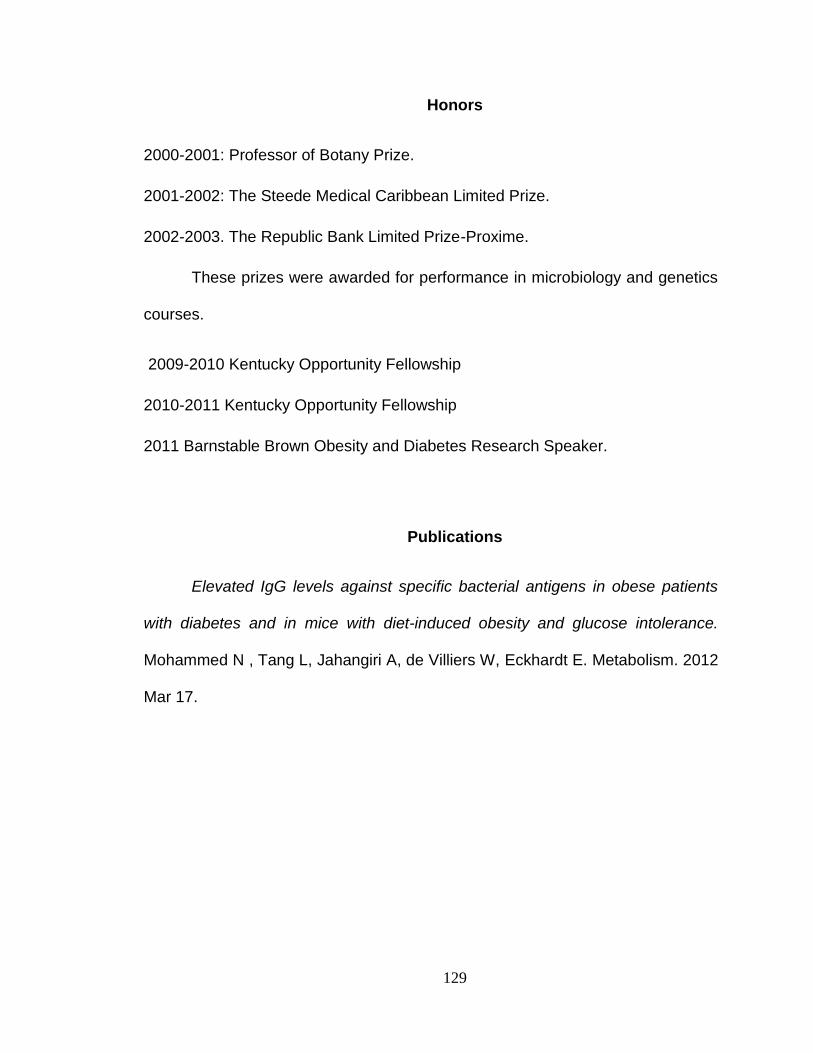
129
Honors
2000-2001: Professor of Botany Prize.
2001-2002: The Steede Medical Caribbean Limited Prize.
2002-2003. The Republic Bank Limited Prize-Proxime.
These prizes were awarded for performance in microbiology and genetics
courses.
2009-2010 Kentucky Opportunity Fellowship
2010-2011 Kentucky Opportunity Fellowship
2011 Barnstable Brown Obesity and Diabetes Research Speaker.
Publications
Elevated IgG levels against specific bacterial antigens in obese patients
with diabetes and in mice with diet-induced obesity and glucose intolerance.
Mohammed N , Tang L, Jahangiri A, de Villiers W, Eckhardt E. Metabolism. 2012
Mar 17.