Embed Size (px)
Citation preview
Phy~ochemisrry, Vol. 26. No. 9. pp. 2495-2498. 1987. 003 l-9422187 $3.00 + 0.00 printed in Great Britain. 0 1987 Pcrgamon Journals Ltd.
NADPH: BIOCHANIN A OXIDOREDUCTASE FROM THE FUNGUS FUSARIUM JAVANICUM
DIITMAR SCHLIEPER and WOLFGANG BARZ*
Westfiilische Wilhclms-Univcrsitit, Lehrstuhl fiir Biochemic der Ptlanzen, Hindenburgplatz 55, D-4400 Miinster, F.R.G.
(Received 23 Janumy 1987)
Key Word Index-Fusariurn jauunicum; Hyphomyceta; NADPH: biochanin A oxidoreductase; isoflavone metabolism; bicchanin A.
Abstract-A soluble enzyme which catalyses the NADPHdependent reduction of the heterocyclic double bond of the isotlavone biochanin A (5,7dihydroxy-4’-methoxy-isoflavone) yielding the corresponding isoflavanone was isolated from the fungus Fusurium jauanicum. The NADPH: biochanin A oxidoreductase was constitutively present in the mycelium with an extractable average activity of 4 pkat/g fresh weight. The enzyme was purified ca 4500 fold to apparent homogeneity. The native enzyme had M, of ca 87 000 and consisted of two identical subunits of M, 43 000. The enzyme reaction showed a pH-optimum at pH 7.5 and a temperature optimum between 30 and 35”. The apparent K, values were 43 PM for biochanin A and 190 PM for NADPH with a maximum velocity of 4 mkat/kg protein. The enzyme exhibited a remarkable substrate specificity for biochanin A.
INTRODUCTION
Among the defence mechanisms which plants have de- veloped against the attack of micro-organisms, the iso- llavonoids are of considerable interest, due to their differential fungitoxic properties [l-3]. Numerous phyto- pathogenic fungi were shown to be potent degraders of such isoflavonoid pre- or post-infectional inhibitors r391.
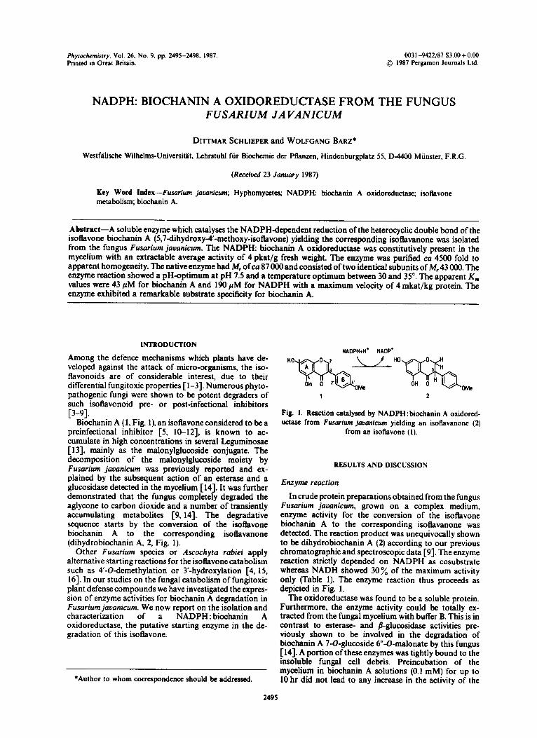
Biochanin A (1, Fig. l), an iso!lavone considered to be a preinfectional inhibitor [5. 10-121, is known to ac- cumulate in high concentrations in several Leguminosae [13], mainly as the malonylglucoside conjugate. The decomposition of the malonylglucoside moiety by Fusarium jauanicum was previously reported and ex- plained by the subsequent action of an esterase and a glucosidase detected in the my&urn [14]. It was further demonstrated that the fungus completely degraded the aglycone to carbon dioxide and a number of transiently accumulating metabolites [9,14]. The degradative sequence starts by the conversion of the isoflavone biochanin A to the corresponding isoflavanone (dihydrobiochanin A, 2, Fig. 1).
Other Fusurium species or Ascochyta rabiei apply alternative starting reactions for the isotlavone catabolism such as 4’-Odemethylation or 3’-hydroxylation [4, 15, 161. In our studies on the fungal catabolism of fungitoxic plant defense compounds we have investigated the expres- sion of enzyme activities for biochanin A degradation in Fusurium jauunicum. We now report on the isolation and characterization of a NADPH : biochanin A oxidoreductase, the putative starting enzyme in the de- gradation of this isoflavone.
*Author to whom correspondcncc should be addressed.
NA0PH.H. NAOP’
Fig. 1. Reaction catalysed by NADPH: biochanin A oxidored- uctasc from Fusurium juuanicwn yielding an isoflavanone (2)
from an isoflavone (I).
RESULTS AND DISCUSSION
Enzyme reaction
In crude protein preparations obtained from the fungus Fusarium javanicum, grown on a complex medium, enzyme activity for the conversion of the isoflavone biochanin A to the corresponding isoilavanone was detected. The reaction product was unequivocally shown to be dihydrobiochanin A (2) according to our previous chromatographic and spectroscopic data [9]. The enzyme reaction strictly depended on NADPH as cosubstmte whereas NADH showed 30% of the maximum activity only (Table 1). The enzyme reaction thus proceeds as depicted in Fig. 1.
The oxidoreductase was found to be a soluble protein. Furthermore, the enzyme activity could be totally ex- tracted from the fungal mycelium with buffer B. This is in contrast to esterase- and /?-glucosidase activities pre- viously shown to be involved in the degradation of biochanin A 7-O-glucoside 6”-O-malonate by this fungus 1143. A portion of these enzymes was tightly bound to the insoluble fungal cell debris. Preincubation of the mycclium in biochanin A solutions (0.1 mM) for up to 10 hr did not lead to any increase in the activity of the
2495
24% D. SCHLIEPER and W. BARZ
oxidoreductase. We must thus conclude that this enxyme is constitutively expressed in F. jauanicum. Data obtained from in uiuo studies [9,14] and from other investigations on the enzymes involved in biochanin A degradation by Fusarium jauanicum (unpublished) support the assump tion that the enzymes subsequent to dihydrobiochanin A are inducrble either by biochanin A or by the correspond- ing substrate [9, 143.
Enzyme purification
Attempts to purify the oxidoreductase were hampered by the very low enzyme activity in the mycelium (4 pkat/g fr. wt), due to the low concentration of the enzyme (ca 0.02 % of the soluble proteins). Despite this, a 46tXXfold increase in specific activity with a 12 y0 yield of activity and a 0.003 y0 yield of total protein was finally reached (Table 2). Affinity chromatography on Blue Sepharose CL 6 B and subsequent elution with NADPH represented the best purification step (Table 1). When applied in a 50 mM potassium phosphate buffer at pH 7.5 the enzyme was tightly bound to the triazin dye and could not be eluted from the gel with salt solutions up to &fold higher in ionic strength. Though numerous compounds were tested (see Experimental) only NADPH was a successful ehrant for the enzyme from the Blue Sepharose ligands. This competitive behaviour of ligand and NADPH is in agreement with the high specificity of the enzyme for NADPH and also indicates that the enzyme possesses specific binding sites for the folded dinucleotide structure of the reduced cosubstrate [17-191. The pronounced difference in the elution potential between NADPH and NADP further supports the assumption that the enzyme may form a stable complex with the reduced form of the cosubstrate and that the oxidized form is readily released from it after hydride transfer. The enzyme finally obtained after the affinity chromatography step (Table 1) was
Table 1. Relative activities of the purified oxidoreduc- tase with various substrates and cosubstrates
Substrate (0.2 mM)
Cosubstrate (2 mM)
Ref. activity
(%)
Biochanin A Biochanin A Pratenscin Cknistein
NADPH NADH NADPH ‘NAPDH
100 30* 4 78klO 44It 5
shown to be homogeneous as judged by SDS-PAGE and was used for further characterization.
When the blue sepharose step was replaced by Fast Protein Liquid Chromatography (FPLC) on a Mono Q column, an efhcient preparative purification was obtained. With this method a 700-fold increase in specific activity was achieved which resulted in a final value of 2.5 mkat/kg protein, a yield of 2.5 % total activity and a 0.003 % yield of protein respectively. The enzyme could he stored at - 25” in the presence of 20% (v/v) glycerol without significant loss of activitv. Enzvme solutions kent at 4” showed a decrease in enzyme activity with a weeks.
Structure of the enzyme
Gel chromatography indicated a for the native enzyme as estimated experiments. SDS-PAGE with the
&If life of about 2
M, of 87OC0-88ooO from 4 independent purified enzyme re-
vealed one single protein band with an M, of co 43 000-44 000. No other proteins could be detected, either with Coomassie Blue staining or by the silver staining method. These results suggest a dimeric structure with monomers of 43OCKI. Sedimentation analysis by ultra- centrifugation with alcohol dehydrogenase as reference protein resulted in much higher values for the M, in the range of 240000. Though only one single, distinct band for the reductase activity could be detected in the sucrose gradients, polymerization cannot be excluded, but more likely the enzyme contains metal atoms leading to a high density, and thus an overestimated M, could have been shown by this method.
Kinetic properties
The purified NADPH: biochanin A oxidoreductase exhibited a broad pH optimum at pH 7.5 with con- siderable enzyme activities also in the range of pH 5-8. The temperature optimum was between 30-35”, while a temperature above 40” rapidly inactivated the enzyme. The enzyme reaction was linear up to 50 % conversion of biochanin A, both for the amount of protein in the assay and for incubation time (up to 4 hr at 30”). The values for K, of biochanin A and NADPH were (43 f 5) PM and (19Of 20) PM, respectively, and V,, was measured as (4.2 f 0.2) mkat/kg. Several flavones, chalcones and iso- tlavones (see Experimental) were tested to determine the substrate specificity of the purified oxidoreductase. In addition to biochanin A, an enzymic reduction could only be measured with the isotlavones pratensein (5,7,3’-
Table 2. Puritkation of the NADPH: Biochanin A oxidoreductase from Fusarium jouanicum
Puritication step
Crude extract (NH,),SO, fractionation 40-60 % saturation DUE-Scphacel Ultrogel AcA 34 Blue Sepharosc CL 6B (Elution with NADPH)
Protein (mg)
Total activity @kst)
specific activity
01 hat/kg) Recovery
(%)
Puritication (- fold)
857 1105 1.3 100 1
327 803 2.5 73 1.9
39.3 583 14.8 53 11.4 6.8 475 69.9 43 54 0.023 134 5920 12 4550
NADPH: Bicchanin A oxidoreductase from Fusurium javanicum 2497
trihydroxy4’-methoxy-isotlavone) and genistein (5,7,4’- (column I5 x 50 mm). Unbound protein was eluted with 600 ml trihydroxy-isoflavone) (Table 1). Flavones, chakones and buffer A. A selective elution of the oxidoreductase a&vity could 5deoxy_isotIavones, respectively, were not accepted as be achieved by the use of 5 ml 10 mM NADPH in butTer A substrates by the oxidoreductase. The enzyme is thus (Rowrate 50 ml/hr, fraction size 1.4 ml). The bulk of tnyme characterized by an absolute specificity for ring A (5,7- activity was distributed over two fractions only. Other com- dihydroxy pattern) and a pronounced specificity for ring B pounds such as 0. I M KCI. O-2 M NaCi, 0.01 M 5’-AMP, 0.02 M (4’~substituent) of isotlavoaes. These results on substrate 2’-AMP, 0.1 M nicotinamide, 0.01 M NAD, 0.01 M NADP, specificity are in agreement with the observation that the 0.01 M NADH were unable lo remove the enzyme from the dye fungus readily degrades the isoflavones listed in Table 1, whereas Sdeoxy-isoflavones are not catabolized [ 16,201.
ligands. The eluants were routinely withdrawn from the fractions by gelfiltration on PD-IO columns as mentionai above. In order
The NADPH : biochanin A oxidoreductase represents lo maintain the binding capacity of the gel it had lo be another exampte of a highly specific fungal reductase [2 11. regenerated with 8 M urea following each purification procedure.
The enzymatic reaction shown in the Fig. 1 is com- parable with another oxidoreductase reaction recently found in a plant system where 2’-hydroxy4deoxy- isofiavones are converted to the corresponding iso- flavanones [22]. This enzyme is part of the pterocarpan phytoalexin biosynthesis in chickpea and offers to be absolutely specific for a Sdeoxy-2’-hydroxy substitution pattern.
Molecular moss. The M, of the native enzyme was determined in the course of gel filtration steps using hexokinase, chymotrypsinogen (Serva, Heidelberg, F.R.G.), bovine serum albumine and ovalbumine (Sigma, Munich, F.R.G.) as marker proteins. The void vol. and the pore volume (Ultrogel AcA 34.26 x 860mm) were determined with Blue Dextran 2ooO (Pharmacia. Freiburg, F.R.G.) and DNP-Alanine (Serva, Heidelberg, F.R.G.), respectively.
EXPERIMENTAL
Chemjcols. 2,5,7-Trihydroxy4’-methoxy-isoflavone was ob- tained from Prof. Grisebach (University of Frciburg, F.R.G.), all other phenolic compounds were from our previous studies [S, 9.14.203. DTE (dithio-erythritol), NADPH. NADH, NADP, NAD, 2’-AMP and nicotinamide were purchased from Serva (Heidelberg, F.R.G.). DEAE-Sephacel, Blue Sepharose CL 6 B and Sephadex G-25 (medium) came from Pharmacia (Freiburg, F.R.G.). Ultrogel AcA 34 was a product of LKB (Griifelfing, F.R.G.).
Analytical SDS-PAGE was performed after each purification step under denaturating conditions according to ref. [23]. The stacking gel consisted of 3% and the running gel of 12.5 % acrylamide. Gels (80 x 80 mm) were stained with Serva Blue G. Some gels were also stained by the silver method [24] using Bio- Rad reagents (Munich, F.R.G.). Bovine serum albumin. ovalbumin (Sigma, Munich, F.R.G.), chymotrypsinogen and myoglobulin (Serva, Heidelberg, F.R.G.) wtre used as markers. The mixture contained 0.2 % Bromophenol Blue as tracking dye.
Bufir systems The buffers were: buffer A, 50 mM K-Pi, pH 7.5, containing 2 mM DTE; buffer B, 100 mM K-Pi, pH 7.5, containing 2 mM DTE. For estimating the pH-optimum of the enxyme reaction the following buffers were used: pH range 2-3, 100 mM glycine/HCl; pH 45. 100 mM tri-Na&rate; pH 5-8, buffer B; pH 7-8.5, 1OOmM Tris-HCl; pH 9-12, 1OOmM glycine/NaOH; each buffer included 2 mM DTE.
Sedimentation analysis was carried out according to ref. [25] using linear, isokinetic sucrose gradients [%?O% (w/v)] in buffer A with a vol. of 12 ml and protein samples of 0.1 ml. A pnparative ultracentrifuge (Damon/IEC B60) equipped with the swinging bucket rotor 488 was employed. After ccntrifugation (24 hr. 4”, 40000rpm) the tubes were fractionated dropwise from the bottom and fractions of 0.4ml each were collected. Alcohol dehydrogenase from horseliver (Boehringer, Mannheim, F.R.G.) was used as int. ref. protein.
Fungus. Fusmium jauanicum (Koordb CBS 203.32 obtained from the Centraalbureau voor Schimmelcultures (Baam, Netherlands), was stored, grown and harvested as previously described [14].
Enzyme pwjficcrtion. The mycclium harvested 4 days after inoculation was washed in buffer A and stored in liquid N,. After thawing in buffer B (l-2 ml/g fr. wt) it was further homogenized in a chilled mortar with 20 y0 (w/w) quartz sand. All procedures were carried out at 4”. The homogenate wascentrifuged (30 000 g, 30min) and the supernatant was fractionated by dropwise addition of a saturated (NH&SO, soln in two steps between 040% and 4&60x satn. The precipitated protein was removed by centrifugation (30000 g, 20 min). Protein from the second precipitation step was dissolved in buffer A and the soln was desalted on Sephadex G-25 (PD-IO) using buffer A. The protein material was then applied to a DEAE-Sephacel column (29 x 450 mm). After elution of upbound protein (500 ml bulTer A) a linear gradient of NaCl in buffer A with a slope of O-O.7 M and a total vol. of 500ml was started. Proteins were eluted with a flowrate of 50 ml/hr and fractions of 4 ml each were collected. Fractions with pronounced oxidoreductase activity were pooled and concentrated by ultrafiltration (PM 10, Amicon, Osterhout, Netherlands) to a vol. of 10 ml. This sample was immediately subjected to Ultrogel AcA 34 chromatography (column 26 x 860 mm) using buffer A and fraction sizes of 4 ml. The fractions with oxidoreductase activity were pooled and used for the final afiinity chromatography on Blue Sepharom CL 6 B
Enzyme assays. Standard assays consisted of 50-200~1 enzyme preparation, 100 nmol biochanin A and MO nmol NADPH in a total vol. of 500~1 buffer A. The phenolic compounds were added with 104 2-metboxycthanol. The assays were started with NADPH, incubated for 1 hr at 30” and stopped by adding 500~1 MeOH. Denaturated protein was removed by centrifugation and the resulting methanolic solns were directly used for HPLC analysis. Alternatively tests were stopped by the addition of 600 4 EtOAc, and the phenols were extracted by shaking for 1 min. After removal of proteins by centrifugation the EtOAc phase was recovered and evaporated lo dryness. The residue was dissolved in 100~1 MeOH and sub- mitted lo HPLC analysis. The analytical system for HPLC separation of isollavones and other phenolic compounds has bxn described [13.14,28]. The separation of biochanin A and dihydrobiochanin A was achieved with a linear gradient of 40 “/. B in A lo 60% B in A within 15 min a1 a Bow of 0.8 ml/min, performed on a RP I8 column (Lichrosorb, Merck, Darmstadt, F.R.G.), with 1.5% HJQ (A) and CHsCN (Bj A gradient of 20 % B in A to 80 % B in A of the same eluants was used for chromatography of assays including other isoflavones, fiavones and the chalcone as substrates. Quantitation of substrates and products was perforn~at at 260,290 and 320 nm, respectively, by exterunl standardization. Alcohol dehydrogenase was assayed according to the recommendations of the producer. The data quoted are the average values obtained by three difTerent methods of linear transformations (Lineweaver-Burk, Eadie-Hofstee+ and Hanes [26,27]).
2498 D. SCHLI~PER and W. BARZ
Product idenrijicarion. The identity of the reduction product 8. Barz, W., W&kc, U. and Weltring, K.-M. (1980) Ann. obtained from biochanin A wax chrcidatcd by co- Phytoparh. 12, 135. chromatography and GC/MS analyses after preparative HPLC 9. Willeke, U. and Barr, W. (1982) Z. Naturforsch. 37C. 861. purification. MS data identical to our previous reports [9] were 10. Brcdenburg, J. B. (1961) AC& Chum. 34b, 23. obtained. 11. Debnam, J. R. and Smith, 1. M. (1976) Physiol. Plant Parhol.
Substrate spec~fiity. The following compounds were not ac- 9, 9. cepted as substrates by the puriBcd oxidorcductascunder 12. Kosugc, T. (1969) Ann. Rev. Phytoparhol. 7, 195. standard incubation conditions: (a) the &ones apigcnin. 13. K&&X, J., Strack, D. and Barz, W. (1983) Plonta Med. 48, quercctin, (b) 2’,4’dihydroxy4methoxy-chakonc and (c) the 131. isohvones orobol (3’,4’,5,7-tctrahydroxy-isotlavonc), 2,5,7-u-i- 14. Schliepcr, D., KomoDa, D. and Batl, W. (1984) Z. hydroxy-t’-mcthoxy-isofiavonc, 3’,4’,5-trihydroxy-isotlavonc, Naurforsch. 39C, 882. tcxasin (6,7dihydroxy4’-methoxy-isoBavone), formononetin (7- 15. Willeke, U. and Ban, W. (1982) Arch. Microbial. 132,266. tiydroxy-t’-mcthoxy-isotlavone), calycosin (3’,7dihydroxy-4’- 16. Weltring, K.-M., Mackcnbrock, K. and Barz W. (1982) Z. mcthoxy-ho&one), daidxein (4’,7dihydroxy+o5vonc), and Naturforsch. 37C, 570. 4’-hydroxy-7-methoxy-isoBavone. 17. Dean, P. D. G. and Watson, D. H. (1973)J. Chromorogr. 165.
301. Ackaowled~ement-Financial support by Deutschc Forschungs- 18. Limbach, B. and Schmidt, H. L. (1973) J. Chromatogr. 285, gemeinschaft and Fonds dcr chcmischen Industrie is gratefully 457. acknowledged. 19. Thompson, S. T.. Cass, K. H. and Stellwagen, E. (1975) Rot.
1.
2.
3.
4. 5.
6.
7.
REFERENCES
Kramer, R. P., Hindorf, H., Jha, H. C., Kallage, J. and Zillikcn, F. (1984) Phytochemistry 22, 2203. Amoldi, A., Farina, G., Galli, R, Merlini, L. and Parino, M. G. (1986) J. A&z. Food C/tern. 34, 185. VanEtten, H. D., Matthews, P. S., Tegtmcicr, K. J., Dieter?, M. F. and Stein, J. 1. (1980) Physiol. P/ant Pathol. 16,257. Kraft, 9. and Barx W. (1985) Appl. Environ. Microbial. So, 45. Wilkke. U.. Weltring, K.-M., Bar-x, W. and VanEtten, H. D. (1983) Phytochemisrry 22 1539. VanEtten, H. D., Matthews, P. S. and Smith D. A. (1982) in Phytoalexins (Bailey, J. A. and Mansticld, J. W., a-Is) Blackie, Glasgow. Weltring, K.-M., Ban, W. and Dcwick, P. M. (1983) Phytochemistry 22, 2883.
Nat. Acad. Sci. U.S.A. 72, 669. 20. Willcke, U. (1981) Doctoral thesis, University of Miinster,
F.R.G. 21. Kwan-sa, Y. (1985) CRC Critical Rev. Biochem. 17, 313. 22. Tiemann, K.. Hinderer, W. and Barx, W. (1987) FEBS Letters
213, 324. 23. Lummli, U. K. (1970) Nature 227,680. 24. Meriil, C. R., Coldman, D., sadmann, S. A. and Ebcrt, M. H.
(1981) Science 211, 1437. 25. Martin,R.G.andAmcs,B. N. (l%l)J. Biol.Chem. 236,1372. 26. &gel, J. H. (1976) in Biochemical Ca/cu~ions. Wiley, New
York. 27. Dixon, M. and Webb, E. C. (1980) in Enzymes 3rd edn.
Academic Press, New York. 28. Kiistcr, J., Zuxok, A. and Bat-x W. (1983) J. Chmmarogr. 270,
392.


![Pyrethrin Biosynthesis: The Cytochrome P450 Oxidoreductase ...Pyrethrin Biosynthesis: The Cytochrome P450 Oxidoreductase CYP82Q3 Converts Jasmolone To Pyrethrolone1[OPEN] Wei Li,a](https://img.pdfslide.net/doc/110x75/5e2d08c0200c602a86070292/pyrethrin-biosynthesis-the-cytochrome-p450-oxidoreductase-pyrethrin-biosynthesis.jpg)