Embed Size (px)
Citation preview
174 175 174 175
INTRODUCTION
Microbial, mechanical or chemical irritation of the pulp and periradicular tissues lead to inflam-mation. In cases of vital pulp therapy that requires placement of an intracanal medicament between appointments, the choice of this agent depends on its biological characteristics: non-irritant, pulp vitality preservation, control of intensity and dura-tion of inflammatory processes and infection, and potential healing induction9,12.
Several chemicals and therapeutic agents have been suggested as intracanal medicaments. The
Braz Oral Res 2004;18(2):174-9
Natural medicaments in endodontics – a comparative study of the anti-inflammatory action
Medicamentos naturais na Endodontia – estudo comparativo da ação antiinflamatória
Fabiane Bortoluci da Silva* Juliano Milanezi de Almeida** Simone Maria Galvão de Sousa***
ABSTRACT: The objective of this study was to evaluate the irritant potential of propolis, Casearia sylvestris, Otos-porin and saline solution (control). Twenty-eight male Wistar rats were selected, anesthetized and four experimen-tal sites were designed on their backs. Injections of 2% Evans blue were intravenously administered in the lateral caudal vein and 0.1 ml of the tested solutions was injected intradermally into the experimental sites. The animals were killed 1/2, 1, 3 and 6 hours after the injection of the solutions. Each piece of skin containing the lesion was immersed in formamide and incubated at 45oC for 72 h. After filtration, optical density was measured in a spectro-photometer. Data were statistically analyzed by a 2-way non-parametric test. The highest values of extracted dye were observed at 3 hours characterizing a peak in the inflammatory process. Propolis was the least irritant solu-tion. The natural medicaments tested in this study may be a valuable alternative for endodontic treatment.
DESCRIPTORS: Endodontics; Dental pulp cavity; Naturopathy; Anti-inflammatory agents.
RESUMO: O objetivo desta pesquisa foi avaliar o potencial irritativo de própolis, Casearia sylvestris, Otosporin e soro fisiológico (controle). Foram utilizados 28 ratos machos da linhagem Wistar. Os animais foram anestesiados e, em seguida, receberam a injeção do corante azul de Evans (2%) por via intravenosa na veia caudal. Em quatro pontos predeterminados e depilados da região dorsal de cada animal, foram injetados 0,1 ml das substâncias teste. Os animais foram sacrificados meia, uma, três e seis horas após a injeção das substâncias, e cada pedaço de pele contendo a lesão foi colocado em frascos contendo formamida, que foram incubados a 45oC por 72 h. Após esse período, as amostras foram filtradas e submetidas à análise em espectrofotômetro. Os dados foram submetidos ao teste estatístico não-paramétrico de medidas repetidas. No período de 3 h, foram observados os maiores valores de corante extraído, caracterizando, assim, o pico do processo inflamatório. A própolis foi a substância que apre-sentou menor potencial irritativo. Os produtos naturais testados neste trabalho podem ser uma alternativa para o tratamento endodôntico.
DESCRITORES: Endodontia; Cavidade da polpa dentária; Naturopatia; Antiinflamatórios.
most commonly employed are calcium hydroxide and the combination of steroid and antibiotic12. Otosporin (Zest, Rio de Janeiro, RJ, Brazil) reduces vasodilatation, decreases liquid exudation, and also presents a direct vasoconstrictive action on small blood vessels9. Holland et al.13 (1980) com-pared three commercially prepared corticosteroid-antibiotic solutions after pulpectomy and biome-chanical preparation in dogs’ teeth. Otosporin was more effective as an intracanal medication than Panotil and Otosynalar.
* Bio�of Dentistry – University of Sacred Heart, Bauru.
** Postgraduate in Periodontology, School of Dentistry of Araçatuba, São Paulo State University.
Endodontics
174 175
Silva FB, Almeida JM, Sousa SMG. Natural medicaments in endodontics – a comparative study of the anti-inflammatory action. Braz Oral Res 2004;18(2):174-9.
174 175
Medicinal plants constitute a promising source of phytotherapy drugs and new molecules. The number of studies on this alternative therapeutic system increased in the last decades, as well as their use for several purposes7.
Casearia sylvestris is a medicinal plant that offers a wide range of uses: healing, antiseptic, antiulcerative, diuretic, tonic, stimulant, antimi-crobial, and depurative. This species is very com-mon in the tropical America and Brazil; one of its popular names is “guacatonga”20.
Studies have demonstrated controversial re-sults about its anti-inflammatory action. Ruppelt et al.18 (1991) analyzed the analgesic and anti-in-flammatory action of 10 plants similar to C. syl-vestris by means of Evans blue diffusion in the peritoneal cavity, and observed reduced activity for C. sylvestris. Furthermore, another study in-dicated that the alcoholic extract of C. sylvestris prolonged the acute phase of the inflammatory process5. The C. sylvestris extract constitutes a rich source of phospholipase A
2 inhibitors. The
components of this plant inhibit the enzymatic and toxic activities of several venoms, are effective in inhibiting isolated class I and II phospholipases A
2,
and the extract is partially effective against edema formation3. A clinical investigation demonstrated progressive healing of intra and extraoral lesions of herpes simplex after topic application of C. syl-vestris extract4.
Propolis is a resin widely used in folk medicine for centuries. The chemical composition of this atoxic natural substance is complex. Flavonoids and cinnamic acid derivatives have been consid-ered as the main primary biologically active com-ponents10,14,16. It is known that propolis exhibits several pharmacological properties such as anti-microbial, anti-inflammatory, healing, anesthetic, cytostatic and cariostatic properties. Ethanolic ex-tract of propolis inhibits hyaluronidase activity. This enzyme is responsible for several inflamma-tory processes and, if a certain substance is able to inhibit its activity, such substance will have a great potential as an anti-inflammatory agent17. In Dentistry, propolis has been used for the treat-ment of aphthous ulcers, Candida albicans, acute necrotizing ulcerative gingivitis (ANUG), gingivitis and periodontitis8,10,15,16.
The method proposed by Udaka et al.21 (1970) has often been employed in an attempt to quantify the irritant potential of several substances injected intradermally (or inoculated in vivo) and also to evaluate the effectiveness of anti-inflammatory
drugs. This method analyzes the plasma exudate produced after an increase in vascular permeabil-ity that can be inferred by means of spectrophoto-metric measurement of Evans blue dye.
The purpose of this study was to comparatively evaluate the biocompatibility of propolis, Casearia sylvestris, Otosporin and saline solution (control) using the physicochemical method for quantifica-tion of the enhanced vascular permeability (Evans blue test).
MATERIALS AND METHODS
Twenty-eight adult male Wistar rats (Rattus norvegicus) weighting approximately 320 g were used. The rats were anesthetized with an asso-ciation of ketamine (Francotar, Virbac do Brasil Indústria e Comércio Ltda., São Paulo, Brazil) and xylazine (Virbaxyl 2%, Virbac do Brasil Indústria e Comércio Ltda., São Paulo, Brazil). Their backs were shaved and four experimental sites were des-ignated. Their tails were washed and dried in order to facilitate the injection of 2% Evans blue (20 mg/kg; Merck, Darmstadt, Germany), administered intravenously in the lateral caudal vein.
Immediately after this, 0.1 ml of each tested solution – propolis (10% alcoholic solution; Propo-vit, Bionatus Laboratório Botânico Ltda., São José do Rio Preto, Brazil), aqueous/alcoholic extract of Brazilian Casearia sylvestris (10 g of fresh plant in 100 ml of alcohol, Extratoline 115, A Natureza Produtos Farmacêuticos Ltda., Brazil), Otosporin (Zest Pharmaceutics Ltda., Rio de Janeiro, Bra-zil) and saline solution (0.9% NaCl, Darrow Labo-ratório S/A, Rio de Janeiro, Brazil) – was injected intradermally into the experimental sites following a rotational system. Both natural medicaments were prepared by dissolving 1 ml of the drug in 9 ml of distilled water.
Evaluation of the inflammatory exudate was performed after ½, 1, 3 and 6 hours. Subsequently, subgroups were established in each group accord-ing to the period of time elapsed before sacrifice. After the determined period, the animals were killed by injecting an excessive dose of anesthetic (100 mg/kg). Dorsal skin was dissected and skin lesions were punched out with a standard steel punch set (3 cm in diameter). Each piece of skin containing the lesion was cut into small pieces and the dye was extracted with 10 ml of formamide (Vetec Química Fina Ltda, Rio de Janeiro, Brazil) for 72 hours at 45oC.
Silva FB, Almeida JM, Sousa SMG. Natural medicaments in endodontics – a comparative study of the anti-inflammatory action. Braz Oral Res 2004;18(2):174-9.
176 177 176 177
Optical density was measured after filtration with glass wool. Initially, the curve of the absorp-tion spectrum of Evans blue was determined to achieve the best wavelength and calibration curve for the Evans blue. Measures were made at 620 µm (A620) in a spectrophotometer (Ultrospec 2000, Pharmacia Biotech, Cambridge, USA). Transfor-mation of each sample or group mean absorption, in µg, was obtained using the formula: µg = ab-sorption × calculation factor (68) × total volume of formamide. Data were statistically analyzed by a 2-way non-parametric test (Friedman Repeated Measures Analysis of Variance on Ranks).
RESULTS
The means and standard deviations of the amount of dye extracted (µg) for all experimen-tal groups are presented in Table 1. Medians and semi-interquartile range of the amount of dye extracted (A620) and the respective results of the
non-parametric statistical test comparing time and substance are expressed in Table 2.
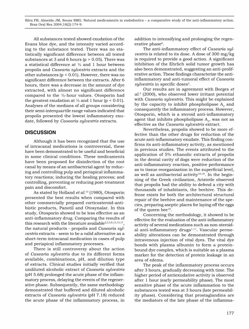
Graph 1 illustrates the medians of the amount of dye extracted (µg) for all experimental groups, showing an enhanced vascular permeability at the 3-hour experimental period.
0
100
200
300
400
500
600
700
800
1/2 h 1 h 3 h 6 hTime
Dye
ext
ract
ed (
�g)
Casearia sylvestris PropolisOtosporin Saline
GRAPH 1 - Medians of the quantification of dye extract-ed (µg) from the experimental groups along time.
TABLE 1 - Means and standard deviations (SD) of the amount of dye extracted (µg) in each experimental group, as to substance and time.
Time
Substance
Propolis Casearia sylvestris Otosporin Saline solution
Mean SD Mean SD Mean SD Mean SD
1/2 h 100.17 51.94 127.55 39.20 443.82 154.77 181.60 96.21
1 h 105.35 22.85 189.07 33.49 496.10 197.37 132.62 33.12
3 h 175.62 37.97 297.00 108.92 641.16 383.27 199.26 35.81
6 h 162.81 55.34 212.51 29.58 630.54 334.63 204.02 90.28
TABLE 2 - Medians, semi-interquartile range and minimum and maximum values of absorption of the dye extracted (A620) as to substance and time.
SubstanceTime Result -
test of time1/2 h 1 h 3 h 6 h
Propolis90.10 ± 15.00
(60.32;213.18) aA119.54 ± 26.12
(83.57;197.47) aB430.10 ± 94.98
(266.90;726.78) aC137.97 ± 84.04
(92.07;324.09) aB16.20
(p < 0.01)
Casearia sylvestris
94.59 ± 13.35(82.69;148.24) abA
175.44 ± 19.29(148.92;247.32) abB
434.18 ± 151.71(282.47;824.02) aC
129.95 ± 26.54(90.03;181.20) aA
17.91 (p < 0.01)
Otosporin186.66 ± 23.75
(102.54;209.17) cA258.67 ± 68.33
(110.02;412.28) bB685.44 ± 260.43
(49.98;1,188.64) aB191.62 ± 23.78
(162.18;263.23) aA9.34
(p < 0.05)
Saline solution
136.61 ± 36.08(117.37;258.67) bcA
208.15 ± 23.27(166.53;246.70) bA
542.16 ± 116.39(379.37;1,351.9) aB
167.55 ± 67.30(131.10;355.50) aA
14.14 (p < 0.01)
Result - test of substance 12.94 (p < 0.01) 15.08 (p < 0.01) 2.93 (p > 0.05) 7.32 (p > 0.05)
Two medians followed by a same lower case letter do not differ (p > 0.05) regarding the substances at a given period. Two medi-ans followed by a same capital letter do not differ (p > 0.05) regarding periods of time for the same substance.
176 177
Silva FB, Almeida JM, Sousa SMG. Natural medicaments in endodontics – a comparative study of the anti-inflammatory action. Braz Oral Res 2004;18(2):174-9.
176 177
All substances tested showed exudation of the Evans blue dye, and the intensity varied accord-ing to the substance tested. There was no sta-tistically significant difference between all tested substances at 3 and 6 hours (p > 0.05). There was a statistical difference at ½ and 1 hour between propolis and Casearia sylvestris extracts and the other substances (p < 0.01). However, there was no significant difference between the extracts. After 6 hours, there was a decrease in the amount of dye extracted, with almost no significant difference compared to the ½-hour values. Otosporin had the greatest exudation at ½ and 1 hour (p < 0.01). Analyses of the medians of all groups considering their semi-interquartile range values indicated that propolis presented the lowest inflammatory exu-date, followed by Casearia sylvestris extracts.
DISCUSSION
Although it has been recognized that the use of intracanal medications is controversial, these have been demonstrated to be useful and beneficial in some clinical conditions. These medicaments have been proposed for disinfection of the root canal by means of an antibacterial agent by reduc-ing and controlling pulp and periapical inflamma-tory reactions; inducing the healing process; and controlling, preventing or reducing post-treatment pain and discomfort.
As stated by Holland et al.13 (1980), Otosporin presented the best results when compared with other commercially prepared corticosteroid-anti-biotic products, Panotil and Otosynalar. In this study, Otosporin showed to be less effective as an anti-inflammatory drug. Comparing the results of this research with the literature available2,3,5,8,13,15,17, the natural products - propolis and Casearia syl-vestris extracts - seem to be a valid alternative as a short-term intracanal medication in cases of pulp and periapical inflammatory processes.
There is still controversy about the action of Casearia sylvestris due to its different forms available, combinations, pH, and dilution type of extracts. Clinical studies initially verified that undiluted alcoholic extract of Casearia sylvestris (pH 5.68) prolonged the acute phase of the inflam-matory process, delaying the events of the regener-ative phase. Subsequently, the same methodology demonstrated that buffered and diluted alcoholic extracts of Casearia sylvestris (pH 7.18) reduced the acute phase of the inflammatory process, in
addition to intensifying and prolonging the regen-erative phase6.
The anti-inflammatory effect of Casearia syl-vestris is related to its dose. A dose of 300 mg/kg is required to provide a good action. A significant inhibition of the Ehrlich solid tumor growth has also been demonstrated, suggesting an anti-prolif-erative action. These findings characterize the anti-inflammatory and anti-tumoral effect of Casearia sylvestris in specific doses2.
Our results are in agreement with Borges et al.3 (2000), who observed lower irritant potential with Casearia sylvestris. This might be explained by the capacity to inhibit phospholipase A
2 and
consequently the inflammatory process. However, Otosporin, which is a steroid anti-inflammatory agent that inhibits phospholipase A
2, was not as
effective as the Casearia sylvestris extract.Nevertheless, propolis showed to be more ef-
fective than the other drugs for reduction of the acute anti-inflammatory exudate. This finding con-firms its anti-inflammatory activity, as mentioned in previous studies. The events attributed to the application of 5% ethanolic extracts of propolis in the dental cavity of dogs were reduction of the anti-inflammatory reaction, positive performance as to tissue reorganization in the superficial level, as well as antibacterial activity16,19. In the begin-ning of the Greek civilization, Aristotle observed that propolis had the ability to defend a city with thousands of inhabitants, the beehive. This de-fense exists for both the architectural structural repair of the beehive and maintenance of the spe-cies, preparing aseptic places for laying off the eggs of the queen bee10.
Concerning the methodology, it showed to be effective for the evaluation of the anti-inflammatory activity of several medications such as non-steroid-al anti-inflammatory drugs1,11. Vascular perme-ability alterations can be demonstrated through intravenous injection of vital dyes. The vital dye bonds with plasma albumin to form a protein-bound dye complex, which is suitable as a plasma marker for the detection of protein leakage in an area of edema.
The peak of the inflammatory process occurs after 3 hours, gradually decreasing with time. The higher period of antiexudative activity is observed after 1 hour (early permeability phase). The most sensitive phase of the acute inflammation to the substances tested was at 3 hours (late permeabil-ity phase). Considering that prostaglandins are the mediators of the late phase of the inflamma-
Silva FB, Almeida JM, Sousa SMG. Natural medicaments in endodontics – a comparative study of the anti-inflammatory action. Braz Oral Res 2004;18(2):174-9.
178 179 178 179
Brazilian Casearia sylvestris extracts may offer a
good alternative as short-term intracanal medica-
ments. Our results indicated that propolis pre-
sented the lowest value of inflammatory exudate,
followed by Casearia sylvestris extract. However,
this study did not allow the establishment of the
nature of the bioactive compound(s) responsible
for the anti-inflammatory activity. Further chemi-
cal and pharmacological investigations in animal
models using different dilutions and pH values are
under way in an attempt to better identify their
effectiveness and clinical applicability.
ACKNOWLEDGEMENT
The authors would like to thank Carlos Rober-
to Padovani for the statistical analysis and Thelma
Lopes Silva for her technical assistance.
REFERENCES
1. Akatsu T, Catanzaro Guimarães SA. Comparative effects of non-steroidal anti-inflammatory drugs, mefenamic acid, metiazinic acid and glucametacin on the inflammatory response induced by subcutaneous implantation of hu-man dental plaque and on the mitotic activity of isoprot-erenol-stimulated parotid glands of rats. Cell Mol Biol 1986;32:619-26.
2. Almeida A. Atividades antiinflamatória e antitumoral do extrato hidroalcoólico de Casearia sylvestris: estudo com-parativo com os antiinflamatórios piroxicam e meloxicam [Tese de Mestrado]. São Paulo: Instituto de Ciências Bio-médicas da USP; 2000.
3. Borges MH, Soares AM, Rodrigues VM, Andrião-Escarso SH, Diniz H, Hamaguchi A, et al. Effects of aqueous extract of Casearia sylvestris (Flacourtiaceae) on action of snake and bee venoms and on activity of phospholipases A
2. Comp
Biochem Physiol B Biochem Mol Biol 2000;127:21-30. 4. Camargo FG, Gomes E, Pannunzio E, Bueno VS. Uso tópico
de extrato fluido de folha de guaçatonga (Casearia sylves-tris Swartz) topicamente em lesões de estomatite herpética. LECTA - USF Brag Pta 1993;1:121-7.
5. Camargo FG, Pereira JA, Bueno VS, Gomes E, Ando T. Ação do extrato alcoólico de guaçatonga (Casearia sylves-tris sw) em subcutâneo de camundongo. Parte I. Estudo histológico. LECTA-USF 1995;13:47-68.
6. Camargo FG, Pereira JA, Bueno VS, Gomes E, Ando T. Ação do extrato alcoólico de guaçatonga diluído e tamponado em subcutâneo de camundongo. Parte II. Estudo histológico. LECTA-USF 1996;14:61-86.
7. di Stasi LC. Plantas medicinais: arte e ciência; um guia de estudo. São Paulo: UNESP; 1996.
8. Duarte S, Koo H, Bowen WH, Hayacibara MF, Cury JA, Ikegaki M, et al. Effect of a novel type of propolis and its chemical fractions on glucosyltransferases and on growth
and adherence of mutans streptococci. Biol Pharm Bull 2003;26:527-31.
9. Fava LRG. Human pulpectomy: incidence of postoperative pain using two different intracanal dressings. Int Endod J 1992;25:257-60.
10. Geraldini CAC, Salgado EGC, Rode SM. Ação de dife-rentes soluções de própolis na superfície dentinária – ava-liação ultra-estrutural. RPG Rev Pos Grad 2000;3:37-42.
11. Guimarães SA, Akatsu T, Tago EM, Consolaro A. As-sessment of the antiexudative and antiproliferative activities of non-steroidal anti-inflammatory drugs in inflammatory models developed in rats by subcutaneous implantation of bacterial cell walls from the dental plaque. Inflammation 1996;20:623-36.
12. Hidalgo MM, Kawana GRC, Gonçalves CEB, Oliveira RMMW, Bersani-Amado CA. Avaliação da tolerância teci-dual a algumas substâncias utilizadas como curativo de demora no tratamento endodôntico. Rev Fac Odontol Lins 1999;11:5-9.
13. Holland R, Souza V, Nery MJ. Emprego da associação corticosteróide-antibiótico durante o tratamento endodôn-tico. Rev Paul Endod 1980;2:4-7.
14. Koo H, Gomes BPFA, Rosalen PL, Ambrosano GMB, Park YK, Cury JA. In vitro antimicrobial activity of propolis and Arnica montana against oral pathogens. Arch Oral Biol 2000;45:141-8.
15. Magro Filho O, de Carvalho ACP. Application of propo-lis to dental sockets and skin wounds. J Nihon Univ Sch Dent 1990;32:4-13.
16. Manara LRB, Anconi SI, Gromatzky A, Conde MC, Bretz WA. Utilização da própolis em Odontologia. Rev FOB 1999;7:15-20.
17. Park YK, Ikegaki M, Alencar SM. Classificação das própolis brasileiras a partir de suas características fisico-
tory process, it is possible to understand that the substances used in this study do not inhibit the biosynthesis of prostaglandins and consequently do not effectively act in this phase. All solutions tested were time-dependent.
Currently, the development and accessibil-ity of information on phytopharmaceuticals and natural medications are gradually gaining the re-spect of some patients and health professionals. Besides, the exploitation of these substances has a socioeconomic impact. It leads to an increase in cultivation fields and in the market of informal herbs and beekeepers, as well as in an expansion of small and medium national pharmaceutical laboratories dedicated to manufacturing medica-ments of natural and vegetable origin.
CONCLUSIONS
According to the methodology of this prelimi-nary study, the natural medicaments propolis and
178 179
Silva FB, Almeida JM, Sousa SMG. Natural medicaments in endodontics – a comparative study of the anti-inflammatory action. Braz Oral Res 2004;18(2):174-9.
178 179
químicas e propriedades biológicas. [periódico online] 2002. Disponível em: URL: http://www.naturapi.com.br/artigo.htm.
18. Ruppelt BM, Pereira EFR, Gonçalves LC, Pereira NA. Pharmacological screening of plants recommended by folk medicine as anti-snake venom – I. Analgesic and anti-inflammatory activities. Mem Inst Oswaldo Cruz 1991;86:203-5.
19. Scheller S, Ilewicz L, Luciak M, Skrobidurska D, Sto-jko A, Matuga W, et al. Biological properties and clinical
application of propolis. IX. Experimental observation on the influence of ethanol extract of propolis (EEP) on dental pulp regeneration. Arzneimittelforschung 1978;28:289-91.
20. Teske M, Trentini AMM. Herbarium – Compêndio de Fitoterapia. 3ª ed. Curitiba: Herbarium Laboratório Botâ-nico; 1995.
21. Udaka K, Takeuchi Y, Movat HZ. Simple method for quantitation of enhanced vascular permeability. Proc Soc Exp Biol Med 1970;133:1384-7.
Received for publication on Nov 12, 2003 Sent for alterations on Mar 29, 2004
Accepted for publication on May 05, 2004