Embed Size (px)
Citation preview
Negative regulators of integrin activity
Jeroen Pouwels1,2, Jonna Nevo1,2, Teijo Pellinen3, Jari Ylanne4 and Johanna Ivaska1,2,5,*1Centre for Biotechnology, University of Turku, Tykistokatu 6 B, FIN-20521 Turku, Finland2VTT Technical Research Centre of Finland, P.O. Box 1000, FI-02044 VTT, Finland3FIMM Finnish Institute for Molecular Medicine, University of Helsinki, P.O. Box 20, FI-00014 Helsinki, Finland4Department of Biological and Environmental Science, Division of Cell and Molecular Biology, University of Jyvaskyla, P.O. Box 35, FI-40014Jyvaskyla, Finland5Department of Biochemistry and Food Chemistry, University of Turku, Tykistokatu 6 A 6. Krs, FI-20520 Turku, Finland
*Author for correspondence ([email protected])
Journal of Cell Science 125, 1–10� 2012. Published by The Company of Biologists Ltddoi: 10.1242/jcs.093641
SummaryIntegrins are heterodimeric transmembrane adhesion receptors composed of a- and b-subunits. They are ubiquitously expressed and have
key roles in a number of important biological processes, such as development, maintenance of tissue homeostasis and immunologicalresponses. The activity of integrins, which indicates their affinity towards their ligands, is tightly regulated such that signals inside the cellcruicially regulate the switching between active and inactive states. An impaired ability to activate integrins is associated with many human
diseases, including bleeding disorders and immune deficiencies, whereas inappropriate integrin activation has been linked to inflammatorydisorders and cancer. In recent years, the molecular details of integrin ‘inside-out’ activation have been actively investigated. Binding ofcytoplasmic proteins, such as talins and kindlins, to the cytoplasmic tail of b-integrins is widely accepted as being the crucial step in integrin
activation. By contrast, much less is known with regard to the counteracting mechanism involved in switching integrins into an inactiveconformation. In this Commentary, we aim to discuss the known mechanisms of integrin inactivation and the molecules involved.
Key words: Activation, Adhesion, Endocytosis, Integrin, SHARPIN, Talin
IntroductionIntegrins are heterodimeric transmembrane proteins composed of a-
and b-subunits. They are ubiquitously expressed, often in high
numbers, and mediate cell–cell adhesion, as well as adhesion of cells
to extracellular matrix (ECM) proteins (Hynes, 2002; Legate et al.,
2009). The affinity of integrins for their ligands (integrin activation)
is allosterically regulated such that the intracellular and extracellular
domains of both subunits undergo conformational changes (Moser
et al., 2009; Shattil et al., 2010). A controlled regulation of integrin
activity is fundamentally important during embryogenesis and is
central to many physiological processes in adults. Impaired integrin
activation has been linked to diseases, including bleeding disorders,
skin blistering and immune-deficiencies (Hogg and Bates, 2000;
Legate et al., 2009). Conversely, increased integrin activity is
associated with chronic inflammation, thrombosis and cancer (Moser
et al., 2009; Shattil et al., 2010).
Integrins are unique in their ability to function as bidirectional
signalling molecules. Binding of extracellular matrix (ECM)
molecules or other ligands to the extracellular domain of
integrins transmits a variety of signals into the cell. This
‘outside-in’ activation (Fig. 1) regulates many important
cellular processes, including migration, survival, proliferation,
gene expression and receptor tyrosine kinase signalling (Ivaska
and Heino, 2010; Zaidel-Bar et al., 2007). Conversely, changes in
the intracellular environment of the cell can alter the binding of
integrins to their ligands through so-called ‘inside-out’ signalling
(Fig. 1). Recently, the individual steps involved in integrin
inside-out activation have been the focus of intense investigation.
Despite some controversy over the details, it is now widely
accepted that binding of the cytoplasmic proteins talin-1 and -2
(TLN1, TLN2) and of kindlins (the fermitin family members 1–3,
FERMT1–FERMT3; also known as KIND1–KIND3) to the
cytoplasmic tail of the integrin b-subunit are crucial for integrin
activation (Calderwood et al., 2004; Moser et al., 2009).
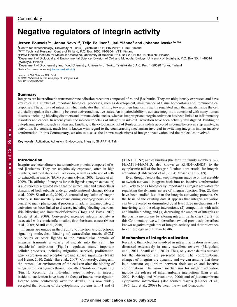
Even though factors that keep integrins inactive or that are able
to switch activated integrins back into an inactive conformation
are likely to be as biologically important as integrin activators for
regulating the dynamic nature of integrin function (Fig. 2), they
have been studied less than the integrin activating proteins. On
the basis of the existing data it appears that integrin activation
can be prevented or diminished by at least three mechanisms: (1)
interfering with the clasp interactions, (2) competition with talin
and kindlin binding, and (3) decreasing the amount of integrins at
the plasma membrane by altering integrin trafficking (Fig. 2). In
this Commentary, we will describe new and previously described
known negative regulators of integrin activity and their relevance
to cell biology and human health.
Mechanism of integrin activationRecently, the molecules involved in integrin activation have been
discussed extensively in many excellent reviews (Margadant
et al., 2011; Shattil et al., 2010). Thus, only some details relevant
for the discussion are presented here. The conformational
changes of integrins are dynamic and we can assume that there
is a constant equilibrium between their active and inactive
conformations. The known mechanisms for integrin activation
include the release of intramembrane interactions (Lau et al.,
2009; Partridge and Marcantonio, 2006) and of juxtamembrane
cytoplasmic interactions (also termed clasps) (Hughes et al.,
1996; Lau et al., 2009) between the a- and b-subunits.
Commentary 1
Journ
alof
Cell
Scie
nce
JCS online publication date 20 July 2012
Talin is a large cytoplasmic protein composed of an integrin-binding head domain and a rod domain, which links talin to
vinculin (VCL) and the actin cytoskeleton (Critchley, 2009;
Grashoff et al., 2010; Humphries et al., 2007). Talin binds to
integrin-b tails and induces conformational activation of the
integrin by disrupting the integrin clasp (Anthis et al., 2009; Kalliet al., 2011; Wegener et al., 2007). For the platelet integrin aIIb3
and for the a–b2 integrin heterodimers, expressed, for example, on
leucocytes, there is compelling evidence that a clasp formed by a
salt-bridge between the a- and b-tails is crucial for maintaining
these receptors in their inactive conformation (Springer andDustin, 2012; Ye et al., 2012). For the b1 integrins, which
predominantly facilitate matrix binding of adherent cell types, the
role of the juxtamembrane clasp is not that well established
(Czuchra et al., 2006). This might reflect the fact that for most a–
b1-integrin-mediated biological processes the switching betweenintegrin activation and inactivation is implicated in adhesion
modulation rather than a complete transition between non-adherent
and adherent states. Nevertheless, talin binding is also required for
b1-integrin activation (Calderwood, 2004). On the basis of in vitro
and in vivo data, kindlins are also crucial for integrin activation;they bind to integrin-b subunits and co-activate integrins together
with talin (Czuchra et al., 2006; Karakose et al., 2010; Montanez
et al., 2008; Moser et al., 2008). Interestingly, talin is not important
for the initial binding of b1 integrins to the matrix, but it is
absolutely required for subsequent cell spreading given thatfibroblasts lacking both the talin-1 and -2 isoforms fail to spread
fully (Zhang et al., 2008). In Drosophila, the talin-null phenotype
has defects in the connections between integrin and the
cytoskeleton, but the binding of integrin to the extracellularmatrix is not abolished (Brown et al., 2002). However, subsequent
work has also demonstrated a role for talin binding to theDrosophila integrins in their activation (Tanentzapf and Brown,2006). These data indicate that integrins can bind the ECM and
become activated in the absence of talin, but talin binding tointegrin-b tails is crucial for the ability of integrins to fulfil theirrole as integrators between the ECM and the cytoskeleton.
Inhibitors interacting with integrin-b tailsThe role of the integrin b-subunit in the regulation of integrinactivity has been the main focus in the field (reviewed byHarburger and Calderwood, 2009; Kim and McCulloch, 2011;
Shattil et al., 2010) and important inhibitors interacting with theb-tails, such as filamin and integrin cytoplasmic domain-associated protein 1 (ICAP1; encoded by ITGB1BP1) (Liu
et al., 2000), have been identified (Fig. 3; Table 1). The mainfocus of our Commentary is the role of the integrin-a tail in themodulation of integrin activity. However, in the following
sections we will also describe the b-tail-binding integrininhibitors. As we are not aware of clasp-stabilising b-subunit-binding proteins, we will divide the inhibitors in two groups;
those that compete with talin and kindlin binding, and those thataffect integrin trafficking (as described in the Introduction).
b-tail-binding inhibitors that compete with the binding oftalins and kindlins
Different integrin b-subunit cytoplasmic domains share twohighly conserved NxxY motifs, which are referred to as the
Closed headpiece, bent
Low affinity
α
α
ICAP1Filamin
SHARPINMDGI
Open headpiece, extended
High affinity
α
Kindlin
Ligand
‘Outside-in’ signalling
‘Inside-out’ signalling
α
Kindlin
Ligand binding
Ligand bindingTalin or kindlinbinding
Talin or kindlinbinding
Ligand
α
α
α
Talin
Talin
Fig. 1. A schematic representation of
integrin conformation switching. In the
inactive conformation (i.e. low-affinity for
ECM components; shown on the left),
integrin a- and b-subunits are in close
proximity, with the headpiece bent towards
the plasma membrane. Binding of, for
example, SHARPIN or MDGI (to a-
subunits), or ICAP1 or filamin (to b-
subunits) to the cytoplasmic domain of
integrins stabilises the integrin heterodimer
in this low-affinity conformation. The
formation of the high-affinity conformation
requires both the extension of the
extracellular domains and the separation of
the integrin a- and b-subunits, resulting in
the so-called open headpiece conformation
(shown on the right). Formation of this open
high-affinity conformation can be triggered
by binding of ECM components to the
extracellular domain of the integrin, termed
‘outside-in’ signalling, or by association of
talin and kindlins with the cytoplasmic
domain of integrin b-subunits in response to
an intracellular signal, called ‘inside-out’
signalling as shown in the centre. In the
schematic depicting ‘inside out’ signalling,
the extracellular domains of the integrins
have been omitted for clarity.
Journal of Cell Science 125 (0)2
Journ
alof
Cell
Scie
nce
membrane-proximal and membrane-distal motifs. Mutational
analyses of these sequences have suggested that they have a
crucial role both in integrin ‘inside-out’ and ‘outside-in’
signalling (Liu et al., 2000). Talin binds to the membrane-
proximal NPxY motif in the integrin b-subunits through its
FERM domain (Goldmann, 2000; Horwitz et al., 1986; Knezevic
et al., 1996; Pfaff et al., 1998) and there are at least two classes of
proteins that inhibit integrin activity by competing with talin for
binding to this motif.
The first class of talin inhibitors are proteins that contain a
phosphotyrosine-binding (PTB) domain (Calderwood et al., 2003).
The F3 subdomain of the talin FERM domain is similar to PTB
domains, and talin interacts with integrins in a similar way to that
of PTB domains binding to phosphorylated tyrosine motifs
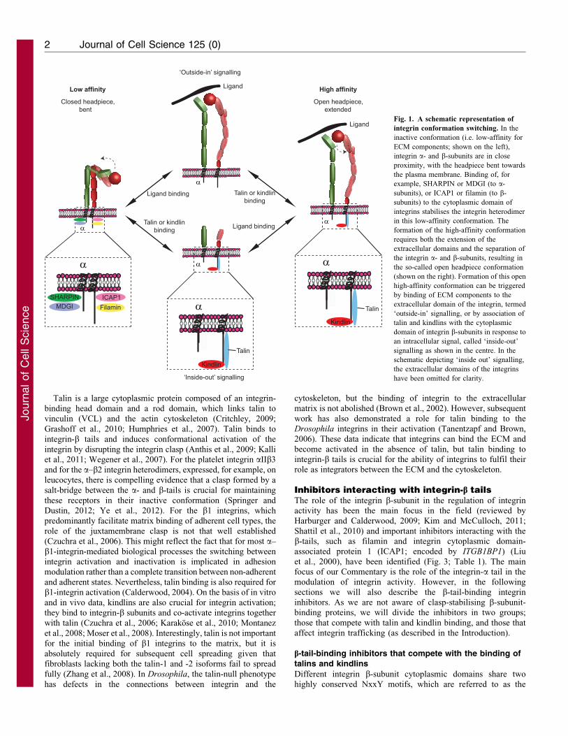
Table 1. Overview of all integrin inhibitors described in this Commentary
Integrin inhibitor a-tail binding b-tail binding Actin-binding Type of inhibition Reference(s)
ICAP1 2 + 2 Competition with talin (Bouvard et al., 2003)Filamin A 2 + + Competition with talin (Kiema et al., 2006)Numb 2 + ? Competition with talin?, endocytosis (Calderwood et al., 2003;
Nishimura and Kaibuchi,2007)
DAB2 2 + ? Endocytosis (Teckchandani et al., 2009)PKCa + 2 (Ng et al., 1999; Parsons et al.,
2002; Upla et al., 2004)Nischarin + 2 a-Subunit-specific inhibitor (Alahari and Nasrallah, 2004)CIB1 + 2 a-Subunit-specific inhibitor (Gushiken et al., 2008; Naik
et al., 1997)PP2A + + 2 Other (Gushiken et al., 2008; Kim
et al., 2004)SHARPIN + ? Clasp stabiliser or competition with talin
and/or kindlin?(Rantala et al., 2011)
MDGI + 2 Competition with kindlin (Nevo et al., 2010)Rab21 + 2 Endocytosis (Pellinen et al., 2006)p120RasGAP + 2 b1 integrin recycling to the plasma
membrane(Mai et al., 2011)
ACAP1 + + b1 integrin recycling to the plasmamembrane
(Li et al., 2005)
PKD1 + + b3 integrin recycling to the plasmamembrane
(Woods et al., 2004)
GIPC1 ? ? 2 Active a5b1 integrin; endocytosis (Valdembri et al., 2009)MMP8 ? ? 2 Inhibits b1 integrin by binding to the
extracellular domain(Pellinen et al., 2012)
+, positive interaction; 2, no interaction; ?, not determined
SHARPIN
MDGIICAP1
Filamin
(1) Clasp stabilisers
(2) -tail-bindingtalin competitors
(2) -tail-bindingtalin competitors
R D
α
SHARPIN
(3) Traffickingregulators
Rab21
TalinTalin
Talin
Talin
α α
α
α Fig. 2. Schematic illustration of three possible mechanisms of integrin
inactivation. Negative integrin regulators can inactivate integrins by
using three different mechanisms. In the first, clasp stabilisers shield the
salt bridge between an arginine residue in the a-integrin cytoplasmic
domain and an aspartate residue in the b-integrin cytoplasmic domain (1).
The presence of this salt bridge prevents separation of the cytoplasmic
domains of integrin a- and b-subunits and therefore stabilises the integrin
in its inactive form. Second, binding of talin competitors to integrin
cytoplasmic domains prevent talin, which is an essential integrin activator,
from associating with the cytoplasmic domain of the integrin b-subunit
(2). Finally, integrin binding by proteins involved in receptor endocytosis
(such as the Rab21 small GTPase) can reduce the amount of active
receptor on the cell surface and thus influence integrin-dependent
biological functions (3). Some of the integrin-inactivating proteins that
have been shown or are suspected (see text for details) to inhibit integrin
activity according to these mechanisms are shown here. Inactivators
binding to the cytoplasmic domains of integrin a- and b-subunits are
shown separately.
Integrin inactivators 3
Journ
alof
Cell
Scie
nce
(Forman-Kay and Pawson, 1999). However, unlike most PTB-
mediated protein–protein interactions, the binding of talin F3 to
integrin does not require tyrosine phosphorylation of the integrin
NPxY motif. By contrast, the talin–integrin interaction is inhibited
by tyrosine phosphorylation (Anthis et al., 2009; Oxley et al., 2008).
At least 17 PTB domain proteins interact with integrin-b tails
(Calderwood et al., 2003). The PTB domains of tensins 1–4, numb
and docking protein 1 (DOK1) bind to the same membrane-
proximal NPxY motif in integrins as talin and are thus expected to
compete with talin for integrin binding (Fig. 3). However, the
relative binding affinities of these interactions are not known.
Kindlins interact with the distal NxxY sequence of integrin tails
(Moser et al., 2009) and have been shown to activate integrins,
possibly by stabilising talin–integrin interactions. However, in the
case of b1 integrins, overexpression of kindlin-2 (FERMT2) has
been shown to interfere with talin binding (Harburger et al., 2009),
suggesting that in some cases the ability of kindlins to activate
integrins is cell type and integrin heterodimer specific. Some PTB
domain proteins preferentially bind to the distal NxxY motif of the
b-tails (Calderwood et al., 2003). Of these, Shc binding to integrin
is phosphorylation dependent, whereas disabled homolog 1 and 2
(DAB1 and DAB2) and ICAP1 do not require integrin tyrosine
phosphorylation. Of these, the role of ICAP1 as a negative
regulator of b1 integrin activity (ICAP1 does not bind b3 integrins)
is well-characterized in vitro and in vivo. ICAP1 competes for talin
binding in vitro (Bouvard et al., 2003) and regulates focal adhesion
(FA) formation (Millon-Fremillon et al., 2008) such that increased
integrin activity in the absence of ICAP1 slows down FA
dynamics. In addition, mice lacking ICAP1 have defective
osteoclast proliferation (Bouvard et al., 2007), which could be
integrin dependent. Interestingly, mice null for SHARPIN (see
below) show a similar defect in osteoclast proliferation (Xia et al.,
2011).
Thus, proteins that bind to either the proximal NPxY or the
distal NxxY motifs of the integrin tail can negatively regulate
integrin activation by competing with talin or kindlin binding.
However, elucidating the details of how these interactions are
regulated in time and space still requires more research.
Filamins form the second class of inhibitors of talin binding. The
filamin-binding site is located close to the talin-binding site on the
b-integrin tails and filamin and talin compete with each other,
whereby filamin inhibits integrin activity, as opposed to talin
(Kiema et al., 2006). Filamin depletion increases the amount of
active integrins on the cell surface, but is it unclear whether any of
the developmental defects caused by mutations in filamin genes is
the consequence of increased integrin activation (reviewed by
Zhou et al., 2010). The genetic analysis of filamin function is
complicated by the existence of multiple genes that have partially
overlapping expression patterns. In addition to this, migfilin, a
filamin-binding protein that is enriched at cell–cell and cell–ECM
contact sites, can displace filamin from b1 and b3 integrins and
promote integrin activation (Das et al., 2011; Ithychanda et al.,
2009; Lad et al., 2008). Therefore, the balance between filamin and
migfilin expression will influence the extent to which filamin
inhibits integrin activity, and, thus, the filamin–migfilin interaction
provides an additional regulatory layer for filamin-induced
inhibition of integrins.
Integrin trafficking regulators that bind the integrin-b tail
In addition to the regulation of integrin receptor activity on
the cell surface by ‘inside-out’ signalling as described above,
selective integrin endocytosis can affect the availability of active
integrin receptors on the cell surface. Most integrins are
constantly endocytosed and recycled back to the plasma
membrane in adherent cells and this is regulated in part by
proteins interacting with the integrin cytoplasmic tails
(Margadant et al., 2011; Pellinen and Ivaska, 2006). However,
to date the relationship between integrin activity and integrin
endocytic trafficking remains unclear. As mentioned above,
many PTB-domain-containing proteins have been shown to
interact with integrin-b tails in vitro (Calderwood et al., 2003)
and, interestingly, several of these are clathrin adaptor proteins
that are involved in endocytosis. According to one report, DAB2
is important for the endocytosis of active integrins from
disassembling FAs (Ezratty et al., 2009), whereas another study
suggests that there is a role for DAB2 in the endocytosis of
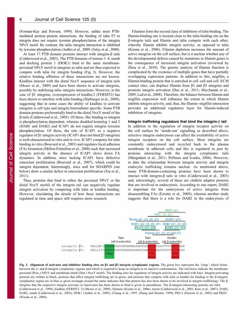
Integrin β1: WKLLMIIHDRREFAKFEKEKMNAKWDTGENPIYKSAVTTVVNPKYEGKIntegrin β3: WKLLITIHDRKEFAKFEEERARAKWDTANNPLYKEATSTFTNITYRGT
Clasp Prox.NPxY
Dist.NxxY
Talin(all β-subunits)
Talin(all β-subunits)
Kindlin(all β-subunits)
Filamin (β1, β2, β3 and β7)
ICAP1 (β1)PKC (β1, β3)
Tensin (β1, β3, β5, β7)DAB1 (β1, β3, β5)
DAB2 (β3, β5)DOK1 (β2, β3, β5, β7)
Numb (β3, β5)
PKD1 (β3)
Fig. 3. Alignment of activator and inhibitor binding sites on b1 and b3 integrin cytoplasmic regions. The green box represents the ‘clasp’, which forms
between the a- and b-integrin cytoplasmic regions and which is required to keep an integrin in its inactive conformation. The red boxes indicate the membrane-
proximal (Prox.) NPxY and membrane-distal (Dist.) NxxY motifs. The binding sites for regulators of integrin activity are indicated with lines. Integrin-activating
proteins are written in black, proteins that affect integrin trafficking are in green, and proteins that compete with talin or kindlin for binding to the b-integrin
cytoplasmic region are in blue (a green rectangle around the name indicates that that protein has also been shown to be involved in integrin trafficking). The b-
integrins that the respective integrin activator or inactivator has been shown to bind is given in parentheses. The b-integrin-interacting proteins are talin
(Calderwood et al., 1999), kindlins (FERMT1–3) (Moser et al., 2009), filamins (Kiema et al., 2006), tensin (Calderwood et al., 2003; Katz et al., 2007), DAB1,
DAB2, numb (Calderwood et al., 2003), DOK1 (Anthis et al., 2009), (Chang et al., 1997; Zhang and Hemler, 1999), PKCa (Parsons et al., 2002) and PKD1
(Woods et al., 2004).
Journal of Cell Science 125 (0)4
Journ
alof
Cell
Scie
nce
inactive integrins from the dorsal surface of cells (Teckchandani
et al., 2009). On the basis of the available structural information,it is conceivable that DAB2 could interact with both active andinactive integrins, as DAB2 binding to the b3-tail involves the
membrane distal NPxY motif, which is not bound by talin inactive integrins. The PTB-containing clathrin adaptor numb hasalso been shown to regulate integrin endocytosis and cellmigration (Nishimura and Kaibuchi, 2007). Interestingly, the
binding sites of talin and numb on the integrin b3 tail overlap(Calderwood et al., 2003), which suggests that numb couldspecifically regulate endocytosis of inactive integrin receptors by
specifically recruiting non-talin-binding inactive receptors forendocytosis. This possibility, however, has not been investigated.
In addition to these clathrin adaptors, other b-tail-interactingproteins can also trigger integrin endocytosis. For example, protein
kinase C alpha (PKCa) binds directly to the integrin b1 tail and itsbinding sequence spans both NPxY motifs. PKCa inducesendocytosis of b1 integrin in migrating cancer cells (Ng et al.,
1999; Parsons et al., 2002) and in response to echovirus-1 (EV-1)binding to a2b1 integrin or integrin clustering (Upla et al., 2004).As talin and PKCa probably cannot bind integrin simultaneously
due to the overlapping binding sites on the b-integrin cytoplasmictail (Fig. 3) (Parsons et al., 2002), it is not surprising that PKCa-dependent EV-1-induced integrin endocytosis is specific for the
inactive a2b1 conformer (Jokinen et al., 2010). Recently, it hasalso been shown that PKCa affects integrin endocytosis throughanother pathway. Binding of transmembrane proteoglycansyndecan-4 to fibronectin triggers PKCa-dependent RhoG
binding to a5b1 integrins (the fibronectin-binding integrin) andsubsequent clearance of the integrin from the membrane byaccelerated endocytosis (Bass et al., 2011). This transient
reduction of integrin levels at the cell surface is important toregulate the adhesive strength of the cells to favour migration(Bass and Humphries, 2002).
The cell surface integrin levels are also influenced by the
rate of recycling of the endocytosed receptors to the plasmamembrane. For b3 integrins, which predominantly recyclethrough a Rab4-dependent pathway, the direct binding of
protein kinase D1 (PKD1) is required for return of theendocytosed receptor to the plasma membrane (Woods et al.,2004). The recycling of the endocytosed b1 integrins, by contrast,
is regulated by binding of ACAP1 (coiled-coil, ANK repeat andPH domain-containing protein 1) to the integrin b1 cytoplasmicdomain (Li et al., 2005). Therefore, these proteins contribute to
the levels of active integrin present on the cell surface throughtheir effect on receptor recycling.
Inhibitors interacting with integrin a-tailsFor years, the main focus of the integrin field has been on the role
of the b-subunits in regulating integrin activity. As several of theb-subunits (especially b1) can pair with many different a-subunits, proteins that are involved in regulating b-subunits are
expected to have important functions in triggering variouscellular functions regulated by all b1 integrins. However, inprinciple, the a-subunits offer two distinct layers of regulating
integrin activity. First, they share conserved sequence elements inthe membrane-proximal regions of their cytoplasmic tails, whichallows for proteins to interact with and regulate all of the
different a-subunits at once (Liu et al., 2000; Rantala et al.,2011). In addition, the poorly conserved specific membrane-distal parts of the a-subunit cytoplasmic tails facilitate protein
interactions and could regulate integrin activity in a heterodimer-
specific manner. At present, our insight into the molecular
mechanisms of a-tail-mediated integrin activity regulation is far
from that of the b-tail-binding activators or inactivators.
Therefore it is not possible to categorise a-tail-interacting
activity regulators into the three mechanistic classes used above
to describe different b-tail-binding regulators. Thus, we have
opted to group them into common a-tail-binding inhibitors, a-
tail-interacting regulators of integrin trafficking and a-subunit-
specific inhibitors (Fig. 4; Table 1), as discussed below.
Integrin inhibitors binding to several or all a-subunits
SHARPIN
Until recently very little was known about SHARPIN, except that
it localises in the postsynaptic density of excitatory synapses in the
brain, where it binds Shank proteins (Lim et al., 2001) and that a
spontaneous mutation in the Sharpin gene results in eosinophilic
proliferative dermatitis and multiorgan inflammation (Liang et al.,
2011; Seymour et al., 2007). During the last few years, however,
new roles of SHARPIN have emerged as it was shown to bind to
and inhibit the lipid-phosphatase activity of phosphatase and
tensin homolog (PTEN) (He et al., 2010), act as a transcriptional
co-activator of eyes absent homolog 1 (EYA1) (Landgraf et al.,
2010), and be part of the linear ubiquitin chain assembly complex
[LUBAC, composed of heme-oxidised IRP2 ubiquitin ligase 1
(HOIL1)-interacting protein (HOIP), HOIL1 and SHARPIN
proteins], regulating nuclear factor kappa B (NFkB) activity and
apoptosis (Gerlach et al., 2011; Ikeda et al., 2011; Tokunaga et al.,
2011).
In addition to these functions, a small interfering RNA
(siRNA) screen for integrin inhibitors identified SHARPIN as a
ubiquitously expressed integrin-inactivating protein (Rantala
et al., 2011). Depletion of SHARPIN increases active b1
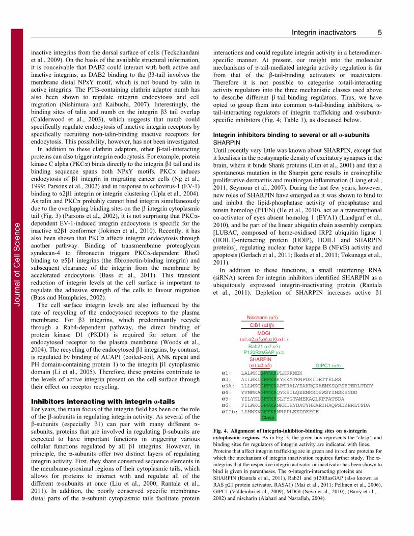
α1: LALWKIGFFKRPLKKKMEKα2: AILWKLGFFKRKYEKMTKNPDEIDETTELSSα3A: LLLWKCGFFKRARTRALYEAKRQKAEMKSQPSETERLTDDYα4: YVMWKAGFFKRQYKSILQEENRRDSWSYINSKSNDDα5: YILYKLGFFKRSLPYGTAMEKAQLKPPATSDAα6: FILWKCGFFKRNKKDHYDATYHKAEIHAQPSDKERLTSDAαIIb: LAMWKVGFFKRNRPPLEEDDEEGE
Clasp
MDGI(α1,α2,α5,α6,α10,α11)
SHARPIN(α1,α2,α5)
CIB1 (αΙΙβ)Nischarin (α5)
Rab21 (α2,α5)P120RasGAP (α2)
GIPC1 (α5)
Fig. 4. Alignment of integrin-inhibitor-binding sites on a-integrin
cytoplasmic regions. As in Fig. 3, the green box represents the ‘clasp’, and
binding sites for regulators of integrin activity are indicated with lines.
Proteins that affect integrin trafficking are in green and in red are proteins for
which the mechanism of integrin inactivation requires further study. The a-
integrins that the respective integrin activator or inactivator has been shown to
bind is given in parentheses. The a-integrin-interacting proteins are
SHARPIN (Rantala et al., 2011), Rab21 and p120RasGAP (also known as
RAS p21 protein activator, RASA1) (Mai et al., 2011; Pellinen et al., 2006),
GIPC1 (Valdembri et al., 2009), MDGI (Nevo et al., 2010), (Barry et al.,
2002) and nischarin (Alahari and Nasrallah, 2004).
Integrin inactivators 5
Journ
alof
Cell
Scie
nce
integrin at the cell surface without affecting the total amount of
integrins. SHARPIN binds to the highly conserved WKxGFFKR(Fig. 4) sequence present in the cytoplasmic tail of a-integrins(Rantala et al., 2011) and this interaction is required for
SHARPIN-mediated integrin inactivation. The essential aminoacids within the integrin cytoplasmic tail are WKxGFF (aminoacids 1154–1159 in a2 integrin), whereas the KR residues (thelysine and arginine residues immediately adjacent to the
WKxGFF sequence) are not required for SHARPIN binding(Rantala et al., 2011). This observation is compatible with afunction of SHARPIN as an integrin inhibitor, as the arginine
residue (R) within the a-tail WKxGFFKR sequence has beensuggested to form a salt bridge (clasp) with the cytoplasmic tailof the b-integrin subunit, keeping the integrin in an inactive state
(O’Toole et al., 1991).
Mechanistically, SHARPIN inhibits the binding of talin andkindlin to the cytoplasmic domain of b1integrin (Rantala et al.,2011). Thus, on the one hand, the ‘inside-out’ integrin activation
is controlled by the balance of counteracting forces that areexerted by SHARPIN (and possibly other integrin inhibitors), andon the other hand and talins and kindlins. It remains to be
determined whether this inhibition is direct (through sterichindrance or by stabilising the integrin inactivating clasp) orindirect (for example by recruiting inhibiting kinases). In cells,
SHARPIN colocalises with inactive, but not active, integrins indetached ruffles (Rantala et al., 2011), consistent with SHARPINacting as an integrin-inactivating protein. SHARPIN bindingto integrin a-subunits also inhibits a–b1-integrin-dependent
functions, such as cell spreading and cell migration.
Epidermal homeostasis is regulated by integrin expression, anddownregulation of b1 integrin expression in the suprabasal
keratinocytes is a prerequisite for keratinocyte differentiation(Watt, 2002). Interestingly, the proliferative dermatitis phenotypeof mice null for SHARPIN resembles that of the transgenicmouse models with forced suprabasal b1 integrin expression
under the control of the involucrin promoter (Carroll et al.,1995). Thus, loss of the b1 integrin inactivator SHARPINand overexpression of b1 integrin results in very similar
physiological outcomes in vivo. In addition, the function ofSHARPIN as a b1-integrin inhibitor in vivo is supported by theobservation that keratin-14-positive keratinocytes from Sharpin-
null mice contain higher active b1-integrin levels thankeratinocytes from wild-type mice (Rantala et al., 2011).
Importantly, the integrin inhibitory effect of SHARPIN isindependent of the aforementioned other functions of SHARPIN.
SHARPIN silencing induces integrin activation in PC3 cells,which are null for PTEN (Gustin et al., 2001) and lack EYA1expression (Kilpinen et al., 2008). Furthermore, silencing of HOIP
(the catalytic subunit of LUBAC) does not affect integrin activityin cells (Rantala et al., 2011). In addition, mice lacking HOIL1(another member of the LUBAC complex) (i.e. after knockout of
the HOIL1-encoding gene Rbck) do not have any obvious integrin-related phenotype (Tokunaga et al., 2009), indicating thatSHARPIN has important, LUBAC-independent, functions in vivo.
The identification of SHARPIN opens a new paradigm
in integrin regulation as it demonstrates that the dynamicswitching between inactive and active integrin conformations isphysiologically controlled in vivo by a protein that interacts with
the a-subunit cytoplasmic domain. As SHARPIN is expressed inmost human tissues and several cancer cell types (Rantala et al.,2011), and it binds several (and potentially all) a-integrins,
SHARPIN might represent a general means for cells to suppress
integrin activity.
MDGI
Mammary-derived growth inhibitor (MDGI, also known as FABP3)
has been named after its origin from lactating bovine mammary
glands and its growth inhibitory properties in human mammary
carcinoma cell cultures (Bohmer et al., 1987). MDGI is almost
ubiquitously expressed, and is particularly abundant in muscle and
mammary gland (Haunerland and Spener, 2004). Recently, MDGI
has been shown to interact with several different integrin a-subunits
(a1, a2, a5, a6, a10 and a11), probably through the highly
conserved WKxGFFKR sequence that is present in most, if not all,
a-subunits (Nevo et al., 2010). MDGI overexpression reduces
adhesion of cells to type I collagen and fibronectin and impairs both
migration and invasion specifically in human breast cancer cell lines
but not in other cell types (Nevo et al., 2010). These effects are due
to an MDGI-induced significant reduction in the active b1integrin
conformation on the cell surface as determined with conformation-
sensitive antibodies against b1 integrin (Nevo et al., 2010). The
exact molecular details of the function of MDGI as a negative
regulator of integrin activity remain unsolved, but MDGI
overexpression in MDA-MB-231 breast cancer cells reduces the
association of kindlin with active b1 integrin, suggesting that MDGI
binding to cytoplasmic integrin-a tails could indirectly hamper the
binding of integrin agonists to integrin-b tails (Nevo et al., 2010). It
remains unclear why these effects of MDGI on integrin activity are
specific to breast cancer cells. One possibility is that they are linked
to epidermal growth factor receptor (EGFR) overexpression, a
common genetic alteration for breast cancer cells. MDGI influences
EGFR trafficking so that the receptor is increasingly localised on
endosomes (Nevo et al., 2009). As EGFR and b1 integrins
participate in extensive crosstalk (Ivaska and Heino, 2011),
the altered EGFR dynamics could also influence b1 integrin
conformation in breast cancer cells. Regardless of the molecular
details, the ability of MDGI to inhibit breast cancer cell invasion in
vitro appears to be clinically relevant. Analysis of a tissue
microarray of 1331 breast carcinomas revealed that patients with
MDGI-positive tumours have a more favourable 10-year disease-
free survival prognosis compared with that of patients with MDGI-
negative tumours (Nevo et al., 2010).
Integrin trafficking regulators that bind the integrin a-tail
As discussed above, the conserved membrane-proximal segment
of integrin a-tails is important for SHARPIN and MDGI binding
and integrin inactivation. Interestingly, this binding site overlaps
with the site on the integrin a-tails, at which the small GTPase
Rab21 binds many integrin a–b1 heterodimers and subsequently
induces their endocytosis (Pellinen et al., 2006). Furthermore, on
endosomes, the GTPase-activating protein (GAP) p120RasGAP
(also known as RASA1) competes with Rab21 for integrin
binding and regulates the recycling of the receptor to the plasma
membrane (Mai et al., 2011). The possible selectivity of Rab21
(and p120RasGAP) for active integrins has not been investigated
but, as in the case of the clathrin adaptor Numb, one could
envision a scenario, in which integrin activity regulators (for
example talin and SHARPIN) compete with endocytosis
regulators (Numb and Rab21) for integrin binding. This would
provide a mechanism to control the relative amounts of active
and inactive integrin receptors on the cell surface.
Journal of Cell Science 125 (0)6
Journ
alof
Cell
Scie
nce
In endothelial cells, endocytosis of the active a5b1 integrin is
also specifically regulated by the a-tail interacting homomultimeric
endocytic adaptor GAIP interacting protein C terminus member 1
(GIPC1). In addition to the a5b1 integrin, GIPC1 also binds to
Neuropilin-1 (Nrp-1) and Myosin-6, and formation of this complex
triggers endocytosis of the active a5b1-integrin into Rab5-positive
endosomes (Valdembri et al., 2009). This results in increased
adhesion of endothelial cells to fibronectin, in line with the
previously established link between increased integrin traffic and
faster cell spreading and adhesion (Pellinen et al., 2006). At present
it is not clear how stimulation of endothelial cells with Nrp-1
ligands triggers active-conformer specific endocytosis. However,
the selective uptake of the active receptor from the cell surface is
absolutely dependent on Neuropilin-1 as endocytosis of the inactive
a5b1 is Neuropilin-1 independent. This suggests that in addition to
the ability of cells to modulate their adhesion receptor activity on
the plasma membrane via ‘inside-out’ signalling, selective
endocytosis of a specific integrin conformer may be an important
mechanism to regulate the availability of active integrins at the cell
surface in a spatially controlled or polarised manner. In addition,
very recent data indicate that active and inactive b1-integrins also
use distinct recycling pathways to return to the plasma membrane
(Arjonen et al., 2012).
Integrin inhibitors binding to specific a-subunits
There are a few proteins that have been described to bind to
a specific integrin a-subunit and inhibit integrin function.
However, the negative regulatory role of these proteins is
controversial, and data demonstrating their actual regulation of
integrin activity on the cell surface is lacking. Nevertheless, these
examples are discussed here briefly as integrin-heterodimer-
specific regulation of integrin activity is an understudied area and
at present better examples are lacking.
Nischarin is a ubiquitously expressed cytosolic protein that
interacts with the cytoplasmic domain of integrin a5 (Alahari
et al., 2000). The interaction involves the a5 membrane proximal
sequence IYILYKLGFFKRSL (residues 1017–1030), for which
residues Tyr1018 and Lys1022 are crucial (Alahari and
Nasrallah, 2004). As these residues are relatively conserved in
other integrin a-subunits it is possible that nischarin could also
interact with other integrin-a tails; however, this has not been
studied. Overexpression of nischarin inhibits both cell migration
and invasion, but not adhesion to fibronectin (Alahari et al., 2000;
Alahari, 2003). This is owing to alterations in Rac activation and
the organisation of peripheral actin filaments in nischarin-
overexpressing cells. Overexpression of the integrin a5 subunit
enhances coprecipitation of the serine/threonine-protein kinase
PAK1 [p21 protein (Cdc42/Rac)-activated kinase 1] with
nischarin, which inhibits PAK1 kinase activity (Alahari and
Nasrallah, 2004). This suggests that a5 has a role in the localised
control of PAK1 function. Although it appears that nischarin
inhibits integrin-dependent processes, data that formally address
the effect of nischarin expression on integrin activation are
missing. Interestingly, recently nischarin expression has been
shown to correlate with low-grade less-aggressive tumours in
breast cancer and with reduced tumour growth and lung
metastasis in a nude mouse model (Baranwal et al., 2011),
which could be linked to reduced integrin activity.
Fine-tuned platelet aggregation is responsible for physiological
homeostasis and aberrant platelet function can lead to pathological
thrombosis. The ubiquitously expressed calcium- and integrin-
binding protein 1 (CIB1) has a key role in the regulation of the
platelet integrin aIIbb3. CIB1 interacts directly with the
cytoplasmic domain of aIIb (Naik et al., 1997), of which the aIIb
membrane-proximal sequence LVLAMWKVGFFKRNR (residues
983 to 997) forms a minimal-binding domain for CIB1 (Barry et al.,
2002). Residues Leu983, Trp988, Phe992 and Phe993 and Ca2+-
binding are crucial for the complex formation (Barry et al., 2002;
Shock et al., 1999). CIB1 has been reported to function as an
endogenous inhibitor of agonist-induced aIIbb3 integrin activation
(Yuan et al., 2006). However, CIB1 has several other binding
partners apart from integrins and they might explain the variable
roles that have been described for the CIB1–aIIb complex. For
example, in contrast to the proposed role as an integrin inactivator,
CIB1 has been shown to activate aIIbb3 integrin ‘inside-out’
signalling by converting the integrin from the resting state into its
active conformation (Tsuboi, 2002) and to have an important role in
platelet spreading on immobilised fibrinogen (Naik and Naik,
2003). Finally, the physiological consequences of loss of CIB1 (in
knockout mice) are impaired thrombosis and increased mouse tail-
bleeding time (Naik et al., 2009), suggesting that in vivo CIB1
functions as a positive regulator of aIIbb3.
Serine-threonine phosphatase 2A (PP2A) has been shown to
interact directly with the membrane-proximal KVGFFKR
sequence of aIIb integrin (Gushiken et al., 2008). In human
embryonal kidney (HEK) 293 cells, PP2A overexpression
decreases aIIbb3-mediated adhesion to immobilised fibrinogen
(Gushiken et al., 2008). By contrast, PP2A association with b1
integrin followed by dephosphorylation of crucial residues within
the b1 integrin cytoplasmic tail is required for integrin
localisation to focal contacts and subsequent activation of
downstream signalling (Kim et al., 2004; Mulrooney et al., 2000).
Taken together, at present there are no clear examples of
proteins binding to specific a-subunits as bona fide integrin
inhibitors. For all the examples discussed above there are data
indicating context dependent roles for these proteins in both
integrin inactivation and activation.
Matrix metalloproteinase 8 (MMP8)As described here, integrin activity can also be regulated by
extracellular proteins. Recently, we reported an siRNA screen
that revealed new genes that were involved in regulating b1
integrin activity (Pellinen et al., 2012). One particularly
interesting finding is that the secreted collagenase MMP8 is a
negative regulator of b1 integrin activity in several prostate
cancer and one breast cancer cell line. MMP8 has been
previously associated with tumour suppression owing to its
anti-inflammatory or anti-metastatic role in several cancers
(Dejonckheere et al., 2011). The increase in integrin activity
caused by MMP8 silencing is counteracted by the addition of
recombinant MMP8 to the growth medium and correlates with
increased in vitro invasion through Matrigel (Pellinen et al.,
2012). In a mouse metastasis assay, MMP8-silenced cancer cells
extravasate to the lungs to a much greater extent than do control
cells. In addition, MMP8 co-precipitates with b1 integrin,
implying that binding of MMP8 to the extracellular part of the
b1 integrin could modulate integrin activity, although we cannot
rule out that MMP8 binds to integrin indirectly through its
interactions with collagen. It is also possible that the collagenase
activity of MMP8 modifies the integrin ligand collagen in a way
Integrin inactivators 7
Journ
alof
Cell
Scie
nce
that abrogates integrin binding. In the future, it will be interesting
to study the exact mechanism underlying the effect of MMP8
on integrin activity and how it might be related to the anti-
inflammatory role of MMP8.
In vivo roles of integrin activators andinactivatorsAbove we have discussed several integrin activity inhibitors
which all have the ability to induce the inactive integrin
conformation on the cell surface, albeit the mechanisms
involved differ between the proteins. In cells, these inactivators
are counteracting the integrin-activating talin and kindlin proteins
such that switching of the integrins between active and inactive
states is inhibited. However, a more complex picture emerges
when the phenotypes of mice lacking these activators or
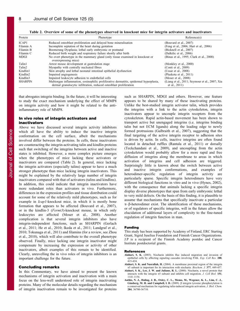
inactivators are compared (Table 2). In general, mice lacking
the integrin activators (especially talins) appear to have a much
stronger phenotype than mice lacking integrin inactivators. This
might be explained by the relatively large number of integrin
inactivators compared with that of activators (talins and kindlins).
In addition, this could indicate that integrin inactivators have
more redundant roles than activators in vivo. Furthermore,
differences in the expression profiles and tissue distribution could
account for some of the relatively mild phenotypes, such as for
example in Icap1-knockout mice, in which it is mostly bone
formation that appears to be affected (Bouvard et al., 2007),
or in the kindlin-3 (Fermt3)-knockout mouse, in which only
leukocytes are affected (Moser et al., 2008). Another
complication is that several integrin inhibitors also have
integrin-independent functions, such as SHARPIN (Gerlach
et al., 2011; He et al., 2010; Ikeda et al., 2011; Landgraf et al.,
2010; Tokunaga et al., 2011) and filamins (for a review, see Zhou
et al., 2010), which will also contribute to the overall phenotype
observed. Finally, mice lacking one integrin inactivator might
compensate by increasing the expression or activity of other
inactivators, albeit examples of this remain to be identified.
Clearly, unravelling the in vivo roles of integrin inhibitors is an
important challenge for the future.
Concluding remarksIn this Commentary, we have aimed to present the known
mechanisms of integrin activation and inactivation with a main
focus on the less-well understood role of integrin inactivating
proteins. Many of the molecular details regarding the mechanism
of integrin inactivation remain to be investigated for proteins
such as SHARPIN, MDGI and others. However, one feature
appears to be shared by many of these inactivating proteins.Unlike the best-studied integrin activator talin, which providesthe integrins with a link to the actin cytoskeleton, integrininactivators appear to uncouple integrin receptors from the
cytoskeleton. Rapid actin-based movement has been shown totransport active but unengaged integrins (i.e. integrins bindingtalin but not ECM ligands) along the leading edge to newly
formed protrusions (Galbraith et al., 2007), suggesting that thefinal targeting of the active integrin receptor to adhesion sitesis driven by actin. In cells, inactive integrins are often found
located in detached ruffles (Rantala et al., 2011) or dorsally(Teckchandani et al., 2009), and uncoupling from the actincytoskeleton would probably allow for a more rapid and freediffusion of integrins along the membrane to areas in which
activation of integrins and cell adhesion are triggered.Surprisingly little is known about the switch between activeand inactive integrin conformations, and examples of
heterodimer-specific regulation of integrin activity areparticularly sparse. Specific integrin heterodimers have verydifferent biological functions in vitro and in vivo (Hynes, 2002),
with the consequence that animals lacking a specific integrindisplay diverse phenotypes that span from early embryonic lethalto very mild defects. On the basis of this finding, it is plausible to
assume that mechanisms that specifically inactivate a particulara–b-heterodimer exist. The identification of these mechanisms,or of regulators of specific integrins, will in the future allow theelucidation of additional layers of complexity to the fine-tuned
regulation of integrin function in man.
FundingThis study has been supported by Academy of Finland, ERC StartingGrant, Sigrid Juselius Foundation and Finnish Cancer Organizations.J.P is a recipient of the Finnish Academy postdoc and CancerInstitute postdoctorial grants.
ReferencesAlahari, S. K. (2003). Nischarin inhibits Rac induced migration and invasion of
epithelial cells by affecting signaling cascades involving PAK. Exp. Cell Res. 288,415-424.
Alahari, S. K. and Nasrallah, H. (2004). A membrane proximal region of the integrina5 subunit is important for its interaction with nischarin. Biochem. J. 377, 449-457.
Alahari, S. K., Lee, J. W. and Juliano, R. L. (2000). Nischarin, a novel protein thatinteracts with the integrin a5 subunit and inhibits cell migration. J. Cell Biol. 151,1141-1154.
Anthis, N. J., Haling, J. R., Oxley, C. L., Memo, M., Wegener, K. L., Lim, C. J.,
Ginsberg, M. H. and Campbell, I. D. (2009). b integrin tyrosine phosphorylation isa conserved mechanism for regulating talin-induced integrin activation. J. Biol. Chem.
284, 36700-36710.
Table 2. Overview of some of the phenotypes observed in knockout mice for integrin activators and inactivators
Protein Phenotype Reference(s)
ICAP1 Reduced osteoblast proliferation and delayed bone mineralisation (Bouvard et al., 2007)Filamin A Incomplete septation of the heart during gestation (Feng et al., 2006; Hart et al., 2006)Filamin B Boomerang Dysplasia; lethal early embryonic or postnatal (Bicknell et al., 2007)Filamin C Reduced birth weight and respiratory failure shortly after birth (Dalkilic et al., 2006)MDGI No overt phenotype in the mammary gland (only tissue examined in knockout or
overexpressing mice)(Binas et al., 1995; Clark et al., 2000)
Talin1 Arrest mouse development at gastrulation stage (Monkley et al., 2000)Talin2 Myopathy with centrally nucleated fibres (Conti et al., 2009)Kindlin1 Skin atrophy and lethal neonatal intestinal epithelial dysfunction (Ussar et al., 2008)Kindlin2 Impaired angiogenesis (Pluskota et al., 2011)Kindlin3 Impaired leukocyte adhesion to endothelial cells (Moser et al., 2008)SHARPIN Multiorgan inflammation, eosinophilic proliferative dermatitis, epidermal hyperplasia,
dermal granulocytic infiltration, reduced osteoblast proliferation(Liang et al., 2011; Seymour et al., 2007; Xia
et al., 2011)
Journal of Cell Science 125 (0)8
Journ
alof
Cell
Scie
nce
Arjonen, A., Alanko, J., Veltel, S. and Ivaska, J. (2012). Distinct recycling of active
and inactive b1 integrins. Traffic 13, 610-625.
Baranwal, S., Wang, Y., Rathinam, R., Lee, J., Jin, L., McGoey, R., Pylayeva, Y.,
Giancotti, F., Blobe, G. C. and Alahari, S. K. (2011). Molecular characterization ofthe tumor-suppressive function of nischarin in breast cancer. J. Natl. Cancer Inst. 103,1513-1528.
Barry, W. T., Boudignon-Proudhon, C., Shock, D. D., McFadden, A., Weiss, J. M.,
Sondek, J. and Parise, L. V. (2002). Molecular basis of CIB binding to the integrin
aIIb cytoplasmic domain. J. Biol. Chem. 277, 28877-28883.
Bass, M. D. and Humphries, M. J. (2002). Cytoplasmic interactions of syndecan-4
orchestrate adhesion receptor and growth factor receptor signalling. Biochem. J. 368,1-15.
Bass, M. D., Williamson, R. C., Nunan, R. D., Humphries, J. D., Byron, A., Morgan,
M. R., Martin, P. and Humphries, M. J. (2011). A syndecan-4 hair trigger initiates
wound healing through caveolin- and RhoG-regulated integrin endocytosis. Dev. Cell
21, 681-693.
Bicknell, L. S., Farrington-Rock, C., Shafeghati, Y., Rump, P., Alanay, Y., Alembik, Y.,
Al-Madani, N., Firth, H., Karimi-Nejad, M. H., Kim, C. A. et al. (2007). A molecularand clinical study of Larsen syndrome caused by mutations in FLNB. J. Med. Genet. 44,
89-98.
Binas, B., Gusterson, B., Wallace, R. and Clark, A. J. (1995). Epithelial proliferationand differentiation in the mammary gland do not correlate with cFABP geneexpression during early pregnancy. Dev. Genet. 17, 167-175.
Bohmer, F. D., Sun, Q., Pepperle, M., Muller, T., Eriksson, U., Wang, J. L. and
Grosse, R. (1987). Antibodies against mammary derived growth inhibitor (MDGI)
react with a fibroblast growth inhibitor and with heart fatty acid binding protein.Biochem. Biophys. Res. Commun. 148, 1425-1431.
Bouvard, D., Vignoud, L., Dupe-Manet, S., Abed, N., Fournier, H. N., Vincent-
Monegat, C., Retta, S. F., Fassler, R. and Block, M. R. (2003). Disruption of focal
adhesions by integrin cytoplasmic domain-associated protein-1a. J. Biol. Chem. 278,6567-6574.
Bouvard, D., Aszodi, A., Kostka, G., Block, M. R., Albiges-Rizo, C. and Fassler, R.
(2007). Defective osteoblast function in ICAP-1-deficient mice. Development 134,2615-2625.
Brown, N. H., Gregory, S. L., Rickoll, W. L., Fessler, L. I., Prout, M., White, R. A.
and Fristrom, J. W. (2002). Talin is essential for integrin function in Drosophila.
Dev. Cell 3, 569-579.
Calderwood, D. A. (2004). Integrin activation. J. Cell Sci. 117, 657-666.
Calderwood, D. A., Zent, R., Grant, R., Rees, D. J., Hynes, R. O. and Ginsberg,
M. H. (1999). The Talin head domain binds to integrin b subunit cytoplasmic tails
and regulates integrin activation. J. Biol. Chem. 274, 28071-28074.
Calderwood, D. A., Fujioka, Y., de Pereda, J. M., Garcıa-Alvarez, B., Nakamoto, T.,
Margolis, B., McGlade, C. J., Liddington, R. C. and Ginsberg, M. H. (2003).Integrin b cytoplasmic domain interactions with phosphotyrosine-binding domains: a
structural prototype for diversity in integrin signaling. Proc. Natl. Acad. Sci. USA 100,2272-2277.
Calderwood, D. A., Tai, V., Di Paolo, G., De Camilli, P. and Ginsberg, M. H. (2004).Competition for talin results in trans-dominant inhibition of integrin activation. J.
Biol. Chem. 279, 28889-28895.
Carroll, J. M., Romero, M. R. and Watt, F. M. (1995). Suprabasal integrin expressionin the epidermis of transgenic mice results in developmental defects and a phenotype
resembling psoriasis. Cell 83, 957-968.
Chang, D. D., Wong, C., Smith, H. and Liu, J. (1997). ICAP-1, a novel b1 integrin
cytoplasmic domain-associated protein, binds to a conserved and functionallyimportant NPXY sequence motif of b1 integrin. J. Cell Biol. 138, 1149-1157.
Clark, A. J., Neil, C., Gusterson, B., McWhir, J. and Binas, B. (2000). Deletion ofthe gene encoding H-FABP/MDGI has no overt effects in the mammary gland.
Transgenic Res. 9, 439-444.
Conti, F. J., Monkley, S. J., Wood, M. R., Critchley, D. R. and Muller, U. (2009).
Talin 1 and 2 are required for myoblast fusion, sarcomere assembly and themaintenance of myotendinous junctions. Development 136, 3597-3606.
Critchley, D. R. (2009). Biochemical and structural properties of the integrin-associatedcytoskeletal protein talin. Annu. Rev. Biophys. 38, 235-254.
Czuchra, A., Meyer, H., Legate, K. R., Brakebusch, C. and Fassler, R. (2006).Genetic analysis of b1 integrin ‘‘activation motifs’’ in mice. J. Cell Biol. 174, 889-
899.
Dalkilic, I., Schienda, J., Thompson, T. G. and Kunkel, L. M. (2006). Loss of
FilaminC (FLNc) results in severe defects in myogenesis and myotube structure. Mol.
Cell. Biol. 26, 6522-6534.
Das, M., Ithychanda, S. S., Qin, J. and Plow, E. F. (2011). Migfilin and filamin asregulators of integrin activation in endothelial cells and neutrophils. PLoS ONE 6,e26355.
Dejonckheere, E., Vandenbroucke, R. E. and Libert, C. (2011). Matrix metallopro-teinase8 has a central role in inflammatory disorders and cancer progression. Cytokine
Growth Factor Rev. 22, 73-81.
Ezratty, E. J., Bertaux, C., Marcantonio, E. E. and Gundersen, G. G. (2009).
Clathrin mediates integrin endocytosis for focal adhesion disassembly in migratingcells. J. Cell Biol. 187, 733-747.
Feng, Y., Chen, M. H., Moskowitz, I. P., Mendonza, A. M., Vidali, L., Nakamura, F.,
Kwiatkowski, D. J. and Walsh, C. A. (2006). Filamin A (FLNA) is required for cell-
cell contact in vascular development and cardiac morphogenesis. Proc. Natl. Acad. Sci.
USA 103, 19836-19841.
Forman-Kay, J. D. and Pawson, T. (1999). Diversity in protein recognition by PTBdomains. Curr. Opin. Struct. Biol. 9, 690-695.
Galbraith, C. G., Yamada, K. M. and Galbraith, J. A. (2007). Polymerizing actinfibers position integrins primed to probe for adhesion sites. Science 315, 992-995.
Gerlach, B., Cordier, S. M., Schmukle, A. C., Emmerich, C. H., Rieser, E., Haas,
T. L., Webb, A. I., Rickard, J. A., Anderton, H., Wong, W. W. et al. (2011). Linearubiquitination prevents inflammation and regulates immune signalling. Nature 471,591-596.
Goldmann, W. H. (2000). Kinetic determination of focal adhesion protein formation.Biochem. Biophys. Res. Commun. 271, 553-557.
Grashoff, C., Hoffman, B. D., Brenner, M. D., Zhou, R., Parsons, M., Yang, M. T.,
McLean, M. A., Sligar, S. G., Chen, C. S., Ha, T. et al. (2010). Measuring
mechanical tension across vinculin reveals regulation of focal adhesion dynamics.Nature 466, 263-266.
Gushiken, F. C., Patel, V., Liu, Y., Pradhan, S., Bergeron, A. L., Peng, Y. and
Vijayan, K. V. (2008). Protein phosphatase 2A negatively regulates integrin aIIbb3signaling. J. Biol. Chem. 283, 12862-12869.
Gustin, J. A., Maehama, T., Dixon, J. E. and Donner, D. B. (2001). The PTEN tumorsuppressor protein inhibits tumor necrosis factor-induced nuclear factor kB activity. J.
Biol. Chem. 276, 27740-27744.
Harburger, D. S. and Calderwood, D. A. (2009). Integrin signalling at a glance. J. Cell
Sci. 122, 159-163.
Harburger, D. S., Bouaouina, M. and Calderwood, D. A. (2009). Kindlin-1 and -2directly bind the C-terminal region of b integrin cytoplasmic tails and exert integrin-specific activation effects. J. Biol. Chem. 284, 11485-11497.
Hart, A. W., Morgan, J. E., Schneider, J., West, K., McKie, L., Bhattacharya, S.,
Jackson, I. J. and Cross, S. H. (2006). Cardiac malformations and midline skeletaldefects in mice lacking filamin A. Hum. Mol. Genet. 15, 2457-2467.
Haunerland, N. H. and Spener, F. (2004). Fatty acid-binding proteins–insights fromgenetic manipulations. Prog. Lipid Res. 43, 328-349.
He, L., Ingram, A., Rybak, A. P. and Tang, D. (2010). Shank-interacting protein-like 1promotes tumorigenesis via PTEN inhibition in human tumor cells. J. Clin. Invest.
120, 2094-2108.
Hogg, N. and Bates, P. A. (2000). Genetic analysis of integrin function in man: LAD-1and other syndromes. Matrix Biol. 19, 211-222.
Horwitz, A., Duggan, K., Buck, C., Beckerle, M. C. and Burridge, K. (1986).Interaction of plasma membrane fibronectin receptor with talin–a transmembranelinkage. Nature 320, 531-533.
Hughes, P. E., Diaz-Gonzalez, F., Leong, L., Wu, C., McDonald, J. A., Shattil, S. J.
and Ginsberg, M. H. (1996). Breaking the integrin hinge. A defined structural
constraint regulates integrin signaling. J. Biol. Chem. 271, 6571-6574.
Humphries, J. D., Wang, P., Streuli, C., Geiger, B., Humphries, M. J. and
Ballestrem, C. (2007). Vinculin controls focal adhesion formation by directinteractions with talin and actin. J. Cell Biol. 179, 1043-1057.
Hynes, R. O. (2002). Integrins: bidirectional, allosteric signaling machines. Cell 110,673-687.
Ikeda, F., Deribe, Y. L., Skanland, S. S., Stieglitz, B., Grabbe, C., Franz-Wachtel, M.,
van Wijk, S. J., Goswami, P., Nagy, V., Terzic, J. et al. (2011). SHARPIN forms alinear ubiquitin ligase complex regulating NF-kB activity and apoptosis. Nature 471,637-641.
Ithychanda, S. S., Das, M., Ma, Y. Q., Ding, K., Wang, X., Gupta, S., Wu, C., Plow,
E. F. and Qin, J. (2009). Migfilin, a molecular switch in regulation of integrinactivation. J. Biol. Chem. 284, 4713-4722.
Ivaska, J. and Heino, J. (2010). Interplay between cell adhesion and growth factor
receptors: from the plasma membrane to the endosomes. Cell Tissue Res. 339, 111-120.
Ivaska, J. and Heino, J. (2011). Cooperation between integrins and growth factorreceptors in signaling and endocytosis. Annu. Rev. Cell Dev. Biol. 27, 291-320.
Jokinen, J., White, D. J., Salmela, M., Huhtala, M., Kapyla, J., Sipila, K., Puranen,
J. S., Nissinen, L., Kankaanpaa, P., Marjomaki, V. et al. (2010). Molecularmechanism of a2b1 integrin interaction with human echovirus 1. EMBO J. 29, 196-208.
Kalli, A. C., Campbell, I. D. and Sansom, M. S. (2011). Multiscale simulations suggesta mechanism for integrin inside-out activation. Proc. Natl. Acad. Sci. USA 108,11890-11895.
Karakose, E., Schiller, H. B. and Fassler, R. (2010). The kindlins at a glance. J. Cell
Sci. 123, 2353-2356.
Katz, M., Amit, I., Citri, A., Shay, T., Carvalho, S., Lavi, S., Milanezi, F., Lyass, L.,
Amariglio, N., Jacob-Hirsch, J. et al. (2007). A reciprocal tensin-3-cten switchmediates EGF-driven mammary cell migration. Nat. Cell Biol. 9, 961-969.
Kiema, T., Lad, Y., Jiang, P., Oxley, C. L., Baldassarre, M., Wegener, K. L.,
Campbell, I. D., Ylanne, J. and Calderwood, D. A. (2006). The molecular basis of
filamin binding to integrins and competition with talin. Mol. Cell 21, 337-347.
Kilpinen, S., Autio, R., Ojala, K., Iljin, K., Bucher, E., Sara, H., Pisto, T., Saarela, M.,
Skotheim, R. I., Bjorkman, M. et al. (2008). Systematic bioinformatic analysis ofexpression levels of 17,330 human genes across 9,783 samples from 175 types ofhealthy and pathological tissues. Genome Biol. 9, R139.
Kim, H. and McCulloch, C. A. (2011). Filamin A mediates interactions betweencytoskeletal proteins that control cell adhesion. FEBS Lett. 585, 18-22.
Kim, S. M., Kwon, M. S., Park, C. S., Choi, K. R., Chun, J. S., Ahn, J. and Song,
W. K. (2004). Modulation of Thr phosphorylation of integrin b1 during muscledifferentiation. J. Biol. Chem. 279, 7082-7090.
Integrin inactivators 9
Journ
alof
Cell
Scie
nce
Knezevic, I., Leisner, T. M. and Lam, S. C.-T. (1996). Direct binding of the plateletintegrin aIIbb3 (GPIIb-IIIa) to talin. Evidence that interaction is mediated through thecytoplasmic domains of both aIIb and b3. J. Biol. Chem. 271, 16416-16421.
Lad, Y., Jiang, P., Ruskamo, S., Harburger, D. S., Ylanne, J., Campbell, I. D. and
Calderwood, D. A. (2008). Structural basis of the migfilin-filamin interaction andcompetition with integrin b tails. J. Biol. Chem. 283, 35154-35163.
Landgraf, K., Bollig, F., Trowe, M. O., Besenbeck, B., Ebert, C., Kruspe, D.,
Kispert, A., Hanel, F. and Englert, C. (2010). Sipl1 and Rbck1 are novel Eya1-binding proteins with a role in craniofacial development. Mol. Cell. Biol. 30, 5764-5775.
Lau, T. L., Kim, C., Ginsberg, M. H. and Ulmer, T. S. (2009). The structure of theintegrin aIIbb3 transmembrane complex explains integrin transmembrane signalling.EMBO J. 28, 1351-1361.
Legate, K. R., Wickstrom, S. A. and Fassler, R. (2009). Genetic and cell biologicalanalysis of integrin outside-in signaling. Genes Dev. 23, 397-418.
Li, J., Ballif, B. A., Powelka, A. M., Dai, J., Gygi, S. P. and Hsu, V. W. (2005).Phosphorylation of ACAP1 by Akt regulates the stimulation-dependent recycling ofintegrin b1 to control cell migration. Dev. Cell 9, 663-673.
Liang, Y., Seymour, R. E. and Sundberg, J. P. (2011). Inhibition of NF-kB signalingretards eosinophilic dermatitis in SHARPIN-deficient mice. J. Invest. Dermatol. 131,141-149.
Lim, S., Sala, C., Yoon, J., Park, S., Kuroda, S., Sheng, M. and Kim, E. (2001).Sharpin, a novel postsynaptic density protein that directly interacts with the shankfamily of proteins. Mol. Cell. Neurosci. 17, 385-397.
Liu, S., Calderwood, D. A. and Ginsberg, M. H. (2000). Integrin cytoplasmic domain-binding proteins. J. Cell Sci. 113, 3563-3571.
Mai, A., Veltel, S., Pellinen, T., Padzik, A., Coffey, E., Marjomaki, V. and Ivaska, J.(2011). Competitive binding of Rab21 and p120RasGAP to integrins regulatesreceptor traffic and migration. J. Cell Biol. 194, 291-306.
Margadant, C., Monsuur, H. N., Norman, J. C. and Sonnenberg, A. (2011).Mechanisms of integrin activation and trafficking. Curr. Opin. Cell Biol. 23, 607-614.
Millon-Fremillon, A., Bouvard, D., Grichine, A., Manet-Dupe, S., Block, M. R. andAlbiges-Rizo, C. (2008). Cell adaptive response to extracellular matrix density iscontrolled by ICAP-1-dependent b1-integrin affinity. J. Cell Biol. 180, 427-441.
Monkley, S. J., Zhou, X.-H., Kinston, S. J., Giblett, S. M., Hemmings, L., Priddle, H.,Brown, J. E., Pritchard, C. A., Critchley, D. R. and Fassler, R. (2000). Disruption ofthe talin gene arrests mouse development at the gastrulation stage. Dev. Dyn. 219, 560-574.
Montanez, E., Ussar, S., Schifferer, M., Bosl, M., Zent, R., Moser, M. and Fassler, R.
(2008). Kindlin-2 controls bidirectional signaling of integrins. Genes Dev. 22, 1325-1330.
Moser, M., Nieswandt, B., Ussar, S., Pozgajova, M. and Fassler, R. (2008). Kindlin-3is essential for integrin activation and platelet aggregation. Nat. Med. 14, 325-330.
Moser, M., Legate, K. R., Zent, R. and Fassler, R. (2009). The tail of integrins, talin,and kindlins. Science 324, 895-899.
Mulrooney, J., Foley, K., Vineberg, S., Barreuther, M. and Grabel, L. (2000).Phosphorylation of the b1 integrin cytoplasmic domain: toward an understanding offunction and mechanism. Exp. Cell Res. 258, 332-341.
Naik, M. U., Nigam, A., Manrai, P., Millili, P., Czymmek, K., Sullivan, M. and Naik,U. P. (2009). CIB1 deficiency results in impaired thrombosis: the potential role ofCIB1 in outside-in signaling through integrin aIIbb3. J. Thromb. Haemost. 7, 1906-1914.
Naik, U. P. and Naik, M. U. (2003). Association of CIB with GPIIb/IIIa during outside-in signaling is required for platelet spreading on fibrinogen. Blood 102, 1355-1362.
Naik, U. P., Patel, P. M. and Parise, L. V. (1997). Identification of a novel calcium-binding protein that interacts with the integrin aIIb cytoplasmic domain. J. Biol.
Chem. 272, 4651-4654.Nevo, J., Mattila, E., Pellinen, T., Yamamoto, D. L., Sara, H., Iljin, K., Kallioniemi,
O., Bono, P., Heikkila, P., Joensuu, H. et al. (2009). Mammary-derived growthinhibitor alters traffic of EGFR and induces a novel form of cetuximab resistance.Clin. Cancer Res. 15, 6570-6581.
Nevo, J., Mai, A., Tuomi, S., Pellinen, T., Pentikainen, O. T., Heikkila, P., Lundin, J.,
Joensuu, H., Bono, P. and Ivaska, J. (2010). Mammary-derived growth inhibitor(MDGI) interacts with integrin a-subunits and suppresses integrin activity and invasion.Oncogene 29, 6452-6463.
Ng, T., Shima, D., Squire, A., Bastiaens, P. I., Gschmeissner, S., Humphries, M. J.and Parker, P. J. (1999). PKCa regulates b1 integrin-dependent cell motility throughassociation and control of integrin traffic. EMBO J. 18, 3909-3923.
Nishimura, T. and Kaibuchi, K. (2007). Numb controls integrin endocytosis fordirectional cell migration with aPKC and PAR-3. Dev. Cell 13, 15-28.
O’Toole, T. E., Mandelman, D., Forsyth, J., Shattil, S. J., Plow, E. F. and Ginsberg,M. H. (1991). Modulation of the affinity of integrin alpha IIb beta 3 (GPIIb-IIIa) bythe cytoplasmic domain of alpha IIb. Science 254, 845-847.
Oxley, C. L., Anthis, N. J., Lowe, E. D., Vakonakis, I., Campbell, I. D. and Wegener,
K. L. (2008). An integrin phosphorylation switch: the effect of b3 integrin tailphosphorylation on Dok1 and talin binding. J. Biol. Chem. 283, 5420-5426.
Parsons, M., Keppler, M. D., Kline, A., Messent, A., Humphries, M. J., Gilchrist, R.,
Hart, I. R., Quittau-Prevostel, C., Hughes, W. E., Parker, P. J. et al. (2002). Site-directed perturbation of protein kinase C- integrin interaction blocks carcinoma cellchemotaxis. Mol. Cell. Biol. 22, 5897-5911.
Partridge, M. A. and Marcantonio, E. E. (2006). Initiation of attachment andgeneration of mature focal adhesions by integrin-containing filopodia in cellspreading. Mol. Biol. Cell 17, 4237-4248.
Pellinen, T. and Ivaska, J. (2006). Integrin traffic. J. Cell Sci. 119, 3723-3731.
Pellinen, T., Arjonen, A., Vuoriluoto, K., Kallio, K., Fransen, J. A. and Ivaska, J.
(2006). Small GTPase Rab21 regulates cell adhesion and controls endosomal trafficof b1-integrins. J. Cell Biol. 173, 767-780.
Pellinen, T., Rantala, J. K., Arjonen, A., Mpindi, J. P., Kallioniemi, O. and Ivaska, J.
(2012). A functional genetic screen reveals new regulators of b1-integrin activity. J.
Cell Sci. 125, 649-661.
Pfaff, M., Liu, S., Erle, D. J. and Ginsberg, M. H. (1998). Integrin b cytoplasmicdomains differentially bind to cytoskeletal proteins. J. Biol. Chem. 273, 6104-6109.
Pluskota, E., Dowling, J. J., Gordon, N., Golden, J. A., Szpak, D., West, X. Z.,Nestor, C., Ma, Y. Q., Bialkowska, K., Byzova, T. et al. (2011). The integrincoactivator kindlin-2 plays a critical role in angiogenesis in mice and zebrafish. Blood
117, 4978-4987.
Rantala, J. K., Pouwels, J., Pellinen, T., Veltel, S., Laasola, P., Mattila, E., Potter,
C. S., Duffy, T., Sundberg, J. P., Kallioniemi, O. et al. (2011). SHARPIN is anendogenous inhibitor of b1-integrin activation. Nat. Cell Biol. 13, 1315-1324.
Seymour, R. E., Hasham, M. G., Cox, G. A., Shultz, L. D., Hogenesch, H.,Roopenian, D. C. and Sundberg, J. P. (2007). Spontaneous mutations in the mouseSharpin gene result in multiorgan inflammation, immune system dysregulation anddermatitis. Genes Immun. 8, 416-421.
Shattil, S. J., Kim, C. and Ginsberg, M. H. (2010). The final steps of integrinactivation: the end game. Nat. Rev. Mol. Cell Biol. 11, 288-300.
Shock, D. D., Naik, U. P., Brittain, J. E., Alahari, S. K., Sondek, J. and Parise, L. V.
(1999). Calcium-dependent properties of CIB binding to the integrin aIIb cytoplasmicdomain and translocation to the platelet cytoskeleton. Biochem. J. 342, 729-735.
Springer, T. A. and Dustin, M. L. (2012). Integrin inside-out signaling and theimmunological synapse. Curr. Opin. Cell Biol. 24, 107-115.
Tanentzapf, G. and Brown, N. H. (2006). An interaction between integrin and the talinFERM domain mediates integrin activation but not linkage to the cytoskeleton. Nat.
Cell Biol. 8, 601-606.
Teckchandani, A., Toida, N., Goodchild, J., Henderson, C., Watts, J., Wollscheid, B.and Cooper, J. A. (2009). Quantitative proteomics identifies a Dab2/integrin moduleregulating cell migration. J. Cell Biol. 186, 99-111.
Tokunaga, F., Sakata, S., Saeki, Y., Satomi, Y., Kirisako, T., Kamei, K., Nakagawa, T.,
Kato, M., Murata, S., Yamaoka, S. et al. (2009). Involvement of linearpolyubiquitylation of NEMO in NF-kB activation. Nat. Cell Biol. 11, 123-132.
Tokunaga, F., Nakagawa, T., Nakahara, M., Saeki, Y., Taniguchi, M., Sakata, S.,
Tanaka, K., Nakano, H. and Iwai, K. (2011). SHARPIN is a component of the NF-kB-activating linear ubiquitin chain assembly complex. Nature 471, 633-636.
Tsuboi, S. (2002). Calcium integrin-binding protein activates platelet integrin aIIbb3. J.
Biol. Chem. 277, 1919-1923.
Upla, P., Marjomaki, V., Kankaanpaa, P., Ivaska, J., Hyypia, T., Van Der Goot,F. G. and Heino, J. (2004). Clustering induces a lateral redistribution of a2b1integrin from membrane rafts to caveolae and subsequent protein kinase C-dependentinternalization. Mol. Biol. Cell 15, 625-636.
Ussar, S., Moser, M., Widmaier, M., Rognoni, E., Harrer, C., Genzel-Boroviczeny, O.
and Fassler, R. (2008). Loss of Kindlin-1 causes skin atrophy and lethal neonatalintestinal epithelial dysfunction. PLoS Genet. 4, e1000289.
Valdembri, D., Caswell, P. T., Anderson, K. I., Schwarz, J. P., Konig, I., Astanina, E.,Caccavari, F., Norman, J. C., Humphries, M. J., Bussolino, F. et al. (2009).Neuropilin-1/GIPC1 signaling regulates a5b1 integrin traffic and function inendothelial cells. PLoS Biol. 7, e25.
Watt, F. M. (2002). Role of integrins in regulating epidermal adhesion, growth anddifferentiation. EMBO J. 21, 3919-3926.
Wegener, K. L., Partridge, A. W., Han, J., Pickford, A. R., Liddington, R. C.,
Ginsberg, M. H. and Campbell, I. D. (2007). Structural basis of integrin activationby talin. Cell 128, 171-182.
Woods, A. J., White, D. P., Caswell, P. T. and Norman, J. C. (2004). PKD1/PKCmpromotes avb3 integrin recycling and delivery to nascent focal adhesions. EMBO J.
23, 2531-2543.
Xia, T., Liang, Y., Ma, J., Li, M., Gong, M. and Yu, X. (2011). Loss-of-function ofSHARPIN causes an osteopenic phenotype in mice. Endocrine 39, 104-112.
Ye, F., Kim, C. and Ginsberg, M. H. (2012). Reconstruction of integrin activation.Blood 119, 26-33.
Yuan, W., Leisner, T. M., McFadden, A. W., Wang, Z., Larson, M. K., Clark, S.,Boudignon-Proudhon, C., Lam, S. C.-T. and Parise, L. V. (2006). CIB1 is an endogenousinhibitor of agonist-induced integrin aIIbb3 activation. J. Cell Biol. 172, 169-175.
Zaidel-Bar, R., Itzkovitz, S., Ma’ayan, A., Iyengar, R. and Geiger, B. (2007).Functional atlas of the integrin adhesome. Nat. Cell Biol. 9, 858-867.
Zhang, X. A. and Hemler, M. E. (1999). Interaction of the integrin b1 cytoplasmicdomain with ICAP-1 protein. J. Biol. Chem. 274, 11-19.
Zhang, X., Jiang, G., Cai, Y., Monkley, S. J., Critchley, D. R. and Sheetz, M. P.(2008). Talin depletion reveals independence of initial cell spreading from integrinactivation and traction. Nat. Cell Biol. 10, 1062-1068.
Zhou, A.-X., Hartwig, J. H. and Akyurek, L. M. (2010). Filamins in cell signaling,transcription and organ development. Trends Cell Biol. 20, 113-123.
Journal of Cell Science 125 (0)10
Journ
alof
Cell
Scie
nce