Embed Size (px)
Citation preview

Pathologie Prof. Dr. med. Katharina Glatz
Neuroendocrine Neoplasias
2016-02-16


EPIDEMIOLOGY
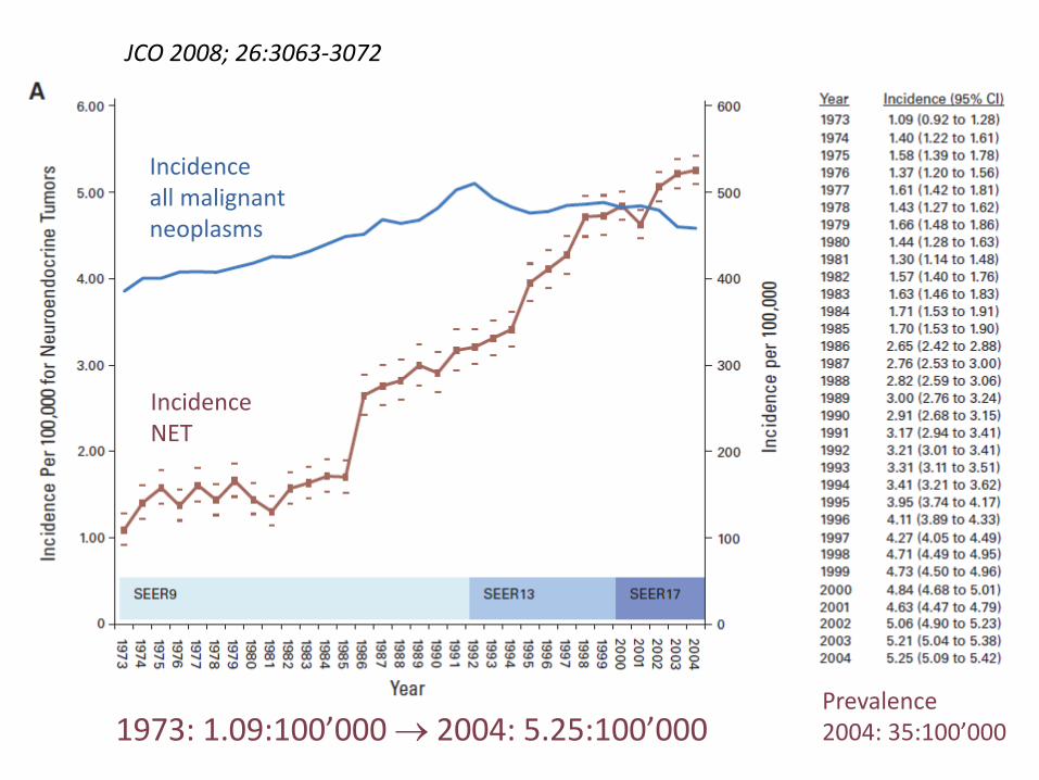
Incidence all malignant neoplasms
Incidence NET
1973: 1.09:100’000 2004: 5.25:100’000 Prevalence 2004: 35:100’000
JCO 2008; 26:3063-3072
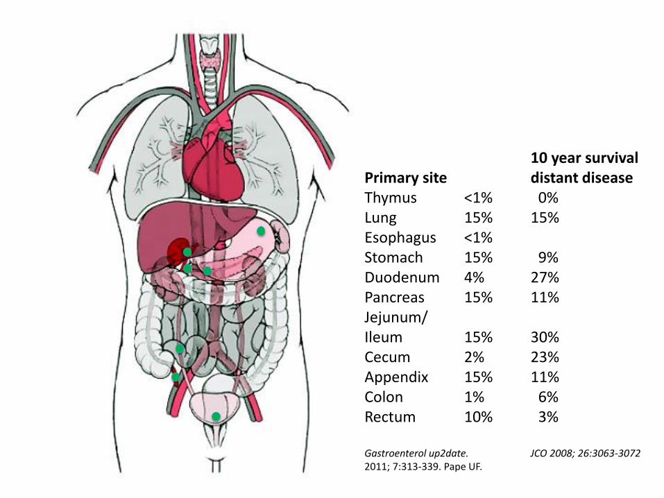
10 year survival Primary site distant disease Thymus <1% 0% Lung 15% 15% Esophagus <1% Stomach 15% 9% Duodenum 4% 27% Pancreas 15% 11% Jejunum/ Ileum 15% 30% Cecum 2% 23% Appendix 15% 11% Colon 1% 6% Rectum 10% 3% Gastroenterol up2date. JCO 2008; 26:3063-3072 2011; 7:313-339. Pape UF.
TUMOR CLASSIFICATION
Nomenclature
Grading
Staging ENETS vs. TNM

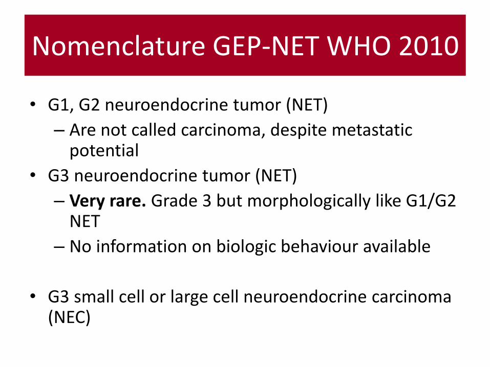
Nomenclature GEP-NET WHO 2010
• G1, G2 neuroendocrine tumor (NET)
– Are not called carcinoma, despite metastatic potential
• G3 neuroendocrine tumor (NET)
– Very rare. Grade 3 but morphologically like G1/G2 NET
– No information on biologic behaviour available
• G3 small cell or large cell neuroendocrine carcinoma (NEC)
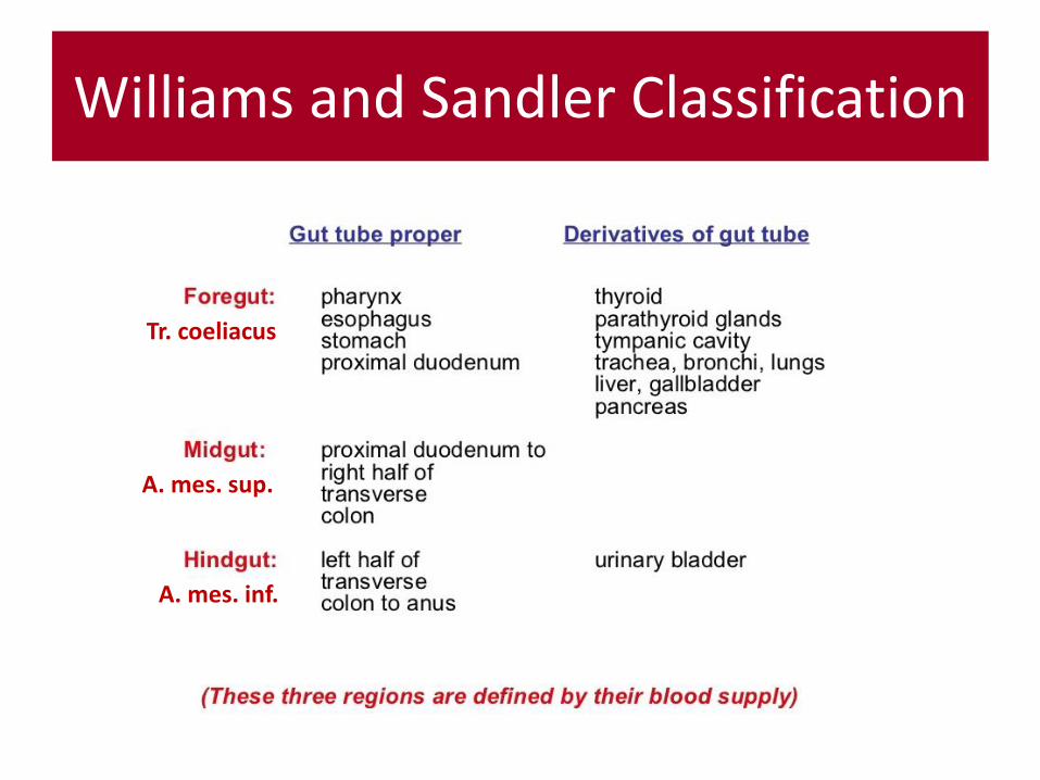
Williams and Sandler Classification
Tr. coeliacus
A. mes. sup.
A. mes. inf.
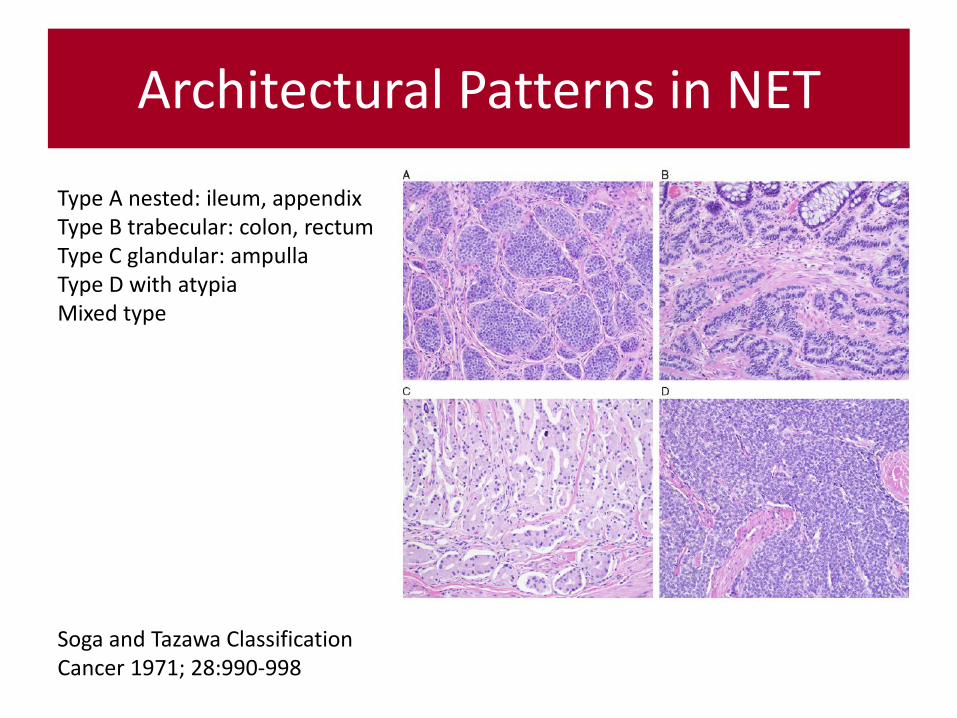
Architectural Patterns in NET
Soga and Tazawa Classification Cancer 1971; 28:990-998
Type A nested: ileum, appendix Type B trabecular: colon, rectum Type C glandular: ampulla Type D with atypia Mixed type
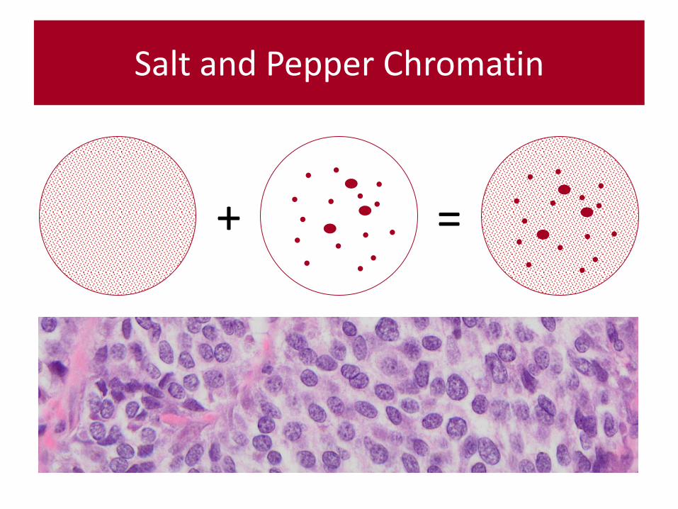
Salt and Pepper Chromatin
+ =
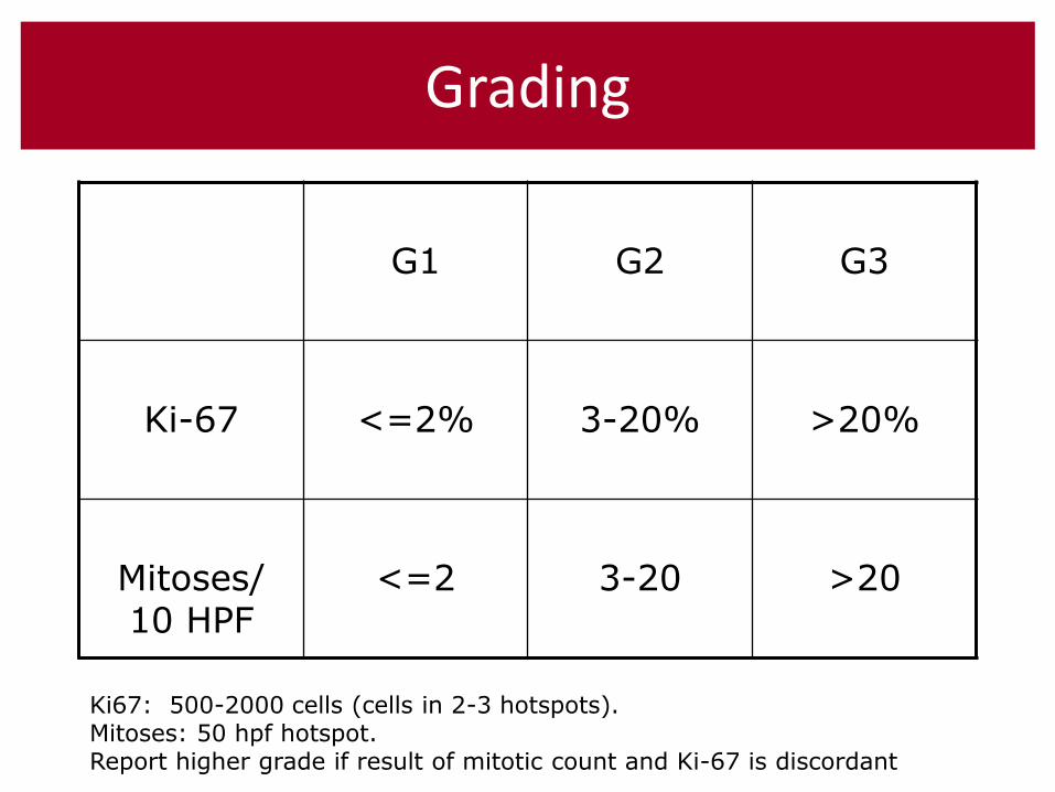
Grading
G1
G2
G3
Ki-67
<=2%
3-20%
>20%
Mitoses/ 10 HPF
<=2
3-20
>20
Ki67: 500-2000 cells (cells in 2-3 hotspots).
Mitoses: 50 hpf hotspot. Report higher grade if result of mitotic count and Ki-67 is discordant
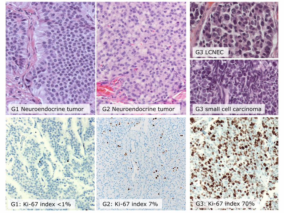
G1 Neuroendocrine tumor G2 Neuroendocrine tumor
G3 LCNEC
G3 small cell carcinoma
G1: Ki-67 index <1% G2: Ki-67 index 7% G3: Ki-67 index 70%
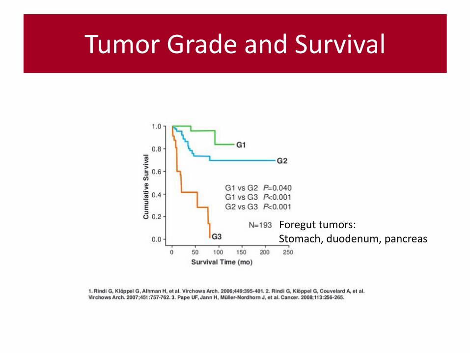
Tumor Grade and Survival
Foregut tumors: Stomach, duodenum, pancreas
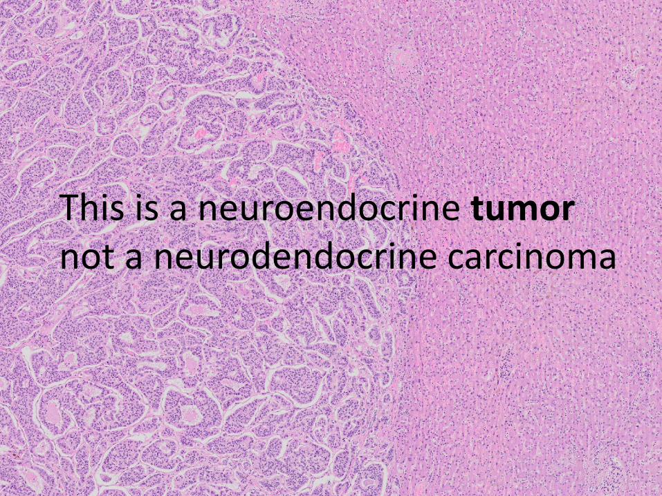
This is a neuroendocrine tumor not a neurodendocrine carcinoma
Staging & TNM Classification
ENETS (European Neuroendocrine Tumor Society) www.enets.org 2006/2007 UICC (International Union against Cancer) 7. Auflage 2010
Differences in pancreatic and appendiceal tumors: indicate both classifications!

GASTROINTESTINAL NEUROENDOCRINE NEOPLASIAS
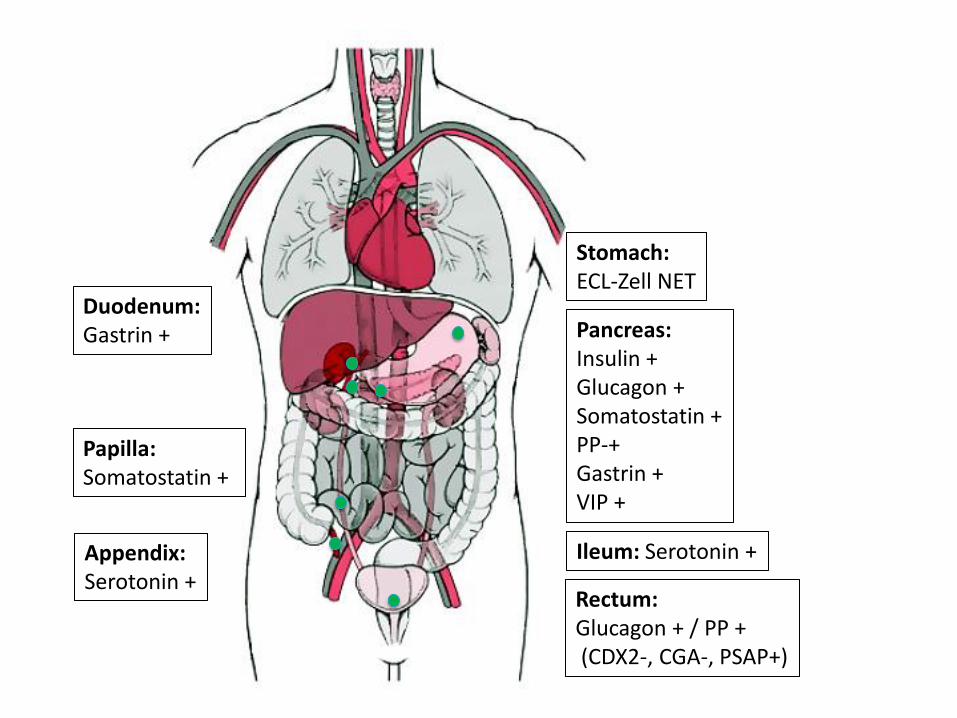
Stomach: ECL-Zell NET
Duodenum: Gastrin +
Papilla: Somatostatin +
Ileum: Serotonin + Appendix: Serotonin +
Rectum: Glucagon + / PP + (CDX2-, CGA-, PSAP+)
Pancreas: Insulin + Glucagon + Somatostatin + PP-+ Gastrin + VIP +
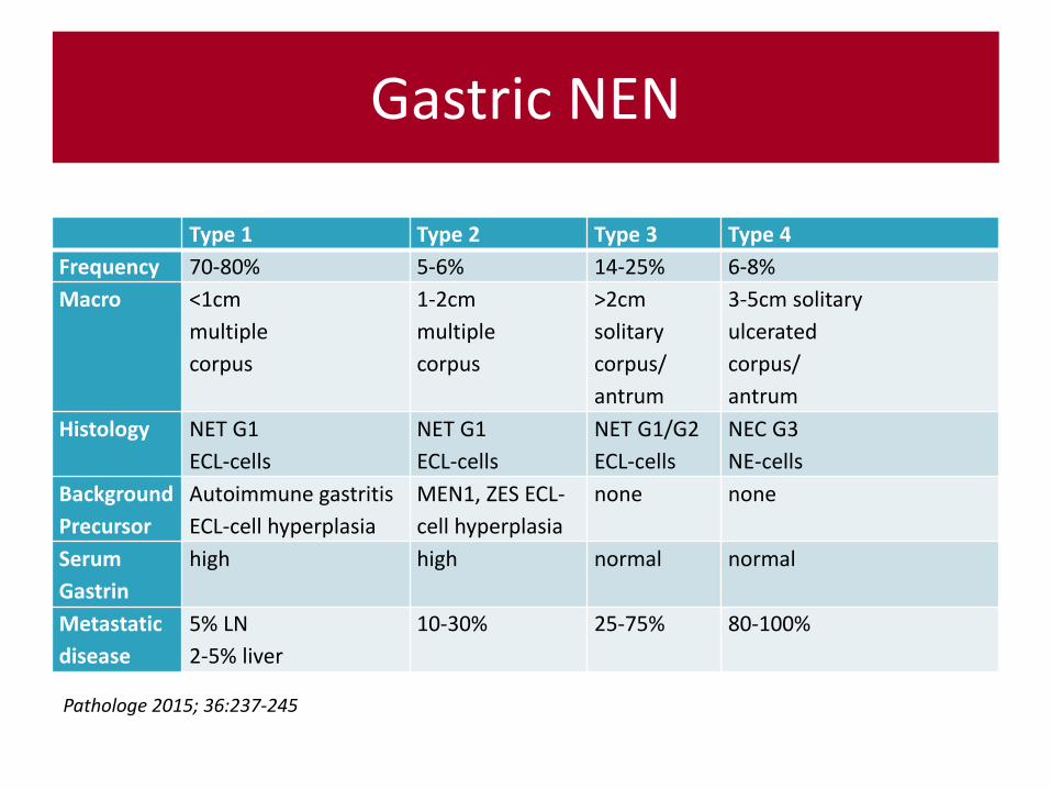
Type 1 Type 2 Type 3 Type 4
Frequency 70-80% 5-6% 14-25% 6-8%
Macro <1cm
multiple
corpus
1-2cm
multiple
corpus
>2cm
solitary
corpus/
antrum
3-5cm solitary
ulcerated
corpus/
antrum
Histology NET G1
ECL-cells
NET G1
ECL-cells
NET G1/G2
ECL-cells
NEC G3
NE-cells
Background
Precursor
Autoimmune gastritis
ECL-cell hyperplasia
MEN1, ZES ECL-
cell hyperplasia
none none
Serum
Gastrin
high high normal normal
Metastatic
disease
5% LN
2-5% liver
10-30% 25-75% 80-100%
Gastric NEN
Pathologe 2015; 36:237-245
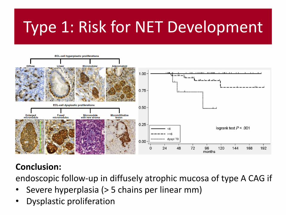
Type 1: Risk for NET Development
Conclusion: endoscopic follow-up in diffusely atrophic mucosa of type A CAG if • Severe hyperplasia (> 5 chains per linear mm) • Dysplastic proliferation
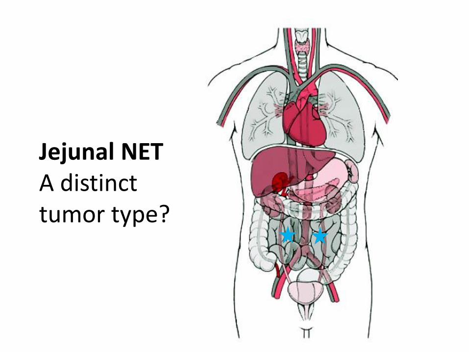
Jejunal NET A distinct tumor type?
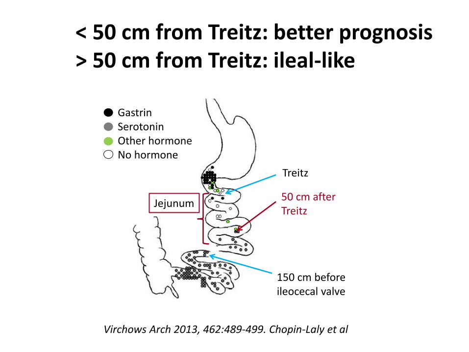
Virchows Arch 2013, 462:489-499. Chopin-Laly et al
Treitz
50 cm after Treitz
150 cm before ileocecal valve
Jejunum
< 50 cm from Treitz: better prognosis > 50 cm from Treitz: ileal-like
Gastrin Serotonin Other hormone No hormone
Mixed Tumors: MANEC
• Mixed adenoneuroendocrine tumors
– Lung
– Prostate
– Breast (% no longer defined)
– Gastroenteropankreatic mixed adenoneuroendocrine carcinoma (MANEC) WHO 2010: each component at least 30% (morphology and immunohistochemistry)
Virchows Arch 2011, 458:393-402. Volante et al. FAQ No. 9

METASTATIC NEN
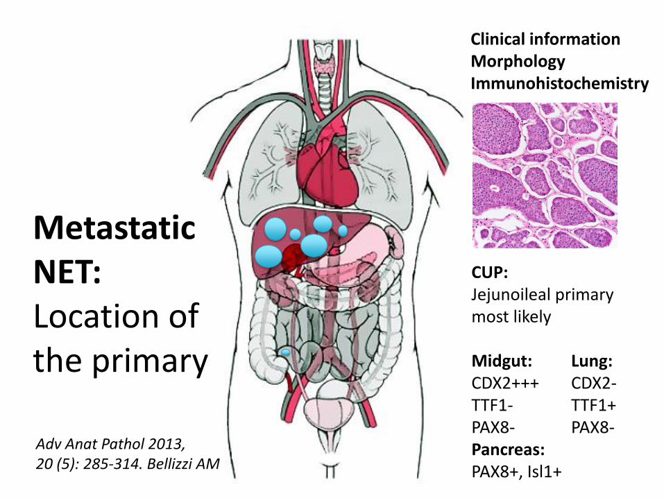
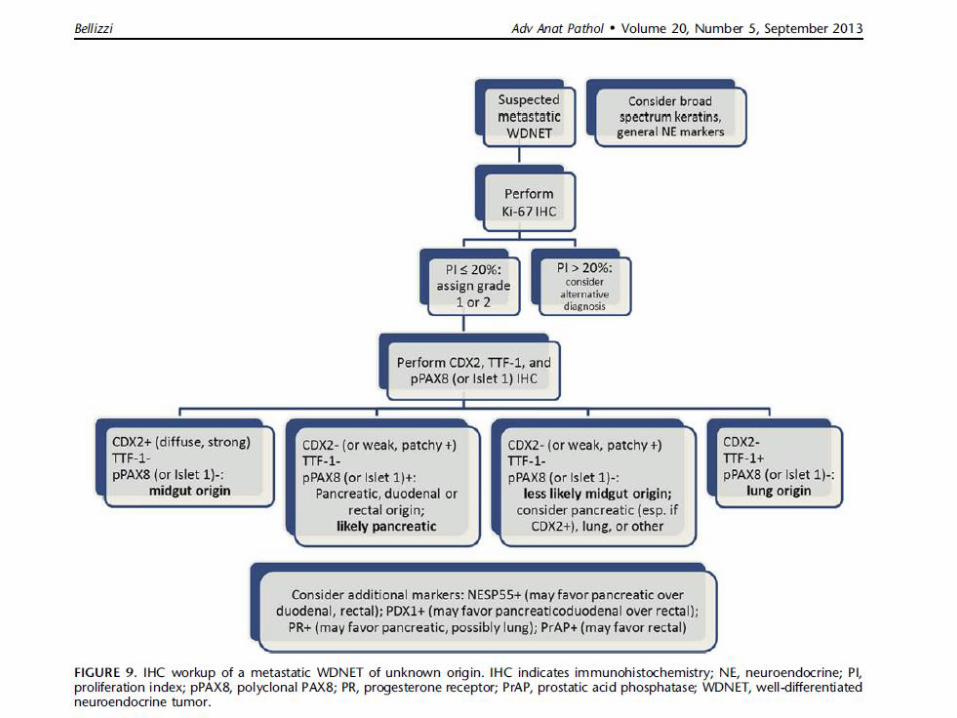
Metastatic NET: Location of the primary
CUP: Jejunoileal primary most likely Midgut: Lung: CDX2+++ CDX2- TTF1- TTF1+ PAX8- PAX8- Pancreas: PAX8+, Isl1+
Adv Anat Pathol 2013, 20 (5): 285-314. Bellizzi AM
Clinical information Morphology Immunohistochemistry
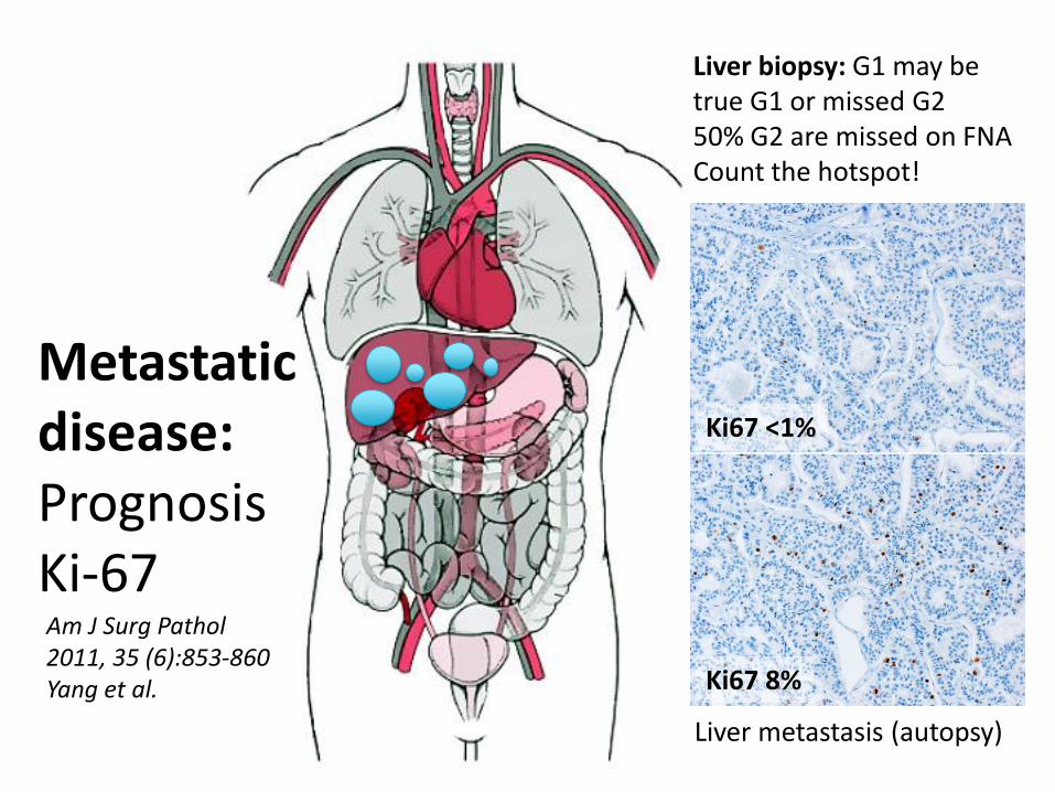
Metastatic disease: Prognosis Ki-67
Ki67 <1%
Ki67 8%
Liver biopsy: G1 may be true G1 or missed G2 50% G2 are missed on FNA Count the hotspot!
Liver metastasis (autopsy)
Am J Surg Pathol 2011, 35 (6):853-860 Yang et al.
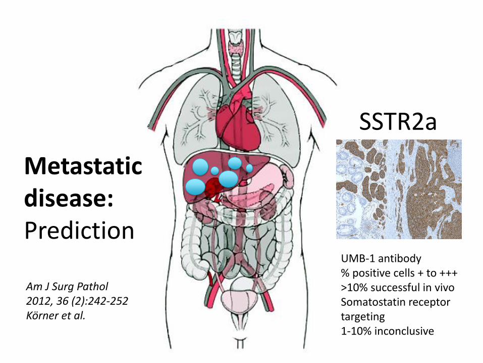
Metastatic disease: Prediction
UMB-1 antibody % positive cells + to +++ >10% successful in vivo Somatostatin receptor targeting 1-10% inconclusive
Am J Surg Pathol 2012, 36 (2):242-252 Körner et al.
SSTR2a
ROLE OF IMMUNOHISTOCHEMISTRY
Confirmation of NE differentiation
Grading
SSTR 2alpha
Site of origin of metastatic NET
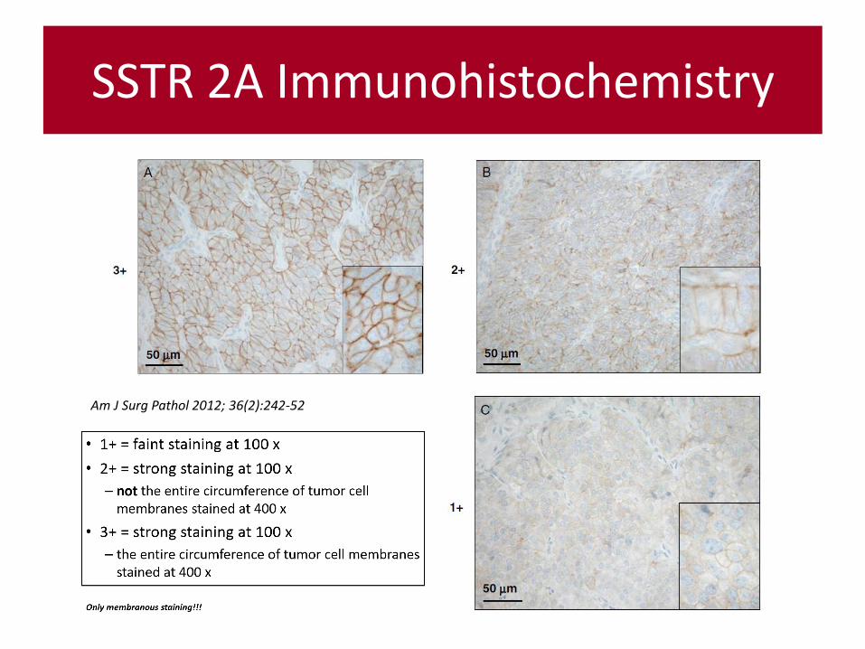
SSTR 2A Immunohistochemistry
Am J Surg Pathol 2012; 36(2):242-52
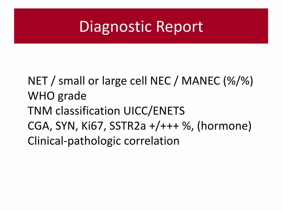
Diagnostic Report
NET / small or large cell NEC / MANEC (%/%) WHO grade TNM classification UICC/ENETS CGA, SYN, Ki67, SSTR2a +/+++ %, (hormone) Clinical-pathologic correlation
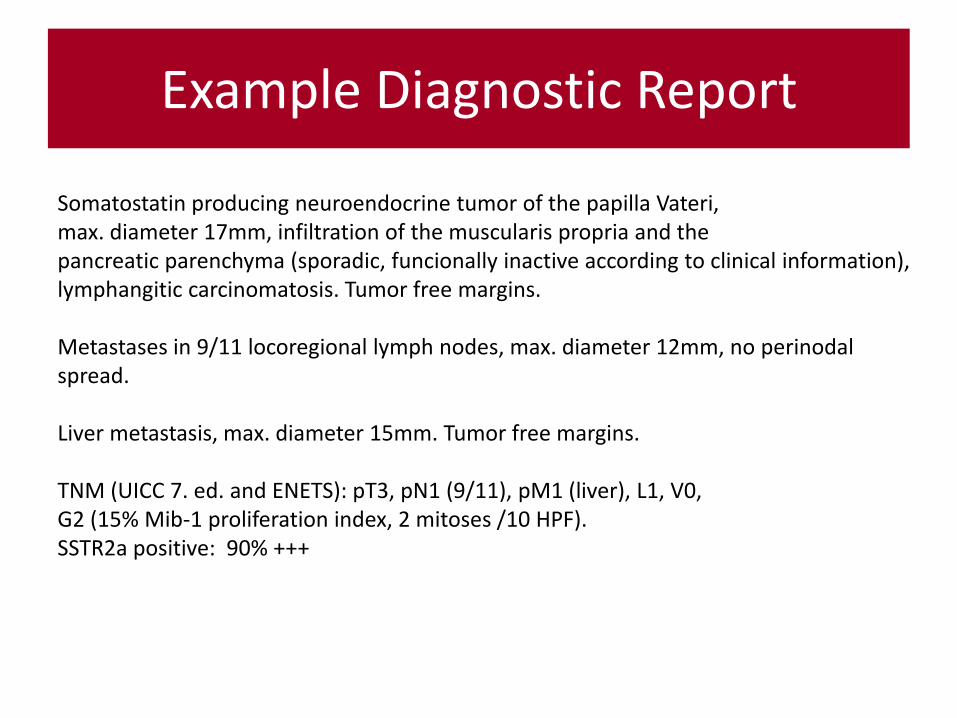
Example Diagnostic Report
Somatostatin producing neuroendocrine tumor of the papilla Vateri, max. diameter 17mm, infiltration of the muscularis propria and the pancreatic parenchyma (sporadic, funcionally inactive according to clinical information), lymphangitic carcinomatosis. Tumor free margins. Metastases in 9/11 locoregional lymph nodes, max. diameter 12mm, no perinodal spread. Liver metastasis, max. diameter 15mm. Tumor free margins. TNM (UICC 7. ed. and ENETS): pT3, pN1 (9/11), pM1 (liver), L1, V0, G2 (15% Mib-1 proliferation index, 2 mitoses /10 HPF). SSTR2a positive: 90% +++