Embed Size (px)
Citation preview
Neurons Containing Hypocretin (Orexin) Project to MultipleNeuronal Systems
Christelle Peyron,1 Devin K. Tighe,1 Anthony N. van den Pol,1,3 Luis de Lecea,2 H. Craig Heller,1J. Gregor Sutcliffe,2 and Thomas S. Kilduff1
1Department of Biological Sciences, Stanford University, Stanford, California 94305, 2Department of Molecular Biology,The Scripps Research Institute, La Jolla, California 92037, and 3Department of Neurosurgery, Yale University,New Haven, Connecticut 06520
The novel neuropeptides called hypocretins (orexins) have re-cently been identified as being localized exclusively in cellbodies in a subregion of the tuberal part of the hypothalamus.The structure of the hypocretins, their accumulation in vesiclesof axon terminals, and their excitatory effect on cultured hypo-thalamic neurons suggest that the hypocretins function in in-tercellular communication. To characterize these peptides fur-ther and to help understand what physiological functions theymay serve, we undertook an immunohistochemical study toexamine the distribution of preprohypocretin-immunoreactiveneurons and fibers in the rat brain. Preprohypocretin-positiveneurons were found in the perifornical nucleus and in the dorsaland lateral hypothalamic areas. These cells were distinct fromthose that express melanin-concentrating hormone. Althoughthey represent a restricted group of cells, their projections werewidely distributed in the brain. We observed labeled fibers
throughout the hypothalamus. The densest extrahypothalamicprojection was found in the locus coeruleus. Fibers were alsoseen in the septal nuclei, the bed nucleus of the stria terminalis,the paraventricular and reuniens nuclei of the thalamus, thezona incerta, the subthalamic nucleus, the central gray, thesubstantia nigra, the raphe nuclei, the parabrachial area, themedullary reticular formation, and the nucleus of the solitarytract. Less prominent projections were found in cortical regions,central and anterior amygdaloid nuclei, and the olfactory bulb.These results suggest that hypocretins are likely to have a rolein physiological functions in addition to food intake such asregulation of blood pressure, the neuroendocrine system, bodytemperature, and the sleep–waking cycle.
Key words: neuropeptide; hypothalamus; immunohistochem-istry; blood pressure; feeding; autonomic functions; hypocretin;orexin; melanin-concentrating hormone; neuroendocrine
The hypothalamus is an essential interface between endocrine,autonomic, and somatomotor systems. In mammals, the hypothal-amus is a hub of central regulatory centers for autonomic andendocrine homeostatic systems such as cardiovascular, tempera-ture, and abdominal visceral regulation, as well as ingestivebehaviors (Swanson, 1987). For some of these systems, particularpeptides have been identified as major products of individualnuclei; the localization of vasopressin and oxytocin to the para-ventricular and supraoptic nuclei is a prime example. It is likelythat other hypothalamic peptides are yet to be identified that maybe similarly localized and that may contribute to the physiologicalfunctions regulated by the hypothalamus.
To identify novel hypothalamic peptides, an analysis of themRNAs whose expression is restricted to or enriched in the rathypothalamus was undertaken using directional tag-PCR subtrac-tion (Gautvik et al., 1996). A nucleotide sequence encoding a 130residue protein called preprohypocretin (hcrt) was isolated from
this hypothalamus-enriched cDNA library (Sutcliffe et al., 1997;de Lecea et al., 1998). In situ hybridization revealed that neuronsexpressing hcrt mRNA were located exclusively in the tuberalregion of the hypothalamus (Gautvik et al., 1996). Sequenceanalysis indicated that hcrt yields two peptides, hcrt-1 (residues28–66) and hcrt-2 (residues 69–97). The structure of hcrt, itsexpression in hypothalamic neurons, and its accumulation invesicles of axon terminals suggested that the hcrt peptides mayhave intercellular signaling activity, and synthetic hcrt-2 wasexcitatory when applied to synaptically coupled rat hypothalamicneurons in vitro (de Lecea et al., 1998).
More recently, screening of high-resolution HPLC fractions oncell lines expressing orphan G-protein–coupled receptors re-sulted in the isolation of two peptides called orexin A and B(Sakurai et al., 1998) that are identical to hypocretin 1 and 2,respectively. Chemical analyses confirmed the identity of orexinB and hcrt-2, defined the N terminal of orexin A (hcrt-1) asresidue 33, and verified that both peptides are amidated at theirC terminals. These two peptides activate two distinct G-protein–coupled receptors, OX1 and OX2 (Sakurai et al., 1998).
To characterize further these new peptides and to obtain cluesabout their potential physiological functions, we undertook animmunohistochemical study to examine the distribution of hcrt-immunoreactive neurons and fibers in the brain. Sakurai et al.(1998) reported that intracerebroventricular injection of hcrtstimulates food intake. However, the widespread distribution ofhcrt fibers observed in the present study suggests that hcrt is likelyto play a role in other physiological functions as well.
Received July 15, 1998; revised Sept. 11, 1998; accepted Sept. 15, 1998.This work was supported by National Institutes of Health Grants AG11084,
GM32355, NS33396, and NS34887 and by Air Force Office of Scientific Research,the Army Research Office, and the Fyssen Fondation. We thank Dr. Peter O’Harafor use of his Neurolucida system (MicroBrightField, Colchester, VT) in the analysisof the distribution of hypocretin-immunoreactive fibers. We also thank Drs. Y. Yangfor technical assistance with the electron microscopy, Chiaki Fukuhara who madethe preprohypocretin used to raise antisera, Masashi Yanagisawa for the gift oforexin A, and Jean-Louis Nahon for the gift of melanin-concentrating hormonecDNA plasmid.
Correspondence should be addressed to Dr. Christelle Peyron, Department ofBiological Sciences, Gilbert Hall, 371 Serra Mall, Stanford University, Stanford, CA94305-5020.Copyright © 1998 Society for Neuroscience 0270-6474/98/189996-20$05.00/0
The Journal of Neuroscience, December 1, 1998, 18(23):9996–10015
Parts of this paper have been published previously in abstractform (Peyron et al., 1997).
MATERIALS AND METHODSPerfusion, fixationAdult male Wistar rats were deeply anesthetized with a lethal dose ofNembutal (80 mg/kg) and perfused transcardially with 0.9% salinefollowed by an ice-cold fixative solution containing 4% paraformalde-hyde and 0 or 0.25% glutaraldehyde in 0.1 M phosphate buffer (PB).Brains were removed, post-fixed overnight by immersion in the samefixative without glutaraldehyde, and cryoprotected with 30% sucrose for2–3 d at 4°C. Brains were rapidly frozen in dry ice and sliced into 20-mm-thick coronal sections on a cryostat (223°C). Free-floating sections wererinsed several times and stored in 0.1 M PB containing 0.9% NaCl and0.3% Triton X-100 plus 0.1% sodium azide (PBST-Az) at 4°C until use.
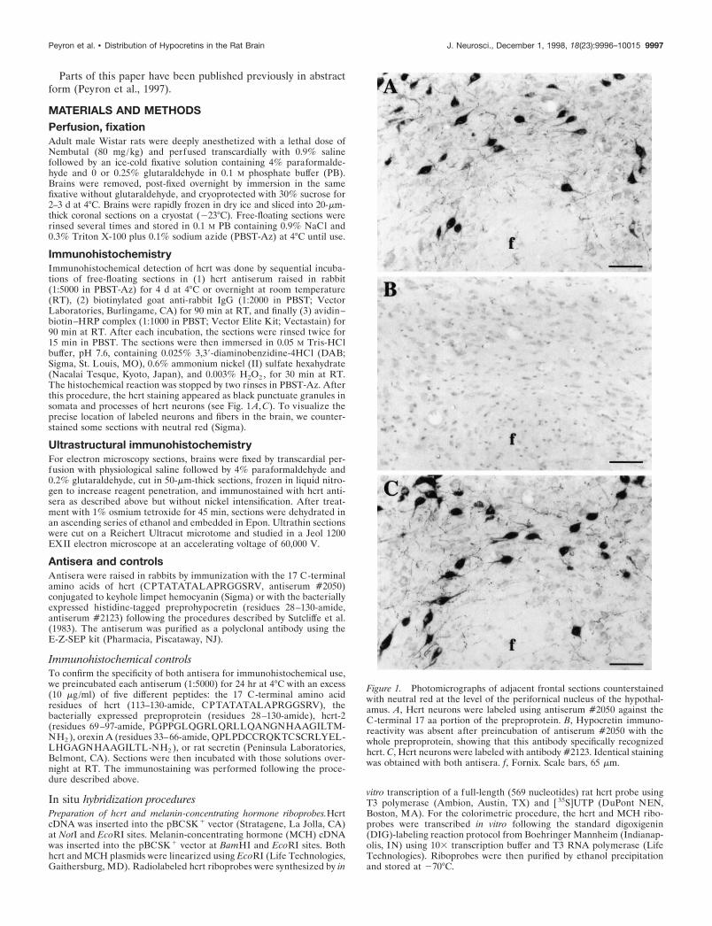
ImmunohistochemistryImmunohistochemical detection of hcrt was done by sequential incuba-tions of free-floating sections in (1) hcrt antiserum raised in rabbit(1:5000 in PBST-Az) for 4 d at 4°C or overnight at room temperature(RT), (2) biotinylated goat anti-rabbit IgG (1:2000 in PBST; VectorLaboratories, Burlingame, CA) for 90 min at RT, and finally (3) avidin–biotin–HRP complex (1:1000 in PBST; Vector Elite Kit; Vectastain) for90 min at RT. After each incubation, the sections were rinsed twice for15 min in PBST. The sections were then immersed in 0.05 M Tris-HClbuffer, pH 7.6, containing 0.025% 3,39-diaminobenzidine-4HCl (DAB;Sigma, St. Louis, MO), 0.6% ammonium nickel (II) sulfate hexahydrate(Nacalai Tesque, Kyoto, Japan), and 0.003% H2O2 , for 30 min at RT.The histochemical reaction was stopped by two rinses in PBST-Az. Afterthis procedure, the hcrt staining appeared as black punctuate granules insomata and processes of hcrt neurons (see Fig. 1 A, C). To visualize theprecise location of labeled neurons and fibers in the brain, we counter-stained some sections with neutral red (Sigma).
Ultrastructural immunohistochemistryFor electron microscopy sections, brains were fixed by transcardial per-fusion with physiological saline followed by 4% paraformaldehyde and0.2% glutaraldehyde, cut in 50-mm-thick sections, frozen in liquid nitro-gen to increase reagent penetration, and immunostained with hcrt anti-sera as described above but without nickel intensification. After treat-ment with 1% osmium tetroxide for 45 min, sections were dehydrated inan ascending series of ethanol and embedded in Epon. Ultrathin sectionswere cut on a Reichert Ultracut microtome and studied in a Jeol 1200EXII electron microscope at an accelerating voltage of 60,000 V.
Antisera and controlsAntisera were raised in rabbits by immunization with the 17 C-terminalamino acids of hcrt (CPTATATALAPRGGSRV, antiserum #2050)conjugated to keyhole limpet hemocyanin (Sigma) or with the bacteriallyexpressed histidine-tagged preprohypocretin (residues 28–130-amide,antiserum #2123) following the procedures described by Sutcliffe et al.(1983). The antiserum was purified as a polyclonal antibody using theE-Z-SEP kit (Pharmacia, Piscataway, NJ).
Immunohistochemical controlsTo confirm the specificity of both antisera for immunohistochemical use,we preincubated each antiserum (1:5000) for 24 hr at 4°C with an excess(10 mg/ml) of five different peptides: the 17 C-terminal amino acidresidues of hcrt (113–130-amide, CPTATATALAPRGGSRV), thebacterially expressed preproprotein (residues 28–130-amide), hcrt-2(residues 69–97-amide, PGPPGLQGRLQRLLQANGNHAAGILTM-NH2 ), orexin A (residues 33–66-amide, QPLPDCCRQKTCSCRLYEL-LHGAGNHAAGILTL-NH2 ), or rat secretin (Peninsula Laboratories,Belmont, CA). Sections were then incubated with those solutions over-night at RT. The immunostaining was performed following the proce-dure described above.
In situ hybridization proceduresPreparation of hcrt and melanin-concentrating hormone riboprobes.HcrtcDNA was inserted into the pBCSK 1 vector (Stratagene, La Jolla, CA)at NotI and EcoRI sites. Melanin-concentrating hormone (MCH) cDNAwas inserted into the pBCSK 1 vector at BamHI and EcoRI sites. Bothhcrt and MCH plasmids were linearized using EcoRI (Life Technologies,Gaithersburg, MD). Radiolabeled hcrt riboprobes were synthesized by in
vitro transcription of a full-length (569 nucleotides) rat hcrt probe usingT3 polymerase (Ambion, Austin, TX) and [ 35S]UTP (DuPont NEN,Boston, MA). For the colorimetric procedure, the hcrt and MCH ribo-probes were transcribed in vitro following the standard digoxigenin(DIG)-labeling reaction protocol from Boehringer Mannheim (Indianap-olis, IN) using 103 transcription buffer and T3 RNA polymerase (LifeTechnologies). Riboprobes were then purified by ethanol precipitationand stored at 270°C.
Figure 1. Photomicrographs of adjacent frontal sections counterstainedwith neutral red at the level of the perifornical nucleus of the hypothal-amus. A, Hcrt neurons were labeled using antiserum #2050 against theC-terminal 17 aa portion of the preproprotein. B, Hypocretin immuno-reactivity was absent after preincubation of antiserum #2050 with thewhole preproprotein, showing that this antibody specifically recognizedhcrt. C, Hcrt neurons were labeled with antibody #2123. Identical stainingwas obtained with both antisera. f, Fornix. Scale bars, 65 mm.
Peyron et al. • Distribution of Hypocretins in the Rat Brain J. Neurosci., December 1, 1998, 18(23):9996–10015 9997
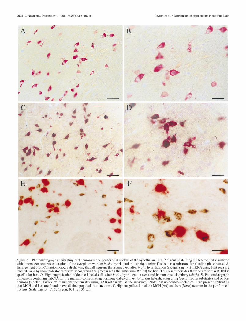
Figure 2. Photomicrographs illustrating hcrt neurons in the perifornical nucleus of the hypothalamus. A, Neurons containing mRNA for hcrt visualizedwith a homogeneous red coloration of the cytoplasm with an in situ hybridization technique using Fast red as a substrate for alkaline phosphatase. B,Enlargement of A. C, Photomicrograph showing that all neurons that stained red after in situ hybridization (recognizing hcrt mRNA using Fast red) arelabeled black by immunohistochemistry (recognizing the protein with the antiserum #2050) for hcrt. This result indicates that the antiserum #2050 isspecific for hcrt. D, High magnification of double-labeled cells after in situ hybridization (red) and immunohistochemistry (black). E, Photomicrographof neurons containing mRNA for the melanin-concentrating hormone (labeled in red by in situ hybridization using Vector red as substrate) and of hcrtneurons (labeled in black by immunohistochemistry using DAB with nickel as the substrate). Note that no double-labeled cells are present, indicatingthat MCH and hcrt are found in two distinct populations of neurons. F, High magnification of the MCH (red) and hcrt (black) neurons in the perifornicalnucleus. Scale bars: A, C, E, 65 mm; B, D, F, 36 mm.
9998 J. Neurosci., December 1, 1998, 18(23):9996–10015 Peyron et al. • Distribution of Hypocretins in the Rat Brain
Estimation of DIG-labeled riboprobe yields. The yield of the DIG-labeled hcrt and MCH riboprobes was approximated by comparison withDIG-labeled control RNA (Boehringer Mannheim). Serial dilutions ofDIG-labeled control RNA and hcrt and MCH riboprobes were spottedonto a nylon membrane (Nytran Plus; Schleicher & Schuell, Keene, NH).The membrane was then incubated in 0.1 M PBS solution containing 1%Triton X-100 and 4% bovine serum albumin (PBST-BSA) for 15 min atRT, followed by an incubation with a sheep antibody against DIGconjugated with alkaline phosphatase (1:5000 in PBST-BSA; BoehringerMannheim) for 30 min at RT. The membrane was rinsed in 0.1 M PBS for15 min followed by a 3 min rinse in a development buffer (100 mM Trisbuffer, 50 mM MgCl2 , and 150 mM NaCl, pH 9). Finally, the reaction wasdeveloped with nitroblue tetrazolium (4.5 ml /ml) and 5-bromo-4-chloro-3-indolyl phosphate (3.5 ml /ml) in the development buffer (Bio-Rad,Hercules, CA) for 30 min at RT.
Hybridization. Twenty-five micrometer thick brain sections preservedin a cryoprotectant solution of 0.1 M PBS with 30% glycerol and 30%ethylene glycol were stored at 270°C. After thawing of frozen sections,free-floating sections were washed in 0.1 M PBS and then incubated inPBS with 0.5% Triton X-100 for 10 min, deproteinized with 0.1N HCl for10 min, and acetylated with acetic anhydride (0.25% in 0.1 M triethanol-amine hydrochoride, pH 8) for 10 min. The sections were then post-fixedin 4% paraformaldehyde for 10 min. Five minute washes in PBS weredone after each of the above steps. The sections were then prehybridizedin a solution containing 43 PIPES, 10% (w/v) dextran sulfate, 50%deionized formamide, 53 Denhardt’s, 50 mM DTT, 0.2% (w/v) SDS, 250mg/ml denatured salmon sperm DNA, and 250 mg/ml yeast tRNA for 3hr at 55°C. Labeled antisense hcrt or MCH riboprobes (10 6 cpm/ml forautoradiographic localization; ;600 ng/ml for colorimetric procedures)were heated at 68°C for 10 min and added to the sections in theprehybridization solution for an overnight incubation at 55°C. The sec-tions were then (1) washed in 23 SSC with 10 mM b-mercaptoethanol(b-ME) for 30 min at RT; (2) digested with 4 mg/ml RNase A in 53 Trisbuffer (50 mM Tris-HCl, 5 mM EDTA, and 0.5 M NaCl) at 37°C for 1 hr;(3) rinsed in 13 SSC, 50% formamide, and 5 mM b-ME at 55°C for 2 hr;(4) incubated in 0.23 SSC, 10% formamide, 1 mM b-ME, and 0.1%Sarkosyl at 68°C for 1 hr; and (5) washed three times in PBS at RT.
For autoradiographic localization, sections were mounted on slides,dehydrated in a series of 50–100% ethanol, defatted in 50%/50% eth-anol–chloroform followed by 100% ethanol, air dried, and then exposedto Kodak X-Omat AR film for 1–7 d. Sections were subsequently dippedin I lford K5 liquid photographic emulsion and exposed for 2–4 weeks at4°C. The resultant autoradiographs were developed in Kodak D-19 andcounterstained with Richardson’s blue.
For the colorimetric procedure, sections were washed in PBST-BSAfor 2 hr at RT and then were incubated with a sheep anti-DIG-alkalinephosphatase antiserum (1:3000 in PBST-BSA) overnight at RT. For colordevelopment, sections were first rinsed twice for 30 min in 0.1 M Tris-HCl buffer, pH 8.2, and then immersed in a Tris-HCl buffer containingFast red (Sigma Fast; Sigma) (see Fig. 2 A–D) or Vector red (VectorLaboratories) (see Fig. 2 E, F ) as a substrate for alkaline phosphatase for5 hr at RT. To increase the intensity of the signal, we added 0.3 M NaClin the Fast red solution as suggested by Chiu et al. (1996). Stainedneurons have an homogeneous red color of the cytoplasm with bothsubstrates.
In situ/immunostainingAfter in situ hybridization, the sections were subjected to the sameimmunohistochemistry procedure as described above but with minorchanges. Anti-hcrt (#2050) antiserum was used at a 1:1000 dilution, andsuccessive incubations were done in PBST-BSA. Double-labeled cellswere identified by the presence of black granulations over a red colora-tion of the cytoplasm (see Fig. 2C,D). To test whether there was cross-reactivity between the sheep anti-DIG-alkaline phosphatase and thesecondary anti-rabbit IgG, we conducted the procedure without theprimary antibodies (#2050 and #2123).
Data analysisThe sections were mounted on gelatin-coated glass slides, dried, dehy-drated, and coverslipped with Mounting medium (Harleco; DiagnosticSystems, Gibbstown, NJ) or rinsed with water and coverslipped withaqueous mounting medium (Scytek Laboratories, Logan, UT). Theywere later observed with an Olympus BH-2 microscope. Mapping ofneurons and fibers immunoreactive to hcrt in rat brain was done using aNikon light microscope equipped with a motorized X/Y stage, position
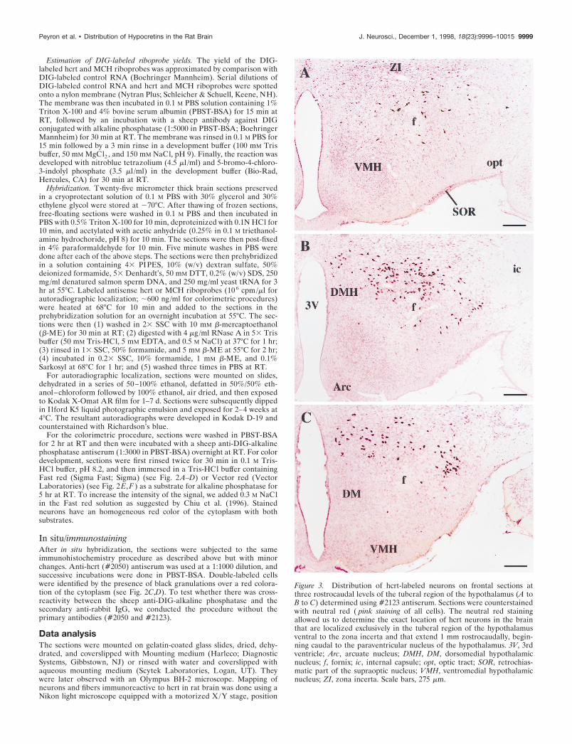
Figure 3. Distribution of hcrt-labeled neurons on frontal sections atthree rostrocaudal levels of the tuberal region of the hypothalamus (A toB to C) determined using #2123 antiserum. Sections were counterstainedwith neutral red ( pink staining of all cells). The neutral red stainingallowed us to determine the exact location of hcrt neurons in the brainthat are localized exclusively in the tuberal region of the hypothalamusventral to the zona incerta and that extend 1 mm rostrocaudally, begin-ning caudal to the paraventricular nucleus of the hypothalamus. 3V, 3rdventricle; Arc, arcuate nucleus; DMH, DM, dorsomedial hypothalamicnucleus; f, fornix; ic, internal capsule; opt, optic tract; SOR, retrochias-matic part of the supraoptic nucleus; VMH, ventromedial hypothalamicnucleus; ZI, zona incerta. Scale bars, 275 mm.
Peyron et al. • Distribution of Hypocretins in the Rat Brain J. Neurosci., December 1, 1998, 18(23):9996–10015 9999
encoders, and a video camera connected to a computerized image dataanalysis system (Neurolucida; MicroBrightField, Colchester, VT). Out-lines of sections and major structures were drawn at a low magnification(43), whereas labeled neurons and fibers were plotted at higher magni-fication (20–403). Finally, the drawings were assembled with AdobeIllustrator 7.0 to obtain the figures depicting the distribution of hcrt
projections. Counting of hcrt neurons was done bilaterally on 15-mm-thick coronal sections taken every 120 mm (one-eighth of the brain). Thenumber of labeled cells in the brain was estimated using the method ofAbercrombie (1946).
Photomicrographs were taken on the Olympus microscope, and thefilms were scanned with a Kodak slide scanner. To obtain optimal
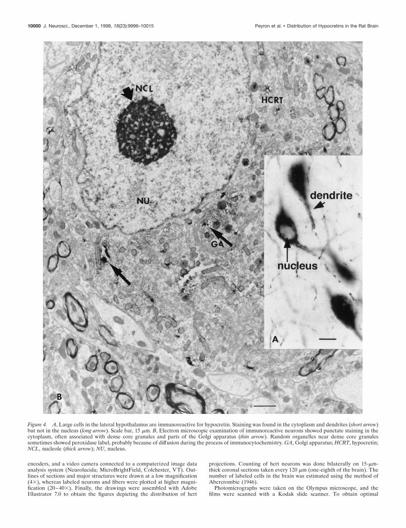
Figure 4. A, Large cells in the lateral hypothalamus are immunoreactive for hypocretin. Staining was found in the cytoplasm and dendrites (short arrow)but not in the nucleus (long arrow). Scale bar, 15 mm. B, Electron microscopic examination of immunoreactive neurons showed punctate staining in thecytoplasm, often associated with dense core granules and parts of the Golgi apparatus (thin arrow). Random organelles near dense core granulessometimes showed peroxidase label, probably because of diffusion during the process of immunocytochemistry. GA, Golgi apparatus; HCRT, hypocretin;NCL, nucleole (thick arrow); NU, nucleus.
10000 J. Neurosci., December 1, 1998, 18(23):9996–10015 Peyron et al. • Distribution of Hypocretins in the Rat Brain
reproduction of the staining, we modified the contrast and luminosity ofthe crude scans with Adobe Photoshop 4.0. The illustration plates wereprinted on a Kodak 7700 dye sublimation printer.
RESULTSAntiserum specificityWe used two antisera, one raised against the C-terminal 17amino acids of preprohypocretin (#2050) and a second raisedagainst the bacterially expressed preproprotein hcrt (#2123).Competition studies showed that the immunohistochemical stain-ing was absent when antiserum #2050 was preincubated for 24 hrat 4°C with the preproprotein or with the 17 aa portion of hcrt. Incontrast, the labeling was still present after preincubation of theantibody with synthetic hcrt-2 or with rat secretin, showing that
antiserum #2050 specifically recognizes the 17 aa portion of hcrtbut not hcrt-2 or rat secretin (Fig. 1). No labeling was obtainedafter preincubation of #2123 with the preproprotein. However,the labeling was still present after preincubation of #2123 withthe 17 aa portion, orexin A, or hcrt-2, indicating that #2123 is apolyclonal antiserum against multiple epitopes of the prepropro-tein and not just the C-terminal 17 aa portion recognized by#2050. The cell body staining obtained with both antisera (#2050and #2123) and by autoradiographic (refer to de Lecea et al.,1998) and colorimetric in situ hybridization is indistinguishable(Figs. 1, 2). The combination of in situ hybridization and immu-nohistochemistry for hcrt showed that neurons that expressed themRNA coding for hcrt also made the protein (Fig. 2C,D).
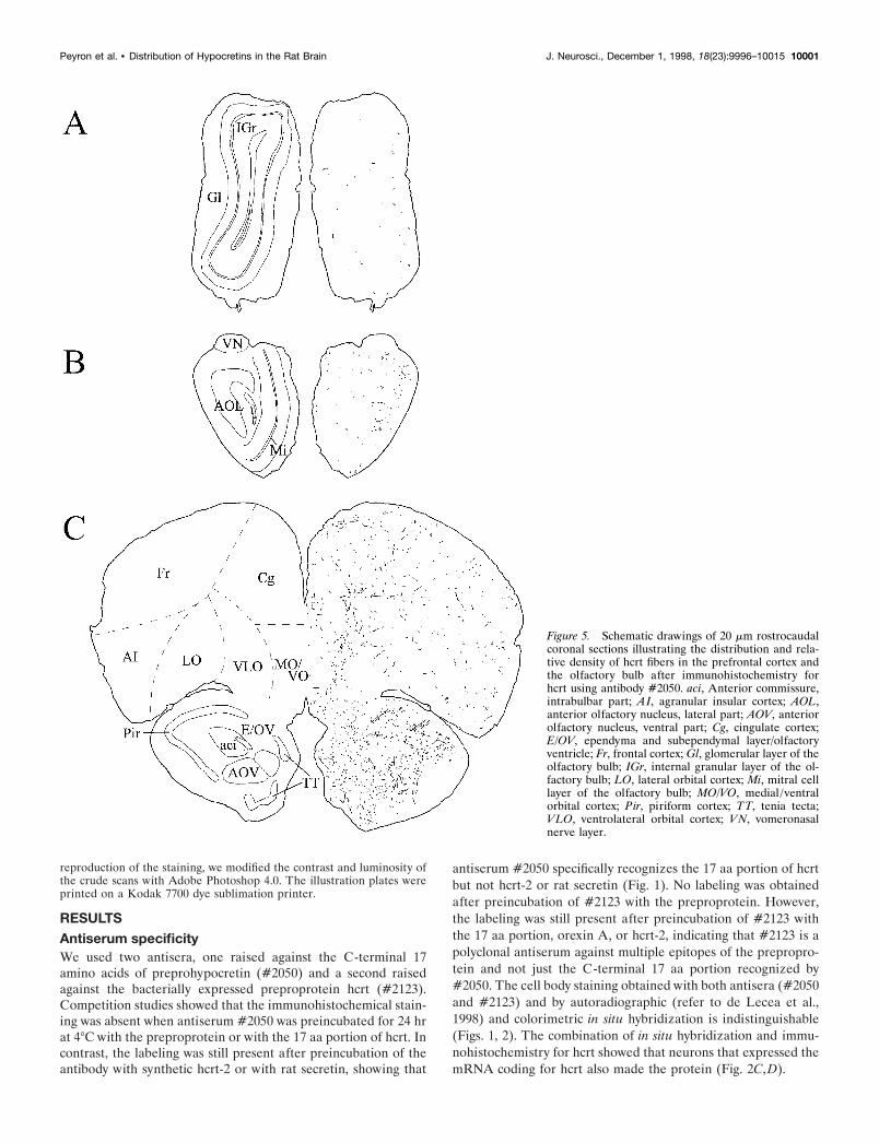
Figure 5. Schematic drawings of 20 mm rostrocaudalcoronal sections illustrating the distribution and rela-tive density of hcrt fibers in the prefrontal cortex andthe olfactory bulb after immunohistochemistry forhcrt using antibody #2050. aci, Anterior commissure,intrabulbar part; AI, agranular insular cortex; AOL,anterior olfactory nucleus, lateral part; AOV, anteriorolfactory nucleus, ventral part; Cg, cingulate cortex;E/OV, ependyma and subependymal layer/olfactoryventricle; Fr, frontal cortex; Gl, glomerular layer of theolfactory bulb; IGr, internal granular layer of the ol-factory bulb; LO, lateral orbital cortex; Mi, mitral celllayer of the olfactory bulb; MO/VO, medial /ventralorbital cortex; Pir, piriform cortex; TT, tenia tecta;VLO, ventrolateral orbital cortex; VN, vomeronasalnerve layer.
Peyron et al. • Distribution of Hypocretins in the Rat Brain J. Neurosci., December 1, 1998, 18(23):9996–10015 10001
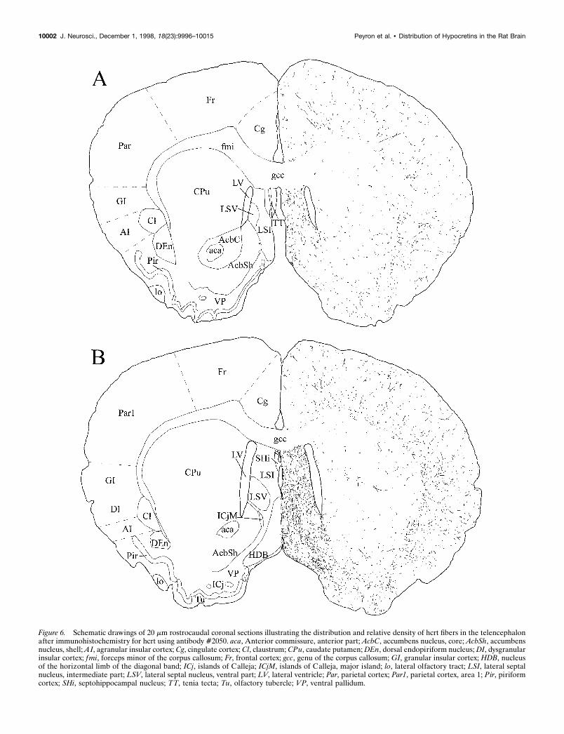
Figure 6. Schematic drawings of 20 mm rostrocaudal coronal sections illustrating the distribution and relative density of hcrt fibers in the telencephalonafter immunohistochemistry for hcrt using antibody #2050. aca, Anterior commissure, anterior part; AcbC, accumbens nucleus, core; AcbSh, accumbensnucleus, shell; AI, agranular insular cortex; Cg, cingulate cortex; Cl, claustrum; CPu, caudate putamen; DEn, dorsal endopiriform nucleus; DI, dysgranularinsular cortex; fmi, forceps minor of the corpus callosum; Fr, frontal cortex; gcc, genu of the corpus callosum; GI, granular insular cortex; HDB, nucleusof the horizontal limb of the diagonal band; ICj, islands of Calleja; ICjM, islands of Calleja, major island; lo, lateral olfactory tract; LSI, lateral septalnucleus, intermediate part; LSV, lateral septal nucleus, ventral part; LV, lateral ventricle; Par, parietal cortex; Par1, parietal cortex, area 1; Pir, piriformcortex; SHi, septohippocampal nucleus; TT, tenia tecta; Tu, olfactory tubercle; VP, ventral pallidum.
10002 J. Neurosci., December 1, 1998, 18(23):9996–10015 Peyron et al. • Distribution of Hypocretins in the Rat Brain
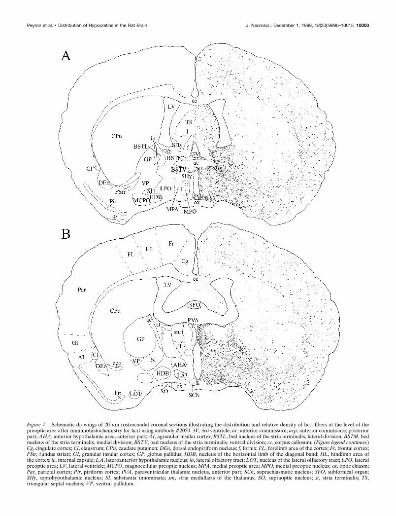
Figure 7. Schematic drawings of 20 mm rostrocaudal coronal sections illustrating the distribution and relative density of hcrt fibers at the level of thepreoptic area after immunohistochemistry for hcrt using antibody #2050. 3V, 3rd ventricle; ac, anterior commissure; acp, anterior commissure, posteriorpart; AHA, anterior hypothalamic area, anterior part; AI, agranular insular cortex; BSTL, bed nucleus of the stria terminalis, lateral division; BSTM, bednucleus of the stria terminalis, medial division; BSTV, bed nucleus of the stria terminalis, ventral division; cc, corpus callosum; (Figure legend continues)Cg, cingulate cortex; Cl, claustrum; CPu, caudate putamen; DEn, dorsal endopiriform nucleus; f, fornix; FL, forelimb area of the cortex; Fr, frontal cortex;FStr, fundus striati; GI, granular insular cortex; GP, globus pallidus; HDB, nucleus of the horizontal limb of the diagonal band; HL, hindlimb area ofthe cortex; ic, internal capsule; LA, lateroanterior hypothalamic nucleus; lo, lateral olfactory tract; LOT, nucleus of the lateral olfactory tract; LPO, lateralpreoptic area; LV, lateral ventricle; MCPO, magnocellular preoptic nucleus; MPA, medial preoptic area; MPO, medial preoptic nucleus; ox, optic chiasm;Par, parietal cortex; Pir, piriform cortex; PVA, paraventricular thalamic nucleus, anterior part; SCh, suprachiasmatic nucleus; SFO, subfornical organ;SHy, septohypothalamic nucleus; SI, substantia innominata; sm, stria medullaris of the thalamus; SO, supraoptic nucleus; st, stria terminalis; TS,triangular septal nucleus; VP, ventral pallidum.
Peyron et al. • Distribution of Hypocretins in the Rat Brain J. Neurosci., December 1, 1998, 18(23):9996–10015 10003
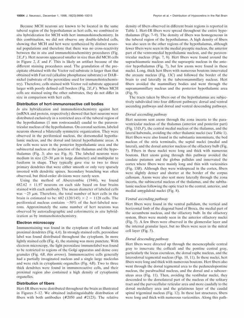
Because MCH neurons are known to be located in the sametuberal region of the hypothalamus as hcrt cells, we combined insitu hybridization for MCH with hcrt immunohistochemistry. Inthis combination, we did not observe any double-labeled cells,showing that MCH and hcrt were synthesized by distinct neuro-nal populations and therefore that there was no cross-reactivitybetween the in situ and immunohistochemistry procedures (Fig.2E,F). Hcrt neurons appeared smaller in size than did MCH cellsin Figure 2, E and F. This is likely an artifact because of thedifferent staining procedures used. The granulation of the pre-cipitate obtained with the Vector red substrate is larger than thatobtained with Fast red (alkaline phosphatase substrate) or DAB–nickel (substrate of the peroxidase used for immunohistochemis-try). Therefore, cells stained with Vector red substrate appearedlarger with poorly defined cell borders (Fig. 2E,F). When MCHcells are stained using the other substrates, they do not differ insize in comparison with hcrt cells.
Distribution of hcrt-immunoreactive cell bodiesIn situ hybridization and immunohistochemistry against hcrt(mRNA and protein, respectively) showed that hcrt neurons weredistributed exclusively in a restricted area of the tuberal region ofthe hypothalamus (1 mm rostrocaudal) caudal to the paraven-tricular nucleus of the hypothalamus. On coronal sections, labeledneurons showed a bilaterally symmetric organization. They wereobserved in the perifornical nucleus, the dorsomedial hypotha-lamic nucleus, and the dorsal and lateral hypothalamic areas. Afew cells were seen in the posterior hypothalamic area and thesubincertal nucleus at the junction of the thalamus and the hypo-thalamus (Fig. 3; also see Fig. 8). The labeled neurons weremedium in size (25–30 mm in large diameter) and multipolar tofusiform in shape. They typically gave rise to two to threeprimary dendrites that were either smooth or only very sparselyinvested with dendritic spines. Secondary branching was oftenobserved, but third-order divisions were rarely seen.
Using the method of Abercrombie (1946), we found682.62 6 11.97 neurons on each side based on four brainsstained with each antibody. The mean diameter of labeled cellswas ;25 mm. Therefore, the total number of hcrt cells in thebrain is estimated to be: 682 (120/145) 3 2 5 1128 cells. Theperifornical nucleus contains ;50% of the hcrt-labeled neu-rons. Approximately the same number of hcrt neurons wasobserved by autoradiographic and colorimetric in situ hybrid-ization as by immunohistochemistry.
Electron microscopyImmunostaining was found in the cytoplasm of cell bodies andproximal dendrites (Fig. 4A). In strongly stained cells, peroxidaselabel was found distributed throughout the cytoplasm. In morelightly stained cells (Fig. 4), the staining was more punctate. Withelectron microscopy, the light peroxidase immunolabel was foundto be restricted to regions of the Golgi apparatus and dense coregranules (Fig. 4B, thin arrows). Immunoreactive cells generallyhad a partially invaginated nucleus and a single large nucleolusand were rich in cytoplasmic organelles (Fig. 4B). Two to threethick dendrites were found in immunoreactive cells, and theirproximal region also contained a high density of cytoplasmicorganelles.
Distribution of fibersHcrt-IR fibers were distributed throughout the brain as illustratedin Figures 5–12. We obtained indistinguishable distribution offibers with both antibodies (#2050 and #2123). The relative
density of fibers observed in different brain regions is reported inTable 1. Hcrt-IR fibers were spread throughout the entire hypo-thalamus (Figs. 7–9). The density of fibers was homogeneous inthe tuberal region of the hypothalamus. A high density of fiberswas also seen in the other regions of the hypothalamus, althoughfewer fibers were seen in the medial preoptic nucleus, the anteriorpart of the ventromedial hypothalamic nucleus, and the paraven-tricular nucleus (Figs. 7, 8). Hcrt fibers were found around thesuprachiasmatic nucleus and the supraoptic nucleus in the ante-rior hypothalamus (Fig. 7), but few axons were found in thesenuclei. Long, thick hcrt fibers with numerous boutons innervatedthe arcuate nucleus (Fig. 13C) and followed the border of thebrain to end laterally in the tuberomammillary nucleus. Hcrtfibers avoided the mammillary bodies and went through thesupramammillary nucleus and the posterior hypothalamic area(Fig. 9).
The tracts taken by fibers out of the hypothalamus are subjec-tively subdivided into four different pathways: dorsal and ventralascending pathways and dorsal and ventral descending pathways.
Dorsal ascending pathwayHcrt neurons sent axons through the zona incerta to the para-ventricular nucleus of the thalamus (anterior and posterior part)(Fig. 13D,F), the central medial nucleus of the thalamus, and thelateral habenula, avoiding the other thalamic nuclei (see Table 1).Hcrt fibers were also found in the substantia innominata, the bednucleus of the stria terminalis, the septal nuclei (medial andlateral), and the dorsal anterior nucleus of the olfactory bulb (Fig.5). Fibers in these nuclei were long and thick with numerousboutons. Hcrt axons going through this pathway avoided thecaudate putamen and the globus pallidus and innervated thecortex where fibers were mainly long and thin with varicosities(Fig. 13B). Although they were widespread in the cortex, fiberswere slightly denser and shorter at the border of the corpuscallosum. Axons were also sent more laterally through the zonaincerta, the subincertal nucleus of the thalamus, and the subtha-lamic nucleus following the optic tract to the central, anterior, andmedial amygdaloid nuclei (Fig. 8).
Ventral ascending pathwayHcrt fibers were found in the ventral pallidum, the vertical andhorizontal limb of the diagonal band of Broca, the medial part ofthe accumbens nucleus, and the olfactory bulb. In the olfactorysystem, fibers were mainly seen in the anterior olfactory nuclei(Fig. 5). A few fibers were observed in the glomerular layer andthe internal granular layer, but no fibers were seen in the mitralcell layer (Fig. 5).
Dorsal descending pathwayHcrt fibers were directed up through the mesencephalic centralgray to innervate the colliculi and the pontine central gray,particularly the locus coeruleus, the dorsal raphe nucleus, and thelaterodorsal tegmental nucleus (Figs. 10, 11). In these nuclei, hcrtfibers were long and thick with numerous boutons. Hcrt fibers alsowent through the dorsal tegmental area to the pedunculopontinenucleus, the parabrachial nucleus, and the dorsal and a subcoer-uleus area (Fig. 11). Then, avoiding the vestibular nuclei, theydescended to the dorsolateral part of the nucleus of the solitarytract and the parvocellular reticular area and more caudally to thedorsal medullary area and the gelatinous layer of the caudalspinal trigeminal nucleus (Fig. 12). In these last structures, fiberswere long and thick with numerous varicosities. Along this path-
10004 J. Neurosci., December 1, 1998, 18(23):9996–10015 Peyron et al. • Distribution of Hypocretins in the Rat Brain
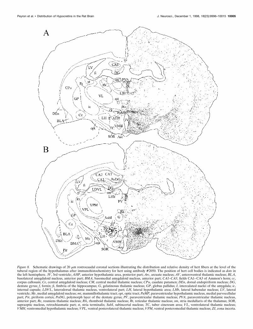
Figure 8. Schematic drawings of 20 mm rostrocaudal coronal sections illustrating the distribution and relative density of hcrt fibers at the level of thetuberal region of the hypothalamus after immunohistochemistry for hcrt using antibody #2050. The position of hcrt cell bodies is indicated as dots inthe left hemisphere. 3V, 3rd ventricle; AHP, anterior hypothalamic area, posterior part; Arc, arcuate nucleus; AV, anteroventral thalamic nucleus; BLA,basolateral amygdaloid nucleus, anterior part; BMA, basomedial amygdaloid nucleus, anterior part; CA1–CA3, fields CA1–CA3 of Ammon’s horn; cc,corpus callosum; Ce, central amygdaloid nucleus; CM, central medial thalamic nucleus; CPu, caudate putamen; DEn, dorsal endopiriform nucleus; DG,dentate gyrus; f, fornix; fi, fimbria of the hippocampus; G, gelatinosus thalamic nucleus; GP, globus pallidus; I, intercalated nuclei of the amygdala; ic,internal capsule; LDVL, laterodorsal thalamic nucleus, ventrolateral part; LH, lateral hypothalamic area; LHb, lateral habenular nucleus; LV, lateralventricle; Me, medial amygdaloid nucleus; mt, mammillothalamic tract; opt, optic tract; PaMP, paraventricular hypothalamic nucleus, medial parvocellularpart; Pir, piriform cortex; PoDG, polymorph layer of the dentate gyrus; PV, paraventricular thalamic nucleus; PVA, paraventricular thalamic nucleus,anterior part; Re, reuniens thalamic nucleus; Rh, rhomboid thalamic nucleus; Rt, reticular thalamic nucleus; sm, stria medullaris of the thalamus; SOR,supraoptic nucleus, retrochiasmatic part; st, stria terminalis; SubI, subincertal nucleus; TC, tuber cinereum area; VL, ventrolateral thalamic nucleus;VMH, ventromedial hypothalamic nucleus; VPL, ventral posterolateral thalamic nucleus; VPM, ventral posteromedial thalamic nucleus; ZI, zona incerta.
Peyron et al. • Distribution of Hypocretins in the Rat Brain J. Neurosci., December 1, 1998, 18(23):9996–10015 10005
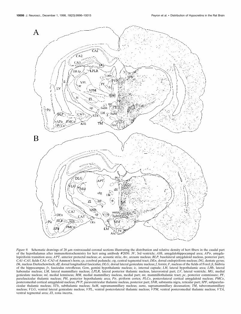
Figure 9. Schematic drawings of 20 mm rostrocaudal coronal sections illustrating the distribution and relative density of hcrt fibers in the caudal partof the hypothalamus after immunohistochemistry for hcrt using antibody #2050. 3V, 3rd ventricle; AHi, amygdalohippocampal area; APir, amygda-lopiriform transition area; APT, anterior pretectal nucleus; ar, acoustic stria; Arc, arcuate nucleus; BLP, basolateral amygdaloid nucleus, posterior part;CA1–CA3, fields CA1–CA3 of Ammon’s horn; cp, cerebral peduncle; ctg, central tegmental tract; DEn, dorsal endopiriform nucleus; DG, dentate gyrus;Dk, nucleus Darkschewitsch; dlf, dorsal longitudinal fasciculus; DLG, dorsal lateral geniculate nucleus; f, fornix; F, nucleus of the fields of Forel; fi, fimbriaof the hippocampus; f r, fasciculus retroflexus; Gem, gemini hypothalamic nucleus; ic, internal capsule; LH, lateral hypothalamic area; LHb, lateralhabenular nucleus; LM, lateral mammillary nucleus; LPLR, lateral posterior thalamic nucleus, laterorostral part; LV, lateral ventricle; MG, medialgeniculate nucleus; ml, medial lemniscus; MM, medial mammillary nucleus, medial part; mt, mammillothalamic tract; pc, posterior commissure; PF,parafascicular thalamic nucleus; PH, posterior hypothalamic area; Pir, piriform cortex; PLCo, posterolateral cortical amygdaloid nucleus; PMCo,posteromedial cortical amygdaloid nucleus; PVP, paraventricular thalamic nucleus, posterior part; SNR, substantia nigra, reticular part; SPF, subparafas-cicular thalamic nucleus; STh, subthalamic nucleus; SuM, supramammillary nucleus; sumx, supramammillary decussation; TM, tuberomammillarynucleus; VLG, ventral lateral geniculate nucleus; VPL, ventral posterolateral thalamic nucleus; VPM, ventral posteromedial thalamic nucleus; VTA,ventral tegmental area; ZI, zona incerta.
10006 J. Neurosci., December 1, 1998, 18(23):9996–10015 Peyron et al. • Distribution of Hypocretins in the Rat Brain
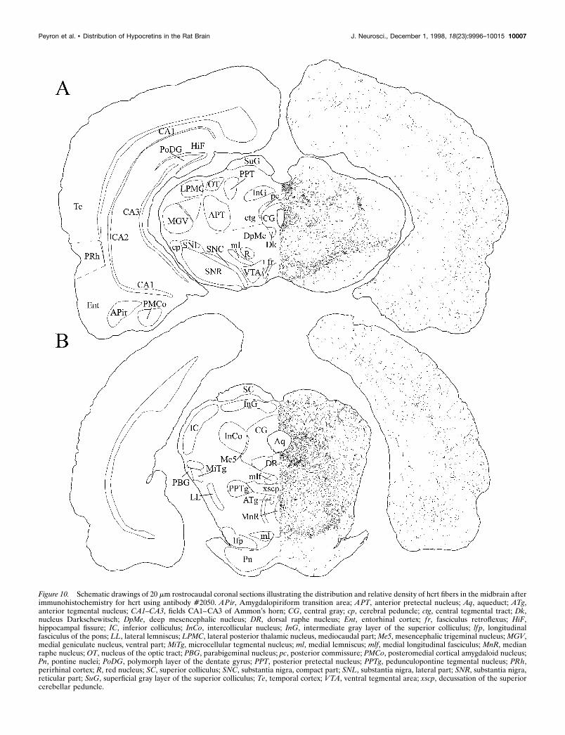
Figure 10. Schematic drawings of 20 mm rostrocaudal coronal sections illustrating the distribution and relative density of hcrt fibers in the midbrain afterimmunohistochemistry for hcrt using antibody #2050. APir, Amygdalopiriform transition area; APT, anterior pretectal nucleus; Aq, aqueduct; ATg,anterior tegmental nucleus; CA1–CA3, fields CA1–CA3 of Ammon’s horn; CG, central gray; cp, cerebral peduncle; ctg, central tegmental tract; Dk,nucleus Darkschewitsch; DpMe, deep mesencephalic nucleus; DR, dorsal raphe nucleus; Ent, entorhinal cortex; f r, fasciculus retroflexus; HiF,hippocampal fissure; IC, inferior colliculus; InCo, intercollicular nucleus; InG, intermediate gray layer of the superior colliculus; lfp, longitudinalfasciculus of the pons; LL, lateral lemniscus; LPMC, lateral posterior thalamic nucleus, mediocaudal part; Me5, mesencephalic trigeminal nucleus; MGV,medial geniculate nucleus, ventral part; MiTg, microcellular tegmental nucleus; ml, medial lemniscus; mlf, medial longitudinal fasciculus; MnR, medianraphe nucleus; OT, nucleus of the optic tract; PBG, parabigeminal nucleus; pc, posterior commissure; PMCo, posteromedial cortical amygdaloid nucleus;Pn, pontine nuclei; PoDG, polymorph layer of the dentate gyrus; PPT, posterior pretectal nucleus; PPTg, pedunculopontine tegmental nucleus; PRh,perirhinal cortex; R, red nucleus; SC, superior colliculus; SNC, substantia nigra, compact part; SNL, substantia nigra, lateral part; SNR, substantia nigra,reticular part; SuG, superficial gray layer of the superior colliculus; Te, temporal cortex; VTA, ventral tegmental area; xscp, decussation of the superiorcerebellar peduncle.
Peyron et al. • Distribution of Hypocretins in the Rat Brain J. Neurosci., December 1, 1998, 18(23):9996–10015 10007
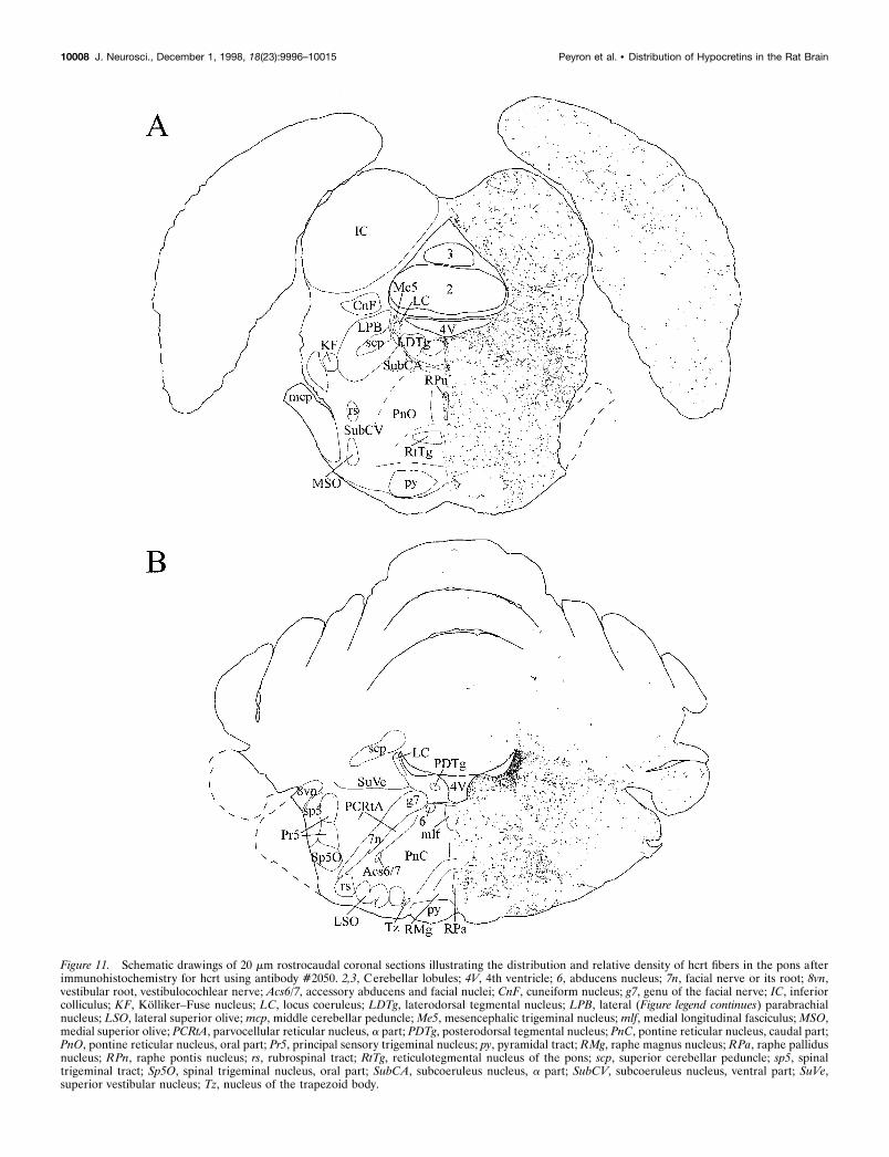
Figure 11. Schematic drawings of 20 mm rostrocaudal coronal sections illustrating the distribution and relative density of hcrt fibers in the pons afterimmunohistochemistry for hcrt using antibody #2050. 2,3, Cerebellar lobules; 4V, 4th ventricle; 6, abducens nucleus; 7n, facial nerve or its root; 8vn,vestibular root, vestibulocochlear nerve; Acs6/7, accessory abducens and facial nuclei; CnF, cuneiform nucleus; g7, genu of the facial nerve; IC, inferiorcolliculus; KF, Kolliker–Fuse nucleus; LC, locus coeruleus; LDTg, laterodorsal tegmental nucleus; LPB, lateral (Figure legend continues) parabrachialnucleus; LSO, lateral superior olive; mcp, middle cerebellar peduncle; Me5, mesencephalic trigeminal nucleus; mlf, medial longitudinal fasciculus; MSO,medial superior olive; PCRtA, parvocellular reticular nucleus, a part; PDTg, posterodorsal tegmental nucleus; PnC, pontine reticular nucleus, caudal part;PnO, pontine reticular nucleus, oral part; Pr5, principal sensory trigeminal nucleus; py, pyramidal tract; RMg, raphe magnus nucleus; RPa, raphe pallidusnucleus; RPn, raphe pontis nucleus; rs, rubrospinal tract; RtTg, reticulotegmental nucleus of the pons; scp, superior cerebellar peduncle; sp5, spinaltrigeminal tract; Sp5O, spinal trigeminal nucleus, oral part; SubCA, subcoeruleus nucleus, a part; SubCV, subcoeruleus nucleus, ventral part; SuVe,superior vestibular nucleus; Tz, nucleus of the trapezoid body.
10008 J. Neurosci., December 1, 1998, 18(23):9996–10015 Peyron et al. • Distribution of Hypocretins in the Rat Brain
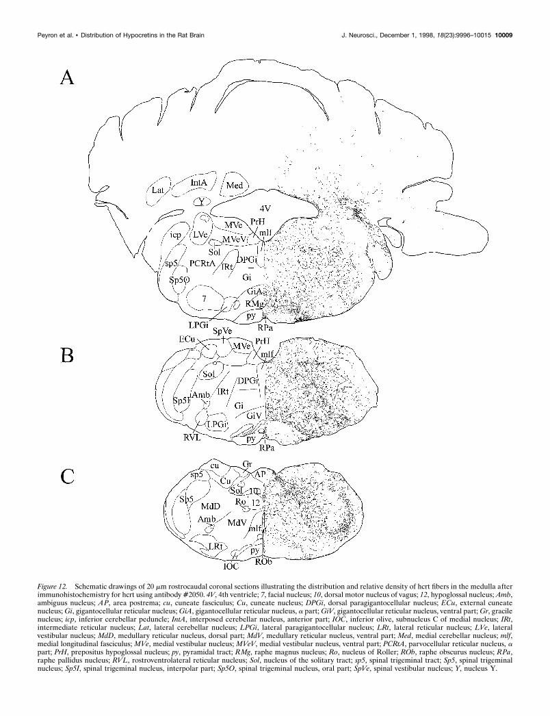
Figure 12. Schematic drawings of 20 mm rostrocaudal coronal sections illustrating the distribution and relative density of hcrt fibers in the medulla afterimmunohistochemistry for hcrt using antibody #2050. 4V, 4th ventricle; 7, facial nucleus; 10, dorsal motor nucleus of vagus; 12, hypoglossal nucleus; Amb,ambiguus nucleus; AP, area postrema; cu, cuneate fasciculus; Cu, cuneate nucleus; DPGi, dorsal paragigantocellular nucleus; ECu, external cuneatenucleus; Gi, gigantocellular reticular nucleus; GiA, gigantocellular reticular nucleus, a part; GiV, gigantocellular reticular nucleus, ventral part; Gr, gracilenucleus; icp, inferior cerebellar peduncle; IntA, interposed cerebellar nucleus, anterior part; IOC, inferior olive, subnucleus C of medial nucleus; IRt,intermediate reticular nucleus; Lat, lateral cerebellar nucleus; LPGi, lateral paragigantocellular nucleus; LRt, lateral reticular nucleus; LVe, lateralvestibular nucleus; MdD, medullary reticular nucleus, dorsal part; MdV, medullary reticular nucleus, ventral part; Med, medial cerebellar nucleus; mlf,medial longitudinal fasciculus; MVe, medial vestibular nucleus; MVeV, medial vestibular nucleus, ventral part; PCRtA, parvocellular reticular nucleus, apart; PrH, prepositus hypoglossal nucleus; py, pyramidal tract; RMg, raphe magnus nucleus; Ro, nucleus of Roller; ROb, raphe obscurus nucleus; RPa,raphe pallidus nucleus; RVL, rostroventrolateral reticular nucleus; Sol, nucleus of the solitary tract; sp5, spinal trigeminal tract; Sp5, spinal trigeminalnucleus; Sp5I, spinal trigeminal nucleus, interpolar part; Sp5O, spinal trigeminal nucleus, oral part; SpVe, spinal vestibular nucleus; Y, nucleus Y.
Peyron et al. • Distribution of Hypocretins in the Rat Brain J. Neurosci., December 1, 1998, 18(23):9996–10015 10009

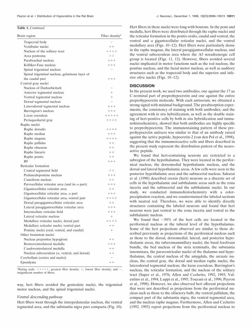
Table 1. Relative density of hypocretin-immunoreactive fibers invarious regions of the rat brain
Brain region Fiber densitya
CortexLayers 1–3 11
Layer 4 1(1)Layers 5–6 11(1)
Olfactory bulbMain bulb 2
Anterior olfactory nuclei 1
Endopiriform nucleus 11
Claustrum 11
Tenia tecta 1
Hippocampus 1
AmygdalaAmygdalohippocampal area 1
Anterior amygdaloid area 111
Basolateral nucleus 1
Basomedial nucleus 1
Cortical nucleus 1
Central nucleus 111
Intercalated nuclei 1
Lateral nucleus 1
Medial nucleus 11
SeptumLateral nucleus 111
Medial nucleus 111
Septofimbrial nucleus 11
Subfornical organ 2
Diagonal band of BrocaHorizontal limb 11
Vertical limb 111
Bed nucleus of stria terminalisLateral nucleus 111
Medial nucleus 111
Ventral nucleus 1111
Posteromedial nucleus 111
Posterolateral nucleus 1111
Basal gangliaAccumbens nucleus, core 1
Accumbens nucleus, shell 11
Caudate putamen 2
Globus pallidus 2
Fundus of the striatum 111
ThalamusAnteroventral thalamic nucleus 2
Anterior pretectal nucleus 2
Central medial nucleus 1111
Medial habenular nucleus 2
Mediodorsal thalamic nucleus 2
Lateral habenular nucleus 111
Laterodorsal nucleus 1
Paraventricular nucleus 1111
Reuniens nucleus 11
Rhomboid nucleus 11
Ventrolateral nucleus 2
Ventroposteriomedial nucleus 2
Ventroposteriolateral nucleus 2
Lateral geniculate nucleus 2
Medial geniculate nucleus 2
Zona incerta, rostral 1111
Table 1. Continued
Brain region Fiber densitya
Zona incerta, caudal 11
Subincertal nucleus 1111
Subthalamic nucleus 1111
Subparafascicular thalamic nucleus 1111
Parafascicular nucleus 11
Central gray of the thalamus 1111
Anterior hypothalamusMedial preoptic area 111
Medial preoptic nucleus 11
Lateral preoptic nucleus 111
Magnocellular preoptic nucleus 111
Supraoptic nucleus 2
Suprachiasmatic nucleus 2
Anterior hypothalamic area, anterior part 11
Lateroanterior nucleus 111
Substantia innominata 1111
Ventral pallidum 111
Posterior hypothalamusParaventricular nucleus 111
Ventromedial nucleus, rostral 11
Ventromedial nucleus, caudal 1111
Dorsomedial nucleus 1111
Dorsal hypothalamic area 1111
Lateral hypothalamic area 1111
Tuberum cinereum 1111
Tuberomammillary nucleus 1111
Perifornical nucleus 11111
Posterior hypothalamic area 1111
Arcuate nucleus 1111
Submammillary nucleus 1111
Mammillary bodies 2
Pineal gland 2
MidbrainSubstantia nigra, compact part 1111
Substantia nigra, reticular part 2
Substantia nigra, lateral part 111
Ventral tegmental area 111
Central gray 1111
Red nucleus 2
Mesencephalic reticular formation 11
Interpeduncular nucleus 111
BrainstemOculomotor nuclei
Abducens nucleus (VI) 2
Oculomotor nucleus (III) 2
Trochlear motor nucleus (IV) 2
Trigeminal motor nucleus (V) 1
Facial motor nucleus (VII) 1
Motor nucleus of the vagus (X) 1
Hypoglossal nucleus (XII) 1
Somatosensory nucleiSuperior colliculus 11
Inferior colliculus 11
Mesencephalic nucleus 2
Parabigeminal nucleus 1
Olivary nuclei 2
Cochlear nucleus, dorsal 11
Cochlear nucleus, ventral 2
Lateral lemniscus 111
10010 J. Neurosci., December 1, 1998, 18(23):9996–10015 Peyron et al. • Distribution of Hypocretins in the Rat Brain
way, hcrt fibers avoided the geniculate nuclei, the trigeminalmotor nucleus, and the spinal trigeminal nuclei.
Ventral descending pathwayHcrt fibers went through the interpeduncular nucleus, the ventraltegmental area, and the substantia nigra pars compacta (Fig. 10).
Hcrt fibers in these nuclei were long with boutons. In the pons andmedulla, hcrt fibers were distributed through the raphe nuclei andthe reticular formation in the pontis oralis, caudal and ventral, theventral and a gigantocellular reticular nuclei, and the ventralmedullary area (Figs. 10–12). Hcrt fibers were particularly densein the raphe magnus, the lateral paragigantocellular nucleus, andthe ventral subcoeruleus area where the A5 noradrenergic cellgroup is located (Figs. 11, 12). However, fibers avoided severalnuclei implicated in motor functions such as the red nucleus, thepontine nucleus, and the facial motor nucleus, as well as auditorystructures such as the trapezoid body and the superior and infe-rior olive nuclei (Figs. 10–12).
DISCUSSIONIn the present work, we used two antibodies, one against the 17 aaC-terminal part of preprohypocretin and one against the entirepreprohypocretin molecule. With each antiserum, we obtained astrong signal with minimal background. The preabsorption exper-iments, the consistency of staining with both antibodies, and theagreement with in situ hybridization, as well as the double stain-ing of hcrt-positive cells by both in situ hybridization and immu-nohistochemistry, showed that both antibodies are highly specificto preprohypocretin. The immunostaining pattern of these pre-prohypocretin antisera was similar to that of an antibody raisedagainst the active peptide, hypocretin-2 (van den Pol et al., 1998),suggesting that the immumoreactive cells and fibers described inthe present study represent the distribution pattern of the neuro-active peptide.
We found that hcrt-containing neurons are restricted to asubregion of the hypothalamus. They were located in the perifor-nical nucleus, the dorsomedial hypothalamic nucleus, and thedorsal and lateral hypothalamic areas. A few cells were seen in theposterior hypothalamic area and the subincertal nucleus. Sakuraiet al. (1998) described orexin (hcrt) neurons as a discrete set ofcells in the hypothalamic and subthalamic areas such as the zonaincerta and the subincertal and the subthalamic nuclei. In ourstudy, we conducted immunohistochemistry with a color-precipitation reaction, and we counterstained the labeled sectionswith neutral red. Therefore, we were able to identify directlystructures containing the labeled neurons and found that hcrtneurons were just ventral to the zona incerta and rostral to thesubthalamic nucleus.
We found that ;50% of the hcrt cells are located in theperifornical nucleus at the tuberal level of the hypothalamus.Some of the hcrt projections observed are similar to those de-scribed previously as projections of the perifornical nucleus suchas those to the dorsal, dorsomedial, lateral, and posterior hypo-thalamic areas, the tuberomammillary nuclei, the basal forebrainbundle, the bed nucleus of the stria terminalis, the substantiainnominata, the paraventricular nuclei of the thalamus and hypo-thalamus, the central nucleus of the amygdala, the arcuate nu-cleus, the central gray, the dorsal and median raphe nuclei, thelaterodorsal tegmental nucleus, the locus coeruleus, Barrington’snucleus, the reticular formation, and the nucleus of the solitarytract (Saper et al., 1976; Allen and Cechetto, 1992, 1993; Val-entino et al., 1994; Luppi et al., 1995; Touzani et al., 1996; Peyronet al., 1998). However, we also observed hcrt efferent projectionsthat were not described as projections from the perifornical nu-cleus such as those to the olfactory bulb, the ventral pallidum, thecompact part of the substantia nigra, the ventral tegmental area,and the nucleus raphe magnus. Furthermore, Allen and Cechetto(1992, 1993) report projections from the perifornical nucleus to
Table 1. Continued
Brain region Fiber densitya
Trapezoid body 2
Vestibular nuclei 11
Nucleus of the solitary tract 1111
Area postrema 11
Parabrachial nucleus 111
Kolliker-Fuse nucleus 111
Spinal trigeminal nucleus 2
Spinal trigeminal nucleus, gelatinous layer ofthe caudal part 111
Central gray nucleiNucleus of Darkschwitsch 2
Anterior tegmental nucleus 1
Ventral tegmental nucleus 1
Dorsal tegmental nucleus 2
Laterodorsal tegmental nucleus 111
Barrington’s nucleus 111
Locus coeruleus 11111
Periaqueductal gray 1111
Raphe nucleiRaphe dorsalis 1111
Raphe median 111
Raphe magnus 1111
Raphe pallidus 11
Raphe obscurus 11
Raphe linearis 11
Raphe pontis 11
B9 111
Reticular formationCentral tegmental field 11
Pedunculopontine nucleus 111
Cuneiform nucleus 11
Parvocellular reticular area (and its a part) 111
Gigantocellular reticular area 11
Gigantocellular reticular area, a part 111
Gigantocellular reticular area, ventral part 1111
Dorsal paragigantocellular reticular area 11
Lateral paragigantocellular reticular area 111
Intermediate reticular field 111
Lateral reticular nucleus 1
Medullary reticular nuclei, dorsal part 1111
Medullary reticular nuclei, ventral part 11
Pontine nuclei (oral, ventral, and caudal) 1
Other brainstem nucleiNucleus prepositus hypoglossi 11
Rostroventrolateral medulla 111
Caudoventrolateral medulla 11
Nucleus subcoeruleus (a, ventral, and dorsal) 11
Cerebellum (cortex and nuclei) 2
Ependyma 11
aRating scale: 11111, greatest fiber density; 1, lowest fiber density; and 2,insignificant number of fibers.
Peyron et al. • Distribution of Hypocretins in the Rat Brain J. Neurosci., December 1, 1998, 18(23):9996–10015 10011
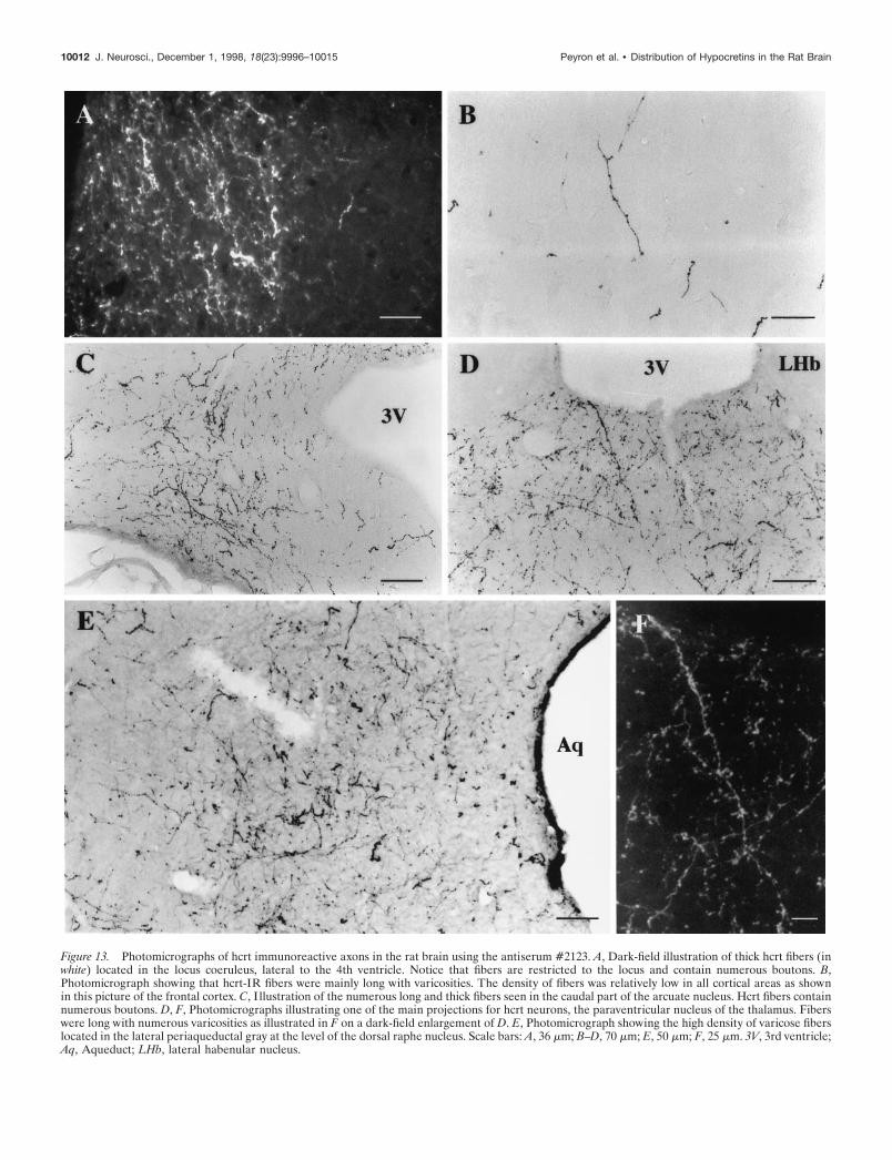
Figure 13. Photomicrographs of hcrt immunoreactive axons in the rat brain using the antiserum #2123. A, Dark-field illustration of thick hcrt fibers (inwhite) located in the locus coeruleus, lateral to the 4th ventricle. Notice that fibers are restricted to the locus and contain numerous boutons. B,Photomicrograph showing that hcrt-IR fibers were mainly long with varicosities. The density of fibers was relatively low in all cortical areas as shownin this picture of the frontal cortex. C, I llustration of the numerous long and thick fibers seen in the caudal part of the arcuate nucleus. Hcrt fibers containnumerous boutons. D, F, Photomicrographs illustrating one of the main projections for hcrt neurons, the paraventricular nucleus of the thalamus. Fiberswere long with numerous varicosities as illustrated in F on a dark-field enlargement of D. E, Photomicrograph showing the high density of varicose fiberslocated in the lateral periaqueductal gray at the level of the dorsal raphe nucleus. Scale bars: A, 36 mm; B–D, 70 mm; E, 50 mm; F, 25 mm. 3V, 3rd ventricle;Aq, Aqueduct; LHb, lateral habenular nucleus.
10012 J. Neurosci., December 1, 1998, 18(23):9996–10015 Peyron et al. • Distribution of Hypocretins in the Rat Brain
the mediodorsal thalamic nucleus, the ventral premammillarynucleus, and the dorsal tegmental nucleus that we did not ob-serve; these projections are therefore unlikely to use hcrt as aneurotransmitter and/or neuromodulator. Consequently, the hcrtcell group is a unique system and not simply a subset of theperifornical nucleus.
Although hcrt-containing neurons represent a relatively smallnumber of cells, their projections are widely distributed in theCNS (Fig. 14), suggesting that hcrt might be involved in multiplefunctions as we discuss in the following paragraphs.
FeedingThe perifornical nucleus contains ;50% of the hcrt-labeled neu-rons. This nucleus has been shown to be intimately involved inthe neural control of food intake (Winn et al., 1984; Stanley et al.,1996) and is the most sensitive brain region for both the orexi-genic effect of neuropeptide Y (Stanley et al., 1993) and thesuppressive effects of catecholamines on feeding (Leibowitz andStanley, 1986). Neurons in the perifornical nucleus and the lateralhypothalamic area also respond to internal signals related to thenutritional state of the animal (Himmi et al., 1988). We observedhcrt fibers in nuclei that are known to be involved in the regula-tion of food intake such as the ventromedial hypothalamic nu-cleus, the arcuate nucleus, the paraventricular nucleus of thehypothalamus, the parabrachial area, the nucleus of the solitarytract, and the area postrema (Leibowitz and Brown, 1980; Rob-erts, 1980; Stanley and Leibowitz, 1984; Luiten et al., 1987; Ritterand Stone, 1987; Akabayashi et al., 1994; Ritter et al., 1994;Nishijo and Norgren, 1997; Shimura et al., 1997). Taken together,these data suggest that hcrt might be involved in the regulation offeeding. Indeed, Sakurai et al. (1998) recently showed that orexinA (hcrt-1) and B (hcrt-2) stimulate food intake when injectedintracerebroventricularly and that the mRNA accumulates duringfasting.
MCH, whose cell bodies are also found in the tuberal region ofthe hypothalamus (Skofitsch et al., 1985; Nahon et al., 1989;Bittencourt et al., 1992), has also been reported to have potent
orexigenic activity (Presse et al., 1996; Qu et al., 1996; Rossi et al.,1997). Based on the absence of colocalization (Fig. 2E,F), MCHand hcrt cells are distinct neuronal populations although they arepartly intermingled anatomically. MCH and hcrt neurons inner-vate some of the same brain regions such as the medial septum/diagonal band, the bed nucleus of the stria terminalis, the zonaincerta, the entire hypothalamus, the arcuate nucleus, the ventraltegmental area, the periaqueductal gray, the locus coeruleus, andthe nucleus of the solitary tract (Bittencourt et al., 1992), suggest-ing that MCH and hcrt might be involved in the same physiolog-ical functions. Håkansson et al. (1998) showed that leptin recep-tors are located in the perifornical nucleus and the lateralhypothalamic area and are found on MCH neurons as well asother cell types. Whether leptin receptors are also present on hcrtcells remains to be determined.
Blood pressure regulationElectrical or chemical stimulation of the perifornical nucleusincreases blood pressure and heart rate and activates neurons ofthe lateral paragigantocellular area (Sun and Guyenet, 1986;Allen and Cechetto, 1992). The perifornical nucleus and adjacentlateral hypothalamic area have been identified as the source ofneurons responsible for producing cardiovascular responses asso-ciated with emotion (Smith et al., 1990). In our study, we foundhcrt fibers located in nuclei that are well known to be involved inblood pressure regulation such as the rostral ventrolateral me-dulla, the lateral paragigantocellular nucleus, the locus coeruleus,the nucleus of the solitary tract, the midbrain periaqueductal gray,the A5 noradrenergic cell group, the parabrachial region, and thearea postrema (for review, see Dampney, 1994). Furthermore, theperifornical nucleus receives ascending afferent inputs mainlyfrom the ventromedial central gray, the dorsal raphe nucleus, thelaterodorsal tegmental region, and the nucleus of the solitary tract(Allen and Cechetto, 1992). These brainstem structures areknown to be closely associated with cardiovascular function(Lindgren, 1961; Reis and Cuenod, 1965; Calaresu and Ciriello,1980; for review, see Dampney, 1994). Consequently, our results
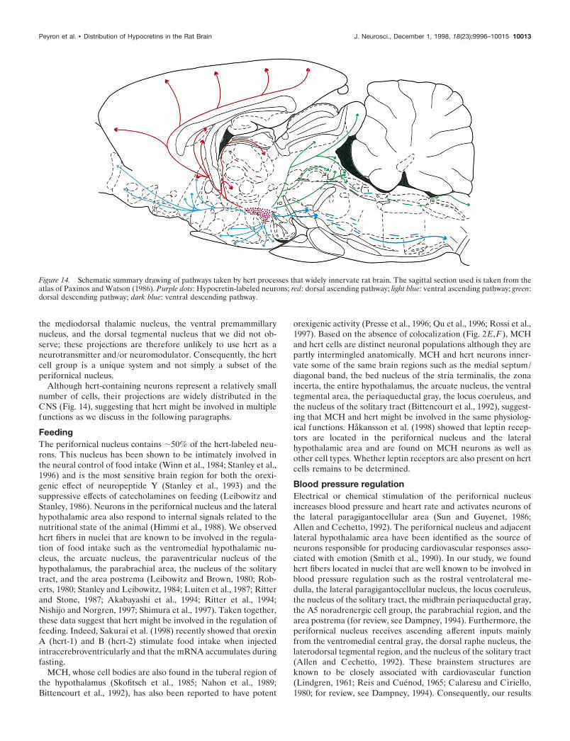
Figure 14. Schematic summary drawing of pathways taken by hcrt processes that widely innervate rat brain. The sagittal section used is taken from theatlas of Paxinos and Watson (1986). Purple dots: Hypocretin-labeled neurons; red: dorsal ascending pathway; light blue: ventral ascending pathway; green:dorsal descending pathway; dark blue: ventral descending pathway.
Peyron et al. • Distribution of Hypocretins in the Rat Brain J. Neurosci., December 1, 1998, 18(23):9996–10015 10013
combined with the literature suggest that hcrt could be involvedin the regulation of blood pressure.
Neuroendocrine regulationThe arcuate nucleus, highly involved in neuroendocrine regula-tion, is among the most densely hcrt-innervated areas of thehypothalamus, suggesting that hcrt might also be involved inhormonal regulation. This hypothesis has recently been testedelectrophysiologically in hypothalamic slices containing the arcu-ate nucleus and the median eminence (van del Pol et al., 1998).Application of hcrt increased presynaptic activity of GABAergiccells terminating on electrophysiologically identified neuroendo-crine neurons. These data indicate that hcrt modulates the affer-ent input controlling the neuroendocrine neurons in the arcuatenucleus. Furthermore, the paraventricular nucleus of the hypo-thalamus (PVN) is innervated by the perifornical nucleus (Allenand Cechetto, 1993; Larsen et al., 1994). The perifornical nu-cleus, along with the dorsomedial hypothalamic nucleus, providesmost of the local excitatory synaptic input to PVN neurons(Boudaba et al., 1997). We have shown previously that hcrt has anexcitatory effect on hypothalamic cells in vitro (de Lecea et al.,1998) and, in the present study, that the perifornical input to thePVN includes hcrt axons. These data suggest that hcrt might alsomodulate hormonal release controlled by PVN neurons.
ThermoregulationThe lateral hypothalamic area, in which we observed hcrt-labeledcells, has been implicated in the behavioral regulation of temper-ature (Corbett et al., 1988). The lateral hypothalamic area is alsoinvolved in heat loss responses, and the neural pathway involvedin shivering passes through this region (Hemingway, 1963). Thepresence of hcrt fibers in the raphe magnus and the subcoeruleusareas, brain regions that also have been implicated in thermoreg-ulation (Werner and Bienck, 1990), suggests that hcrt couldmodulate the regulation of body temperature.
Sleep–waking cycleNeurons in the lateral hypothalamic area have been involved inthe maintenance of waking state (Vanni-Mercier et al., 1984).Our results showed that hcrt fibers were seen in numerous struc-tures involved in the sleep–waking cycle such as the locus coer-uleus, the tuberomammillary nucleus, the pontine reticular for-mation, the raphe nuclei, the preoptic area, and the laterodorsaltegmental nucleus (for review, see Vertes, 1990; Jones, 1994). Thehigh density of fibers in the locus coeruleus and the tuberomam-millary nucleus of the hypothalamus, nuclei containing neuronsresponsible for the maintenance of the waking state, suggests thathcrt might have an effect on arousal.
ConclusionThe hypothalamus has a major role in regulating various behav-iors that contribute to homeostasis such as arousal, feeding, andthermoregulation by integrating external and internal stimuli(Simerly, 1995). As discussed above, hcrt could be involved in theregulation of feeding, blood pressure, hormonal release, temper-ature, and arousal. Animals encounter situations that require orfavor coordinated responses of several autonomic systems. Wesuggest that the hypocretins could provide such a coordinatingsignal.
REFERENCESAbercrombie M (1946) Estimation of nuclear population from mic-
rotome sections. Anat Rec 94:239–247.Akabayashi A, Wahlestedt C, Alexander JT, Leibowitz SF (1994) Spe-
cific inhibition of endogenous neuropeptide Y synthesis in arcuatenucleus by antisense oligonucleotides suppresses feeding behavior andinsulin secretion. Mol Brain Res 21:55–61.
Allen GV, Cechetto DF (1992) Functional and anatomical organizationof cardiovascular pressor and depressor sites in the lateral hypothalamicarea: I. Descending projections. J Comp Neurol 315:313–332.
Allen GV, Cechetto DF (1993) Functional and anatomical organizationof cardiovascular pressor and depressor sites in the lateral hypothalamicarea: II. Ascending projections. J Comp Neurol 330:421–438.
Bittencourt JC, Presse F, Arias C, Peto C, Vaughan J, Nahon JL, Vale W,Sawchenko PE (1992) The melanin-concentrating hormone system ofthe rat brain: an immuno- and hybridization histochemical character-ization. J Comp Neurol 319:218–245.
Boudaba C, Schrader LA, Tasker JG (1997) Physiological evidence forlocal excitatory synaptic circuits in the rat hypothalamus. J Neuro-physiol 77:3396–3400.
Calaresu FR, Ciriello J (1980) Projections to the hypothalamus frombuffer nerves and nucleus tractus solitarius in the cat. Am J Physiol239:R130–R136.
Chiu K-P, Sullivan T, Bursztajn S (1996) Improved in situ hybridization:color intensity enhancement procedure for the alkaline phosphatase/Fast red system. Biotechniques 20:964–968.
Corbett SW, Kaufman LN, Keesey RE (1988) Thermogenesis after lat-eral hypothalamic lesions: contributions of brown adipose tissue. Am JPhysiol 255:E708–E715.
Dampney RAL (1994) Functional organization of central pathways reg-ulating the cardiovascular system. Physiol Rev 74:323–364.
de Lecea L, Kilduff TS, Peyron C, Gao X-B, Foye PE, Danielson PE,Fukuhara C, Battenberg ELF, Gautvik VT, Bartlett II FS, FrankelWN, van den Pol AN, Bloom FE, Gautvik KM, Sutcliffe JG (1998)The hypocretins: hypothalamus-specific peptides with neuroexcitatoryactivity. Proc Natl Acad Sci USA 95:322–327.
Gautvik KM, de Lecea L, Gautvik VT, Danielson PE, Tranque P,Dopazo A, Bloom FE, Sutcliffe JG (1996) Overview of the mostprevalent hypothalamus-specific mRNAs, as identified by directionaltag PCR subtraction. Proc Natl Acad Sci USA 93:8733–8738.
Håkansson ML, Brown H, Ghilardi N, Skoda RC, Meister B (1998)Leptin receptor immunoreactivity in chemically defined target neuronsof the hypothalamus. J Neurosci 18:559–572.
Hemingway A (1963) Shivering. Physiol Rev 43:397–422.Himmi T, Boyer A, Orsini JC (1988) Changes in lateral hypothalamic
neuronal activity accompanying hyper- and hypoglycemias. PhysiolBehav 44:347–354.
Jones BE (1994) Basic mechanisms of sleep-waking states. In: Principlesand practice of sleep medicine, 2nd Edition (Kryger MH, Roth T,Dement WC, eds), pp 145–162. Philadelphia: Saunders.
Larsen PJ, Hay-Schmidt A, Mikkelsen JD (1994) Efferent connectionsfrom the lateral hypothalamic region and the lateral preoptic area to thehypothalamic paraventricular nucleus of the rat. J Comp Neurol342:299–319.
Leibowitz SF, Brown LL (1980) Histochemical and pharmacologicalanalysis of noradrenergic projections to the paraventricular hypothala-mus in relation to feeding stimulation. Brain Res 201:289–314.
Leibowitz SF, Stanley BG (1986) Neurochemical controls of appetite. In:Feeding behavior: neural and humoral controls (Ritter RC, Ritter S,Barnes CD, eds), pp 191–234. Orlando, FL: Academic.
Lindgren P (1961) Localization and function of the medullary vasomo-tor center in intracollicularly decerebrated cats. Circ Res 9:250–255.
Luiten PGM, ter Horst GJ, Steffens AB (1987) The hypothalamus, in-trinsic connections and outflow pathways to the endocrine system inrelation to the control of feeding and metabolism. Prog Neurobiol28:1–54.
Luppi P-H, Aston-Jones G, Akaoka H, Chouvet G, Jouvet M (1995)Afferent projections to the rat locus coeruleus demonstrated by retro-grade and anterograde tracing with cholera-toxin B subunit and Phaseo-lus vulgaris leucoagglutinin. Neuroscience 65:119–160.
Nahon JL, Presse F, Bittencourt JC, Sawchenko PE, Vale W (1989) Therat melanin-concentrating hormone messenger ribonucleic acid en-codes multiple putative neuropeptides coexpressed in the dorsolateralhypothalamus. Endocrinology 125:2056–2065.
Nishijo H, Norgren R (1997) Parabrachial neural coding of taste stimuliin awake rats. J Neurophysiol 78:2254–2268.
Paxinos G, Watson C (1986) The rat brain in stereotaxic coordinates,2nd Edition. Sydney: Academic.
Peyron C, Tighe DK, Lee BS, de Lecea L, Heller HC, Sutcliffe JG, Kilduff
10014 J. Neurosci., December 1, 1998, 18(23):9996–10015 Peyron et al. • Distribution of Hypocretins in the Rat Brain
TS (1997) Distribution of immunoreactive neurons and fibers for ahypothalamic neuropeptide precursor related to secretin. Soc NeurosciAbstr 23:2032.
Peyron C, Petit J-M, Rampon C, Jouvet M, Luppi P-H (1998) Forebrainafferents to the rat dorsal raphe nucleus demonstrated by retrogradeand anterograde tracing methods. Neuroscience 82:443–468.
Presse F, Sorokvsky I, Max JP, Nicolaidis S, Nahon JL (1996) Melanin-concentrating hormone is a potent anorectic peptide regulated byfood-deprivation and glucopenia in the rat. Neuroscience 71:735–745.
Qu D, Ludwig DS, Gammeltoft S, Piper M, Pelleymounter MA, CullenMJ, Mathes WF, Przypek J, Kanarek R, Maratos-Flier E (1996) Arole for melanin-concentrating hormone in the regulation of feedingbehaviour. Nature 380:243–247.
Reis DJ, Cuenod M (1965) Central neural regulation of carotid barore-ceptor reflexes in the cat. Am J Physiol 209:1267–1279.
Ritter S, Stone SL (1987) Area postrema lesions block feeding inducedby systemic injections of monosodium glutamate. Physiol Behav41:21–24.
Ritter S, Dinh TT, Friedman MI (1994) Induction of Fos-like immuno-reactivity (Fos-li) and stimulation of feeding by 2,5-anhydro-D-mannitol(2,5-AM) require the vagus nerve. Brain Res 646:53–64.
Roberts WW (1980) [14C]Deoxyglucose mapping of first-order projec-tions activated by stimulation of lateral hypothalamic sites elicitinggnawing, eating, and drinking in rats. J Comp Neurol 194:617–638.
Rossi M, Choi SJ, O’Shea D, Miyoshi T, Ghatei MA, Bloom SR (1997)Melanin-concentrating hormone acutely stimulates feeding, but chronicadministration has no effect on body weight. Endocrinology 138:351–355.
Sakurai T, Amemiya A, Ishii M, Matsuzaki I, Chemelli RM, Tanaka H,Williams SC, Richardson JA, Kozlowski GP, Wilson S, Arch JRS,Buckingham RE, Haynes AC, Carr SA, Annan RS, McNulty DE, LiuW-S, Terrett JA, Elshourbagy NA, Bergsma DJ, Yanagisawa M (1998)Orexins and orexin receptors: a family of hypothalamic neuropeptidesand G protein-coupled receptors that regulate feeding behavior. Cell92:573–585.
Saper CB, Loewy AD, Swanson LW, Cowan WM (1976) Directhypothalamo-autonomic connections. Brain Res 117:305–312.
Shimura T, Norgren R, Grigson PS, Norgren R (1997) Brainstem lesionsand gustatory function: I. The role of the nucleus of the solitary tractduring a brief intake test in rats. Behav Neurosci 111:155–168.
Simerly RB (1995) Anatomical substrates of hypothalamic integration.In: The rat nervous system, 2nd Edition (Paxinos G, ed), pp 353–376.Academic.
Skofitsch G, Jacobowitz DM, Zamir N (1985) Immunohistochemicallocalization of a melanin concentrating hormone-like peptide in the ratbrain. Brain Res Bull 15:635–645.
Smith OA, DeVito JL, Astley CA (1990) Neurons controlling cardiovas-cular responses to emotion are located in lateral hypothalamus-perifornical region. Am J Physiol 259:R943–R954.
Stanley BG, Leibowitz SF (1984) Neuropeptide Y: stimulation of feed-ing and drinking by injection into the paraventricular nucleus. Life Sci35:2635–2642.
Stanley BG, Magdalin W, Seirafi A, Thomas W, Leibowitz SF (1993)The perifornical area: the major focus of (a) patchily distributed hypo-thalamic neuropeptide Y sensitive feeding system(s). Brain Res604:304–317.
Stanley BG, Willett III VL, Donias HW, Dee II MG, Duva MA (1996)Lateral hypothalamic NMDA receptors and glutamate physiologicalmediators of eating and weight control. Am J Physiol 270:R443–R449.
Sun MK, Guyenet PG (1986) Hypothalamic glutamatergic input to med-ullary sympathoexcitatory neurons in rats. Am J Physiol251:R798–R810.
Sutcliffe JG, Milner RJ, Shinnick TM, Bloom FE (1983) Identifying theprotein products of brain-specific genes with antibodies to chemicallysynthesized peptides. Cell 33:671–682.
Sutcliffe JG, Gautvik KM, Kilduff TS, Horn T, Foye PE, Danielson PE,Frankel WN, Bloom FE, de Lecea L (1997) Two novel hypothalamicpeptides related to secretin derived from a single neuropeptide precur-sor. Soc Neurosci Abstr 23:2032.
Swanson LW (1987) The hypothalamus. In: Handbook of chemical neu-roanatomy, Vol 5, Integrated systems of the CNS (Bjorklund A, Hok-felt T, Swanson LW, eds), pp 1–124. Amsterdam: Elsevier.
Touzani K, Taghzouti K, Velley L (1996) Cellular organization of lat-eral hypothalamic efferents to the central amygdaloid nucleus of the rat.NeuroReport 7:517–520.
Valentino RJ, Page ME, Luppi P-H, Zhu Y, Van Bockstaele E, Aston-Jones G (1994) Evidence for widespread afferents to Barrington’snucleus, a brainstem region rich in corticotropin-releasing hormoneneurons. Neuroscience 62:125–143.
van den Pol AN, Gao X-B, Obrietan K, Kilduff TS, Belousov AB (1998)Presynaptic and postsynaptic actions and modulation of neuroendo-crine neurons by a new hypothalamic peptide, hypocretin/orexin.J Neurosci 18:7962–7971.
Vanni-Mercier G, Sakai K, Salvert D, Jouvet M (1984) “Waking statespecific” neurons in the caudal hypothalamus of the cat. C R Acad SciParis 298:195–200.
Vertes RP (1990) Brainstem mechanisms of slow wave sleep and REMsleep. In: Brainstem mechanisms of behavior (Klemm WR, Vertes RP,eds), pp 535–581. New York: Wiley.
Werner J, Bienek A (1990) Loss and restoration of preoptic thermore-activeness after lesions of the rostral raphe nuclei. Exp Brain Res80:429–435.
Winn P, Tarbuck A, Dunnett SB (1984) Ibotenic acid lesions of thelateral hypothalamus: comparison with the electric lesion syndrome.Neuroscience 12:225–240.
Peyron et al. • Distribution of Hypocretins in the Rat Brain J. Neurosci., December 1, 1998, 18(23):9996–10015 10015