Embed Size (px)
Citation preview
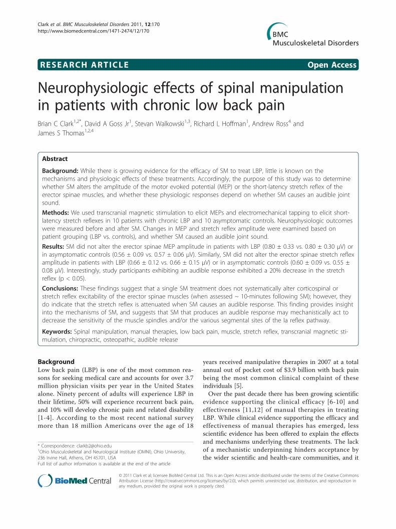
RESEARCH ARTICLE Open Access
Neurophysiologic effects of spinal manipulationin patients with chronic low back painBrian C Clark1,2*, David A Goss Jr1, Stevan Walkowski1,3, Richard L Hoffman1, Andrew Ross4 andJames S Thomas1,2,4
Abstract
Background: While there is growing evidence for the efficacy of SM to treat LBP, little is known on themechanisms and physiologic effects of these treatments. Accordingly, the purpose of this study was to determinewhether SM alters the amplitude of the motor evoked potential (MEP) or the short-latency stretch reflex of theerector spinae muscles, and whether these physiologic responses depend on whether SM causes an audible jointsound.
Methods: We used transcranial magnetic stimulation to elicit MEPs and electromechanical tapping to elicit short-latency stretch reflexes in 10 patients with chronic LBP and 10 asymptomatic controls. Neurophysiologic outcomeswere measured before and after SM. Changes in MEP and stretch reflex amplitude were examined based onpatient grouping (LBP vs. controls), and whether SM caused an audible joint sound.
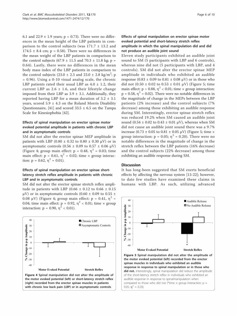
Results: SM did not alter the erector spinae MEP amplitude in patients with LBP (0.80 ± 0.33 vs. 0.80 ± 0.30 μV) orin asymptomatic controls (0.56 ± 0.09 vs. 0.57 ± 0.06 μV). Similarly, SM did not alter the erector spinae stretch reflexamplitude in patients with LBP (0.66 ± 0.12 vs. 0.66 ± 0.15 μV) or in asymptomatic controls (0.60 ± 0.09 vs. 0.55 ±0.08 μV). Interestingly, study participants exhibiting an audible response exhibited a 20% decrease in the stretchreflex (p < 0.05).
Conclusions: These findings suggest that a single SM treatment does not systematically alter corticospinal orstretch reflex excitability of the erector spinae muscles (when assessed ~ 10-minutes following SM); however, theydo indicate that the stretch reflex is attenuated when SM causes an audible response. This finding provides insightinto the mechanisms of SM, and suggests that SM that produces an audible response may mechanistically act todecrease the sensitivity of the muscle spindles and/or the various segmental sites of the Ia reflex pathway.
Keywords: Spinal manipulation, manual therapies, low back pain, muscle, stretch reflex, transcranial magnetic sti-mulation, chiropractic, osteopathic, audible release
BackgroundLow back pain (LBP) is one of the most common rea-sons for seeking medical care and accounts for over 3.7million physician visits per year in the United Statesalone. Ninety percent of adults will experience LBP intheir lifetime, 50% will experience recurrent back pain,and 10% will develop chronic pain and related disability[1-4]. According to the most recent national surveymore than 18 million Americans over the age of 18
years received manipulative therapies in 2007 at a totalannual out of pocket cost of $3.9 billion with back painbeing the most common clinical complaint of theseindividuals [5].Over the past decade there has been growing scientific
evidence supporting the clinical efficacy [6-10] andeffectiveness [11,12] of manual therapies in treatingLBP. While clinical evidence supporting the efficacy andeffectiveness of manual therapies has emerged, lessscientific evidence has been offered to explain the effectsand mechanisms underlying these treatments. The lackof a mechanistic underpinning hinders acceptance bythe wider scientific and health-care communities, and it
* Correspondence: [email protected] Musculoskeletal and Neurological Institute (OMNI), Ohio University,236 Irvine Hall, Athens, OH 45701, USAFull list of author information is available at the end of the article
Clark et al. BMC Musculoskeletal Disorders 2011, 12:170http://www.biomedcentral.com/1471-2474/12/170
© 2011 Clark et al; licensee BioMed Central Ltd. This is an Open Access article distributed under the terms of the Creative CommonsAttribution License (http://creativecommons.org/licenses/by/2.0), which permits unrestricted use, distribution, and reproduction inany medium, provided the original work is properly cited.
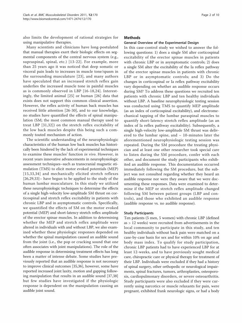
also limits the development of rational strategies forusing manipulative therapies.Many scientists and clinicians have long-postulated
that manual therapies exert their biologic effects on seg-mental components of the central nervous system (e.g.,supraspinal, spinal, etc.) [13-22]. For example, morethan 25 years ago it was noticed that deep somatic orvisceral pain leads to increases in muscle tone/spasm inthe surrounding musculature [23], and many authorshave speculated that an increased stretch reflex gainunderlies the increased muscle tone in painful musclesas is commonly observed in LBP [16-18,24]. Interest-ingly, the limited animal [25] or human [26] data thatexists does not support this common clinical assertion.However, the reflex activity of human back muscles hasreceived little attention [26-30], and to our knowledge,no studies have quantified the effects of spinal manipu-lation (SM; the most common manual therapy used totreat LBP [31,32]) on the stretch reflex excitability ofthe low back muscles despite this being such a com-monly touted mechanism of action.The scientific understanding of the neurophysiologic
characteristics of the human low back muscles has histori-cally been hindered by the lack of experimental techniquesto examine these muscles’ function in vivo. However, inrecent years innovative advancements in neurophysiologicassessment techniques–such as transcranial magnetic sti-mulation (TMS) to elicit motor evoked potentials (MEP)[15,33,34] and mechanically elicited stretch reflexes[26,29,33]– have begun to be applied to the study of thehuman lumbar musculature. In this study we utilizedthese neurophysiologic techniques to determine the effectsof a single high-velocity low-amplitude SM thrust on cor-ticospinal and stretch reflex excitability in patients withchronic LBP and in asymptomatic controls. Specifically,we quantified the effects of SM on the motor evokedpotential (MEP) and short-latency stretch reflex amplitudeof the erector spinae muscles. In addition to determiningwhether the MEP and stretch reflex amplitude werealtered in individuals with and without LBP, we also exam-ined whether these physiologic responses depended onwhether the spinal manipulation caused an audible soundfrom the joint (i.e., the pop or cracking sound that oneoften associates with joint manipulations). The role of theaudible response in determining treatment effects has longbeen a matter of intense debate. Some studies have pre-viously reported that an audible response is not necessaryto improve clinical outcomes [35,36], however, some havereported increased joint laxity, motion and gapping follow-ing manipulation that results in an audible sound [37,38]but few studies have investigated if the physiologicresponse is dependent on the manipulation causing anaudible joint sound.
MethodsGeneral Overview of the Experimental DesignIn this case-control study we wished to answer the fol-lowing questions: 1) does a single SM alter corticospinalexcitability of the erector spinae muscles in patientswith chronic LBP or in asymptomatic controls; 2) doesa single SM alter the excitability of the Ia reflex pathwayof the erector spinae muscles in patients with chronicLBP or in asymptomatic controls; and 3) Do thechanges in corticospinal or Ia reflex pathway excitabilityvary depending on whether an audible response occursduring SM? To address these questions we recruited tenpatients with chronic LBP and ten healthy individualswithout LBP. A baseline neurophysiologic testing sessionwas conducted using TMS to quantify MEP amplitude(as an index of corticospinal excitability), and electrome-chanical tapping of the lumbar paraspinal muscles toquantify short-latency stretch reflex amplitude (as anindex of Ia reflex pathway excitability). Subsequently, asingle high-velocity low-amplitude SM thrust was deliv-ered to the lumbar spine, and ~ 10-minutes later theaforementioned neurophysiologic testing session wasrepeated. During the SM procedure the treating physi-cian and at least one other researcher took special careto listen during the SM procedure, confer with eachother, and document the study participants who exhib-ited an audible response. This documentation occurredimmediately following the SM procedure, but the sub-ject was not consulted regarding whether they heard anaudible response nor were they aware that we were doc-umenting these responses. Data were examined to deter-mine if the MEP or stretch reflex amplitude changedfollowing SM between patient groups (LBP and con-trols), and those who exhibited an audible response(audible response vs. no audible response).
Study ParticipantsTen patients (5 men, 5 women) with chronic LBP (definedas > 12 weeks) were recruited from advertisements in thelocal community to participate in this study, and tenhealthy individuals without back pain were matched on acase-by-case basis for sex and for within 10% on age andbody mass index. To qualify for study participation,chronic LBP patients had to have experienced LBP for atleast 12-weeks, and to have previously sought medicalcare, chiropractic care or physical therapy for treatment oftheir LBP. Individuals were excluded if they had a historyof spinal surgery, other orthopedic or neurological impair-ments, spinal fractures, tumors, arthroplasties, osteoporo-sis, cardiopulmonary disorders, or severe osteoarthritis.Study participants were also excluded if they were cur-rently using narcotics or muscle relaxants for pain, werepregnant, exhibited frank neurologic signs, or had a body
Clark et al. BMC Musculoskeletal Disorders 2011, 12:170http://www.biomedcentral.com/1471-2474/12/170
Page 2 of 10
mass index greater than 32 kg/m2, had clinical depression,if they reported unexplained weight loss or an elevatedtemperature, had received any manual therapy interven-tion in the past 1-month, or if they had pending litigationrelated to an episode of LBP or were receiving disability.Lastly, study participants were excluded if they were takingmedications known to influence TMS parameters [39] orhad any conditions that are contraindicated for exposureto a magnetic field [40]. The asymptomatic controls werematched for age, sex, and body mass index to the LBPpatients. The control subjects were recruited by word ofmouth and electronic mailing to the broader universitycommunity. To be included in the study, the asympto-matic control subjects had to report no history of LBP andrate their current LBP a zero on a 0-10 visual analog scale.The Institutional Review Board at Ohio Universityapproved the study protocol, and study participants gavetheir written informed consent prior to participation.
Characterization of Low Back PainTo characterize the LBP of the patient population, sub-jects were asked to 1) rate their usual LBP on a 0-10visual analog scale, 2) rate their current LBP on a 0-10visual analog scale, 3) rate their lifestyle change imposedby their LBP on a 0-10 visual analog scale, and completethe Roland Morris Disability Questionnaire [41], and theTampa Scale for Kinesiophobia [42]. These surveys werecompleted at the start of the testing session (prior toundergoing any of the physiological testing or the SMprocedure).
History and Physical ExaminationInterested participants completed a standard medicalhistory form during the inclusion and exclusion screen-ing process. On the day of testing, a physical examina-tion was also completed. Here, subjects were assessed inthe standing, seated, and supine positions to evaluate forthe presence of somatic dysfunction in the thoracic,lumbar, sacral, or pelvic regions. This involved a palpa-tory screening assessment for alterations in tissue tex-ture change and alterations in normal regional motion,followed by more detailed palpatory diagnostic proce-dures designed to localize the specific dysfunctionalspinal segment or segments in each of the subjects.These palpatory procedures utilized normal landmarkidentification in the named regions and motion testingat a vertebral segmental level to determine the extentand severity of motion restriction along with increasesin tissue hypertonicity and/or tenderness to palpation.
Electrical RecordingsElectrical signals were recorded bilaterally from theerector spinae (ES) muscles as we have previouslydescribed [33]. In brief, bipolar differential surface
electrodes (Ag-AgCl, potential sensitive area of 22-mm;2015 Nikomed Trace1, Hudson Valley, PA) were placedparallel to the spine’s long axis with one electrode posi-tioned at L2 and the other positioned 6-cm directlybelow (6-cm center-to-center interelectrode distance).These electrodes were placed ~ 2-3 cm lateral of thespine over the belly of the erector spinae muscles. Areference electrode was placed over the anterior superioriliac spine. The electromyogram (EMG) signals wereamplified (1000×), band pass filtered (10-500 Hz), andsampled at 5,000 Hz using a 16-bit data acquisition sys-tem (MP150, BioPac Systems Inc.). Electrodes were leftin place throughout the duration of the testing session.
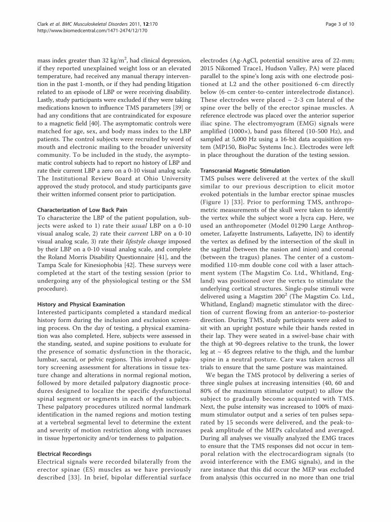
Transcranial Magnetic StimulationTMS pulses were delivered at the vertex of the skullsimilar to our previous description to elicit motorevoked potentials in the lumbar erector spinae muscles(Figure 1) [33]. Prior to performing TMS, anthropo-metric measurements of the skull were taken to identifythe vertex while the subject wore a lycra cap. Here, weused an anthropometer (Model 01290 Large Anthrop-ometer, Lafayette Instruments, Lafayette, IN) to identifythe vertex as defined by the intersection of the skull inthe sagittal (between the nasion and inion) and coronal(between the tragus) planes. The center of a custom-modified 110-mm double cone coil with a laser attach-ment system (The Magstim Co. Ltd., Whitland, Eng-land) was positioned over the vertex to stimulate theunderlying cortical structures. Single-pulse stimuli weredelivered using a Magstim 2002 (The Magstim Co. Ltd.,Whitland, England) magnetic stimulator with the direc-tion of current flowing from an anterior-to-posteriordirection. During TMS, study participants were asked tosit with an upright posture while their hands rested intheir lap. They were seated in a swivel-base chair withthe thigh at 90-degrees relative to the trunk, the lowerleg at ~ 45 degrees relative to the thigh, and the lumbarspine in a neutral posture. Care was taken across alltrials to ensure that the same posture was maintained.We began the TMS protocol by delivering a series of
three single pulses at increasing intensities (40, 60 and80% of the maximum stimulator output) to allow thesubject to gradually become acquainted with TMS.Next, the pulse intensity was increased to 100% of maxi-mum stimulator output and a series of ten pulses sepa-rated by 15 seconds were delivered, and the peak-to-peak amplitude of the MEPs calculated and averaged.During all analyses we visually analyzed the EMG tracesto ensure that the TMS responses did not occur in tem-poral relation with the electrocardiogram signals (toavoid interference with the EMG signals), and in therare instance that this did occur the MEP was excludedfrom analysis (this occurred in no more than one trial
Clark et al. BMC Musculoskeletal Disorders 2011, 12:170http://www.biomedcentral.com/1471-2474/12/170
Page 3 of 10
per a given subject). Following the baseline TMS testingprotocol the stretch reflex testing protocol was per-formed and study participants then received SM. Tenminutes after the study participant received SM theTMS and stretch reflex protocols were then repeated.
Stretch ReflexWhen a muscle is rapidly stretched, a short-latencystretch reflex is elicited due to the excitation of thereceptive endings of the Ia afferent fibers within themuscle spindles [43]. To quantify the erector spinaemuscle stretch reflex responsiveness we determined theEMG activity of the muscle in response to mechanicaltapping as we have previously described (Figure 2) [33].
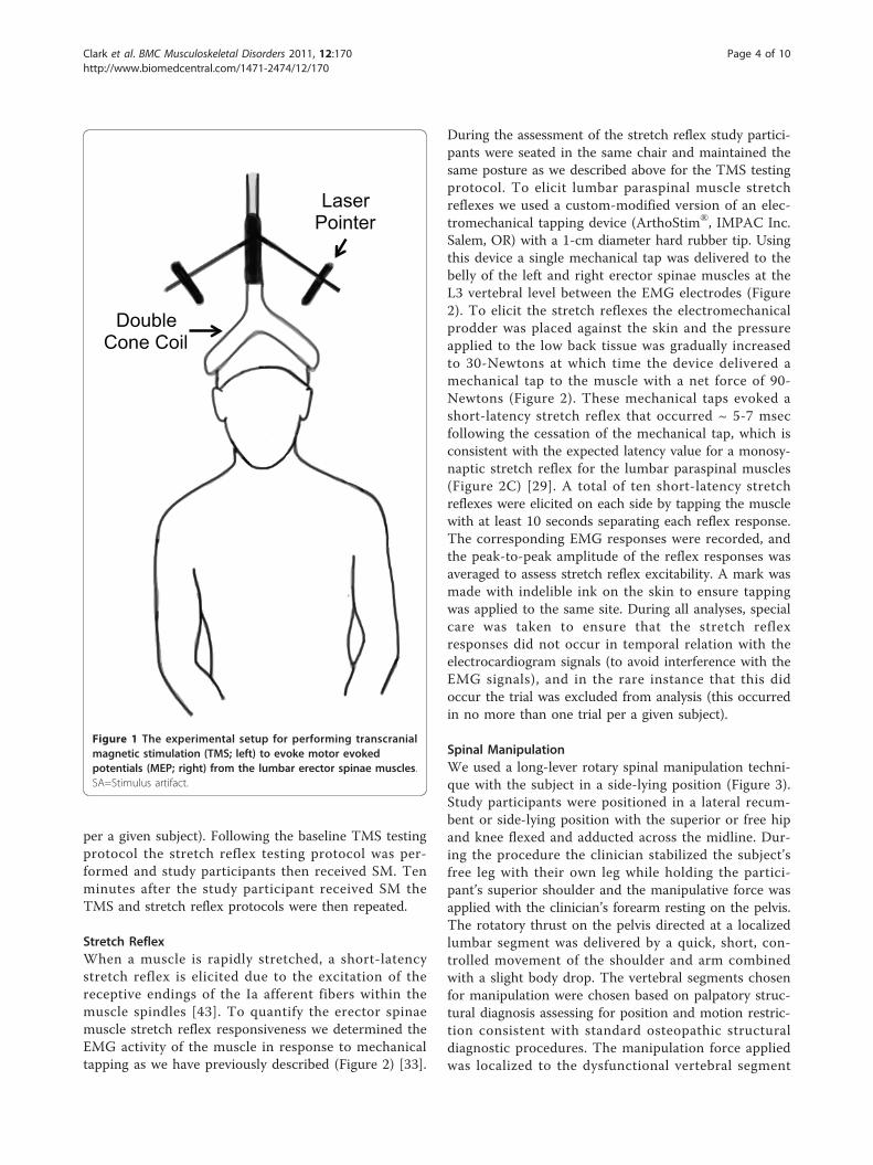
During the assessment of the stretch reflex study partici-pants were seated in the same chair and maintained thesame posture as we described above for the TMS testingprotocol. To elicit lumbar paraspinal muscle stretchreflexes we used a custom-modified version of an elec-tromechanical tapping device (ArthoStim®, IMPAC Inc.Salem, OR) with a 1-cm diameter hard rubber tip. Usingthis device a single mechanical tap was delivered to thebelly of the left and right erector spinae muscles at theL3 vertebral level between the EMG electrodes (Figure2). To elicit the stretch reflexes the electromechanicalprodder was placed against the skin and the pressureapplied to the low back tissue was gradually increasedto 30-Newtons at which time the device delivered amechanical tap to the muscle with a net force of 90-Newtons (Figure 2). These mechanical taps evoked ashort-latency stretch reflex that occurred ~ 5-7 msecfollowing the cessation of the mechanical tap, which isconsistent with the expected latency value for a monosy-naptic stretch reflex for the lumbar paraspinal muscles(Figure 2C) [29]. A total of ten short-latency stretchreflexes were elicited on each side by tapping the musclewith at least 10 seconds separating each reflex response.The corresponding EMG responses were recorded, andthe peak-to-peak amplitude of the reflex responses wasaveraged to assess stretch reflex excitability. A mark wasmade with indelible ink on the skin to ensure tappingwas applied to the same site. During all analyses, specialcare was taken to ensure that the stretch reflexresponses did not occur in temporal relation with theelectrocardiogram signals (to avoid interference with theEMG signals), and in the rare instance that this didoccur the trial was excluded from analysis (this occurredin no more than one trial per a given subject).
Spinal ManipulationWe used a long-lever rotary spinal manipulation techni-que with the subject in a side-lying position (Figure 3).Study participants were positioned in a lateral recum-bent or side-lying position with the superior or free hipand knee flexed and adducted across the midline. Dur-ing the procedure the clinician stabilized the subject’sfree leg with their own leg while holding the partici-pant’s superior shoulder and the manipulative force wasapplied with the clinician’s forearm resting on the pelvis.The rotatory thrust on the pelvis directed at a localizedlumbar segment was delivered by a quick, short, con-trolled movement of the shoulder and arm combinedwith a slight body drop. The vertebral segments chosenfor manipulation were chosen based on palpatory struc-tural diagnosis assessing for position and motion restric-tion consistent with standard osteopathic structuraldiagnostic procedures. The manipulation force appliedwas localized to the dysfunctional vertebral segment
Laser Pointer
Double Cone Coil
Figure 1 The experimental setup for performing transcranialmagnetic stimulation (TMS; left) to evoke motor evokedpotentials (MEP; right) from the lumbar erector spinae muscles.SA=Stimulus artifact.
Clark et al. BMC Musculoskeletal Disorders 2011, 12:170http://www.biomedcentral.com/1471-2474/12/170
Page 4 of 10
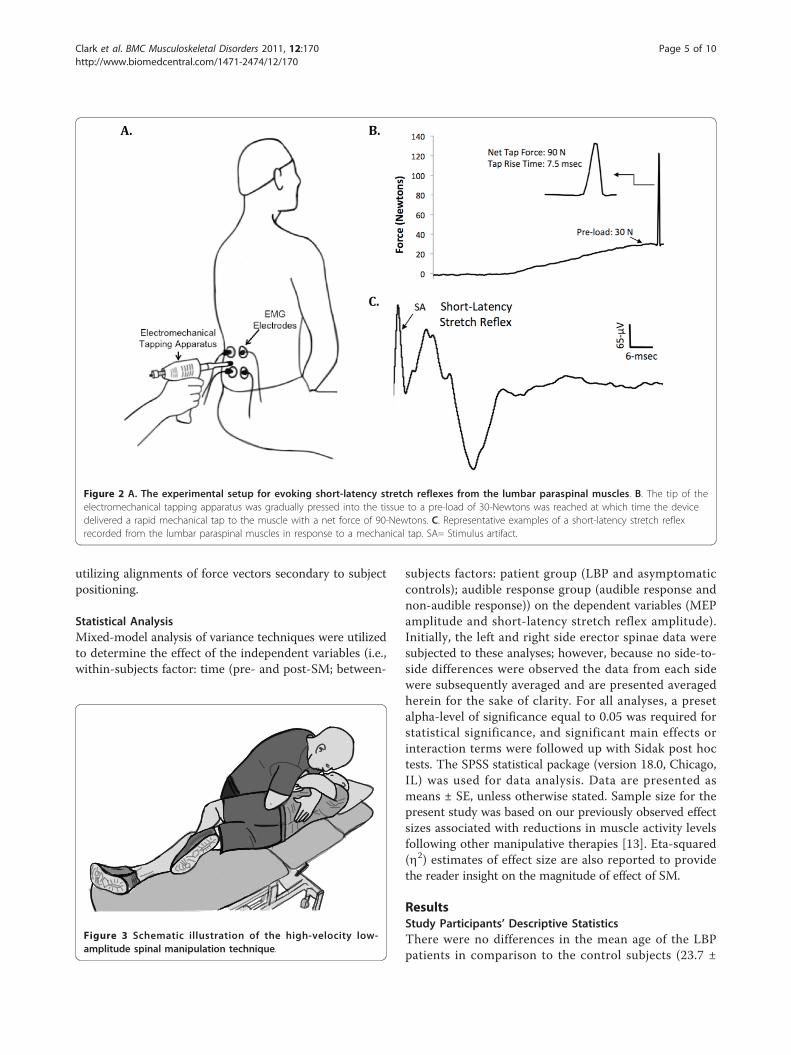
utilizing alignments of force vectors secondary to subjectpositioning.
Statistical AnalysisMixed-model analysis of variance techniques were utilizedto determine the effect of the independent variables (i.e.,within-subjects factor: time (pre- and post-SM; between-
subjects factors: patient group (LBP and asymptomaticcontrols); audible response group (audible response andnon-audible response)) on the dependent variables (MEPamplitude and short-latency stretch reflex amplitude).Initially, the left and right side erector spinae data weresubjected to these analyses; however, because no side-to-side differences were observed the data from each sidewere subsequently averaged and are presented averagedherein for the sake of clarity. For all analyses, a presetalpha-level of significance equal to 0.05 was required forstatistical significance, and significant main effects orinteraction terms were followed up with Sidak post hoctests. The SPSS statistical package (version 18.0, Chicago,IL) was used for data analysis. Data are presented asmeans ± SE, unless otherwise stated. Sample size for thepresent study was based on our previously observed effectsizes associated with reductions in muscle activity levelsfollowing other manipulative therapies [13]. Eta-squared(h2) estimates of effect size are also reported to providethe reader insight on the magnitude of effect of SM.
ResultsStudy Participants’ Descriptive StatisticsThere were no differences in the mean age of the LBPpatients in comparison to the control subjects (23.7 ±
Figure 2 A. The experimental setup for evoking short-latency stretch reflexes from the lumbar paraspinal muscles. B. The tip of theelectromechanical tapping apparatus was gradually pressed into the tissue to a pre-load of 30-Newtons was reached at which time the devicedelivered a rapid mechanical tap to the muscle with a net force of 90-Newtons. C. Representative examples of a short-latency stretch reflexrecorded from the lumbar paraspinal muscles in response to a mechanical tap. SA= Stimulus artifact.
Figure 3 Schematic illustration of the high-velocity low-amplitude spinal manipulation technique.
Clark et al. BMC Musculoskeletal Disorders 2011, 12:170http://www.biomedcentral.com/1471-2474/12/170
Page 5 of 10
6.1 and 22.9 ± 1.9 years; p = 0.73). There were no differ-ences in the mean height of the LBP patients in com-parison to the control subjects (was 171.7 ± 13.2 and174.5 ± 8.4 cm; p = 0.58). There were no differences inthe mean weight of the LBP patients in comparison tothe control subjects (67.9 ± 11.5 and 70.3 ± 11.8 kg; p =0.64). Lastly, there were no differences in the meanbody mass index of the LBP patients in comparison tothe control subjects (23.0 ± 2.3 and 23.0 ± 2.8 kg/m2; p= 0.96). Using a 0-10 visual analog scale, the chronicLBP patients rated their usual LBP as 4.0 ± 1.2, theircurrent LBP as 2.6 ± 1.6, and their lifestyle changeimposed from their LBP as 3.9 ± 3.1. Additionally, theyreported having LBP for a mean duration of 3.2 ± 3.1years, scored 5.9 ± 4.3 on the Roland Morris DisabilityQuestionnaire, [41] and scored 33.5 ± 6.5 on the TampaScale for Kinesiophobia [42].
Effects of spinal manipulation on erector spinae motorevoked potential amplitude in patients with chronic LBPand in asymptomatic controlsSM did not alter the erector spinae MEP amplitude inpatients with LBP (0.80 ± 0.32 to 0.80 ± 0.30 μV) or inasymptomatic controls (0.56 ± 0.09 to 0.57 ± 0.06 μV)(Figure 4; group main effect: p = 0.48, h2 = 0.03; timemain effect: p = 0.61, h2 = 0.02; time × group interac-tion: p = 0.62, h2 = 0.01).
Effects of spinal manipulation on erector spinae short-latency stretch reflex amplitude in patients with chronicLBP and in asymptomatic controlsSM did not alter the erector spinae stretch reflex ampli-tude in patients with LBP (0.66 ± 0.12 to 0.66 ± 0.15μV) or in asymptomatic controls (0.60 ± 0.09 to 0.55 ±0.08 μV) (Figure 4; group main effect: p = 0.41, h2 =0.04; time main effect: p = 0.92, h2 < 0.01; time × groupinteraction: p = 0.90, h2 < 0.01).
Effects of spinal manipulation on erector spinae motorevoked potential and short-latency stretch reflexamplitude in which the spinal manipulation did and didnot produce an audible joint soundEleven study participants exhibited an audible jointsound to SM (5 participants with LBP and 6 controls),whereas nine did not (5 participants with LBP, and 4controls). SM did not alter the erector spinae MEPamplitude in individuals who exhibited an audibleresponse (0.83 ± 0.09 to 0.81 ± 0.08 μV) or in those whodid not (0.50 ± 0.02 to 0.53 ± 0.01 μV) (Figure 5; timemain effect: p = 0.88, h2 < 0.01; time × group interaction:p = 0.58, h2 = 0.02). There were no notable differences inthe magnitude of change in the MEPs between the LBPpatients (2% increase) and the control subjects (7%decrease) among those exhibiting an audible responseduring SM. Interestingly, erector spinae stretch reflexwas reduced 19.2% when SM caused an audible jointsound (0.54 ± 0.02 to 0.43 ± 0.01 μV), whereas when SMdid not cause an audible joint sound there was a 9.7%increase (0.73 ± 0.05 to 0.81 ± 0.05 μV) (Figure 5; time ×group interaction: p = 0.05; h2 = 0.20). There were nonotable differences in the magnitude of change in thestretch reflex between the LBP patients (16% decrease)and the control subjects (22% decrease) among thoseexhibiting an audible response during SM.
DiscussionIt has long-been suggested that SM exerts beneficialeffects by affecting the nervous system [13-22]; however,to date few studies have examined these claims inhumans with LBP. As such, utilizing advanced
-40
-30
-20
-10
0
10
20
30
40
Motor Evoked Potential Stretch Reflex
% C
han
ge
in p
-p A
mp
litu
de
Chronic LBP
Asymptomatic Controls
Figure 4 Spinal manipulation did not alter the amplitude ofthe motor evoked potential (left) or short-latency stretch reflex(right) recorded from the erector spinae muscles in patientswith chronic low back pain (LBP) or in asymptomatic controls.
-40
-30
-20
-10
0
10
20
30
40
Motor Evoked Potential Stretch Reflex
% C
han
ge
in p
-p A
mp
litu
de
Audible Release
No Audible Release
*
Figure 5 Spinal manipulation did not alter the amplitude ofthe motor evoked potential (left) recorded from the erectorspinae muscles in individuals who exhibited an audibleresponse in response to spinal manipulation or in those whodid not. Interestingly, spinal manipulation did reduce the amplitudeof the short-latency stretch reflex in individuals who exhibited anaudible response in response to spinalmanipulation whencompared to those who did not (*time × group interaction: p =0.05; h2 = 0.20).
Clark et al. BMC Musculoskeletal Disorders 2011, 12:170http://www.biomedcentral.com/1471-2474/12/170
Page 6 of 10
neurophysiologic assessment techniques to investigatethe effects of a commonly used– but poorly under-stood– treatment for LBP is particularly innovative. Themost novel findings of the present study are: i) that asingle spinal manipulation does not systematically altercorticospinal or the short-latency stretch reflex excitabil-ity of the erector spinae muscles in patients withchronic LBP or asymptomatic controls (at least whenassessed ~ 10-min following manipulation), and ii) thatonly when spinal manipulation induces an audible jointsound the erector spine short-latency stretch reflex isattenuated. Below we will discuss these findings in thecontext of understanding the physiological effects ofspinal manipulation.A recent review of chronic LBP provides evidence for
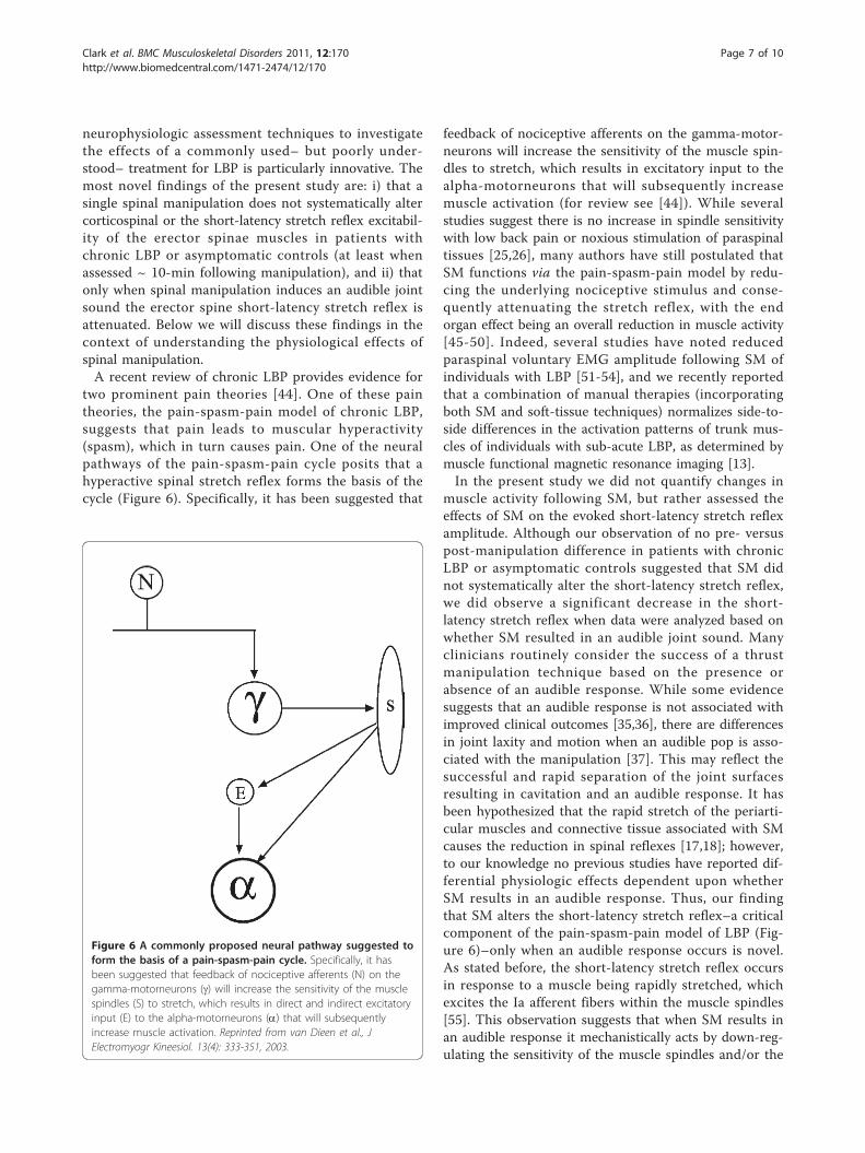
two prominent pain theories [44]. One of these paintheories, the pain-spasm-pain model of chronic LBP,suggests that pain leads to muscular hyperactivity(spasm), which in turn causes pain. One of the neuralpathways of the pain-spasm-pain cycle posits that ahyperactive spinal stretch reflex forms the basis of thecycle (Figure 6). Specifically, it has been suggested that
feedback of nociceptive afferents on the gamma-motor-neurons will increase the sensitivity of the muscle spin-dles to stretch, which results in excitatory input to thealpha-motorneurons that will subsequently increasemuscle activation (for review see [44]). While severalstudies suggest there is no increase in spindle sensitivitywith low back pain or noxious stimulation of paraspinaltissues [25,26], many authors have still postulated thatSM functions via the pain-spasm-pain model by redu-cing the underlying nociceptive stimulus and conse-quently attenuating the stretch reflex, with the endorgan effect being an overall reduction in muscle activity[45-50]. Indeed, several studies have noted reducedparaspinal voluntary EMG amplitude following SM ofindividuals with LBP [51-54], and we recently reportedthat a combination of manual therapies (incorporatingboth SM and soft-tissue techniques) normalizes side-to-side differences in the activation patterns of trunk mus-cles of individuals with sub-acute LBP, as determined bymuscle functional magnetic resonance imaging [13].In the present study we did not quantify changes in
muscle activity following SM, but rather assessed theeffects of SM on the evoked short-latency stretch reflexamplitude. Although our observation of no pre- versuspost-manipulation difference in patients with chronicLBP or asymptomatic controls suggested that SM didnot systematically alter the short-latency stretch reflex,we did observe a significant decrease in the short-latency stretch reflex when data were analyzed based onwhether SM resulted in an audible joint sound. Manyclinicians routinely consider the success of a thrustmanipulation technique based on the presence orabsence of an audible response. While some evidencesuggests that an audible response is not associated withimproved clinical outcomes [35,36], there are differencesin joint laxity and motion when an audible pop is asso-ciated with the manipulation [37]. This may reflect thesuccessful and rapid separation of the joint surfacesresulting in cavitation and an audible response. It hasbeen hypothesized that the rapid stretch of the periarti-cular muscles and connective tissue associated with SMcauses the reduction in spinal reflexes [17,18]; however,to our knowledge no previous studies have reported dif-ferential physiologic effects dependent upon whetherSM results in an audible response. Thus, our findingthat SM alters the short-latency stretch reflex–a criticalcomponent of the pain-spasm-pain model of LBP (Fig-ure 6)–only when an audible response occurs is novel.As stated before, the short-latency stretch reflex occursin response to a muscle being rapidly stretched, whichexcites the Ia afferent fibers within the muscle spindles[55]. This observation suggests that when SM results inan audible response it mechanistically acts by down-reg-ulating the sensitivity of the muscle spindles and/or the
Figure 6 A commonly proposed neural pathway suggested toform the basis of a pain-spasm-pain cycle. Specifically, it hasbeen suggested that feedback of nociceptive afferents (N) on thegamma-motorneurons (g) will increase the sensitivity of the musclespindles (S) to stretch, which results in direct and indirect excitatoryinput (E) to the alpha-motorneurons (a) that will subsequentlyincrease muscle activation. Reprinted from van Dieen et al., JElectromyogr Kineesiol. 13(4): 333-351, 2003.
Clark et al. BMC Musculoskeletal Disorders 2011, 12:170http://www.biomedcentral.com/1471-2474/12/170
Page 7 of 10
various other segmental sites of the Ia stretch reflexpathway. It is also possible that the change in reflexactivity associated with subjects having an audiblerelease during SM may relate to gapping in the jointsurfaces, as it was recently shown that vertebral seg-ments that cavitated during SM gapped (separated)more than those that did not [38]. This greater jointgapping could result in the break-up of small adhesionspresent even in normal joints, or due to increased mus-cle or connective tissue tension surrounding thosejoints, before SM. Consequently, SM that results in anaudible response may conceivably function to restoregreater motion to a vertebral segment (as opposed toSM that does not result in an audible response), andthis biomechanical effect could result in subsequentchanges in reflex activity as we observed.We did not observe changes in MEP amplitude follow-
ing SM in patients with LBP, asymptomatic controls, orwhen data were grouped according to whether an audibleresponse was observed. When a single pulse transcranialmagnetic stimulation stimuli is applied to the motor cor-tex at an intensity above motor threshold, high-frequencyindirect waves (I waves) are elicited in the corticospinaltract [56], which are modifiable by many mechanisms (i.e., glutatmate, GABA, acetylcholine, etc.) [39] that influ-ence the amplitude of the MEP. Thus, our finding of nochange in the MEP indicates that a single SM treatmentin patients with chronic LBP does not alter global excit-ability of the corticospinal tract, at least when assessed ~10-min following the manipulative intervention. To dateonly one other study has examined the effects of SM oncorticospinal excitability of the low back muscles usingtranscranial magnetic stimulation [15]. In this studyDishman and colleagues examined the effects of a singleSM treatment on MEP amplitude in asymptomatic youngadults, and observed a transient increase in the MEP fol-lowing SM. The MEP facilitation was short-lived how-ever– as MEP amplitude was increased 10-secs followingSM but had returned to baseline levels less than 20-sec-onds after SM. Thus, in the present we would havemissed any short-term, transient effects that occurred asa result of SM. Additionally, in our work as well as thatconducted by Dishman et al. it is possible that segmentalchanges in the nervous systems excitability (e.g., corticallevel changes) may have been confounded by no changein or opposite changes in excitability at a different seg-mental level (e.g., spinal level changes) as the MEP ampli-tude elicited using single-pulse transcranial magneticstimulation can be influenced at both the cortical andspinal levels. To more fully explore the effects of SM oncortico-cortical excitability it is suggested that futureinvestigations utilize paired-pulse transcranial magneticstimulation to measure intracortical facilitation andinhibition.
There are several limitations of the present study thatshould be mentioned. First, it should be noted that thepresent work was conducted in patients with mild-to-moderate chronic LBP and asymptomatic controls, andthat these individuals only received a single high-velocitylow-amplitude SM thrust with outcome measuresassessed shortly after the manipulative treatment. Assuch, it is possible that i) SM may result in differentphysiologic responses in other populations (e.g., sub-acute LBP), ii) that a course of SM treatment may havea more pronounced effect (e.g., three weeks of SM twiceper week), and/or iii) that greater or lesser effects mayhave been observed at various time points followingmanipulation. Additionally, we chose to study chronicLBP patients (as opposed to acute or sub-acute LBPpatients) due to the staggering economic costs that areassociated with chronic LBP and the fact that manypatients with chronic LBP seek manipulation therapy asa treatment option for their LBP [5]. However, it is pos-sible that the neurophysiologic responses may be differ-ent if other groups of LBP patients had been studied aspatients with LBP symptom duration for < 16 days arereported to be more likely to respond favorably to SM[57]. Further, we cannot rule out the potential for a pla-cebo effect to influence our observed reduction in thestretch reflex in individuals who exhibited an audibleresponse to SM. However, with this stated, it seemsunlikely that this finding was driven by a placebo effectas one would likely expect to observe a concomitantchange in corticospinal excitability– as a placebo effectwould likely be assumed to have systemic effects (asopposed to having a local, selective effect on the stretchreflex only).
ConclusionIn summary, this study examined the effects of spinalmanipulation on the motor evoked potential and short-latency stretch reflex amplitudes of the erector spinaemuscles in patients with chronic low back pain andasymptomatic controls. We did not observe changes inthese outcomes in either group when assessed ~ 10-minutes following a single spinal manipulative thrust.Interestingly, when data were analyzed according towhether spinal manipulation caused an audible jointsound, regardless of patient group, we observed thatstudy participants exhibiting an audible response exhib-ited a significant reduction in the short-latency stretchreflex. These findings suggest that a single SM treatmentdoes not systematically alter corticospinal or stretchreflex excitability of the erector spinae muscles; how-ever, they do indicate that the stretch reflex is attenu-ated when spinal manipulation causes an audible jointsound. This finding provides insight into the mechanism(s) of action of spinal manipulation, and suggests that
Clark et al. BMC Musculoskeletal Disorders 2011, 12:170http://www.biomedcentral.com/1471-2474/12/170
Page 8 of 10
spinal manipulation may mechanistically act by downregulating the gain of the muscle spindles and/or thevarious segmental sites of the Ia reflex pathway. Devel-oping a better understanding of the physiologic effectsof various manual therapies to treat low back pain willin the long-term assist in optimizing and developingstrategic treatment strategies for specific patient popula-tions with LBP.
AcknowledgementsThis work was funded in part by a grant from the OsteopathicHeritage Foundations to BC Clark. A Research and Scholarly AdvancementFellowship from Ohio University supported DJ Goss’ work on this project.
Author details1Ohio Musculoskeletal and Neurological Institute (OMNI), Ohio University,236 Irvine Hall, Athens, OH 45701, USA. 2Department of Biomedical Sciences,Ohio University Heritage College of Osteopathic Medicine, 228 Irvine Hall,Athens, OH 45701, USA. 3Department of Family Medicine, Ohio UniversityHeritage College of Osteopathic Medicine, Grosvenor Hall, Athens, OH45701, USA. 4School of Rehabilitation and Communication Sciences, GroverCenter, Ohio University, Athens, OH 45701 USA.
Authors’ contributionsBC participated in the conception and design of the study, supervised thedata collection, helped secure grant funding, and drafted the manuscript.DG, RH, and AR recruited and screened subjects and were actively involvedin all data collection procedures. DG and SW performed the spinalmanipulative procedures. JT participated in the conception and design ofthe study, supervised the data collection, and helped secure grant funding.All authors read and approved the final manuscript.
Competing interestsThe authors declare that they have no competing interests.
Received: 31 March 2011 Accepted: 22 July 2011Published: 22 July 2011
References1. Andersson GB: Epidemiological features of chronic low-back pain. Lancet
1999, 354(9178):581-585.2. Carey TS, Garrett JM, Jackman AM: Beyond the good prognosis.
Examination of an inception cohort of patients with chronic low backpain. Spine (Phila Pa 1976) 2000, 25(1):115-120.
3. Klenerman L, Slade PD, Stanley IM, Pennie B, Reilly JP, Atchison LE,Troup JD, Rose MJ: The prediction of chronicity in patients with an acuteattack of low back pain in a general practice setting. Spine (Phila Pa1976) 1995, 20(4):478-484.
4. Von Korff M: Studying the natural history of back pain. Spine (Phila Pa1976) 1994, 19(18 Suppl):2041S-2046S.
5. Nahin RL, Barnes PM, Stussman BJ, Bloom B: Costs of complementary andalternative medicine (CAM) and frequency of visits to CAM practitioners:United States, 2007. Natl Health Stat Report 2009, , 18: 1-14.
6. Haas M, Groupp E, Kraemer DF: Dose-response for chiropractic care ofchronic low back pain. Spine J 2004, 4(5):574-583.
7. Hoiriis KT, Pfleger B, McDuffie FC, Cotsonis G, Elsangak O, Hinson R,Verzosa GT: A randomized clinical trial comparing chiropracticadjustments to muscle relaxants for subacute low back pain. JManipulative Physiol Ther 2004, 27(6):388-398.
8. Hondras MA, Long CR, Cao Y, Rowell RM, Meeker WC: A randomizedcontrolled trial comparing 2 types of spinal manipulation and minimalconservative medical care for adults 55 years and older with subacuteor chronic low back pain. J Manipulative Physiol Ther 2009, 32(5):330-343.
9. Licciardone JC, Brimhall AK, King LN: Osteopathic manipulative treatmentfor low back pain: a systematic review and meta-analysis of randomizedcontrolled trials. BMC Musculoskelet Disord 2005, 6:43.
10. Licciardone JC, Stoll ST, Fulda KG, Russo DP, Siu J, Winn W, Swift J Jr:Osteopathic manipulative treatment for chronic low back pain: arandomized controlled trial. Spine (Phila Pa 1976) 2003, 28(13):1355-1362.
11. Hurwitz EL, Morgenstern H, Harber P, Kominski GF, Belin TR, Yu F,Adams AH: A randomized trial of medical care with and without physicaltherapy and chiropractic care with and without physical modalities forpatients with low back pain: 6-month follow-up outcomes from theUCLA low back pain study. Spine (Phila Pa 1976) 2002, 27(20):2193-2204.
12. MacDonald RS, Bell CM: An open controlled assessment of osteopathicmanipulation in nonspecific low-back pain. Spine (Phila Pa 1976) 1990,15(5):364-370.
13. Clark BC, Walkowski S, Conatser RR, Eland DC, Howell JN: Muscle functionalmagnetic resonance imaging and acute low back pain: a pilot study tocharacterize lumbar muscle activity asymmetries and examine theeffects of osteopathic manipulative treatment. Osteopath Med Prim Care2009, 3:7.
14. Cote P, Mior SA, Vernon H: The short-term effect of a spinal manipulationon pain/pressure threshold in patients with chronic mechanical lowback pain. J Manipulative Physiol Ther 1994, 17(6):364-368.
15. Dishman JD, Greco DS, Burke JR: Motor-evoked potentials recorded fromlumbar erector spinae muscles: a study of corticospinal excitabilitychanges associated with spinal manipulation. J Manipulative Physiol Ther2008, 31(4):258-270.
16. Johansson H, Sojka P: Pathophysiological mechanisms involved ingenesis and spread of muscular tension in occupational muscle painand in chronic musculoskeletal pain syndromes: a hypothesis. MedHypotheses 1991, 35(3):196-203.
17. Knutson GA: The role of the gamma-motor system in increasing muscletone and muscle pain syndromes: a review of the Johansson/Sojkahypothesis. J Manipulative Physiol Ther 2000, 23(8):564-572.
18. Korr IM: Proprioceptors and somatic dysfunction. J Am Osteopath Assoc1975, 74(7):638-650.
19. Pickar JG: Neurophysiological effects of spinal manipulation. Spine J 2002,2(5):357-371.
20. Sung PS, Kang YM, Pickar JG: Effect of spinal manipulation duration onlow threshold mechanoreceptors in lumbar paraspinal muscles: apreliminary report. Spine (Phila Pa 1976) 2005, 30(1):115-122.
21. Terrett AC, Vernon H: Manipulation and pain tolerance. A controlledstudy of the effect of spinal manipulation on paraspinal cutaneous paintolerance levels. Am J Phys Med 1984, 63(5):217-225.
22. Vernon HT, Aker P, Burns S, Viljakaanen S, Short L: Pressure pain thresholdevaluation of the effect of spinal manipulation in the treatment ofchronic neck pain: a pilot study. J Manipulative Physiol Ther 1990,13(1):13-16.
23. Travell JG, Simons DG: Myofascial pain and dysfunction: the trigger pointmanual. Baltimore: Willams and Wilkins; 1983.
24. Matre DA, Sinkjaer T, Svensson P, Arendt-Nielsen L: Experimental musclepain increases the human stretch reflex. Pain 1998, 75(2-3):331-339.
25. Kang YM, Wheeler JD, Pickar JG: Stimulation of chemosensitive afferentsfrom multifidus muscle does not sensitize multifidus muscle spindles tovertebral loads in the lumbar spine of the cat. Spine 2001,26(14):1528-1536.
26. Zedka M, Prochazka A, Knight B, Gillard D, Gauthier M: Voluntary and reflexcontrol of human back muscles during induced pain. J Physiol 1999,520(Pt 2):591-604.
27. Hjortskov N, Essendrop M, Skotte J, Fallentin N: The effect of delayed-onset muscle soreness on stretch reflexes in human low back muscles.Scand J Med Sci Sports 2005, 15(6):409-415.
28. Kugelberg E, Hagbarth KE: Spinal mechanism of the abdominal anderector spinae skin reflexes. Brain 1958, 81(3):290-304.
29. Skotte J, Hjortskov N, Essendrop M, Schibye B, Fallentin N: Short latencystretch reflex in human lumbar paraspinal muscles. J Neurosci Methods2005, 145(1-2):145-150.
30. Dimitrijevic MR, Gregoric MR, Sherwood AM, Spencer WA: Reflex responsesof paraspinal muscles to tapping. J Neurol Neurosurg Psychiatry 1980,43(12):1112-1118.
31. Beeton KS: Manual therapy classes: the vertebreal column. EdinburghLondon New York: Churchhill Lingston; 2003.
32. Christensen MG, Kollasch MW: Job analysis of chiropractic 2005: a projectreport, survey analysis, and summary of the practice of chriopractic care
Clark et al. BMC Musculoskeletal Disorders 2011, 12:170http://www.biomedcentral.com/1471-2474/12/170
Page 9 of 10
in the United States. Greeley, CO: National Board of ChiropracticExaminers; 2005.
33. Goss DA, Thomas JS, Clark BC: Novel methods for quantifyingneurophysiologic properties of the human lumbar paraspinal muscles. JNeurosci Methods .
34. Strutton PH, Theodorou S, Catley M, McGregor AH, Davey NJ: Corticospinalexcitability in patients with chronic low back pain. J Spinal Disord Tech2005, 18(5):420-424.
35. Cleland JA, Flynn TW, Childs JD, Eberhart S: The audible pop from thoracicspine thrust manipulation and its relation to short-term outcomes inpatients with neck pain. J Man Manip Ther 2007, 15(3):143-154.
36. Flynn TW, Fritz JM, Wainner RS, Whitman JM: The audible pop is notnecessary for successful spinal high-velocity thrust manipulation inindividuals with low back pain. Arch Phys Med Rehabil 2003,84(7):1057-1060.
37. Brodeur R: The audible release associated with joint manipulation. JManipulative Physiol Ther 1995, 18(3):155-164.
38. Cramer GD, Ross K, Pocius J, Cantu JA, Laptook E, Fergus M, Gregerson D,Selby S, Raju PK: Evaluating the relationship among cavitation,zygapophyseal joint gapping, and spinal manipulation: an exploratorycase series. Journal of manipulative and physiological therapeutics 2011,34(1):2-14.
39. Ziemann U: TMS and drugs. Clin Neurophysiol 2004, 115(8):1717-1729.40. [http://www.mrisafety.com].41. Roland M, Morris R: A study of the natural history of back pain. Part I:
development of a reliable and sensitive measure of disability in low-back pain. Spine (Phila Pa 1976) 1983, 8(2):141-144.
42. Kori KS, Miller RP, Todd DD: Kinesiophobia: A new view of chronicbehavior. Pain Management 1990, 35-43.
43. Schuurmans J, de Vlugt E, Schouten AC, Meskers CG, de Groot JH, van derHelm FC: The monosynaptic Ia afferent pathway can largely explain thestretch duration effect of the long latency M2 response. Exp Brain Res2009, 193(4):491-500.
44. van Dieen JH, Selen LP, Cholewicki J: Trunk muscle activation in low-backpain patients, an analysis of the literature. J Electromyogr Kinesiol 2003,13(4):333-351.
45. Gillette RG: A speculative argument for the coactivation of diversesomatic receptor populations by forceful chiropractic adjustments.Manual Med 1987, 3:1-14.
46. Haldeman S: Spinal manipulative therapy in sports medicine. Clin SportsMed 1986, 5:277-293.
47. Herzog W: Clinical Biomechanics of Spinal Manipulation. New York:Churchill Livingstone; 2000.
48. Raftis K: Spinal manipulation for back pain. Hosp Pract 1989, 15:95-108.49. Reinert OC: Fundamentals of chriopractic techniques. Chesterfield, MO:
Marian Press; 1983.50. Zusman M: Spinal manipulative therapy: review of some proposed
mechanisms, and a new hypothesis. Aust J Physiother 1986, 32:89-99.51. DeVocht JW, Pickar JG, Wilder DG: Spinal manipulation alters
electromyographic activity of paraspinal muscles: a descriptive study. JManipulative Physiol Ther 2005, 28(7):465-471.
52. Ellestad SM, Nagle RV, Boesler DR, Kilmore MA: Electromyographic andskin resistance responses to osteopathic manipulative treatment forlow-back pain. J Am Osteopath Assoc 1988, 88(8):991-997.
53. Krekoukias G, Petty NJ, Cheek L: Comparison of surface electromyographicactivity of erector spinae before and after the application of centralposteroanterior mobilisation on the lumbar spine. J Electromyogr Kinesiol2009, 19(1):39-45.
54. Lehman GJ, McGill SM: Spinal manipulation causes variable spinekinematic and trunk muscle electromyographic responses. Clin Biomech(Bristol, Avon) 2001, 16(4):293-299.
55. Liddell EGT, Sherrington CS: Reflexes in response to stretch (myotaticreflexes). Proc Roy Soc 1924, 96B:212-242.
56. Di Lazzaro V, Oliviero A, Pilato F, Saturno E, Dileone M, Mazzone P, Insola A,Tonali PA, Rothwell JC: The physiological basis of transcranial motorcortex stimulation in conscious humans. Clin Neurophysiol 2004,115(2):255-266.
57. Childs JD, Fritz JM, Flynn TW, Irrgang JJ, Johnson KK, Majkowski GR,Delitto A: A clinical prediction rule to identify patients with low backpain most likely to benefit from spinal manipulation: a validation study.Ann Intern Med 2004, 141(12):920-928.
Pre-publication historyThe pre-publication history for this paper can be accessed here:http://www.biomedcentral.com/1471-2474/12/170/prepub
doi:10.1186/1471-2474-12-170Cite this article as: Clark et al.: Neurophysiologic effects of spinalmanipulation in patients with chronic low back pain. BMCMusculoskeletal Disorders 2011 12:170.
Submit your next manuscript to BioMed Centraland take full advantage of:
• Convenient online submission
• Thorough peer review
• No space constraints or color figure charges
• Immediate publication on acceptance
• Inclusion in PubMed, CAS, Scopus and Google Scholar
• Research which is freely available for redistribution
Submit your manuscript at www.biomedcentral.com/submit
Clark et al. BMC Musculoskeletal Disorders 2011, 12:170http://www.biomedcentral.com/1471-2474/12/170
Page 10 of 10