Embed Size (px)
Citation preview
Neurosurgery Resident Manual 2008-2009
- 2 -
The Department of Neurosurgery Resident Manual should be used in conjunction with the Louisiana State University Health Sciences Center - Shreveport (LSUHSC-S) GME House Staff Manual. The LSUHSC-S GME House Staff Manual should be reviewed for an explanation of benefits, policies, and additional information. This manual is updated annually and distributed to all residents. It is posted and intermittently updated on the GME website at http://www.sh.lsuhsc.edu/gme/manuals.htm The Department of Neurosurgery Resident Manual should also be used in conjunction with the resident information and self study links available on the resident server as well as with the ACGME information regarding Program Requirements, Institutional Requirements, Case Logs, and other pertinent resident education information. The ACGME is located at www.ACGME.org
- 3 -
TABLE OF CONTENTS
SECTION I: Department of Neurosurgery Full Time Faculty of the Department of Neurosurgery ………. 4 Contact Information ………. 5 Maps to Louisiana State University Health Sciences Center and Affiliated Hospitals ………. 6 SECTION II: Resident Education Neurosurgery Residency Goals and Objectives ………. 10 Resident Experience in Stereotactic Radiosurgery and Endovascular Procedures ………. 65 SECTION III: Policies & Procedures Residency Education Committee and Curriculum Committee ………. 65 Resident Curriculum for Neurosurgery ………. 65 Supervision of Residents General ………. 66 LSUHSC-S Medical Staff Resident Supervision Guidelines ………. 68 Supervision of Residents Specific Activities ………. 68 Moonlighting ………. 69 Resident Stress and Fatigue ………. 69 Duty Hours ………. 70 Call Schedules ………. 71 Vacation ………. 71 On-Call Quarters ………. 71 Offices ………. 72 Parking ………. 73 Meal Program ………. 73 Additional Information ………. 73 SECTION IV: Evaluations Evaluation and Promotion ………. 75 Resident Evaluation of Faculty ………. 78 Resident Evaluation of Program ………. 78 Resident self Assessment and Self Reflection ………. 79 Block Rotational Diagram ……….. 80 LSUHSC-S Department of Neurosurgery Conferences ……….. 81

- 4 -
Anil Nanda, MD, FACS Professor and Chairman of the
Department of Neurosurgery/Neurosurgeon
Brian K. Willis MD, FACS
Professor/Neurosurgeon
Donald R. Smith, MD
Clinical Professor/Neurosurgeon
Bharat
Guthikonda, MD Assistant Professor/Neurosurgeon
Anthony Sin, MD
Assistant Professor/Neurosurgeon
Michael D. Williams, MD
Assistant Professor/Interventional Neuroradiologist
Prasad SSV.
Vannemreddy, MD Assistant Professor of Research
Guohong Li, MD Assistant Professor - Neurosurgery
& Physiology
Debi Mukerjhee, Sc.D Associate Professor – Orthopedics & Neurosurgery
- 5 -
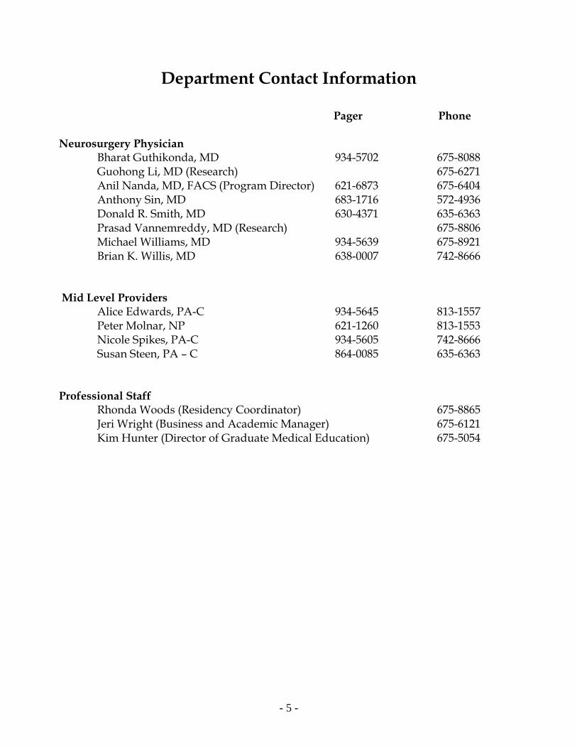
Department Contact Information
Pager Phone
Neurosurgery Physician Bharat Guthikonda, MD 934-5702 675-8088 Guohong Li, MD (Research) 675-6271 Anil Nanda, MD, FACS (Program Director) 621-6873 675-6404 Anthony Sin, MD 683-1716 572-4936 Donald R. Smith, MD 630-4371 635-6363 Prasad Vannemreddy, MD (Research) 675-8806 Michael Williams, MD 934-5639 675-8921 Brian K. Willis, MD 638-0007 742-8666
Mid Level Providers Alice Edwards, PA-C 934-5645 813-1557 Peter Molnar, NP 621-1260 813-1553 Nicole Spikes, PA-C 934-5605 742-8666 Susan Steen, PA – C 864-0085 635-6363
Professional Staff Rhonda Woods (Residency Coordinator) 675-8865 Jeri Wright (Business and Academic Manager) 675-6121 Kim Hunter (Director of Graduate Medical Education) 675-5054
- 6 -
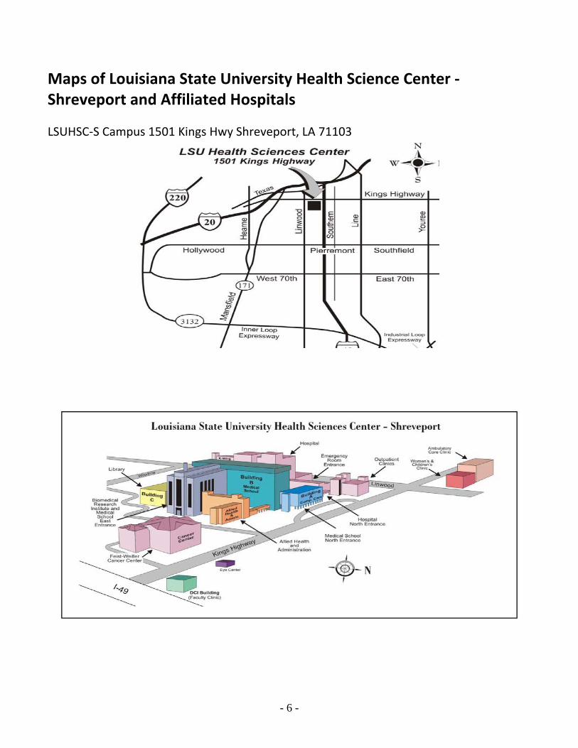
Maps of Louisiana State University Health Science Center ‐Shreveport and Affiliated Hospitals LSUHSC‐S Campus 1501 Kings Hwy Shreveport, LA 71103
7
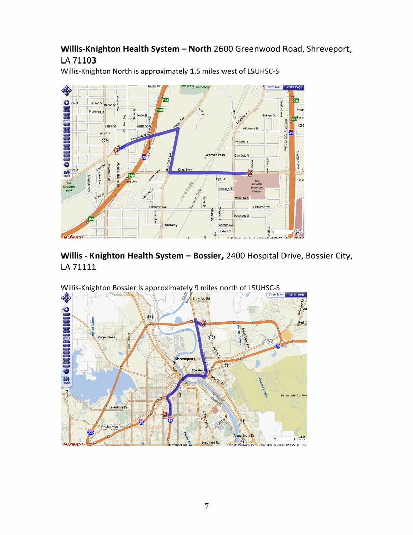
Willis‐Knighton Health System – North 2600 Greenwood Road, Shreveport, LA 71103 Willis‐Knighton North is approximately 1.5 miles west of LSUHSC‐S
Willis ‐ Knighton Health System – Bossier, 2400 Hospital Drive, Bossier City, LA 71111 Willis‐Knighton Bossier is approximately 9 miles north of LSUHSC‐S
8
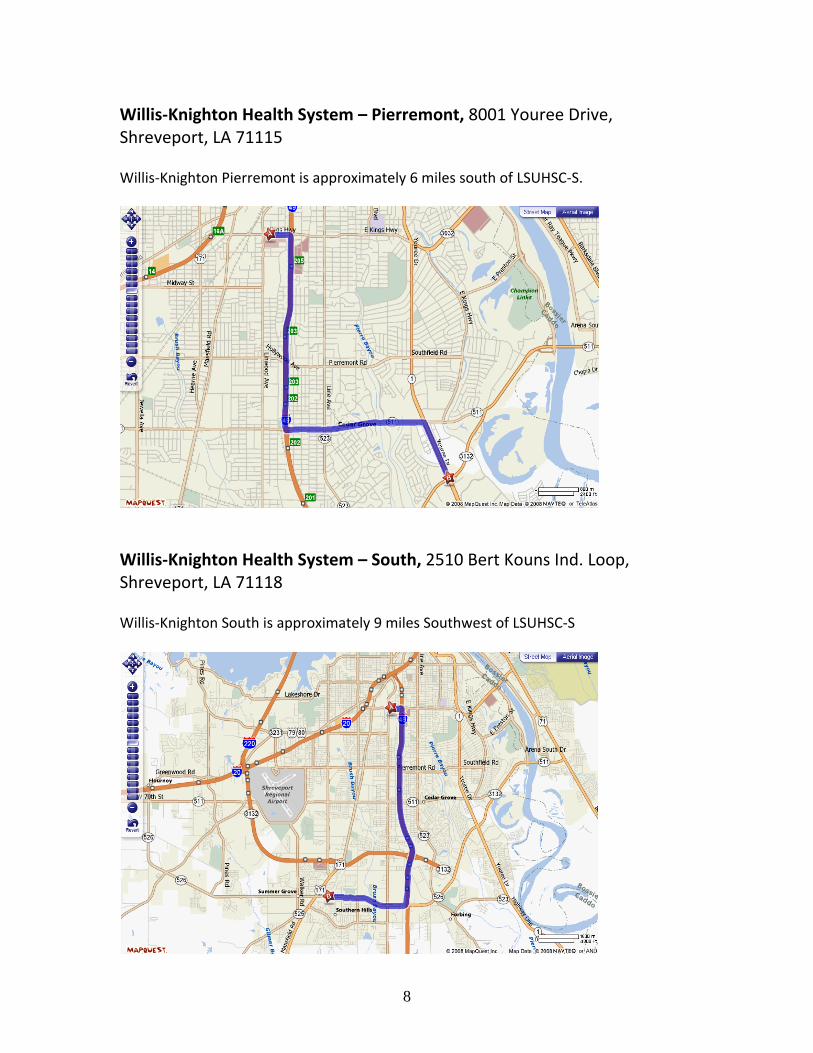
Willis‐Knighton Health System – Pierremont, 8001 Youree Drive, Shreveport, LA 71115 Willis‐Knighton Pierremont is approximately 6 miles south of LSUHSC‐S.
Willis‐Knighton Health System – South, 2510 Bert Kouns Ind. Loop, Shreveport, LA 71118 Willis‐Knighton South is approximately 9 miles Southwest of LSUHSC‐S
9
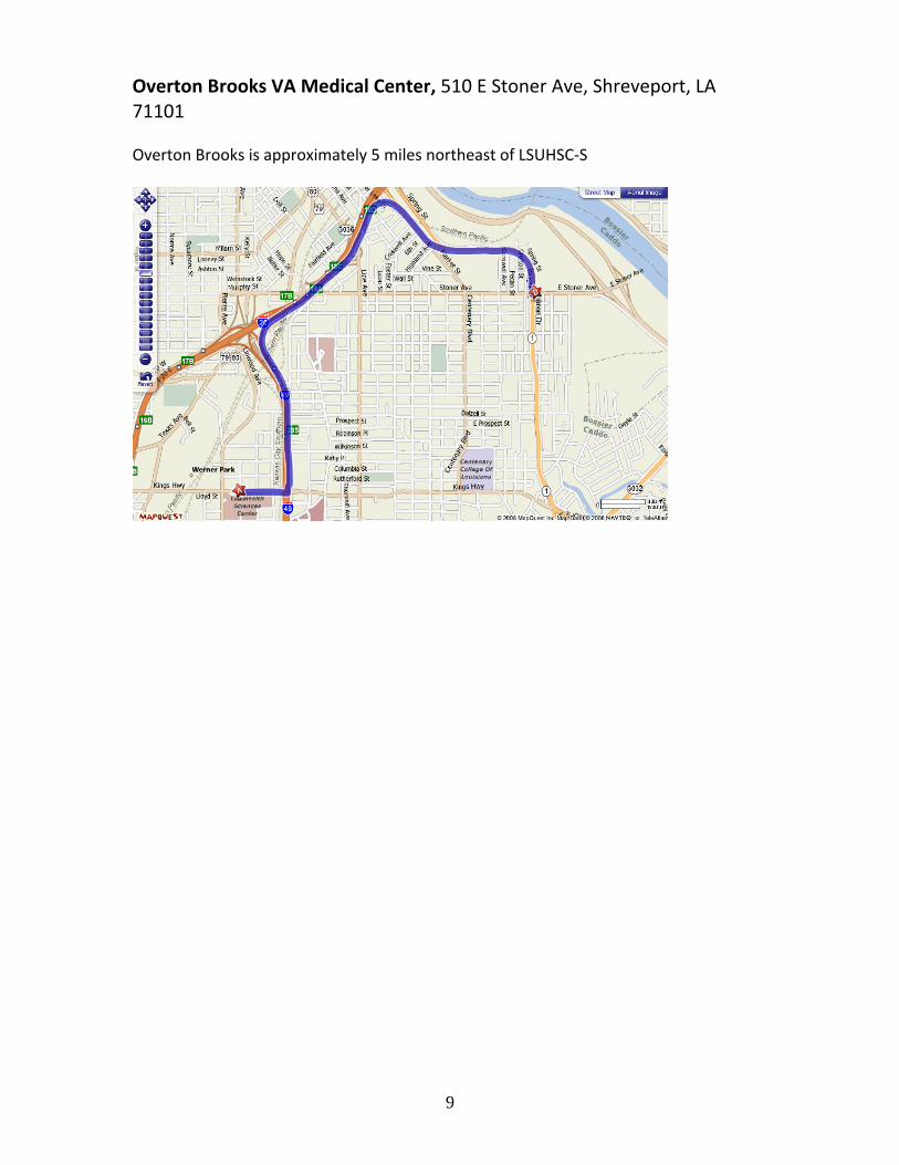
Overton Brooks VA Medical Center, 510 E Stoner Ave, Shreveport, LA 71101 Overton Brooks is approximately 5 miles northeast of LSUHSC‐S
10
SECTION II: Resident Education NEUROSURGERY RESIDENCY EDUCATIONAL GOALS AND OBJECTIVES The Neurosurgery Residency at the Louisiana State University Health Sciences Center (LSUHSC-S) includes services at Louisiana State University Health Sciences Center, The Veteran’s Administration Medical Center (VA), and Willis Knighton Health System (WK). The seven-year program consists of one fundamental clinical year as determined by the Program Director of the Department of Neurosurgery to include surgical experience and other experiences to prepare for neurosurgical training, four years of adult / pediatric neurosurgical training, one year of research, and at least one year as Chief resident to allow for acquisition of graduated experience in all aspects of neurological surgery. The Chief year is a mandatory twelve months split between the University, Willis-Knighton Health Care System, and Veteran’s Administration Mecial Center service. Neuro-critical care experience is emphasized throughout the training, and there is extensive exposure to subspecialty services including neuroendovascular, neurovascular, neuro-oncology, spinal neurosurgery, stereotactic radiosurgery, neurotrauma and pediatric neurosurgery. All residents beyond the PGY 1 year are expected to produce an average of two published manuscripts per year. Preparation of scientific manuscripts, review articles, book chapters and abstracts, along with presentation skills are fostered with multi-disciplinary input and mentorship. Leadership and administrative skills are learned as residents progress in their training and are honed during the chief year. Conferences include rotations of morbidity and mortality conference, board training conference, editorial conference, subspecialty training, journal club, Chairman’s complication conference, professional development, Grand Rounds, the annual Mary Louise and Ben Levy Endowed Visiting Professorship, weekly multi-disciplinary case conference, and stroke, spine, and nursing symposiums.
11
Goals
PGY 1 (NS 0) Successfully complete rotations in general surgery, neurology, neuropathology, neurosurgery, and neuroradiology. Coordinate with chairman to formulate a research project. Become knowledgeable in the physiology of pre and post operative care and develop a knowledge base in surgical disease pertaining to various organ systems. On-call as required by the rotations in compliance with regulations. Show evidence of learning and undertaking progressively responsible patient management. May take the ABNS written boards for practice.
PGY 2 (NS 1) Become comfortable with minor neurosurgical procedures,
specifically lumbar puncture, external ventricular drain placement, and burr hole placement. Develop a knowledge base in basic and clinical neurosurgery, including a thorough understanding of basic neurological examinations for spinal cord injury, the criteria for brain death determination, the anatomy of the cranial vault, leptomeninges, subarachnoid space, and major cerebral blood vessels, and the anatomy, physiology, and pathology of cranial nerves and spinal cord. Learn fundamentals of head trauma, spinal cord injury and critical care management. Become proficient in basic post-operative neurosurgical management. Become comfortable with placement of head-frames for stereotactic radiosurgery procedures and learn basic radiosurgery planning. On call responsibilites will be limited to three months of night float during the entire year and no more than two weekend calls per month during all other months. Discuss a preliminary individual learning plan with the Chairman. Publish two papers. Teach medical students in lecture and on daily rounds. Edit video from OR cases and present at least 36 cases with evidence of literature search in Case Conference. Take the ABNS written boards for practice.
PGY 3 (NS 2) Become comfortable with basic cranial and spine surgery in both
adult and pediatric patients, assuming a more active role in the OR. Further develop knowledge base in clinical neurosurgery and critical care. Augment skills in managing complex neurosurgical conditions such as head and spinal cord trauma, aneurysms and other neurovascular disorders, and skull base tumors. Become comfortable with simple and complex planning for stereotactic radiosurgery procedures. On call responsibilities will be limited to three months of night float during the entire year and no more than two weekend calls per month during all other months. Discuss and revise (as necessary) an individual learning plan with the Chairman. Publish two papers. Teach medical students in lecture and on daily rounds. Edit video from OR cases and present at least 36 cases, in more depth and with literature search, at Case Conference. Take the ABNS written boards for practice. Show evidence of learning and undertake progressively responsible patient management.
PGY 4 (NS 3) Become comfortable with most neurosurgical cases and perform surgery with moderate supervision. Develop a deeper understanding of the differences in treating pediatric patients, learn modes of seizure localization, understand the principles involved in management of movement disorders. Become proficient in the comprehensive management of all categories of neurosurgical patients, including those with cerebrovascular disorders. On-call average for the LSU
12
service is no more than two weekend nights a month and for the VA/WK service, home call with at least one in seven days off per week. Discuss and revise (as necessary) the individual learning plan with the Chairman. Publish two papers. Teach medical students in lecture and on daily rounds. Develop an organized plan for the research year. Edit video from OR cases and present at least 36 cases, in more depth and with literature search, at Case Conference. Take and pass the ABNS written boards for credit. Show evidence of learning and undertake progressively responsible patient management.
PGY 5 (NS 0) Complete research project. Be involved in basic science neuro
lab and complete an anatomical project in skull base lab and a spine project in the biomechanics lab. Discuss and revise the individual learning plan with the Chairman, and take the ABNS written boards for credit if not passed in the PGY4 year. Publish two papers. Teach medical students in lecture. Home chief call taken approximately one weekend per month.
PGY 6 (NS 4) Perform most neurosurgical cases with minimal supervision.
Develop knowledge base concerning more complex neurosurgical procedures, focusing attention on subspecialties. Show thorough knowledge of the management of trauma, spine, and complex cerebrovascular patients. Have an appropriate understanding of advanced neuro-critical care, and knowledge of the biomechanics of the spine, cranial base anatomy, management of all tumors encountered in neurosurgical practice. Demonstrate proficiency in stereotactic radiosurgery planning and treatment. Formulate evidence-based treatment plans. Publish research project and at least one additional paper. Teach medical students in lecture and on daily rounds. Function as alternate chief resident, rotating at-home call. Discuss and revise (as necessary) the individual learning plan with the Chairman. Edit video from OR cases and present at least 36 cases, in depth and with literature search, at Case Conference. Show evidence of learning and undertaking progressively responsible patient management.
PGY 7 (NS 5) Perform all neurosurgical approaches with minimal supervision;
perform independently with attending physicians acting as consultants. Demonstrate independent proficiency in stereotactic radiosurgery planning and treatment. Adequately function as chief resident, showing mastery of administrative duties. Responsible for call and leave schedules. Teach medical students in lecture and on daily rounds. Successfully complete publication of research project and all publication requirements. Present at least 36 cases, in depth and with literature search, at Case Conference, and show evidence of learning and undertaking progressively responsible patient management.
13
Objectives All rotations are located within a short drive from LSUHSC-S. LSUHSC-S and WK offer both adult and pediatric neurosurgery training; the VA offers adult neurosurgery training. Residents do not exclusively cover WK and VA rotations as their proximity to LSUHSC-S allows them to attend procedures, training, and clinics at all locations.
Adult Neurosurgery Cerebrovascular/Endovascular Neurosurgery
Patient Care NS 1-2:
1. Perform a comprehensive neurological history and clinical examination. 2. Perform a comprehensive systemic evaluation. 3. Adapt comprehensive evaluation to specific pertinent positives and negatives
with regard to ischemic and hemorrhagic stroke. 4. Demonstrate an understanding of urgency and the ability to prioritize during
emergent aspects of hemorrhagic and ischemic disease states. 5. Demonstrate the ability to manage cardiac and pulmonary complications
following cerebrovascular illness and therapy, and review the need for specialty and subspecialty consultations.
6. Apply the principles of perioperative care following common endovascular and surgical procedures directed at cerebrovascular disease.
7. Demonstrate the ability to be vigilant in the clinical detection of subtle neurological change during the acute and subacute phases of illness.
8. Demonstrate the ability to place an arterial catheter , central venous catheter, and pulmonary artery catheter. Perform placement of a ventricular catheter via a burr hole or twist-drill craniostomy.
9. Perform lumbar puncture and cerebrospinal fluid (CSF) reservoir tapping. 10. Define the proper placement of a craniotomy flap in the planned surgical
evacuation of hematoma. This should be performed using both topographical as well as stereotactic-assisted navigation techniques.
11. Assist in the opening, exposure, and closure of cervical carotid procedures. 12. Assist during pterional craniotomy for vascular disease. 13. Assist in the performance of intracranial hematoma evacuation.
NS 3-4:
1. Perform pterional craniotomy for vascular disease. 2. Demonstrate the ability to make independent management decisions regarding
ischemic and hemorrhagic stroke states. 3. Supervise care delivered by PGY1 and junior resident physicians for
cerebrovascular patients. 4. Demonstrate efficient prioritization skills for clinical assessment of multiple
simultaneous problems in the same or different patients. Display a clear sense of prioritization regarding timing and urgency of medical and surgical
14
intervention for ischemic and hemorrhagic stroke states. Recognize the impact of systemic conditions on prioritization and timing issues.
5. Correctly interpret and respond to changes in patient status related to systemic and neurological parameters.
6. Implement patient-care protocols regarding perioperative management. 7. Display independence in making decisions regarding the critical care of
cerebrovascular patients. Recognize the need for reporting to senior resident and attending staff such decisions.
8. Demonstrate the ability to obtain appropriate medical and surgical consultation. 9. Display skills in prioritization of diagnostic interventions, including the choice
and sequence of studies in the setting of ischemic and hemorrhagic states. 10. Interpret invasive and noninvasive diagnostic imaging studies in relationship to
cerebrovascular disease. 11. Formulate preliminary and surgical planning. 12. Perform frameless navigation procedures. 13. Perform routine and complicated twist drill or burr-hole procedures for the
drainage of the ventricular system or intracranial hematomas. 14. Perform exposure of the cervical carotid artery for endarterectomy or proximal
arterial control. 15. Observe and assist in the performance of plaque removal and arterial closure
during carotid endarterectomy. 16. Practice microsurgical techniques in the laboratory setting. 17. Demonstrate a mature understanding of the planning and performance of
pterional craniotomy for intracranial vascular pathology. Perform pterional craniotomy with initiation of microsurgical clinical skills. Observe the microsurgical dissection of the Sylvian fissure and basal cisterns for vascular pathology.
18. Perform the surgical approach to vascular structures via a craniotomy other than pterional.
19. Supervise and assist junior residents in burr-hole and twist-drill procedures for ventricular access or intracranial pressure monitoring.
NS 5-6:
1. Review fundamental concepts of cerebrovascular disease during conferences and clinical rounds with the house staff and medical student.
2. Demonstrate a mature clinical judgment related to the spectrum of problems encountered in hemorrhagic and ischemic stroke states.
3. Formulate independent plans for patient assessment and management, including prioritization in cerebrovascular disease while maintaining a clear reporting relationship with faculty.
4. Supervise house staff and medical student team in daily patient assessment and care.
5. Identify the indications and controversies of endovascular catheter procedures, perioperative management, and follow-up. Implement and supervise patient care protocols related to these procedures.
6. Display a mature and detailed understanding of indications, principles, and interpretation of the full spectrum of neurodiagnostic armamentarium.
15
Formulate independent management plans based on sophisticated interpretation of diagnostic studies for concise presentation to faculty.
7. Demonstrate a mature understanding of surgical strategies and approaches to common and unusual vascular disease.
8. Apply the principles of intraoperative anesthetic management, proximal and distal control, temporary arterial occlusion, brain protective strategies, and intraoperative localization as applied to vascular disease.
9. Complete the planning, positioning, and execution of pterional craniotomy for common vascular disease.
10. Perform microsurgical dissection of the Sylvian fissure and exposure of the basal cisterns for vascular disease.
11. Perform microsurgical exposure and clipping of intracranial aneurysm. 12. Complete the planning, positioning, and execution of non-pterional craniotomy
for intracranial vascular disease. 13. Assist in the microsurgical management of highly complex cerebrovascular
disease. 14. Plan and execute the craniotomy for the evacuation of intracranial hematomas. 15. Supervise other house staff in meeting their surgical objectives. 16. Describe the exposure and treatment of intraspinal vascular lesions. Assist in
such operations. Medical Knowledge NS 1-2: 1. Describe the anatomy of the extracranial and intracranial vessels, including
the carotid, vertebral, and spinal arteries. 2. Describe the location of key perforating arteries involving the anterior and
posterior circulation, their target distribution, and the consequence of occlusion or injury.
3. Review the anatomy of the venous circulation as it pertains to the central nervous system.
4. Identify the classic syndromes of vessel occlusion of the following: a. internal carotid artery b. middle cerebral artery c. anterior cerebral artery d. recurrent artery of Heubner e. anterior choroidal artery f. vertebral artery g. posterior inferior cerebellar artery (PICA) h. lower and upper basilar trunk
5. Identify the classic brain stem ischemic syndromes. 6. Explain the concepts of cerebral blood flow, cerebral autoregulation
(hemodynamic and metabolic), ischemic thresholds, intracranial pressure, and cerebral perfusion pressure. Describe the impact of intracranial hypertension with and without mass lesion on cerebral blood flow.
7. Recognize the common causes of brain ischemic states including: a. cardiac embolism
16
b. embolism from proximal vasculature c. large vessel occlusion d. intracranial conducting vessel occlusion e. small vessel disease
8. Associate computed tomography (CT) and magnetic resonance (MR) evidence of ischemic injury with likely anatomic substrate.
9. Describe the epidemiology, physiology, and underlying pathophysiology of ischemic brain injury, including concepts of critical therapeutic window.
10. Recognize the common causes of intracranial and intraspinal hemorrhage including:
a. aneurysmal disease b. vascular malformations c. hypertension d. vasculopathies e. degenerative diseases f. hemorrhagic arterial infarction g. venous infarction.
11. Relate typical imaging characteristics of central nervous system hemorrhagic lesions to probable causes.
12. Categorize common causes of intracranial hemorrhage, subarachnoid hemorrhage, and ischemic stroke.
13. Explain the principles of fluid and electrolyte resuscitation and maintenance, respiratory physiology, cardiac physiology, and nutritional physiology, as applied to the neurological patient following ischemic or hemorrhagic stroke. Integrate this knowledge with the specific issues of the perioperative period.
14. Recognize the need for laboratory evaluation for systemic illness. 15. List the appropriate diagnostic neuro-imaging studies utilized to evaluate
ischemic and hemorrhagic stroke. 16. Recognize the typical clinical course of patients with ischemic and
hemorrhagic stroke, including peak risk intervals for edema, vasospasm, re-bleeding, etc.
17. Identify the periods of high vulnerability to systemic complications of cerebrovascular illness, including deep venous thrombosis, pulmonary embolism, bacterial pneumonia, aspiration, congestive heart failure, etc.
18. Explain the principles of augmentation of cerebral blood flow during cerebral vasospasm.
19. Discuss the principles and indications for medical, endovascular, and surgical interventions for ischemic and hemorrhagic stroke.
20. Relate the principles of timing of medical, endovascular, and surgical intervention in these same disease states.
21. Explain the principles, indications for, and complications of barbiturate coma.
22. Recognize the principles and interpretation of normal and common abnormal findings on skull, chest, and abdominal x-rays in the Critical Care Unit.
23. Describe the fundamentals of CT scanning, including the typical appearance of acute, subacute, and chronic blood, calcification, ventricular anatomy, and mass effect.
17
24. Describe the typical CT appearance of hemorrhagic and ischemic stroke. Provide a detailed explanation for the typical delay between the onset of stroke and appearance of confirmatory CT findings.
25. Explain the fundamentals of MR imaging. Distinguish between normal and abnormal findings within the realm of cerebrovascular disease. Recognize the classic MR appearance of:
a. arteriovenous malformations b. venous angiomas c. cavernous malformations d. aneurysms
26. List the indications for non-invasive vascular imaging, including ultrasound, magnetic resonance angiography (MRA), and CT angiography. Recite the limitations of non-invasive studies.
27. Describe the practical application of commonly employed non-invasive studies, such as transcranial Doppler, in the setting of cerebral vasospasm.
28. List the indications for catheter angiography. Interpret the findings of angiography in ischemic and hemorrhagic cerebrovascular conditions. Identify the key segments of the internal carotid artery including the upper cervical, petrous, cavernous, and supraclinoid components.
29. Recite the principles of localizing focal intracranial and spinal vascular pathology by the use of traditional topographic measurements and the application of stereotactic guidance.
30. Describe the surgical anatomy and the principles of exposure of the cervical carotid artery.
31. Describe the principles of pterional craniotomy, including scalp and bony anatomy, as well as the anatomy of the sphenoid ridge.
32. Explain the principles of cerebrovascular surgery detailed in the previous objectives to medical students and allied health personnel during conferences.
NS 3-4: 1. Recognize controversies regarding the basic neuroscience knowledge concepts
mastered during junior residency. 2. Explain the principles of ischemic neuronal protection and salvage. 3. Review the principles of guideline development and outcome assessment related
to the basic knowledge objectives achieved during junior residency. 4. Display an understanding of the principles of hypothesis development and
testing, and statistical analysis as applied to clinical research trials, as well as the critique of scientific manuscripts.
5. Recognize areas of controversy related to management protocols in cerebrovascular patients achieved during junior residency.
NS 5-6: 1. Demonstrate a sophisticated understanding of current literature related to basic
neuroscience knowledge objectives acquired as a junior and middle resident. Define scientific hypotheses in relationship to controversies and evolving knowledge regarding these same objectives and demonstrate the ability to interpret and adapt new knowledge to evolving patient-care paradigms.
18
2. Demonstrate a mature fundamental knowledge in clinical and teaching conferences, specialty conferences, and in publications and scientific presentations.
3. Understand the guidelines, protocols, and literature controversies regarding the diagnostic imaging modalities available in cerebrovascular disease.
Adult Neurosurgery Spinal Neurosurgery: Patient Care NS 1-2: 1. Perform a complete history and physical examination on patients with spinal
disorders. 2. Interpret plain x-rays, dynamic x-rays, myelograms, CT scans and MR scans of
patients with spinal disorders. 3. Prepare patients for spinal surgery, including proper positioning, protection to
pressure points, and placement of indicated arterial and central venous catheters, indwelling urinary catheters and anti-embolism devices.
4. Perform lumbar punctures and placement of lumbar drains. 5. Demonstrate the ability to place and manage cranial traction devices for
reduction and immobilization of the unstable cervical spine. 6. Demonstrate the ability to place and manage a halo vest, including indications
for placement and criteria for removal. 7. Demonstrate the ability to properly place the Mayfield head holder and other
headrests. 8. Demonstrate the ability to harvest autologous bone graft from the calvarium and
anterior or posterior iliac crest. 9. Perform dorsal exposure of the spinous processes, laminae, and facets of the
cervical, thoracic, and lumbar spine. 10. Demonstrate the ability to close dorsal, ventral, and lateral spinal incisions. 11. Demonstrate proper postoperative wound care. 12. Demonstrate appropriate postoperative management of patients who have
undergone spinal procedures. 13. Demonstrate the ability to perform, with supervision, a lumbar decompressive
laminectomy for spinal stenosis. 14. Demonstrate the ability to excise, with supervision, a herniated lumbar disc. 15. Demonstrate the ability to perform a minimally invasive lumbar discectomy. 16. Demonstrate the appropriate use of the operating microscope. NS 3-4: 1. Demonstrate the ability to prepare structural allografts for use in spinal surgery. 2. Determine the need for postoperative inpatient or outpatient rehabilitation in
patients with spinal disorders. 3. Demonstrate the ability to perform a ventral exposure of the cervical spine
followed by anterior cervical discectomy. 4. Demonstrate the ability to perform an anterior cervical interbody arthrodesis. 5. Demonstrate the ability to place anterior cervical instrumentation.
19
6. Demonstrate the ability to perform posterior cervical decompressive laminectomy.
7. Demonstrate the ability to perform posterior cervical foraminotomy with or without discectomy.
8. Demonstrate the ability to perform medial and lateral approaches to a far lateral lumbar disc herniation.
9. Demonstrate appropriate surgical technique in the management of recurrent lumbar disc herniations and recurrent lumbar stenosis.
10. Demonstrate the ability to perform posterior lumbar arthrodesis with or without the use of interbody instrumentation.
11. Demonstrate exposure of the cervical lateral masses, thoracic and lumbar transverse processes, and the sacral ala.
12. Demonstrate the ability to perform posterior/intertransverse arthrodesis in the cervical, thoracic and lumbar regions.
13. Demonstrate the ability to perform a laminectomy with or without transpedicular decompression for tumor, infection, or trauma.
14. Demonstrate techniques for spinous process arthrodesis of the subaxial cervical spine for fracture or dislocation.
15. Demonstrate the ability to manage postoperative complications of spinal surgery including:
a. hematoma b. infection c. spinal fluid leak d. new neurologic deficit
16. Demonstrate the ability to perform a tethered cord release. 17. Demonstrate the use of intraoperative spinal neuronavigation devices, included
Iso-C. NS 5-6: 1. Demonstrate the ability to function independently in all phases of management
of patients with spinal disorders. 2. Demonstrate the ability to perform occipital-cervical arthrodesis. 3. Demonstrate the ability to properly place sublaminar wires, lateral mass screws,
lower cervical/upper thoracic pedicle screws, C2 pars interarticularis screws, and C1-2 transarticular screws for the management of cervical spine disorders.
4. Demonstrate the ability to perform, with assistance if necessary, transoral odontoidectomy.
5. Demonstrate common techniques for performing C1-2 arthrodesis. 6. Demonstrate the ability to perform anterior cervical corpectomy followed by
arthrodesis. 7. Demonstrate the ability to perform, with assistance if necessary, transthoracic,
thoracoabdominal, retroperitoneal, and transabdominal approaches to the thoracic and lumbar spine.
8. Demonstrate the ability to perform costotransverse and lateral extracavitary approaches to the thoracolumbar spine.
9. Demonstrate the ability to excise a herniated thoracic disc by use of the above-mentioned approaches.
20
10. Demonstrate the ability to perform vertebral corpectomy of the thoracolumbar spine for tumor, infection, or trauma, utilizing the above-mentioned approaches.
11. Demonstrate the ability to perform anterior arthrodesis of the thoracolumbar spine.
12. Demonstrate the proper placement of transpedicular screws in the thoracic and lumbar spine.
13. Demonstrate the proper placement of laminar, transverse process, and pedicle hooks in the thoracic and lumbar spine.
14. Demonstrate the ability to resect intradural spinal neoplasms. 15. Demonstrate the ability to perform methylmethacrylate vertebroplasty. 16. Demonstrate techniques of open reduction of fractures and dislocations of the
cervical, thoracic, and lumbar spine. 17. Demonstrate the ability to surgically manage arachnoid cysts and spinal cord
syrinx. 18. Demonstrate the ability to perform intradural procedures for congenital,
neoplastic, and vascular 19. Demonstrate the ability to perform a minimally invasive fusion procedure. Medical Knowledge NS 1-2: 1. Review the anatomy of the craniocervical junction, cervical, thoracic, and lumbar spine, sacrum. 2. Interpret plain and dynamic radiographs, bone scans, myelograms, computerized
tomographic (CT) scans, and magnetic resonance (MR) scans of patients with spinal disorders.
3. Review the signs, symptoms, and pathophysiology of common syndromes of degenerative spinal disorders: radiculopathy, myelopathy, instability, and neurogenic claudication.
4. Identify the common syndromes of spinal cord injury, including complete transverse injury, anterior cord injury, Brown-Sequard injury, central cord injury, cruciate paralysis, syringomyelia, conus syndrome, and sacral sparing. Describe the pathophysiology of spinal cord injury.
5. Describe the cauda equina syndrome. 6. Recite the differential diagnosis of cervical, thoracic, and lumbar pain. 7. Discuss the indications for cervical, thoracic, and lumbar discectomy. 8. Identify non-surgical spinal cord syndromes including amyotrophic lateral
sclerosis, demyelinating conditions, and combined systems disease. 9. Review the initial management of spine and spinal cord injured patients
including immobilization, traction, reduction, appropriate radiographic studies, and medical management.
10. Classify fractures, dislocations, and ligament injuries of the craniocervical region, subaxial cervical spine, thoracic, thoracolumbar junction, lumbar, and sacral spine. Describe the mechanism of injury and classify the injuries as stable or unstable. Review the indications for surgical management.
11. Discuss briefly the concept of grading schemes for spinal cord injury and myelopathy.
21
NS 3-4: 1. Review the biomechanics of the craniocervical junction, cervical spine, and
thoracolumbar and lumbar spine. 2. Review the biomechanics of common internal spinal fixators. 3. Review the definition of spinal instability based upon the principles of Punjabi
and White and other authors. 4. Recognize the radiographic signs of degenerative neoplastic, traumatic, and
congenital spinal instability. 5. Review the indications for, and uses, and relative effectiveness of common
spinal orthoses. Discuss the degree of segmental and regional immobilization these orthoses provide.
6. Review the indications for, and physiology of intraoperative spinal cord monitoring. Describe the technical aspects of intraoperative spinal cord monitoring.
7. Compare and contrast indications for anterior and posterior approaches to the cervical spine for the treatment of herniated cervical discs, spondylosis, and instability.
8. Discuss the role of corpectomy in the management of cervical disorders. 9. Compare and contrast the indications for anterior cervical discectomy with and
without anterior interbody fusion. 10. Discuss the indications and techniques for anterior and posterior cervical spinal
internal fixators. 11. Explain the biology of bone healing and options for bone grafting in spinal
surgery. 12. Review the diagnosis and management of primary spinal tumors, spinal cord
tumors, and spinal metastatic disease including indications for dorsal decompression, ventral decompression, and radiotherapy.
13. Discuss the management principles for gunshot and other penetrating wounds to the spine.
14. Review the signs, symptoms, and management options in the treatment of the adult tethered cord syndrome and syringomyelia.
15. Review management principles for spontaneous and postoperative spinal infections.
16. Review the management principles for intraoperative and postoperative cerebrospinal fluid leaks.
17. Discuss the surgical management of intradural congenital, neoplastic, and vascular lesions.
NS 5-6: 1. Describe indications for the use of angiography and endovascular procedures in
the management of spinal disorders.
22
2. Discuss the management of cervical degenerative disease secondary to rheumatoid arthritis. Describe factors which make it different from the management of non-rheumatoid disease.
3. Compare and contrast the treatment options for cervical spondylotic myelopathy and ossification of the posterior longitudinal ligament, including multilevel anterior cervical corpectomy and fusion, laminectomy, laminectomy and fusion, laminoplasty, and nonoperative therapies.
4. Discuss the indications for posterior cervical spinal internal fixators. 5. Compare and contrast the transthoracic, transpedicular, costotransverse, and
lateral extracavitary approaches to a herniated thoracic disc, thoracic tumor, or thoracic spinal injury.
6. Discuss the indications for lumbar fusion for congenital disorders, iatrogenic disease, and degenerative disease, ranking indications from least to most controversial.
7. Compare and contrast the indications for anterior or posterior lumbar interbody fusion and intertransverse fusion for lumbar disease.
8. Discuss internal fixation options for posterior lumbar interbody fusion and intertransverse fusion.
9. Summarize the most common types of spinal tumors in the following categories: a. intradural/intramedullary b. intradural/extramedullary c. extradural/extramedullary.
10. Discuss nonoperative and operative treatment options for fractures and dislocations affecting the atlas and axis.
11. Compare and contrast the indications for nonoperative treatment, anterior approaches, and posterior operative approaches for the treatment of fractures and dislocations of the subaxial cervical spine.
12. Describe the indications for anterior, posterior, and posterolateral procedures in the management of thoracolumbar tumor, trauma, or infection.
13. Compare and contrast the indications for anterior and posterior spinal fixators in the management of thoracolumbar tumor, trauma, or infection.
14. Discuss reconstruction options for vertebral body defects after corpectomy for tumor, trauma, or infection.
Adult Neurosurgery Neurotrauma: Patient Care NS 1-2: 1. Perform and document pertinent history, physical findings, and radiologic
findings in a multitrauma patient. 2. Differentiate central from peripheral nervous system injuries. 3. Insert intravascular monitoring devices for use in the hemodynamic management
of critically ill patients, including central venous lines, pulmonary artery catheters, and arterial catheters.
4. Insert intracranial pressure monitoring devices, including ventriculostomy catheters and electronic (fiberoptic or miniaturized strain gauge) devices.
23
5. Perform twist-drill or burr-hole drainage of subdural fluid collections. 6. Decide appropriately which patients require emergency craniotomy and other
procedures. 7. Position patients appropriately for procedures/surgery and begin emergency
procedures if more experienced neurosurgeons have not yet arrived. 8. Assist with opening and closure of craniotomies. 9. Perform elective tracheotomies and be able to perform emergency
tracheotomies. 10. Be able to intubate patients in both emergency and elective situations. NS 3-4: 1. Perform the following surgical procedures in uncomplicated cases:
a. craniotomy for subdural and/or epidural hematoma b. craniotomy for penetrating head injury c. craniotomy for intracerebral hematoma or contusion d. craniotomy for depressed skull fracture e. decompressive craniectomy f. repair/cranialization of frontal sinus fracture g. craniotomy/craniectomy for posterior fossa epidural, subdural, or
intracerebral hematoma h. simple cranioplasty
2. Manage traumatic skull base fractures with CSF leak. 3. Manage infections associated with open CNS injuries. NS 5-6: 1. Perform the above procedures (listed under #1 for “NS 3-4”) in complicated
cases. 2. Reconstruct complex cranial defects, with assistance from other specialties as
indicated. 3. Reconstruct traumatic skull base defects, with assistance from other
specialties as indicated. 4. Explore and repair peripheral nerve injuries. Medical Knowledge NS 1-2: 1. Describe the systematic assessment of multitrauma patients. 2. Rank management priorities in multitrauma patients appropriately. 3. Discuss principles of resuscitation of multitrauma patients including appropriate
fluid resuscitation, and explain the anticipated effects of shock and resuscitation on fluid shifts and on electrolyte balance.
4. Name an initial choice for intravenous fluids for a newly admitted Intensive Care Unit (ICU) patients with the following diagnoses and explain changes in that choice based upon specific changes in the patient’s diagnosis, clinical condition, electrolyte and volume status:
a. head injury b. stroke c. tumor d. infection
24
e. hydrocephalic 5. Propose appropriate initial ventilator settings for patients with different types of
common neurosurgical conditions and explain changes in that choice based upon specific changes in the patient’s metabolic or pulmonary status.
6. List the mechanisms of action and potential complications of commonly used pressors and hypotensive agents.
7. Discuss indications, pharmacologic mechanism, duration of action, and effect on the neurologic examination for sedative, paralytic, and analgesic agents commonly used in the ICU.
8. Explain the indications, advantages, and risks for various hemodynamic monitoring tools (e.g., pulmonary artery catheters, indwelling arterial lines) used in critically ill patients.
9. Discuss the pathophysiology and management of coagulopathy after head injury. 10. Describe basic principles of nutritional management in neurosurgical critical
care. 11. Explain the treatment of posttraumatic seizures. 12. Outline basic principles of ICU management of patients with spinal cord injury. 13. Name the major structures supplied by the major vessels of the brain and spinal
cord. 14. Discuss the evaluation, treatment, and prognosis of subarachnoid hemorrhage,
both traumatic and spontaneous. 15. Explain the pathophysiology and treatment of cerebral vasospasm. 16. Formulate a diagnostic and treatment plan for patients with cerebral ischemia. 17. Explain the evaluation and management of birth-related intracranial hemorrhage,
spinal cord injury, and brachial plexus injury. 18. Describe a systematic approach to the examination of the peripheral nervous
system. 19. Describe basic principles of management of peripheral nerve injuries. 20. List principles of rehabilitation of different types of neurosurgical patients. 21. Define brain death and discuss methods of making such a diagnosis. 22. Describe the pathophysiology of electrical injuries to the nervous system and
review treatment of same. NS 3-4: 1. All of the above and describe the pathophysiology of intracranial hypertension
and explain a plan for its management, including arguments for and against various treatments.
NS 5-6: 1. All of the above and manage priorities in polytrauma patients with severe
neurological and systemic trauma. Adult Neurosurgery Neurosurgical Oncology Patient Care NS 1-2:
25
1. Perform a complete history and physical examination on patients with intracranial neoplasms. 2. Review appropriate radiographic studies with a radiologist and formulate a
differential diagnosis for patients with intracranial neoplasms. 3. Prepare patients for cranial tumor surgery. 4. Understand the positioning of patients for craniotomy and craniectomy. 5. Assist in the opening and closing of craniotomies and craniectomies for
neoplasms. 6. Place lumbar drains. 7. Demonstrate the ability to open and close scalp incisions. 8. Perform ventriculostomies. 9. Demonstrate proper postoperative wound care. NS 3-4: 1. Independently determine a differential diagnosis based on the patient’s history,
physical examination, and radiographic studies. 2. Position patients for craniotomy and craniectomy. 3. Perform the opening and closing of craniotomies and craniectomies. 4. Assist in the resection of intracranial neoplasms. 5. Resect skull lesions. 6. Operatively treat supra- and infratentorial brain abscess. 7. Demonstrate the ability to manage postoperative complications including but not
limited to: a. brain edema b. meningitis c. cranial flap infection d. postoperative seizures
8. Assess the need for appropriate pre-, intra-, and postoperative monitoring. NS 5-6: 1. Demonstrate the capability to function independently in all phases of
management of patients with intracranial neoplasms. 2. Perform resection of supra- and infratentorial intra-axial and extra-axial
neoplasms. 3. Perform resection of pituitary lesions. 4. Perform or serve as first assistant for skull base procedures. 5. Oversee the pre- and postoperative management of patients with intracranial
neoplasms. Medical Knowledge NS 1-2: 1. Summarize the epidemiology, incidence, and risk factors for intracranial
neoplasms. 2. Summarize the tenets of tumor biology including genetic factors, molecular and
biochemical processes associated with invasion. Describe the natural history of intracranial neoplasms.
26
3. List a differential diagnosis of lesions requiring biopsy and describe their pathophysiology.
4. List the various types of bone tumors involving the calvarium. 5. Describe and differentiate:
a. astrocytomas, including the accepted World Health Organization (WHO)
1. grading scheme b. gliomas other than astrocytomas c. metastatic tumors, including location and common origins d. infectious, granulomatous, and cystic lesions that may present in a
tumor-like 1. manner
6. Define the cell or origin of meningioma, its common intracranial locations, and the expected presentation for each location.
7. Define the embryological origin of arachnoid cysts and their natural history; list the etiologies of other cystic lesions of the brain, including tumoral and infectious.
8. Describe the anatomic location, cell of origin, clinical presentation, age at presentation, and natural history of common intrinsic posterior fossa neoplasms, including cerebellar astrocytoma, medulloblastoma, and ependymoma.
9. Describe the anatomy of the posterior fossa and the relation of the cranial nerves to the brain stem and skull.
10. Illustrate the relationship of the facial, vestibular, and cochlear components of the acoustic nerve at the internal auditory meatus.
11. Describe the various tumors that may arise in the cerebellopontine angle (CPA).
12. Describe the management of a patient with a brain abscess, including the role of stereotactic drainage or open drainage.
13. Explain the medical workup of a patient with a diagnosed brain abscess. 14. Specify the follow-up and evaluation of the patient with a brain abscess
following surgical treatment. 15. Describe the embryological origin of craniopharyngioma. List the common
locations of the tumor. 16. Describe the common presentations of pituitary tumors, the cell of origin, and
endocrinopathies associated with: a. null cell adenomas b. somatotrophic adenomas c. prolactinomas d. corticotrophic secreting adenomas e. thyrotrophic-secreting adenoma
17. Define the medical management of the secreting pituitary tumors. Explain the role of surgery in each of the tumors above.
18. Describe the etiology of fibrous dysplasia, its presentation and general management. List the indications for surgery for benign tumors of bone at the base of the skull, and potential adjuvant therapy.
19. List the tumors that may be routinely approached through a transtemporal route.
27
20. Describe the indications for use of lumbar spinal drainage in skull base surgery, and its implementation. List all complications associated with continuous lumbar spinal drainage.
21. Illustrate the general principles of stereotaxis and the underlying localization techniques used in the presently used frame-based and frameless systems.
NS 3-4: 1. Describe appropriate postoperative management with drainage of brain abscess
or cyst. 2. Describe the appropriate surgical management and postoperative treatment of
bony skull lesions. 3. Describe the role of surgery in arachnoid cysts, infectious cysts, and tumor-
related cystic lesions. Describe the adjuvant treatment of parasitic cysts. 4. Explain the rationale and indications for various skull base approaches to the
anterior, middle and posterior cranial fossae. Identify the important anatomical landmarks for each approach. Illustrate the general principles used in prophylaxis of CSF leaks employed in skull base surgery.
5. Describe the neurosurgical management for the following tumors involving the anterior cranial fossa:
a. meningioma b. fibrous dysplasia c. esthesioneuroblastoma d. osteoma of the frontal sinus e. chondroma, chordoma f. mucocele g. bony metastasis
6. Explain the use of the balloon occlusion test of the carotid artery, its indication for use in skull base tumor surgery, how it is performed, and how the information gained influences surgical management.
7. Explain the surgical advantage of transposing the facial nerve during a transtemporal skull base approach.
8. Describe the transcondylar approach, the relationship of the lower cranial nerves, and the exposure gained over a routine suboccipital craniectomy.
9. Illustrate the transpetrosal approach and the relationship of the transverse and sigmoid sinuses with skull bony landmarks such as the asterion, mastoid and inion.
10. Describe the intradural course of the trochlear nerve, trigeminal nerve through Meckel’s cave and the abducens nerve and Dorello’s canal.
11. Describe the surgical management of the frontal sinus which has been exposed during craniotomy for anterior skull base surgery. Illustrate the development and use of a frontal vascularized pericranial flap and explain its indication. Similarly, illustrate the use of a myocutaneous flap of the temporalis muscle and list the locations for application.
12. Describe the general methods employed for embolization of tumors of the head and neck, and the indications for such procedures.
13. Compare and contrast the methods for stereotactic radiation, including particle beam, gamma ray or linear accelerator, and the indications for each technique.
28
NS 5-6: 1. Describe the indications for transcranial orbitotomy and list the lesions
which require this approach. 2. Discuss the surgical management and postoperative treatment of
astrocytomas, gliomas other than astrocytomas, metastatic brain tumors, infectious granulomas, and cystic lesions presenting in a tumor-like manner. Review the role of radiotherapy, chemotherapy, and other adjunctive treatments of these neoplasms.
3. Describe the role of surgery for intracranial meningioma, and the relation between the surgical option and location of tumor. Discuss adjuvant treatments of meningioma and their efficacy.
4. Discuss the surgical treatment of common intrinsic posterior fossa neoplasms, including cerebellar astrocytoma, medulloblastoma, and ependymoma including the role of ventricular drainage, and surveillance imaging. Present adjuvant treatment options and outcomes for the various posterior fossa intrinsic tumors.
5. Address the surgical goals of treatment, complications of surgical treatment, and adjuvant therapy for posterior fossa meningioma.
6. List and illustrate the various approaches for removal of a vestibular schwannoma, and the rationale and indication for each approach.
7. Describe the role of stereotactic radiosurgery and microsurgery in the management of vestibular schwannoma.
8. List the various approaches to the midline clivus and review the indications for each approach. Outline the surgical and medical management of tumors of the clivus and midline skull base.
9. Explain the management goal for a patient with craniopharyngioma, and the risks of surgical treatment and conservative treatment. Describe the various surgical approaches used to resect craniopharyngiomas and the options for adjuvant treatment, including radiotherapy and chemotherapy (systemic and local).
10. Illustrate the transnasal-transphenoidal approach and its indications. Define the options for treatment of recurrent pituitary tumors of all types (including medical management). Describe the risks of the approach and the management of the complication of CSF leak.
11. Illustrate the various skull base approaches to the anterior, middle and posterior cranial fossae in detail, explaining the key anatomical landmarks and strict indications for the approach. List the complications relevant to each approach and the management of each complication.
12. List a differential diagnosis of orbital tumors, their usual location within the orbit, medical and surgical management of the tumor and the approach used to remove the tumor if indicated.
13. List the various tumors and their location in which an orbitocranial approach may be indicated for their removal.
14. Compare and contrast the exposure offered by the pre-and postauricular infratemporal approach, and the indications for each approach.
15. Illustrate transposition of the facial nerve during a transtemporal skull base approach.
29
16. Describe the location of meningiomas intracranially which are amenable to preoperative embolization.
Adult Neurosurgery Stereotactic and Functional Neurosurgery Patient Care NS 1-2: 1. Obtain a history and physical examination on a patient with Parkinson's
disease, essential tremor, or epilepsy. 2. Obtain appropriate ancillary tests: MRI, EEG, functional imaging. 3. Formulate a differential diagnosis for movement disorders and causes of
epilepsy. 4. Perform stereotactic frame application. 5. Perform burr hole craniostomy and twist drill craniostomy. 6. Perform stereotactic target determination for frame-based coordinates (for
stereotactic brain biopsy). 7. Perform a stereotactic brain lesion biopsy. NS 3-4: 1. Perform simple radiosurgery dose-planning. NS 5-6: 1. Perform complex radiosurgery dose-planning. 2. Perform stereotactic craniotomies. 3. Perform deep brain stimulation and/or functional lesioning procedures. Medical Knowledge NS 1-2: 1. Discuss the considerations of stereotactic frame placement in regard to target
localization and purpose of procedure (biopsy, craniotomy, functional, radiosurgery).
2. Describe the direct and indirect basal ganglion-thalamocortical motor pathways. 3. Define and distinguish each of the following entities:
a. tremor b. rigidity c. dystonia d. chorea e. athetosis
4. Describe the pathophysiology of Parkinson’s disease and cerebellar tremor. 5. Explain the primary symptoms treated by ventro-lateral (VL) thalamotomy
pallidotomy.
30
6. Discuss the advantages and disadvantages of stereotactic biopsy compared to open biopsy procedures.
7. Discuss the differential diagnosis of a newly discovered ring-enhancing intracranial mass.
8. Discuss the differential diagnosis of a newly discovered non-enhancing intracranial mass.
9. Define different seizure types (partial, partial-complex, generalized, etc). 10. Define medically intractable epilepsy. 11. Describe the anatomy of the mesial temporal lobe. 12. Define brachytherapy. 13. Define conventional care for patients with high-grade gliomas. 14. Review the limitations of conventional care for patients with high-grade
gliomas. 15. Describe the anatomy of the trigeminal nuclei, root, ganglion and divisions. 16. Define typical trigeminal neuralgia, atypical trigeminal neuralgia, and
trigeminal neuropathy. 17. Explain the potential causes for trigeminal neuralgia. 18. Define stereotactic radiosurgery. 19. Explain the differences between radiosurgery and radiation therapy. 20. List the potential indications for radiosurgery. 21. List the reported complications of radiosurgery. 22. Compare advantages and disadvantages of frame-based or frameless
stereotactic craniotomies to non-stereotactic craniotomies. NS 3-4: 1. Describe factors guiding the choice of neuroimaging (CT, MRI, angiography)
for stereotactic procedures. 2. Explain the rationale for various MRI sequences used for tumor localization and
functional procedures. 3. Discuss the benefits and limitations of frame-based stereotactic procedures. 4. Discuss patient selection for VL thalamotomy, pallidotomy, and deep brain
stimulation. 5. Discuss the advantages and disadvantages of ablative procedures. 6. List the potential complications of VL thalamotomy, pallidotomy, and bilateral
thalamotomies or pallidotomies. 7. Discuss technical considerations to minimize the potential for an intracranial
hemorrhage after a stereotactic biopsy. 8. Discuss technical considerations to minimize the potential for a non-diagnostic
stereotactic biopsy. 9. Describe the appropriate trajectories to biopsy a lesion in the pineal region,
midbrain, pons, and medulla. 10. Compare the advantages and disadvantages of radiosurgery and surgical
resection for tumors and vascular malformations. NS 5-6: 1. Identify the microelectrode recordings of the thalamus, globus pallidus, and
subthalamic nucleus.

31
2. Identify the primary indications for medial thalamotomy and cingulotomy. 3. Describe the evaluation of a patient with medically intractable epilepsy. 4. Discuss the indications for placement of depth electrodes. 5. Describe the surgical treatment of epilepsy in detail. 6. Explain the effect of patient selection on the reported results of brachytherapy
for high-grade gliomas. 7. Describe the methods used to localize and percutaneously penetrate the foramen
ovale. 8. List the potential advantages and disadvantages for the following trigeminal
rhizotomy procedures: a. glycerol b. radiofrequency c. balloon compression d. radiosurgery
9. Discuss the dose-volume relationships for radiation-related complications after radiosurgery.
10. Discuss potential sources of inaccuracy for stereotactic procedures. 11. Discuss advantages and disadvantages of deep brain stimulation compared to
ablative techniques.
Pediatric Neurosurgery: Patient Care NS 1-2: 1. Perform complete history, physical examination and assessment on newborns,
infants, and children. 2. Interpret results of the physical examination, laboratory and radiological
studies to arrive at a differential diagnosis. 3. Perform subdural, intraventricular and lumbar punctures in children. 4. Perform a shunt tap. 5. Perform a twist drill or burr hole for subdural, parenchymal, or ventricular
access or as part of a craniotomy. 6. Perform a craniotomy or craniectomy for evacuation of subdural or epidural
lesion. 7. Perform a craniectomy as part of skull biopsy. 8. Perform craniotomy for elevation of depressed skull fracture. 9. Place a ventriculoperitoneal, jugular, or pleural shunt. 10. Revise a ventriculoperitoneal, jugular, or pleural shunt. 11. Perform a cranioplasty with artificial material or homologous material. 12. Perform a laminectomy in a patient with normal spinal anatomy. 13. Position a patient for intracranial or intraspinal surgery. 14. Demonstrate an ability to open and close cranial and spinal wounds to include
dural opening and repair. 15. Complete a sagittal synostectomy. NS 3-4: 1. Close an open spinal or cranial neural tube defect.
32
2. Repair an intracranial encephalocele. 3. Perform the opening for a complex craniofacial repair. 4. Perform the exposure for supratentorial and infratentorial lesions (excluding
pineal, suprasellar and intraventricular locations). 5. Perform the exposure for spinal exploration in a patient with abnormal spinal
anatomy or reoperation. 6. Evacuate an intraparenchymal hematoma. 7. Accomplish endoscopic third ventriculostomy in uncomplicated settings. 8. Apply and utilize frameless or framed stereotactic modalities for lesion
location and shunt placement. 9. Accomplish repair of a Chiari malformation. 10. Accomplish an uncomplicated detethering procedure. 11. Perform a cranial vault expansion. 12. Perform placement of baclofen type pumps. 13. Perform spinal fusion without instrumentation. 14. Apply a vagal nerve stimulator. NS 5-6: 1. Perform exposure for suprasellar, pineal and intraventricular lesion (including
orbito-frontal, transcallosal and supracerebellar). 2. Remove uncomplicated posterior fossa and supratentorial lesions. 3. Repair complex tethered cords (e.g. lipomyelomeningocele, retethering, and
diastematomyelia). 4. Accomplish exposure for intradural spinal neoplasms. 5. Utilize an endoscope to communicate trapped CSF spaces. 6. Remove intracranial vascular malformation less than 3 cm in size and in non-
eloquent brain. 7. Perform placement of grids for seizure monitoring. 8. Perform rhizotomy for spasticity. 9. Perform temporal lobectomy in an uncomplicated patient. 10. Perform stereotactic biopsy of supratentorial lesion. 11. Perform spinal fusion utilizing instrumentation. 12. Accomplish endoscopic third ventriculostomy in uncomplicated settings. 13. Assist with complex craniofacial surgery. 14. Assist with a vascular procedure for moyamoya disease. Medical Knowledge NS 1-2: Myelomeningocele and its Variants, Meningocele, Encephalocele, Chiari Malformations, Occult Spinal Dysraphism, Split Cord Anomalies, Segmentation Anomalies, Craniofacial Syndromes and Phakomatosis 1. Review the embryology of the central nervous system (CNS) and its
supporting structures.
33
2. List the abnormalities a neurosurgeon may treat which are congenital/developmental in nature and classify them with respect to their embryology defect.
3. Describe the incidence, epidemiology and inheritance patterns. 4. State other disorders associated with this set of diseases. 5. Describe the anatomic and pathophysiologic parameters which distinguish
amongst these diseases. 6. Develop a diagnostic treatment plan along with prognostication of outcome
with optimal management. 7. List disorders which may be referred for neurosurgical care but do not require
surgery. 8. Display current knowledge of the molecular basis for these diseases where
known. 9. Describe the expected outcome if treatment is not undertaken. Hydrocephalus and Other Disorders of CSF Circulation 1. Describe the normal physiology of CSF. 2. Delineate the different etiologies of hydrocephalus and their relative
incidence. 3. Explain how to differentiate between CSF collections which require treatment
and those which do not. 4. Indicate the various treatment options for the management of hydrocephalus. 5. Distinguish between treatment options for hydrocephalus with normal CSF
and contaminated (e.g. infection, blood) CSF. 6. List the complications associated with each treatment option for
hydrocephalus and the diagnosis and treatment of same. 7. Differentiate between low-pressure and high-pressure hydrocephalus. 8. Describe the presentation and diagnostic approach to a patient with suspected
shunt malfunction. 9. Define how the diagnosis of hydrocephalus is made. 10. List nonsurgical diseases which may be mistaken for hydrocephalus but
require treatment different than surgery. 11. Review the causes of cerebral atrophy. Neoplasia 1. Delineate the differences between pediatric and adult tumors. 2. List the common tumor types occurring in children and their typical location. 3. Describe the changing tumor type and location based upon age. 4. Identify lesions which require biopsy as part of the treatment/diagnostic plan. 5. Describe the typical presentations of tumors. 6. Describe appropriate evaluation for patients suspected of having a tumor. 7. Classify tumor types as to degree of malignancy, role of surgical vs.
nonsurgical therapy, and outcomes of optimal treatment. 8. Discuss the possible complications associated with specific tumor types.
34
9. Describe the pertinent anatomy for surgical treatment of midline or hemispheric cerebellar tumors and hemispheric cerebral tumors.
10. Discuss appropriate preoperative management of patients with tumors. 11. Compare the role of biopsy, subtotal resection and total resection in the
management of tumors. 12. List possible complications of the treatment options, their diagnostic
evaluation and treatment. Infection 1. Describe the presentations of a shunt infection. 2. List the indications for ventricular lumbar and subarachnoid CSF sampling. 3. List the common organisms seen in shunt infections. 4. Describe treatment plans for shunt infection. 5. List risk factors and risks of shunt infection and the proper diagnostic protocol
to establish the presence of a shunt infection. 6. Describe common presentations of intracranial and intraspinal suppuration. 7. List host risk factors which are associated with CNS infections. 8. Describe appropriate diagnostic protocol to establish the presence of CNS
infection. 9. Discuss the timeliness and utility of surgical therapy for the treatment of CNS
infection both shunt-related and non-shunt-related. Other 1. Delineate the various types of spasticity and movement disorders seen in
children. 2. List seizure types. 3. Describe surgical lesions which may be related to seizures. 4. Describe surgical and non-surgical treatment options regarding the alleviation
of spasticity in children. 5. Discuss the pathophysiology of craniosynostosis. Cerebrovascular 1. Delineate the possible causes of an atraumatic intracerebral or subarachnoid
hemorrhage. 2. Delineate the possible causes of cerebral infarction/ischemia. 3. Discuss the common locations of arteriovenous shunts and their presentation,
evaluation, and treatment (includes dural AVM). 4. Discuss the embryology of the cerebral and spinal vasculature and its possible
role in vascular anomalies in children. 5. Describe the common locations and types of aneurysms seen in children and
how they differ from those seen in adults. 6. List the possible presentations of Vein of Galen aneurysms, their diagnosis
and management. 7. List the possible causes of aneurysms in children which are not congenital in
nature.
35
8. Describe the pathophysiology, treatment, and outcome of intraventricular hemorrhage in the neonate.
Trauma 1. List the appropriate diagnostic tests to evaluate a children who has sustained
multisystem trauma. 2. Describe the Glasgow Coma Scale and its use. 3. List the salient historical and exam features which lead one to the diagnosis of
non-accidental trauma. 4. Discuss the management of the cervical spine in a child who is comatose. 5. Describe the anatomy of the child’s spine which causes the epidemiology of
spinal cord injury to differ from adults. 6. Describe the common injuries seen as a result of birth trauma and discuss their
diagnosis and management. 7. Describe the use of antibiotics and anticonvulsants in CNS trauma. 8. Review the evaluation and management of a child who has sustained a head
injury with loss of consciousness but is now awake. 9. Discuss the management of depressed skull fractures, both open and closed. 10. Describe the diagnosis and management of spinal column injury. 11. Discuss the diagnosis and management of spinal cord injury without
radiologic abnormality (SCIWORA). 12. Describe the intracranial pressure (ICP) compliance curve and discuss its
utility in the management of head injury. 13. List the parameters needed to decide on letting an athlete who has sustained a
CNS injury return to activity. 14. Discuss the concept of “brain death”, its diagnosis and role in organ donation. 15. Discuss the importance and interplay between ICP and cerebral perfusion
pressure (CPP) in the management of head and spinal cord injury. 16. Define the concept of “secondary injury”. 17. Discuss the role of invasive monitoring in all its forms in closed head injury
(CHI). NS 3-4: Myelomeningocele and its Variants, Meningocele, Encephalocele, Chiari Malformations, Occult Spinal Dysraphism, Split Cord Anomalies, Segmentation Anomalies, Craniofacial Syndromes and Phakomatosis 1. Enumerate the indications for surgery, surgical options and expected
outcomes for each disease entity. 2. Explain the indications for and utility of intraoperative monitoring. 3. Describe appropriate timing of intervention and its rationale. 4. Describe the pathophysiology and presentation of the tethered cord syndrome. Hydrocephalus and Other Disorders of CSF Circulation 1. Describe normal ICP dynamics and their relation to establishing a differential
diagnosis of CSF flow disturbance.
36
2. Define “slit ventricle system” and how it is diagnosed and treated. 3. Define “brain compliance” and relate how that can affect ventricular size. 4. List indications for and describe technique of accessing a shunt for CSF
samples. 5. List disease states which are commonly associated with hydrocephalus. Neoplasia 1. Discuss the differential diagnosis and evaluation of tumors located in the
following areas: a. suprasellar b. pineal region c. intraventricular
2. Discuss the treatment/diagnostic options for tumors in each location listed in #1 including surgical approaches.
3. Describe the appropriate evaluation and treatment of patients with neoplastic
processes associated with: a. neurofibromatosis b. tuberous sclerosis c. von Hippel Lindau
4. Discuss the appropriate use of skull base approaches for specific tumor locations.
5. List tumors which will require adjunctive therapy and describe those therapies and potential complications thereof.
6. Discuss the global management of tumoral hydrocephalus. 7. Cite the long-term outcome and complications for treatment of the common
cerebellar and supratentorial hemispheric tumors. Infection 1. Compare the differing patterns of infection as seen in immune-compromised
patients to those with a functioning immune system. 2. Discuss the sequelae of CNS infection, both shunt-related and non-shunt-
related. 3. List all acceptable treatment options for CNS infection with the pros and cons
of each plan. 4. Demonstrate an understanding of the different etiologies for subdural and
epidural empyema and brain abscess and differing treatments thereof. 5. Provide a complete differential diagnosis in regard to infectious disease for
ring enhancing brain lesions. 6. Discuss the role of osteomyelitis in CNS infection. 7. Differentiate radiographically between infection and tumor of bone. Other

37
1. Discuss variance in the surgical management of tumoral vs non-tumoral seizure foci.
2. Discuss surgical options, indications and outcome for non-lesional approaches (e.g., callosotomy).
3. Discuss various surgical options for the management of spasticity. 4. Discuss preoperative evaluation and planning for seizure treatment. 5. Discuss preoperative evaluation and planning for treatment of spasticity and
postoperative management. Cerebrovascular 1. Describe the nomenclature for congenital vascular anomalies and what, if any,
role inheritance plays. 2. Describe the pathology, risk factors, diagnosis and treatment of moyamoya in
children. 3. List the phakomatoses which have vascular anomalies associated with them
and their treatment. Trauma 1. Discuss the role of apoptosis in brain and spinal cord injury. 2. Compare the utility of epidural, subdural, parenchymal, and intraventricular
ICP monitoring. 3. Differentiate between retinal hemorrhages and Terson’s syndrome. 4. Describe the role of electrophysiological monitoring in the management and
prognostication of the CNS injured patient. 5. Discuss the evidence for and role of steroid therapy in CNS injury. 6. Discuss the prognosis and management of penetrating injuries to the brain and
spine. 7. Discuss the management of CSF leaks after head injury. 8. Describe the diagnosis and treatment of a traumatic leptomeningeal cyst. NS 5-6: Myelomeningocele and its Variants, Meningocele, Encephalocele, Chiari Malformations, Occult Spinal Dysraphism, Split Cord Anomalies, Segmentation Anomalies, Craniofacial Syndromes and Phakomatosis 1. Differentiate between the use of rigid and non-rigid skeletal fixation in the
appropriate surgical setting for this group of disorders. 2. Explain the rationale for surgical treatment of a symptomatic disease.
Hydrocephalus and Other Disorders of CSF Circulation 1. Discuss the utility of expansion craniotomy in the treatment of hydrocephalus. 2. Differentiate between ventriculomegaly, compensated hydrocephalus, and
pseudotumor cerebri.
38
3. Describe the pertinent anatomy of the ventricular system and prepontine cisterns.
4. Describe the role of venous outflow obstruction in hydrocephalus. Neoplasia 1. Describe the pertinent surgical anatomy for approaches to tumors in the
following locations: a. suprasellar b. pineal region c. intraventricular
2. Discuss the role of endoscopic third ventriculostomy in management of tumoral hydrocephalus.
3. Cite the long-term outcome and complications of all treatment options for tumors arising in the following locations:
a. suprasellar b. pineal region c. intraventricular
4. Discuss the utility of preoperative embolization and/or chemotherapy in the surgical management of specific tumors.
5. Discuss the role of stereotactic radiosurgery in the management of selected tumors.
6. Describe the presentations of hypothalamic hamartomas and the role of surgery in management.
7. Describe options for CNS monitoring during surgical therapy and their efficacy.
8. Discuss options for treatment and expected outcomes for recurrent tumors. Infection 1. Describe in detail the differential diagnosis, evolution and treatment options
of an immune-compromised patient with a ring enhancing brain lesion. 2. List the important aspects of the patient’s history which may lead one to
entertain the diagnosis of CNS infection, both shunt-related and non-shunt-related.
3. List diagnostic tools, other than CSF culture, which are utilized to diagnose a shunt infection.
Cerebrovascular
1. List the locations for traumatic vascular lesions and their risk factors,
diagnosis, and treatment. 2. Discuss management options and controversies in the treatment of vascular
disease in children.
39
Trauma 1. Discuss the potential complications and evaluation of comatose patients with
skull base fractures. 2. Discuss the utility of lumbar drains and expansion craniectomy and the
removal of frontal or temporal lobe in the management of refractory elevated ICP.
3. Describe the approaches to the management of traumatic ICH and its supporting data, both surgical and non-surgical.
4. List the vascular and endocrine complications seen after head injury. 5. Discuss the long-term management of a child who has sustained CNS trauma
including rehabilitation and neuro-cognitive issues. 6. Discuss the management of peripheral nerve injuries in a child. Peripheral Nerve Surgery: Patient Care NS 1-2: 1. Obtain a history and perform a motor and sensory examination of the peripheral
nervous system. 2. Based on history and physical, anatomically localize the lesion. 3. Obtain appropriate ancillary tests:
a. EMG/NCV b. metabolic screens c. imaging studies
4. Formulate a differential diagnosis for common entrapments. 5. Position and prep for common entrapment releases. 6. Perform a diagnostic nerve and muscle biopsy. 7. Obtain sural nerve for grafting. NS 3-4: 1. Perform pre- and postoperative care of the patient with a peripheral nerve injury. 2. Evaluate a child with birth palsy. 3. Position a patient for nerve surgery:
a. all entrapment sites b. brachial plexus surgery
4. Perform a neurolysis/decompression. 5. Expose the brachial plexus. 6. Manage the pain associated with nerve injury:
a. use of medications b. use of rehabilitation c. use of stimulation
NS 5-6: 1. Perform a consultation concerning a nerve injury.
40
2. Discuss the risks versus benefits of a surgical repair of a given nerve injury. 3. Determine the parameters confirming anticipated nerve regeneration:
a. anticipated advancing Tinel’s sign b. order of muscle re-innervation
4. Perform a nerve decompression: a. carpal tunnel b. ulnar nerve at elbow c. peroneal nerve
5. Perform a nerve repair: a. neurolysis b. internal neurolysis c. intraoperative nerve conductions d. neuroma excision e. placement and suture of nerve graft
6. Excise a nerve sheath tumor. 7. Expose a brachial plexus injury:
a. determine possible repairs including nerve transfers b. expose the spinal accessory nerve
Medical Knowledge NS 1-2: 1. Define the peripheral nervous system versus the central nervous system. 2. Discuss the major structural elements of a peripheral nerve:
a. epineurium b. perineurium c. endoneurium d. axon e. fascicle f. Schwann cell g. connective tissue h. motor end plate i. sensory receptor
3. Discuss the blood supply of the peripheral nerves. 4. Discuss the blood-nerve barrier. 5. Define axonal transport and differentiate fast from slow. 6. Describe an action potential including the flow of ions. 7. Describe the various nerve fibers in terms of size. 8. Discuss the significance of fiber size in terms of function ( e.g., c-fiber -
nociceptive). 9. Discuss the various forms of action potential propagation. 10. Discuss the pathophysiological response to various injuries by a nerve:
a. compression b. ischemia c. metabolic d. concussive e. stretch
11. Define and discuss apoptosis.
41
12. Define Wallerian degeneration. 13. Discuss nerve regeneration:
a. sprouting b. nerve growth factors c. rate of growth d. remyelination
14. Define neuroma: a. axonal tangle b. mechano-sensitivity c. neuroma-in-continuity
15. Define and discuss the pathophysiology and clinical significance of the Tinel’s sign.
16. Describe the symptoms and signs of typical nerve injuries: a. entrapment syndromes b. stretch injuries c. laceration injuries d. concussive injuries e. injection injuries
17. Distinguish upper versus lower motor neuron symptoms and signs in nerve injury:
a. anatomical definition b. degree of atrophy c. distribution of weakness d. reflex changes e. potential for recovery
18. Describe the classification of nerve injury: a. Seddon classification b. Sunderland classification
19. List the major peripheral nerves of body. Describe the motor and sensory innervation of each.
20. Draw the major components of the brachial plexus. 21. Describe the rating scales for motor power. 22. Describe the various sensory modalities and how to examine each. 23. Describe the symptoms and signs of common nerve entrapments:
a. carpal tunnel b. ulnar entrapment at the elbow c. lateral femoral cutaneous nerve d. peroneal at fibular head
24. Define EMG and NCV. 25. Describe the changes in EMG and NCV in nerve entrapment. 26. Describe the nonoperative and operative treatment of entrapment syndromes. 27. Define:
a. coaptation b. neurorrhaphy c. neurotization d. nerve transfer
42
NS 3-4: 1. Define the autonomic nervous system:
a. differentiate sympathetic and parasympathetic b. discuss anatomic distribution c. identify the various neurotransmitters d. discuss Horner’s syndrome
2. Compare and contrast a peripheral nerve to a cranial nerve: a. Histology b. Response to injury c. Root entry zone
3. Describe nerve regeneration in terms of: a. specificity b. pruning of sprouts c. end to side sprouting
4. Draw the complete brachial plexus. 5. Discuss the lumbar plexus. 6. Discuss stretch injury, missile injury and avulsion injury:
a. definition b. typical etiology c. physical findings d. electrical findings e. nonoperative management f. indications for surgery g. intraoperative findings h. potential for recovery
7. Describe the anatomical location of the common entrapment sites. List the various bands and arcades that produce entrapment.
8. Provide a differential diagnosis for common entrapment syndromes: 9. differentiate radiculopathies from entrapments 10. discuss repetitive strain disorder 11. Discuss uncommon entrapment neuropathies:
a. Guyon’s canal b. suprascapular entrapment c. radial tunnel/PIN d. median nerve in forearm/AIN e. tarsal tunnel (anterior and posterior) f. pyriformis syndrome
12. Explain the use of EMG/NCV in the management of peripheral nerve disorders:
a. physiology b. typical findings in neuropathy c. typical findings in nerve injury d. typical findings in nerve regeneration
13. Discuss the common metabolic/inherited neuropathies. 14. Discuss burn and electrical injury effects on nerves. 15. Classify peripheral nerve tumors. 16. Discuss the pathophysiology of NF1 and NF2. 17. Discuss the timing of peripheral nerve surgery:
43
a. laceration injury b. blunt injury c. missile injury d. iatrogenic injury e. surgical injury f. injection injury
18. Discuss outcome priorities in brachial plexus surgery: a. motor versus sensory b. functional outcome- elbow flexion, shoulder abduction, etc.
19. Discuss tension at the nerve repair site. 20. Discuss nerve repair techniques:
a. direct coaptation b. nerve graft c. nerve transfer d. donor (graft) nerves e. epineurial repair f. fascicular repair g. Describe intra-operative nerve evaluation: h. visual i. palpation j. internal neurolysis k. nerve conduction l. biopsy
NS 5-6: 1. Discuss with the aid of diagrams the anatomy of the peripheral nervous
system: a. common sites of entrapments b. the brachial and lumbar plexus c. innervation of the bladder
2. Discuss the use of nerve grafting: a. types of fixation (suture/glue) b. types of grafts (nerve, vein, artificial) c. end to side
3. Discuss entrapment syndromes: a. thoracic outlet b. double crush syndrome c. repetitive strain
4. Discuss ulnar nerve decompression: a. in situ decompression b. transposition (subcutaneous/intramuscular/ c. submuscular) d. medial epicondylectomy
5. Differentiate brachial plexus injury from brachial plexitis. 6. Formulate a management plan for:
a. birth brachial plexus injury b. acute nerve injury (stretch/compression/laceration/injection) c. chronic nerve injury
44
d. failed nerve decompression e. painful nerve/neuroma
7. Describe the management of nerve tumors: a. imaging techniques, including MR neurography b. indications for surgery in NF1 c. operative and adjuvant treatment for malignant peripheral nerve sheath
tumors d. use of monitoring during tumor surgery e. fascicular dissection
8. Describe adjuvant therapies in nerve injury: a. muscle and tendon transfers b. prosthesis c. joint fusion
All Adult and Pediatric Neurosurgical Training Practice Based Learning and Improvement 1. Analyze and assess your cerebrovascular, spinal, trauma, peripheral nerve,
pediatric, functional, and general neurosurgery practice experience through the morbidity and mortality conference, multi-discliplinary case conference, discussion with faculty mentors, and through thoughtful self reflection. Maintenance of a resident portfolio is required. Participate in analysis of complications and formulate means to improve patient outcomes.
2. Utilize online resources, including PubMed and evidence based medicine databases to locate the latest and best information neurosurgical problems.
3. Apply knowledge of study design and statistical methods to appraise the literature on neurosurgical issues, to conduct your own scientific studies (with faculty mentors), and to participate in monthly journal club and other conferences. Use such knowledge in preparing preoperative and core lecture conferences.
4. Participate in the education of other residents, medical students, and health care professionals regarding adult and pediatric neurosurgical patients.
Interpersonal and Communication Skills/ Professionalism 1. Develop an effective and respectful relationship with patients and families. 2. Help to lead the health care team involved in neurosurgical management. 3. Maintain relevant medical records in a timely manner. 4. Constantly work on effective communication skills. Professionalism 1. Learn to treat patients and families with respect. 2. Demonstrate sensitivity to the patient’s problems. 3. Learn to deal constructively with severe complications and death.
45
Systems Based Practice
1. Utilize all components of the health care system in the care of neurosurgical
patients (i.e.consultants, physical therapy, speech therapy, discharge planning).
2. Learn to practice cost-effective health care by ordering only those tests, medications, and therapies which are effective for specific disorders.
3. Learn to identify and intervene in patient safety issues pertaining to these patients.
Neurology Service
Patent Care 1. Demonstrate an ability to perform a comprehensive neurological
history/physical. 2. Demonstrate the ability to correctly diagnose and treat neurological
emergencies. 3. Participate as an effective member of the neurology consult team.
Medical Knowledge NS 1- 4: 1. Discuss electroencephalography (EEG). Recognize normal and abnormal EEG
patterns. Identify specific epileptic conditions by EEG findings. 2. Describe the principles of sensory evoked potential testing (SEPs). Discuss
how SEPs may be useful diagnostically. 3. List the indications for using intraoperative SEP monitoring and describe in
detail how the procedure may be performed. 4. Describe the principles of visual evoked potential testing (VEPs). Discuss how
VEPs may be useful diagnostically. 5. Describe the principles of motor evoked potential testing (MEPs). Discuss how
MEPs may be useful diagnostically. 6. List the indications for using intraoperative MEP monitoring and describe in
detail how the procedure may be performed. 7. Discuss electromyographic (EMG) testing in detail. Describe how the testing is
performed and review the diagnostic capabilities of EMG testing. Describe the EMG changes associated with neuromuscular pathology.
8. List the indications for using intraoperative EMG testing and describe in detail how the procedure may be performed.
9. Discuss nerve conduction velocity (NCV) testing in detail. Describe how the testing is performed and review its diagnostic capabilities. List the transmission velocities of the major nerves. Describe NCV changes observed in neuropathy.
46
10. Define delirium and dementia. List the differential diagnoses for each. 11. Define and discuss coma and altered states of conciousness. 12. Describe the evaluation of a patient with syncope. 13. Describe the etiology and pathogenesis of cerebrovascular disease. 14. Review the clinical presentation and discuss the radiographic evaluation, clinical
evaluation, and management of the following: a. transient ischemic attacks b. cerebral infarction c. cerebral and cerebellar hemorrhage d. subarachnoid hemorrhage e. venous infarction
15. Identify the primary causes of stroke in the pediatric population. 16. Comprehensively discuss the etiology, clinical presentation, diagnostic
evaluation, and management of cerebral vasculitis. 17. Differentiate between basal occlusive disease with and without telangiectasia.
Review the prognosis and treatment options for each. 18. Describe the acute and chronic effects of ionizing radiation on the central
nervous system. 19. Review the diagnosis and management of pseudotumor cerebri. 20. Discuss the diagnosis and management of normal pressure hydrocephalus. 21. Discuss the management of hyperosmolar hyperglycemic nonketotic diabetic
coma. 22. Review the neurological manifestations of altitude sickness. 23. List the neurological manifestations of decompression sickness. 24. Describe autism. 25. Review the general topic of chromosomal abnormalities as they may relate to the
central nervous system including etiology, inheritance patterns, penetrance, and laboratory diagnosis.
26. List the major syndromes characterized by obesity and hypogonadism, including Prader-Willi syndrome.
27. Discuss agenesis of the corpus callosum. 28. Discuss anencephaly, microencephaly, and megalencephaly. 29. List the major disorders of amino acid and purine metabolism. Discuss the
neurological manifestations of each. 30. Review each of the major storage diseases including:
a. GM1-Gangliosidoses b. GM2-Gangliosidoses c. Fabry disease d. Gaucher disease e. Niemann-Pick disease f. Farber disease g. Wolman disease h. Refsum disease i. Cerebrotendinous Xanthomatosis j. Neuronal ceroid lipofuscinoses
31. Review each of the major leukodystrophies including: a. Krabbe leukodystrophy b. metachromatic leukodystrophy
47
c. X-linked leukodystrophies with and without adrena involvement. d. Review each of the major mucopolysaccharidoses including: e. Hurler syndrome (MPS IH) f. Hunter syndrome (MPS II) g. Sanfilippo syndrome (MPS III) h. Morquio syndrome (MPS IV) i. Maroteaux-Lamy syndrome (MPS VI)
32. Review the disorders of carbohydrate metabolism including: a. glycogen storage diseases b. Lafora disease and other polyglucosan storage diseases
33. Discuss hyperammonemia as it relates to neurological dysfunction. 34. Discuss adrenoleukodystrophy as it relates to neurological dysfunction including
Reye’s syndrome. 35. Review the major syndromes of dysfunctional copper metabolism including:
a. hepatolenticular degeneration (Wilson disease) b. trichopoliodystrophy (Menkes’ syndrome)
36. Review the pathogensis, clinical presentation, diagnosis, and treatment of acute intermittent porphyria. List drugs to avoid in patients with porphyria (i.e., sulfa drugs, etc.).
37. Review the pathogensis, clinical presentation, diagnosis, and treatment of abetalipoproteinemia.
38. List the neurological disorders associated with xeroderma pigmentosum. 39. List the major cerebral degenerative disorders of childhood including:
a. progressive sclerosing poliodystrophy b. spongy degeneration c. infantile neuraxonal dystrophy d. Hallervorden-Spatz disease e. Pelizaeus-Merzbacher disease f. Alexander disease g. Cockayne syndrome h. peroxisomal diseases i. Leigh disease
40. Review in detail the major neurocutaneous disorders including: a. neurofibromatosis, Type 1 and Type 2 b. encephalotrigeminal angiomatosis c. incontinentia pigmenti d. tuberous sclerosis
41. Discuss Leber Herditary Optic Atrophy. 42. Review the salient features of progressive external ophthalmoplegia. 43. Define peripheral neuropathy, polyneuropathy, mononeuropathy,
mononeuropathy multiplex, and neuritis. 44. Review the major inherited neuropathies including:
a. peroneal muscle atrophy b. Dejerine-Sottas disease c. Refsum disease d. hereditary sensory neuropathy e. porphyric neuropathy
48
45. Discuss the etiology, clinical presentation, diagnosis, treatment, and prognosis of Guillain-Barre syndrome.
46. List the major acquired neuropathies other than Guillain-Barre syndrome including:
a. chronic demyelinating polyneuritis b. acute and chronic idiopathic sensory neuropathy c. acute pandysautonomia d. tick paralysis e. brachial neuropathy (neuralgic amyotrophy) f. radiation neuropathy g. cold neuropathy h. cryoglobin neuropathy i. diabetic neuropathy j. hypothyroid neuropathy k. acromegalic neuropathy l. vasculitic neuropathy m. uremic neuropathy n. hepatic neuropathy o. infectious neuropathies p. leprosy q. acquired immunodeficiency virus r. Lyme s. herpes zoster t. sarcoid neuropathy u. paraneoplastic neuropathy v. amyloid neuropathy w. polyneuropathy associated with plasma cell dyscrasia x. polyneuropathy associated with dietary deficiencies y. neuropathy induced by metals z. arsenic aa. lead bb. mercury cc. thallium dd. drug-induced neuropathy ee. neuropathy produced by aliphatic chemicals
47. Discuss the major hereditary ataxias including: a. Friedreich ataxia b. Levy-Roussy syndrome c. hereditary cerebellar ataxia
48. Review the major noninherited forms of cerebellar ataxia including: a. acute cerebellar ataxia in children b. ataxia telangiectasia c. Marinesco-Sjögren syndrome d. Ramsay-Hunt syndrome e. Joseph disease
49. Discuss the pathophysiology, clinical presentation, treatment, and prognosis of Alzheimer’s disease, Pick disease, and diffuse Lewy body disease.
50. Define hemichorea and hemiballismus.
49
51. Review the pathophysiology, clinical presentation, treatment, and prognosis of Syndenham chorea, Huntington’s disease, and senile chorea.
52. Define myoclonus. 53. Review Tourette’s syndrome. 54. Review the major general and focal dystonic conditions. 55. Define benign essential tremor. 56. Discuss the pathophysiology, clinical presentation, diagnosis, treatments and
prognosis of Parkinsonism in detail. 57. Define progressive supranuclear palsy. 58. Review the pathophysiology, clinical presentation, diagnosis, and treatment of
tardive dyskinesia. 59. Discuss hereditary spastic paraplegia. 60. List the major generalized and focal forms of spinal muscular atrophy including:
a. Wernig-Hoffmann disease b. Kugelberg-Welander syndrome c. benign focal amyotrophy
61. Describe the pathophysiology and neurological manifestations of poliomyelitis. 62. Review the pathophysiology, clinical presentation, diagnosis, treatment, and
prognosis of amyotrophic lateral sclerosis. 63. Review the pathophysiology, clinical presentation, diagnosis, treatment, and
prognosis of subacute combined degeneration of the spinal cord. 64. Review the pathophysiology, clinical presentation, diagnosis, treatment, and
prognosis of juvenile and adult myasthenia gravis. 65. Review the pathophysiology, clinical presentation, diagnosis, treatment, and
prognosis of botulism. 66. Review the common muscular dystrophies including:
a. Duchenne muscular dystrophy b. fascioscapulohumeral muscular dystrophy c. myotonic muscular dystrophy d. myotonia congenita e. congenital muscular dystrophy
67. Review the major periodic paralysis syndromes including: a. familial periodic paralysis b. hypokalemic periodic paralysis c. hyperkalemic periodic paralysis d. paramyotonia congenita
68. Discuss polymyositis. 69. Review the epidemiology, pathophysiology, clinical presentation, diagnosis,
treatment, and prognosis of multiple sclerosis. 70. Define Marchiafava-Bignami disease. 71. Review central pontine myelinolysis in detail. 72. Discuss multiple system atrophy. 73. Review the pathophysiology, clinical presentation, diagnosis, treatment, and
prognosis of migraine headaches. 74. Discuss the diagnosis and management of non-migrainous headache syndromes. 75. Review the pathophysiology, clinical presentation, diagnosis, treatment, and
prognosis of the common epileptic disorders in detail. 76. Define status epilepticus and discuss the medical treatment of same.
50
77. Describe the neurological implications of the common collagen-vascular diseases.
78. Describe the neurological implications of alcoholism. 79. Discuss the neurological aspects of pregnancy. 80. Review malignant hyperthermia. Practice Based Learning and Improvement 1. Analyze and assess your ability to perform an outstanding neurological history
and examination. Learn from your errors and improve. 2. Analyze and assess your ability to formulate a neurological differential
diagnosis. Learn from your mistakes and improve. 3. Learn the appropriate indications for ordering neurological consultations and/or
electrophysiological testing. Interpersonal and Communication Skills 1. Develop an effective approach to efficiently communicating with new and return
patients in the outpatient clinic. 2. Learn to communicate effective with patients with neurological diseases. Professionalism 1. Learn to treat patients and families with respect. 2. Demonstrate sensitivity to patient and their family in the outpatient clinic
setting. 3. Demonstrate the ability to work professionally as a member of a neurological
care team. Systems Based Practice 1. Learn which tests are appropriate and cost-effective in the workup of common
neurological problems.
51
Neuropathology
Patient Care: 1. Learn enough about neuropathology that you can participate meaningfully in
the pathologic analysis of your patients. 2. Participate in all frozen sections and brain cutting procedures during this
rotation. Medical Knowledge NS 3-4: General Neuropathology 1. Describe the techniques available for examination of surgical specimens from
central nervous system, peripheral nervous system, skeletal muscle, pineal and pituitary.
2. Review the use of standard chromatic, histochemical and selected immunohistochemical stains employed in the evaluation of surgical specimens from the central nervous system, peripheral nervous system, skeletal muscle, pineal and pituitary.
3. List the techniques available for morphological examination of cerebrospinal fluid and the abnormalities observed in cerebrospinal fluid from patients with meningeal carcinomatosis, meningeal lymphomatosis, pyogenic meningitis and aseptic meningitis.
Central Nervous System 1. Describe the gross and histopathological features and, when applicable, the
genetic basis of the following congenital and perinatal disorders: a. encephaloceles and cranial meningoceles b. myelomeningoceles and meningoceles c. hydromyelia d. diastematomyelia and diplomyelia e. syringomyelia and syringobulbia f. Chiari I malformation g. Chiari II malformation h. Dandy-Walker malformation i. arachnoid cysts j. porencephaly k. aqueductal stenosis l. subependymal germinal matrix hemorrhages m. posthemorrhagic hydrocephalus n. periventricular leukomalacia (white matter infarcts)
2. Describe the gross and histopathological features and characteristics of the causative agents of the following infectious diseases:
a. cranial and spinal epidural abscesses b. cranial and spinal subdural abscesses c. pyogenic bacterial meningitis and ventriculitis
52
d. brain abscesses e. tuberculous meningitis and tuberculomas f. central nervous system sarcoidosis g. central nervous system cryptococcosis h. central nervous system mucormycosis i. central nervous system toxoplasmosis j. central nervous system cysticercosis k. Herpes simplex encephalitis l. central nervous system HIV infections m. central nervous system cytomegalovirus infection
3. Describe the gross and histopathological features of the following vascular lesions:
a. acute, subacute, and remote infarcts b. border zone and watershed infarcts c. manifestations of embolic infarcts including those secondary to
atheromatous embolization and embolization from extracoporeal pumps
d. vasculitis including temporal arteritis, primary central nervous system vasculitis, granulomatous angiitis, and Wegener's granulomatosis
e. moyamoya f. hypertensive intracerebral hemorrhages g. lobar intracerebral hemorrhages h. amyloid angiopathy i. malformations including arteriovenous malformations, cavernous
angiomas, venous angioma and capillary telangiectases j. Vein of Galen "aneurysms" k. saccular aneurysms l. infectious ("mycotic") aneurysms m. giant aneurysms n. traumatic and dissecting aneurysms o. venous and dural sinus occlusive disease p. vascular malformations of the spinal cord q. spinal cord infarcts
4. Describe the gross and histopathological features of the following traumatic lesions:
a. skull fractures b. entrance and exit gunshot wounds of the skull c. gunshot wounds of the brain including internal ricochet d. epidural hematomas e. acute subdural hematomas f. chronic subdural hematomas g. recent and remote cerebral contusions h. traumatic intraparenchymal hemorrhages i. diffuse axonal injury j. traumatic cranial nerve injuries k. spinal cord injuries l. cerebral herniation syndromes m. fat embolization
53
n. central nervous system trauma in infancy o. central nervous system radiation injuries p. manifestations of prior surgical intervention
5. Describe the gross and histopathological features and, when applicable, the metabolic basis for the following intoxications and deficiency states:
a. hypoxic-anoxic encephalopathy b. carbon monoxide intoxication c. ethanol intoxication d. alcoholic cerebellar degeneration e. central pontine myelinolysis f. CNS complications of diagnostic agents including contrast material g. CNS complications of antimicrobial therapy h. CNS complications of antineoplastic therapy i. CNS complications of "street drugs" j. Wernicke's encephalopathy and thiamine deficiency k. Subacute combined degeneration and B12 deficiency
6. Describe the gross and histopathological features of the following demyelinating diseases:
a. multiple sclerosis b. progressive multifocal leukoencephalopathy c. HIV vacuolar myelopathy d. postinfectious encephalomyelitis
7. Describe the gross and histopathological features and the metabolic basis for the following leukodystrophies:
a. adrenoleukodystrophy and adrenomyeloneuropathy b. Krabbe's disease c. metachromatic leukodystrophy
8. Describe the gross and histopathological features and, when applicable, the genetic basis for the following dementias and degenerations:
a. Alzheimer's disease including familial forms b. vascular dementia including Binswanger's disease and cerebral
autosomal dominant arteriopathy (CADASIL) c. Pick's disease d. other fronto-temporal dementias e. Creutzfeldt-Jacob disease and other prion diseases f. Parkinson's disease g. diffuse Lewy body disease h. Huntington's disease i. amyotrophic lateral sclerosis j. paraneoplastic degenerative diseases
9. Describe the gross and histopathological features and, when applicable, the biochemical and genetic basis for the following metabolic diseases:
a. Wilson's disease b. Tay Sachs disease and other GM-2 gangliosidoses c. neuronal ceroid-lipofuscinoses d. hepatic encephalopathy e. Reye's syndrome
54
10. Describe the gross and histopathological features and, when applicable, the grading criteria for the following central nervous system neoplasms:
a. diffuse fibrillary astrocytomas b. gemistocytic astrocytomas c. anaplastic astrocytomas d. glioblastoma multiforme including giant cell glioblastoma and
gliosarcomas e. pilocytic astrocytomas including cerebellar, diencephalic, dorsal
exophytic pontine, and cerebral pilocytic astrocytomas f. subependymal giant cell astrocytomas g. pleomorphic xanthoastrocytoma h. oligodendrogliomas including anaplastic oligodendrogliomas and
mixed oligoastrocytomas i. ependymomas including myxopapillary ependymomas j. subependymomas k. choroid plexus tumors l. colloid cysts m. gliomatosis cerebri n. gangliocytomas and gangliogliomas o. dysembryoplastic neuroepithelial neoplasms p. central neurocytomas q. medulloblastomas r. atypical teratoid/rhabdoid tumors s. primitive neuroectodermal tumors and cerebral neuroblastomas t. olfactory neuroblastoma u. spinal paragangliomas v. meningiomas including meningothelial (syncytial) fibrous,
transitional, psammomatous, angiomatous, and papillary meningiomas w. anaplastic and malignant meningiomas x. meningeal hemangiopericytomas y. other meningeal mesenchymal tumors z. meningeal melanomatosis and melanomas aa. hemangioblastomas bb. lipomas cc. primary central nervous system lymphomas dd. metastatic carcinomas including leptomeningeal carcinomatosis ee. teratomas ff. dermoids and epidermoids gg. schwannomas including acoustic neurinomas or vestibular
schwannomas, schwannomas of other cranial nerves, and spinal root schwannomas
11. Describe the gross and histopathological features and the genetic basis for the following tumor syndromes:
a. Neurofibromatosis type 1 b. Neurofibromatosis type 2 c. von Hippel-Lindau syndrome d. Tuberous sclerosis e. Cowden syndrome
55
f. Turcot syndrome Peripheral Nervous System 1. Describe the gross and histopathological features and, when applicable, the
genetic and biochemical basis for the following disorders of peripheral nerves: a. compressive and traumatic neuropathies b. leprosy c. diabetic and uremic neuropathy d. Charcot-Marie-Tooth disease e. Guillain-Barre syndrome f. sympathetic dystrophy
2. Describe the gross and histopathological features of the following neoplastic and tumorous disorders of peripheral nerves:
a. peripheral schwannoma b. neurofibromas c. malignant peripheral nerve sheath tumors d. spinal root and peripheral nerve root cysts
Pituitary and Pineal 1. Describe the gross and histopathological features of the following pituitary
conditions: a. pituitary adenomas including null cell adenomas, growth hormone
secreting adenomas, prolactin secreting adenomas, ACTH secreting adenomas, and oncocytomas
b. craniopharyngiomas including adamantinomatous and squamopapillary craniopharyngiomas
c. Rathke pouch (cleft) cysts d. pituitary involvement by metastatic neoplasms e. lymphocytic hypophysitis f. pituitary infarcts including pituitary "apoplexy" g. pituitary lesions resulting from closed head trauma h. empty sella syndromes
2. Describe the gross and histopathological features of the following lesions of the pineal:
a. germinomas b. teratomas and embryonal carcinomas c. pineoblastomas and pineocytomas d. metastatic carcinoma
Skull and Spine (including intervertebral discs) 1. Describe the gross and histopathological features of the following disorders of
the skull: a. dermoids and epidermoids b. hemangiomas c. osteomas d. chordomas e. solitary and multifocal eosinophilic granuloma f. Paget's disease including secondary osteosarcoma g. metastatic carcinomas
56
h. plasmacytoma including myeloma 2. Describe the gross and histopathological features of the following disorders of
the spine and intervertebral discs: a. herniated intervertebral discs b. pyrophosphate disease including involvement of ligamentum flavum c. tumoral calcinosis d. hemangiomas e. chordomas f. eosinophilic granulomas g. metastatic carcinomas including epidural metastases h. plasmacytoma including myeloma i. lymphomas j. primary bone tumors k. spinal osteomyelitis including tuberculous and fungal spinal
osteomyelitis Eye and Orbit 1. Describe the gross and histopathological features of the following ocular lesions:
a. retinoblastomas b. ocular melanomas
2. Describe the gross and histopathological features of the following orbital lesions: a. optic nerve gliomas b. optic nerve meningiomas c. orbital lymphomas and pseudotumors d. orbital metastases
Miscellaneous 1. List the gross and histopathological features found in temporal lobectomy and
cerebral hemispherectomy specimens removed during epilepsy surgery. 2. Review the gross, histopathological, and cytopathological features that can be
observed in shunt revision specimens. 3. Describe the gross, histopathological, and cytopathological features that can be
observed with indwelling pump and intrathecal catheter specimens. 4. Cite the techniques for examination of foreign objects removed from the nervous
system and the need for documentation of chain of custody when of potential legal significance.
5. Describe the histopathological features of myotonic dystrophy and central core myopathy and list the potential implications of these diseases with regard to adverse anesthetic reactions including development of malignant hyperthermia.
Practice Based Learning and Improvement 1. Explore and understand the implications of correct neuropathological diagnosis.
Investigate incorrect diagnoses and the steps which might be taken to prevent further errors.
2. Prepare and lead at least one neuropathology conference. 3. Participate in all brain tumor board meetings.
57
4. Participate in the education of other residents, medical students, and health care professionals regarding neuropathology.
Interpersonal and Communication Skills 1. Learn to communicate effectively with the neuropathologist. Professionalism 1. Learn to interact professionally with the neuropathology staff. Systems Based Practice 1. Learn when frozen sections are appropriate and when they are not cost-
effective. 2. Learn to interpret neuropathology reports. 3. Learn how to function as part of a team, including neuropathologists, radiation
oncologists, medical oncologists, and surgeons in comprehensive care of brain tumor patients.
Neuroradiology
Patient Care NS 1-2: 1. Order appropriate radiological evaluations in a timely fashion. 2. Complete radiological requisitions properly. 3. Demonstrate the ability to accurately interpret the radiographic studies of trauma
patients. NS 3-4: 1. Demonstrate the ability to accurately interpret carotid and vertebral
angiograms. 2. Demonstrate the ability to accurately interpret spinal angiograms. 3. Demonstrate the ability to accurately interpret spinal myelograms and post-
myelogram CT scans. 4. Demonstrate the ability to accurately interpret cranial and spinal CT and MR
scans of nontraumatic lesions. 5. Demonstrate the ability to accurately interpret CT angiograms. 6. Demonstrate the ability to perform a cerebral angiogram. NS 5-6: 1. Demonstrate the ability to accurately interpret radiological examinations of
neurosurgical patients.
58
2. Demonstrate the ability to perform intraoperative angiography. 3. Assist with endovascular neurosurgical procedures. Medical Knowledge NS 1-2: 1. Describe the precautions which should be taken when performing radiologic
examinations. 2. Identify the normal anatomical structures of the skull on antero-posterior, lateral,
Towne, and submental vertex radiographs. 3. List the indications for carotid and cerebral angiography. 4. Review the potential complications to intravenous contrast agents and discuss
the management of same. 5. Identify the major arteries and veins of the neck and brain on angiograms. 6. Describe the concepts of computerized tomographic (CT) scanning. 7. Identify the normal anatomical structures of the scalp, skull, dura, brain, and
cranial vasculature on CT scans. 8. Describe the concepts of magnetic resonance (MR) scanning. Review the
various imaging sequences which may be obtained. 9. Identify the normal anatomical structures of the scalp, skull, dura, brain, and
cranial vasculature on MR scans. 10. Recognize common traumatic injuries which may be detected by skull
radiographs including: a. linear skull fractures b. depressed skull fractures c. pneumocephalus d. foreign bodies
11. Recognize common pathologic conditions which may be detected by skull radiographs including:
a. neoplasms b. fibrous dysplasia c. congenital bone diseases d. metabolic bone disorders e. infections
12. Recognize common traumatic injuries which may be detected by head CT including:
a. skull fractures b. pneumocephalus c. intracranial hematomas d. epidural e. acute subdural f. chronic subdural g. intraparenchymal h. intraventricular i. cerebral contusions j. subarachnoid hemorrhage k. foreign bodies
59
13. Recognize common pathologic conditions which may be detected by head CT including:
a. ischemic infarction b. venous infarction c. hydrocephalus d. cysts e. tumors f. cerebral edema g. infections h. congenital abnormalities i. infections
14. Recognize common traumatic injuries which may be detected by head MR scans including:
a. pneumocephalus b. intracranial hematomas c. epidural d. acute subdural e. chronic subdural f. intraparenchymal g. intraventricular h. cerebral contusions i. diffuse axonal injury
15. Recognize common pathologic conditions which may be detected by head MR scans including:
a. ischemic infarction b. venous infarction c. hydrocephalus d. cysts e. tumors f. cerebral edema g. vascular occlusions h. infections i. congenital abnormalities
16. Identify the normal anatomical structures of the craniovertebral junction on plain radiographs.
17. Review the radiographic diagnoses of platybasia and cranial settling. 18. Describe the plain radiographic findings of common traumatic injuries to the
craniovertebral junction including: a. occipital condyle fractures b. atlanto-occipital dislocation c. Jefferson fractures d. posterior atlas fractures e. dens fractures f. axis body fractures g. hangman’s fracture h. atlas and axis facet fractures i. atlanto-axial rotatory dislocation
19. Distinguish between orthotropic and dystropic os odontoideum.
60
20. Describe the common congenital abnormalities of the craniovertebral junction.
21. Recognize common spinal congenital abnormalities on plain radiographs. 22. Recognize common spinal traumatic injuries which may be detected by plain
radiographs including: a. vertebral body fractures b. facet fractures and dislocations c. posterior element fractures d. transverse process fractures e. vertebral subluxation/dislocation
23. Recognize common spinal degenerative conditions which may be detected by plain radiographs.
24. Discuss the indications for CT and MR scanning of the spine in the setting of trauma.
25. Describe the CT scan appearance of each of the traumatic spinal lesions previously listed.
26. Describe the MR scan appearance of: a. spinal ligament injury b. traumatic disc herniation c. spinal cord contusion d. spinal epidural hematoma
27. Recognize common spinal degenerative conditions which may be detected by MR including:
a. disc degeneration b. disc herniation c. degenerative spinal stenosis d. facet hypertrophy e. osteophyte formation f. foraminal stenosis g. degenerative spondylolisthesis h. degenerative scoliosis i. ossification of the posterior longitudinal ligament
28. Identify spinal and spinal cord tumors on CT and MR scans. 29. Discuss the indications for spinal myelography. 30. Review the indications for spinal angiography. 31. Discuss the use of both the radiographic contrast and radionuclide
shuntogram in evaluating neurosurgical patients. NS 3-4: 1. Identify the common carotid and vertebral circulation congenital variants on
angiograms. 2. Recognize intracranial aneurysms on angiograms and CT angiograms. 3. Identify and characterize intracranial vascular malformations on angiograms.
Recognize: a. arteriovenous malformations b. venous angiomas c. arteriovenous fistula d. feeding vessels
61
e. draining veins f. associated aneurysms g. degree of shunting
4. Discuss the angiographic evaluation of carotid and vertebral disease. 5. Review the role of MR angiography and venography in the evaluation of
cerebrovascular disease, neoplasms, and trauma. 6. Describe the radiological evaluation of CNS vasculitis. 7. Describe the radiological evaluation of spinal vascular malformations. 8. Discuss the role of myelography in the evaluation of neurosurgical patients. 9. Discuss the radiological evaluation of suspected CNS and spinal infection. 10. Review MR neurography. 11. Describe the appearance of peripheral nerve tumors on MR scans. 12. Review the role of radionuclide scans in the evaluation of patients with
suspected cranial and spinal disease. 13. Discuss the use of intraoperative radiographs and fluoroscopy. 14. List the indications for CT- and MR-guided biopsies. 15. Describe the concepts of ultrasonography. 16. Review the findings of normal and abnormal neonatal cranial ultrasound.
17. Review the findings of normal and abnormal carotid ultrasounds. 18. Discuss the use of transcranial doppler ultrasonography in the management of
patients with subarachnoid hemorrhage, trauma, and occlusive vascular disease. NS 5-6: 1. Review the indications for interventional endovascular therapies for:
a. aneurysms b. vasospasm c. cranial vascular malformations d. spinal vascular malformations e. tumor embolization f. carotid and vertebral stenosis g. carotid and vertebral dissection
2. Describe the indications and techniques of endovascular trial occlusions. 3. Review the role of quantitative cerebral blood flow studies in the management of
neurosurgical patients. 4. Describe the concepts of positron emission tomography. Review the indications
for obtaining such scans. 5. Describe the concepts of functional MR imaging. Review the indications for
obtaining such scans. 6. Describe the concepts of MR spectroscopy. Review the indications for
obtaining such evaluations in neurosurgical patients. 7. Discuss the indications and technique of discography. Describe the procedure. 8. Discuss the indications for percutaneous vertebroplasty. Describe the procedure. Practice Based Learning and Improvement 1. Understand the importance of correct interpretation of neuroradiological
procedures. Investigate the causes of error and their correction
62
2. Learn which neuroradiological procedures are indicated for a variety of neurosurgical diseases.
3. Participate in the education of other residents and medical students,in neuroradiology.
Interpersonal and Communication Skills 1. Learn to communicate effectively with the neuroradiology team. Professionalism 1. Learn to work harmoniously with the neuroradiologists and the radiologic
technicians. Systems Based Practice 1. Learn which tests are appropriate and cost-effective in the workup of common
neurosurgical problems. 2. Learn to appropriately consult expert neuroradiologists in the comprehensive
management of patients. Chief Resident Rotations
Patient Care The active chief manages the neurosurgical operative service and has first choice of operative cases. Duties include coordinating with neurosurgical, anesthesia, and OR staff to manage the daily operative schedule, assignment of residents to cover operative cases, and ensuring evening cases are staffed to the attending surgeon’s satisfaction. They are responsible for daily patient care with duties to include assisting the services with ICU and floor procedures as well as inpatient and emergency room consults. The active chief will also perform clinical rounds on his postoperative patients and participate in their clinical management. The alternate chief resident is responsible for the WK neurosurgical inpatient and outpatient services. This resident is expected to be familiar and compliant with all current intra- and extra-departmental WK policies. The active and alternate chief residents will split night-time and weekend call duties. The on-call chief will cover the University hospital, WK and the VA. This will be a “backup” call in which non-chief residents provide the primary call duties at main and the WK and VA hospitals. Responsibilities of the chief resident include assisting junior residents
63
with inpatient and emergency room consults and procedures, and serving as the primary resident for all night-time and weekend operative cases. Medical Knowledge
1. Chief Residents are expected to master the medical knowledge categories described as “senior” in all previous subspecialty medical knowledge descriptions.
2. Chief Residents are expected to present the core lectures in
neurosurgery which have been selected to systematically cover all knowledge objectives previously enumerated in this document.
Practice Based Learning and Improvement
1. The chief resident will coordinate the monthly quality assurance and morbidity and mortality conferences. These case reviews provide feedback to the faculty and staff on medical and surgical errors, either as a result of individual or systematic problems. This conference provides a tremendous opportunity for practice based learning and improvement.
Professionalism
1. The Chief Residents are expected to lead the other residents by example. This includes the highest levels of professional behavior when interacting with other residents, nurses, health care personnel, families and patients.
Systems Based Practice
1. Chief Residents coordinate the call schedule. 2. Chief Residents supervise the resident vacation and meeting
schedule. 3. Chief Residents provide overall supervision of resident manpower
assignments on a daily basis for this very busy neurosurgical service.
4. Chief Residents conduct a monthly residents only meetings to discuss programmatic issues in preparation for the monthly resident meeting with the Program Director.
64
Research
Each resident in the program will do a year of research. Each resident has great latitude in choosing their individual laboratory experience, pursuant to the approval of the neurosurgery faculty. Patient Care
1. Residents take some call during research time to maintain clinica acuity and continue to have the objectives applicable to the care rendered. Many research experiences are clinically oriented. Whenever possible the resident should identify clinically relevant correlates of his research experience.
Medical Knowledge
1. The resident should review and understand all principles of the scientific method.
2. The resident should master the area of scientific inquiry involved in his research. This knowledge should be adequate to serve as the basis for future academic activity.
3. The resident should teach the basic principles of his research area to other residents and faculty.
4. The resident should master the principles of scientific manuscript preparation during this rotation.
Practice Based Learning and Improvement
1. The resident should continue to participate, when possible, in the departmental conference schedule.
Interpersonal and communication skills
1. The resident should learn to communicate effectively as part of a clinical or translational research team.
Professionalism
1. The resident should learn to function harmoniously with basic scientists, lab technicians, and other research personnel.
Systems Based Practice
1. The resident should learn how medical scientists can function as a valued member of a team in the development of new medical and surgical therapies.
65
RESIDENT EXPERIENCE IN STEREOTACTIC RADIOSURGERY
1. All residents shall attend the individual training on stereotactic neurosurgery and demonstrate knowledge base in this area through self-study or other educational offering.
2. Residents shall be made aware of scheduled radiosurgical procedures (Gamma knife) and assigned to cover procedures by the chief resident.
3. Each resident is required to participate in at least five (5) such procedures during neurosurgical rotation each year.
ENDOVASCULAR PROCEDURES
It is expected that each resident will become familiar with the trans-catheter management of venous and arterial strokes, aneurysms, intracranial and spinal vascular malformations and tumor embolizations under the tutelage of Dr. Michael Williams. The resident will participate in the procedures and continue to manage the patients in the neuro-intensive care unit.
SECTION III: Policies and Procedures Core Curriculum RESIDENCY EDUCATION COMMITTEE AND CURRICULUM COMMITTEES The Residency Education Committee includes the Program Director, designated faculty members, and resident representation. The Residency Curriculum Committee includes faculty members responsible for teaching conferences and for curriculum based assessment of competence. The Core Curriculum encompasses the recommendations made by the Congress of Neurological Surgeons and basic learning is assessed under the guidelines in the previous section for learning objectives. Additional teaching and self study topics are as follows:
BASIC TOPICS
• Neuroanatomy
• Neurophysiology
• Neuropathology
• Neuropharmacology
• Neurology
66
• Neuroradiology
GENERAL CLINICAL TOPICS
• Fluid, Electrolytes, and Nutrition
• General Critical Care
• Infection
• Practice Management, Legal, and Socioeconomic Issues
NEUROSURGICAL CLINICAL TOPICS
• Cerebrovascular Surgery
• Neurosurgical Oncology
• Neurotrauma and Neurosurgical Critical Care
• Pain Management • Pediatric Neurosurgery
• Surgery of the Peripheral Nervous System
• Spinal Surgery
• Stereotactic and Functional Neurosurgery
SUPERVISION OF RESIDENTS
Purpose: To ensure that residents are provided adequate and appropriate levels of supervision during the course of the educational training experience and to ensure that patient care continues to be delivered in a safe manner. Policy and Procedure: All program faculty members supervising residents must have a faculty or clinical faculty appointment in the School of Medicine or be specifically approved as supervisor by the Program Director. Faculty schedules will be structured to provide residents with continuous supervision and consultation. Residents must be supervised by faculty members in a manner promoting progressively increasing responsibility for each resident according to their level of education, ability and experience. Residents will be provided information addressing the method(s) to access a supervisor in a timely and efficient manner at all times while on duty. The program provides additional information addressing the type and level of supervision for each post-graduate year in the program that is consistent with ACGME program requirements and, specifically, for supervision of residents engaged in performing invasive procedures. 1. To provide patients with quality care and residents with a meaningful learning experience, a supervising attending physician shall be clearly identified for each patient
67
admitted to, or consulted by, the neurosurgical service. It is the responsibility of the resident physician to notify an attending physician that a consultation or admission has been initiated on his/her service, based on the call schedule and back-up mechanisms established in the department. 2. The supervising attending physician is ultimately responsible for all recommendations rendered and care delivered by resident physicians, paramedical personnel and other resident physician on the neurosurgical service. 3. Supervision shall be readily available to all resident physicians on duty. A comprehensive call list of resident physicians and attending physicians is disseminated to all switchboard operators, patient affair coordinators, clinical care areas and all covering resident physicians on a monthly basis. 4. Supervision shall be conducted to ensure that patients receive quality care and resident physicians assume progressively increased responsibility in accordance with their ability and experience, based on curriculum objectives for the respective level of training. 5. Levels of supervision include an attending physician demonstrating a procedure, assisting with the procedure, present physically in the area where intervention is performed, attending available by telephone, senior resident physician or other supervisor present physically or available by telephone. The attending physician in charge of a respective procedure shall determine the level of supervision for a particular resident physician and the specific invasive procedure. 6. The responsible attending physician may delegate supervision of more junior resident physicians to a more senior resident as appropriate. These determinations shall be consistent with the individual resident physician’s knowledge base and skills, the complexity of the case and procedure, and the resident physician’s prior evaluations regarding levels of performance per the residency program core curriculum objectives for each level of training. 7. Resident physicians must request help when the need for assistance is perceived, and responsible attending physicians must respond personally when such help is requested. When a patient’s attending physician is not available, a previously designated physician or the attending on call shall assume all coverage responsibilities for the patients. 8. The Chief Resident shall relay to the Department Chair or the Program Director any incident
68
where another resident physician did not notify a responsible faculty member, a responsible faculty member was not responsive, or any other breach of supervision as outlined in this policy. LSUHSC-S MEDICAL STAFF RESIDENT SUPERVISION GUIDELINES Supervision: The attending physician is responsible for all care delivered by the resident physician. Resident physicians shall always be appropriately supervised and the supervision of resident physicians is ultimately the responsibility of the attending provider, who is accountable to the Medical Board. The attending surgeon has an ethical and a legal responsibility for the overall care of an individual patient and for the supervision of the resident in that care. There is a clear line of responsiblitiy for patient care with graded authority and increasing responsibility from the most junior residents to the most senior residents. The attending surgeon must document their participation in the care of the patient in the medical record. Institutional policy (GME VI.14) states: The Graduate Medical Education Committee (GMEC) supports the Medical Staff Bylaws, Rules, and Regulations, which states:“The attending Medical Staff member (teaching Physician) shall be ultimately responsible for the care of the patients and for the supervision of the patient care rendered by the Resident designee to the case. The attendings’ participation shall be appropriately documented.” The GMEC shall hold Medical Staff Residency Programs responsible for compliance in providing appropriate supervision. Also, during the Internal Review Process the Internal Review Team shall review compliance with the Program and determine if the Program is providing appropriate Resident supervision. Each program shall have developed in writing, goals and objectives for the various levels of Residency training. The programs should also identify methods in which the Residents assume progressively increasing responsibility according to their level of education, ability and experience. A Department’s on-call schedule must be structured to ensure that teaching staff are identified to readily provide supervision to the Residents on duty. If the Internal Review Team determines any level of non-compliance, the issue is reported to the GMEC, Department Chairman, Residency Program Director, and any others necessary to implement corrective action.
Supervision of Residents On inpatient units Residents are supervised at all times in the areas of patient care, patient
evaluation, and during surgical procedures. Supervision of inpatient care is the direct responsibility of the attending physician. Patient rounds are made daily. Daily walking teaching rounds are conducted by the Chairman / Program Director or a faculty member. All decisions made by the residency staff are filtered through the chief resident to the attending staff or directly to the attending staff as applicable. ER and in-house consultations are brought to the attention of the attending on call. It is expected that the chief
69
resident will guide the treatment plan with input from the attending physician.
On outpatient services
1. In the clinics The resident clinic is run by the chief resident with an attending available to discuss treatment plans and surgery options. The junior residents are encouraged and the chief resident is expected to make decisions on the patients in their clinic but in all cases the approval of the attending is obtained. Junior residents review findings and decisions with the chief residents. Residents supervise medical students in clinic.
2. In faculty offices The faculty clinics at affiliated hospitals may have resident participation, but not decision making. The resident will examine and discuss the patient with the attending physician but all final plans are formulated by the attending physician.
3. In other outpatient areas The residents are supervised by appropriate senior staff in all other outpatient areas (ER, X-ray, etc.), with a neurosurgery attending physician who is ultimately responsible.
In the operating room No operation is performed at any of the hospitals without the presence of an attending physician. The attending physician must be available to assist in positioning and planning of the skin incision. The attending must be scrubbed for the critical portion of every procedure. The attending is rapidly available during wound closure and the immediate postoperative course of the patient. The chief resident may instruct the junior resident during routine procedures with the attending assuming primary responsibility. Minor procedures such as ICP monitor placement and lumbar puncture are performed independently by junior residents once the chief resident and attending staff are confident of the individual resident’s ability. Ventriculostomies may be performed by junior residents once proficiency is shown.
MOONLIGHTING As a policy of the Department of Neurosurgery, no moonlighting will be allowed during the clinical or research years. RESIDENT STRESS AND FATIGUE Residents and Teaching Faulty are required to view DVD or attend annual conference on identifying resident stress and fatigue. All residents should be aware of their level of stress and fatigue and should self-report as necessary.
70
Additionally, all residents and faculty are expected to monitor residents for adverse effects of either stress or fatigue. The following procedure should be followed if patient care responsibilities are difficult and/or prolonged or if there is evidence of resident stress and fatigue sufficient to jeopardize patient care:
1. The Chief Resident should be notified by any resident or attending physician if the above occurs and should take action to ensure patient and resident safety. If the Chief Resident is unavailable, the Program Director or attending physcian should be contacted.
2. Patients may be reassigned to another resident 3. Residents may be called from other duties to cover as needed.
The Chief Resident will consult with the Program Director if it has been necessary to implement this policy. Residents should never feel that they are in a situation that endangers patient care. They should contact the Chief Resident immediately if a safety issue exists. DUTY HOURS Duty hours are defined as all clinical and academic activities related to the residency program, ie, patient care (both inpatient and outpatient), administrative duties related to patient care, the provision for transfer of patient care, time spent in-house during call activities, and scheduled academic activities such as conferences. Duty hours do not include reading and preparation time spent away from the duty site. Duty hours will be limited to 80 per week, averaged over a four-week period, inclusive of all in-house call activities. Residents will be provided with 1 day in 7 free from all educational and clinical responsibilities, averaged over a 4-week period. One day is defined as one continuous 24-hour period free from all clinical, educational, and administrative activities. Adequate time for rest and personal activities will be provided. This should consist of a 10-hour time period provided between all daily duty periods and after in-house call.
71
Residents are not to work longer than 30 hours continuous at any time. Some exceptions can be made for unusual cases offering great educational value. These cases must be brought to the attention of the Program Director. CALL SCHEDULES Call Schedules are the responsibility of the Chief Resident and will follow the Duty Hours Rules and Regulations as outlined above and as detailed by the ACGME. Any changes to the call schedule must be approved by the Chief Resident. The Department of Neurosurgery utilizes a night float system (Sunday through Thursday) with other residents fulfilling call duties on Friday and Saturday. A junior resident is assigned to carry the on-call pager during work-day hours, Monday - Friday. This schedule allows all residents to abide by the 80 hour work week and gives enough time on night float for residents to develop a sleep pattern conducive to adequate rest and relaxation. VACATION The following addendum to the University’s House Officer Vacation Policy is in effect for the Department of Neurosurgery. In addition to the annual leave policy as outlined in the Graduate Medical Education Policy Manual, the Department of Neurosurgery will make available up to five (5) additional days of educational leave per year per resident for attendance at away conferences or other out of area educational events as approved by the Chairman and Program Director. Additional educational leave, particularly for conferences at which the resident has an invitation to present, may be considered and approved at the Chairman and Program Director’s discretion. Residents may use vacation leave to attend conferences they are interested in; conference registration and travel may be paid for by the department at the Chairman and Program Director’s discretion. Leave requests should be submitted at least 60 days in advance. Residents should project out leave with the chief resident(s) as far in advance as possible. Please alert the residency coordinator when you submit a leave request to the GME office. All leave requests are subject to approval by the Chairman / Program Director.
72
ANNUAL LEAVE – VACATION GME IV. 7.1 First-year Residents are allowed three weeks (15 weekdays) vacation with pay, and second through sixth year Residents are allowed four weeks (20 weekdays) vacation with pay, except where prohibited by departmental policy or specialty board regulations. You are encouraged to take your vacation in increments of at least one week (5 days). If a Resident applies for one week of vacation (Monday through Friday) it is expected that the Resident will also be free of duty for one of the adjoining weekends (and, the adjoining weekend will not be charged to the Resident’s leave). Vacation leave is non-cumulative—it must be used during the year earned and cannot be carried forward. Vacation Requests Your request for leave is to be submitted to the Medical Education Office on the appropriate application for leave form. It will be circulated for appropriate departmental approval from the Medical Education Office. Leave requests will soon be available on the GMEC website. You will be notified by e-mail when the new leave application process becomes effective. Vacation requests are to be made within the time frame established by the various Departments, but not less than 60 days prior to the leave you are requesting. All leave requests should be submitted in writing. Off service leave requests should be submitted for consideration no later than August 31. Vacation Limitations VACATIONS ARE NOT ALLOWED DURING THE FIRST TWO WEEKS OR THE LAST TWO WEEKS OF YOUR RESIDENCY YEAR OF TRAINING. Exceptions to the above must be approved by the Clinical Department Head to which you are assigned and the Hospital Administrator. EDUCATIONAL LEAVE GME IV.7.2 Educational leave may be granted by the Program Director and/or Clinical Chief. Educational Leave should be recorded with the Office of Medical Education. Residents participating in specialty boards, licensing exams, etc. should record the leave as “educational”. Please see the GME manual for additional leave situations. ON-CALL QUARTERS Neurosurgery residents have a designated double call room in a secured area accessible by card access only on the Ninth (9
th) floor of the hospital. This room is
unlocked by a door code for additional security. Clean linens are provided on a daily basis and housekeeping “Second Cleaning Required” signs are available. Housekeeping may also be contacted at extension 56337 as needed. Each room has a telephone for call-back. An internet-accessible computer, DVD, TV, refrigerator, and shower facilities are also available. The Resident Lounge is also located on 9
th floor. The lounge has several sofas,
microwave, TV, telephone and refrigerator.
73
OFFICES The Residents have shared office space with individual desks and computers located on the third floor of the Medical School near the faculty offices and in close proximity to the OR and ICU’s. PARKING Residents are currently assigned to “P” lot at no charge, but must register to park with the Office of Auxiliary Enterprises. Applications for parking are obtained from the Office of Auxiliary Enterprises, G-112. The registration record of your vehicle must be presented at the time of completing the parking application. Emergency (call-back) parking should not be in designated fire lanes. If you require an escort after hours, please notify University Police. Adherence to the University Parking Rules and Regulations is expected. Violations may result in fines and/or towing of your vehicle. MEAL PROGRAM There will be a payroll deduction of $10.00 per pay period for participation in the resident meal plan which gives you a 250.00 per month inclining balance with the balance returning to $0.00 on the first day of each month. Beyond this $250.00 limit, residents are expected to pay cash for all meals at the time of purchase. The program is offered in both the cafeteria and the deli. ADDITIONAL INFORMATION The LSUHSC- SHREVEPORT GRADUATE MEDICAL EDUCATION COMMITTEE POLICY MANUAL, located online at http://www.sh.lsuhsc.edu/gme/pdf/GMEC%20Policy%20Manual.pdf offers additional detailed information on the topics below. I. PURPOSE 5 II. GOVERNANCE 5 II.1 ACCREDITATION COUNCIL FOR GRADUATE MEDICAL EDUCATION 5 II.2 DEFINITIONS AND DESCRIPTIONS 5, 6 II.3 INSTITUTIONAL COMMITMENT 6, 7 II.4 MISSION STATEMENT 7, 8 II.5 OFFICE OF MEDICAL EDUCATION 8 II.6 ORGANIZATIONAL CHART CLINICAL BOARD 9
74
GRADUATE MEDICAL EDUCATION COMMITTEE 10 III. GRADUATE MEDICAL EDUCATION III.1 GRADUATE MEDICAL EDUCATION COMMITTEE ROLES AND RESPONSIBILITIES 11-13 III.1.1 INTERNAL REVIEW POLICY 13-15 III.1.2 RESIDENCY CLOSURE/REDUCTION 15 III.1.3 RESTRICTIVE COVENANTS 15,16 III.1.4 HOSPITAL SUPPORT SERVICES 16 III.1.5 ACGME SIX COMPETENCIES 16,17 IV. HUMAN RESOURCES IV.1 ACCESS CARD 18 IV.2 BENEFITS 18 IV.2.1 ACLS (INITIAL CERTIFICATION) 18 IV.2.2 MALPRACTICE INSURANCE 18 IV.2.3 DEFERRED COMPENSATION 19 IV.2.4 DISABILITY INSURANCE 19 IV.2.5 HEALTH CARE INSURANCE 19 IV.2.6 MEAL PROGRAM 19,20 IV.2.7 MEDICAL LIBRARY & DEPARTMENT REFERENCE RESOURCES 20
75
IV.2.8 ON-CALL QUARTERS 21 IV.2.9 PARKING 21 IV.2.10 PRESCRIPTION PADS 22 IV.2.11 WHITE COATS/SCRUB SUITS/DRESS CODE 22 IV.3. COUNSELING SERVICES FOR RESIDENTS 22,23 IV.3.1 EMPLOYEE ASSISTANCE PROGRAM 23-25 IV.3.2 PHYSICIAN’S HEALTH COMMITTEE 26 IV.4 DIRECT DEPOSIT 26 IV.5 DUE PROCESS 26-34 IV.6 IDENTIFICATION BADGE 34 IV.7 LEAVE 35 IV.7.1 ANNUAL 35 IV.7.2 EDUCATIONAL 35 IV.7.3 FAMILY AND MEDICAL 36 IV.7.4 FUNERAL 36 IV.7.5 MATERNITY/PATERNITY 37 IV.7.6 LEAVE OF ABSENCE 37 IV.7.7 SICK 37 IV.7.8 SPECIAL 38 IV.8 ORIENTATION 38 IV.8.1 DEPARTMENTAL 38 IV.8.2 RESIDENT 38 IV.8.3 HEALTH SCIENCES CENTER 38 IV.9 PERSONAL DATA UPDATE/CHANGES 38 IV.10 PRE-EMPLOYMENT 39 IV.11 RESIDENT RESPONSIBILITIES 39,40 V. RESIDENT POLICIES 40 V.1 CALL IN 40,41 V.2 CHANGING GRADUATE TRAINING PROGRAMS 41 V.3 DEA CONTROLLED SUBSTANCES 41,42 V.4 DUTY HOURS 42-44 V.5 RESIDENT QUALITY IMPROVEMENT 44 V.6 RESIDENT SIGN OUT 45 V.7 LICENSE 45,46 V.8 LINES OF RESPONSIBILITY 46 V.9 MEDICAID NUMBER ASSIGNMENT 46 V.10 MOONLIGHTING 46,47 V.11 NATIONAL RESIDENT MATCH PROGRAM 47,48 V.12 OUT OF STATE-AWAY ROTATION 48
76
V.13 RESIDENT ELIGIBILITY AND SELECTION 48,49 V.13.1 RESIDENT RECRUITMENT AND SELECTION 49,50 V.13.2 RESIDENT ELIGIBILITY AND REQUIREMENTS FOR RESIDENCY TRAINING 51,52 V.14 RESIDENT SUPERVISION, EVALUATION, AND ADVANCEMENT 52,53 V.14.1 RESIDENT EVALUATION 53-55 V.14.2 RESIDENT PROMTION/ADVANCEMENT 55 V.15 VISA RESTRICTIONS 56 V. 16 GRIEVANCE PROCEDURES 56,57 V.17 RESIDENT DISMISSAL/NON-RENEWAL OF CONTRACT 58 V.18 RESIDENT APPOINTMENT 58 V.19.1 INITIAL RESIDENT APPOINTMENT 59 V.20 COMPLETION OF RESIDENCY TRAINING 60 V1. INSTITUTIONAL POLICIES 60 VI.1 CODE OF ETHICS 60,61 VI.2 COMMUNICATIONS 62 VI.2.1 CELL PHONES 62 VI.2.2 EMAIL AND MAILBOX 63 VI.2.3 PAGERS 63-65 VI.2.4 OVERHEAD PAGE 65 VI.2.5 PERSONAL CALL PROCESS 65,66 VI.3 CONFIDENTIALITY 66-73 VI.4 CUSTOMER RELATIONS 74-79 VI.5 DRUG FREE WORKPLACE (SUBSTANCE AND ALCOHOL ABUSE 79 POLICY) VI.6 FIRE SAFETY AND DISASTER 79 VI.7 IMMUNIZATION REQUIREMENTS 79 VI.8 INCLEMENT WEATHER 79 VI.9 MULTICULTURAL 80 VI.10 POSSESSION OF WEAPONS 80 VI.11 REMOVAL OF STATE PROPERTY 80 VI.12 SEXUAL HARASSMENT & HARASSMENT 80 VI.13 SMOKE FREE FACILITY 80 VI.14 SUPERVISION OF RESIDENTS 81
77
VI.15 VIOLENCE FREE WORKPLACE 81 VII. MISCELLANEOUS 81 VII.1 RESIDENCY APPLICATION 81 VII.2 AGREEMENT FOR POST GRADUATE TRAINING 82-87 VII.3 RESIDENT EVALUATION FORM 87 VII.4 FACULTY EVALUATION FORM 87 VII.5 PROGRAM EVALUATION FORM 87 VII.6 LEAVE REQUEST FORM 87 VIII. OTHER RESOURCES (LISTINGS ONLY) DRUG ENFORCEMENT ADMINISTRATION FEDERAL STATE DEPARTMENTAL POLICY MANUAL RESIDENT MANUAL LOUISIANA STATE BOARD OF MEDICAL EXAMINERS (www.lsbme.louisiana.gov) LSU HEALTH SCIENCES CENTER WEB SITE (www.lsuhsc.edu) LSUHSC-S GME WEB SITE (www.sh.lsuhsc.edu/gme) HEALTH SCIENCES CENTER SAFETY MANUAL HEALTH SCIENCES CENTER ADMINISTRATIVE DIRECTIVES LSUHSC MANUALS, DOCUMENTS & RESOURCES (www.sh.lsuhsc.edu/policies/policy_manuals_via_ms_word/home.htm) MEDICAL STAFF BYLAWS, RULES AND REGULATIONS PRESIDENTIAL MEMORANDUMS SECTION VI: Evaluations EVALUATION AND PROMOTION Purpose The program recognizes the need to provide a structure by which performance related to the training program will be assessed and consideration given for promotion to the next level of training. Evaluation will be provided in accordance with Graduate Medical Education Committee policy and ACGME common program requirements. Note: This policy addresses performance relating to academic program requirements and does not supersede other institutional or legal requirements that must be met by the resident to remain in a training program.
78
Policy Residents will receive goals and objectives for each year of their training program. All residents participating in training will be provided, at a minimum, a semi-annual formal oral and written evaluation by the Program Director including a review of evaluations completed by faculty and non-faculty evaluators. Residents shall be allowed to review semi-annual evaluations contained in permanent records and other evaluations as determined by program policy. The formal evaluation addresses each of the six ACGME core competencies and utilizes assessments with scoring and rating criteria designed to maximize objective performance assessment and becomes a permanent part of the resident file. The evaluation includes but may not be limited to: 360 degree Evaluation Operative Skills Evaluation Clinical Skills Evaluation Review of operative case log Review of publications Chart Review Review of Case Conference Presentations ABNS written board scores Additional Portfolio items Patient evaluations / complaints / compliments Examples of Evaluations are available on the Resident Server RESIDENT EVALUATION OF FACULTY Residents are required to complete evaluations of the neurosurgery faculty every six months. The evaluations are anonymous and disseminated electronically via email, with one resident collecting all typed evaluations to be returned as a group. At the end of the year, the evaluations are summarized onto one form, and given to the faculty member by the Chair at the faculty’s annual evaluation. RESIDENT EVALUATION OF NEUROSURGERY RESIDENCY PROGRAM The Department of Neurosurgery administers an annual program evaluation by the residents. Resident program evaluation forms are completed every six months along with the faculty evaluations. Each resident completes his or her own confidential
79
independent evaluation electronically and printed versions are gathered by one resident to be retuned as a group to maintain anonyminity. These are reviewed at the Education Committee meetings. RESIDENT SELF ASSESSMENT AND SELF REFLECTION Twice per year the residents are requested to complete self assessments and self reflection items designed to assist with an honest assessment and awareness of their participation in their training. Residents are encouraged to discuss their outcomes with faculty mentors. Residents are requested to update their CV’s on an annual basis. This allows them to further reflect on their accomplishments, their academic productivity, and any awards they may have received.
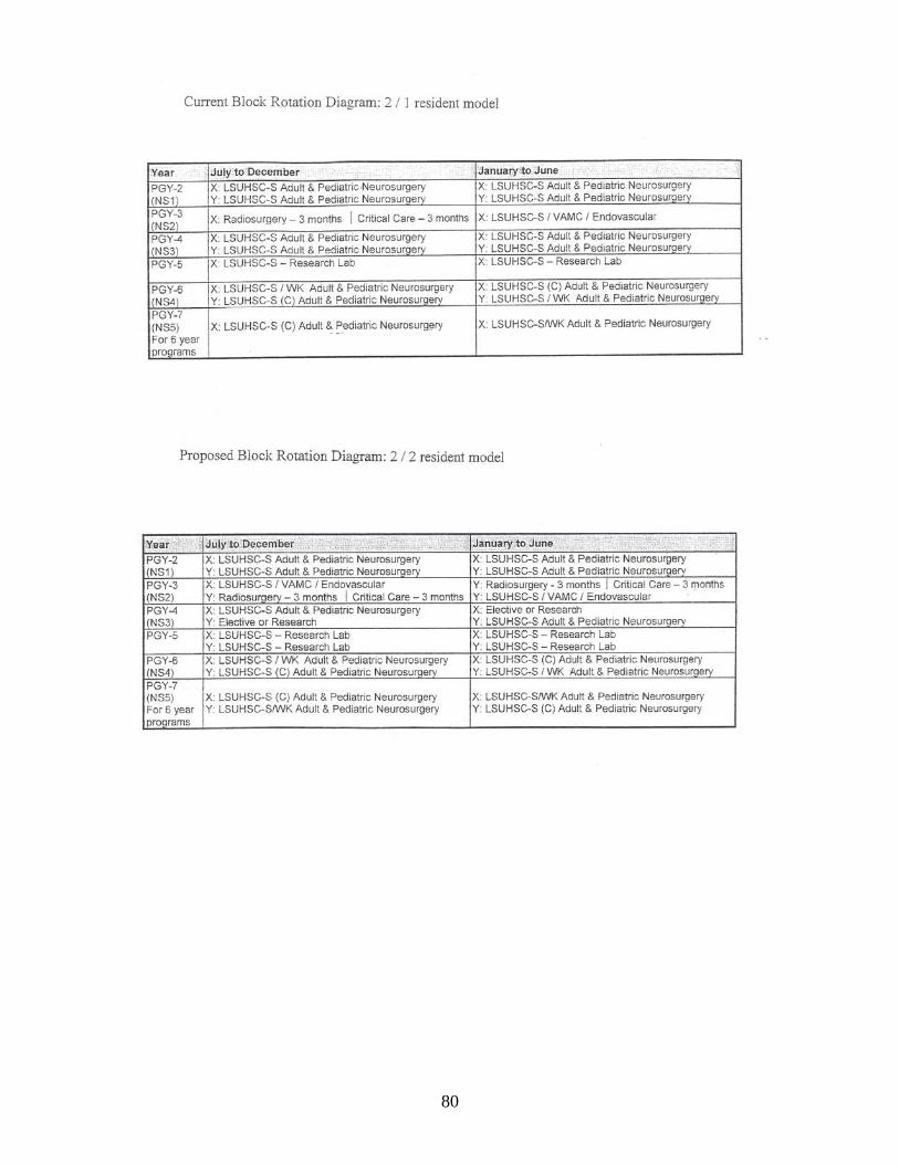
80
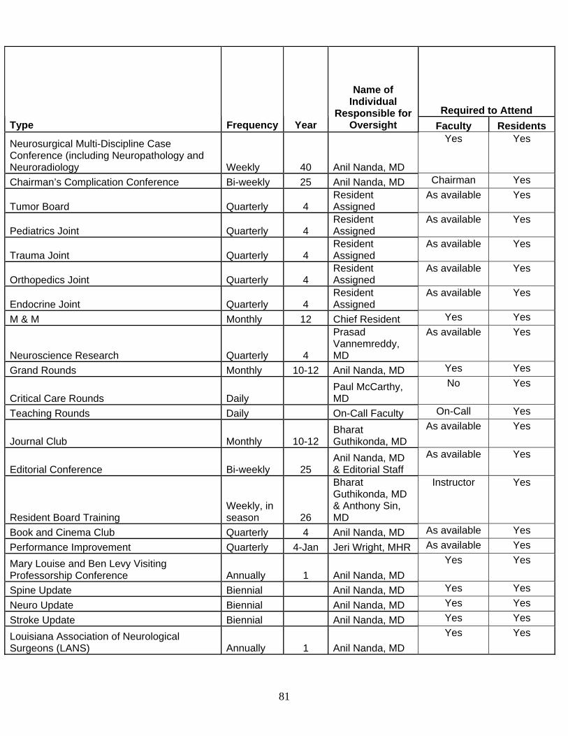
81
Required to Attend Type Frequency Year
Name of Individual
Responsible for Oversight Faculty Residents
Neurosurgical Multi-Discipline Case Conference (including Neuropathology and Neuroradiology Weekly 40 Anil Nanda, MD
Yes Yes
Chairman’s Complication Conference Bi-weekly 25 Anil Nanda, MD Chairman Yes
Tumor Board Quarterly 4 Resident Assigned
As available Yes
Pediatrics Joint Quarterly 4 Resident Assigned
As available Yes
Trauma Joint Quarterly 4 Resident Assigned
As available Yes
Orthopedics Joint Quarterly 4 Resident Assigned
As available Yes
Endocrine Joint Quarterly 4 Resident Assigned
As available Yes
M & M Monthly 12 Chief Resident Yes Yes
Neuroscience Research Quarterly 4
Prasad Vannemreddy, MD
As available Yes
Grand Rounds Monthly 10-12 Anil Nanda, MD Yes Yes
Critical Care Rounds Daily Paul McCarthy, MD
No Yes
Teaching Rounds Daily On-Call Faculty On-Call Yes
Journal Club Monthly 10-12 Bharat Guthikonda, MD
As available Yes
Editorial Conference Bi-weekly 25 Anil Nanda, MD & Editorial Staff
As available Yes
Resident Board Training Weekly, in season 26
Bharat Guthikonda, MD & Anthony Sin, MD
Instructor Yes
Book and Cinema Club Quarterly 4 Anil Nanda, MD As available Yes
Performance Improvement Quarterly 4-Jan Jeri Wright, MHR As available Yes
Mary Louise and Ben Levy Visiting Professorship Conference Annually 1 Anil Nanda, MD
Yes Yes
Spine Update Biennial Anil Nanda, MD Yes Yes
Neuro Update Biennial Anil Nanda, MD Yes Yes
Stroke Update Biennial Anil Nanda, MD Yes Yes
Louisiana Association of Neurological Surgeons (LANS) Annually 1 Anil Nanda, MD
Yes Yes