Embed Size (px)
Citation preview
Copyright ©2001 McGraw-Hil l Victor, Maurice, Ropper, Allan H. Adams & Victors' Principles of Neurology, 7th Edit ion
NEUROVASCULAR SYNDROMES
Part of "Chapter 34 - CEREBROVASCULAR DISEASES"
For reasons already given, the cl inical picture that results from an occlusion of any one artery differs in minor ways from one patient to another. There is suff icient uniformity, however, to justify the assignment of a typical syndrome to each of the major arteries. The following descriptions apply particularly to the cl inical effects of ischemia and infarction due to embolism and thrombosis. Although hemorrhage within a specif ic vascular territory may give rise to many of the same effects, the total cl inical picture is different because it usually involves the territory of more than one artery and, by its deep extension and pressure effects, causes secondary features of headache, vomiting, and hypertension as well as a series of falsely localizing signs, as described in Chap. 17 and Chap. 31.
The Carotid Artery The carotid system consists of three major arteries—the common carotid, internal carotid, and external carotid. As indicated in Fig. 34-1, the right common carotid artery arises at the level of the sternoclavicular notch from the innominate (brachiocephalic) artery, and the left common carotid stems directly from the aortic arch. The common carotids ascend in the neck to the C4 level, just below the angle of the jaw, where each divides into external and internal branches (sometimes the bifurcation is sl ightly above or below this point).
Common carotid occlusion accounts for less than 1 percent of cases of carotid artery syndrome—the remainder being due to disease of the internal carotid artery itself. Nevertheless, the common carotid can be occluded by an atheromatous plaque at its origin, more often on the left side. Atherosclerotic stenosis or occlusion of the midportion of the common carotid may also occur years after radiation therapy for laryngeal or other head and neck cancer. If the bifurcation is patent, few if any symptoms result, in some cases because retrograde flow from the external carotid maintains internal carotid f low and perfusion of the brain. Because the syndromes caused by common carotid occlusion are identical to those of i ts internal branch, the remainder of this discussion is concerned with disease of the internal carotid artery . The territory supplied by this vessel and its main branches is shown in Fig. 34-3, Fig. 34-4, and Fig. 34-5. The clinical manifestations of atherosclerotic thrombotic disease of this artery are the most variable of any cerebrovascular syndrome. Unlike other cerebral vessels, the internal carotid artery is not an end vessel, by virtue of its continuity with the vessels of the circle of Wil l is and those of the orbit, and no part of the brain is completely dependent on it . Therefore occlusion, which occurs most frequently in the f irst part of the internal carotid artery (immediately beyond the carotid bifurcation), is often si lent (30 to 40 percent of cases).
P.830
Página 1 de 31Ovid: Adams & Victors' Principles of Neurology
10/02/05http://gateway.ut.ovid.com/gw2/ovidweb.cgi
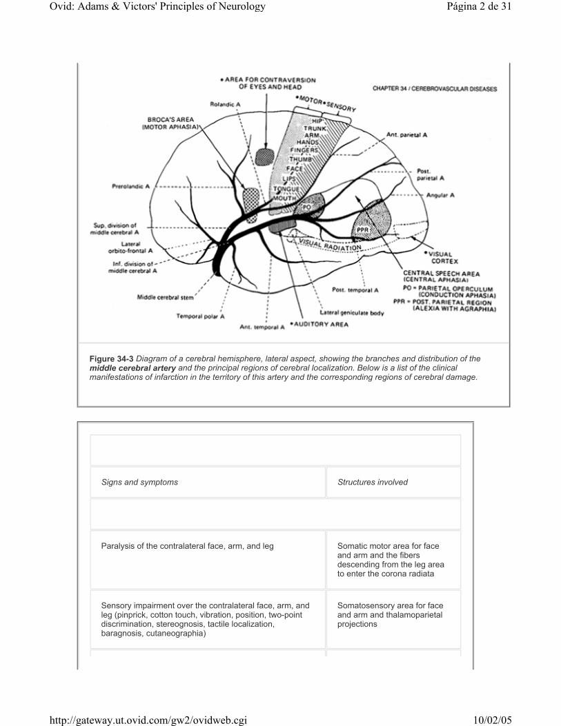
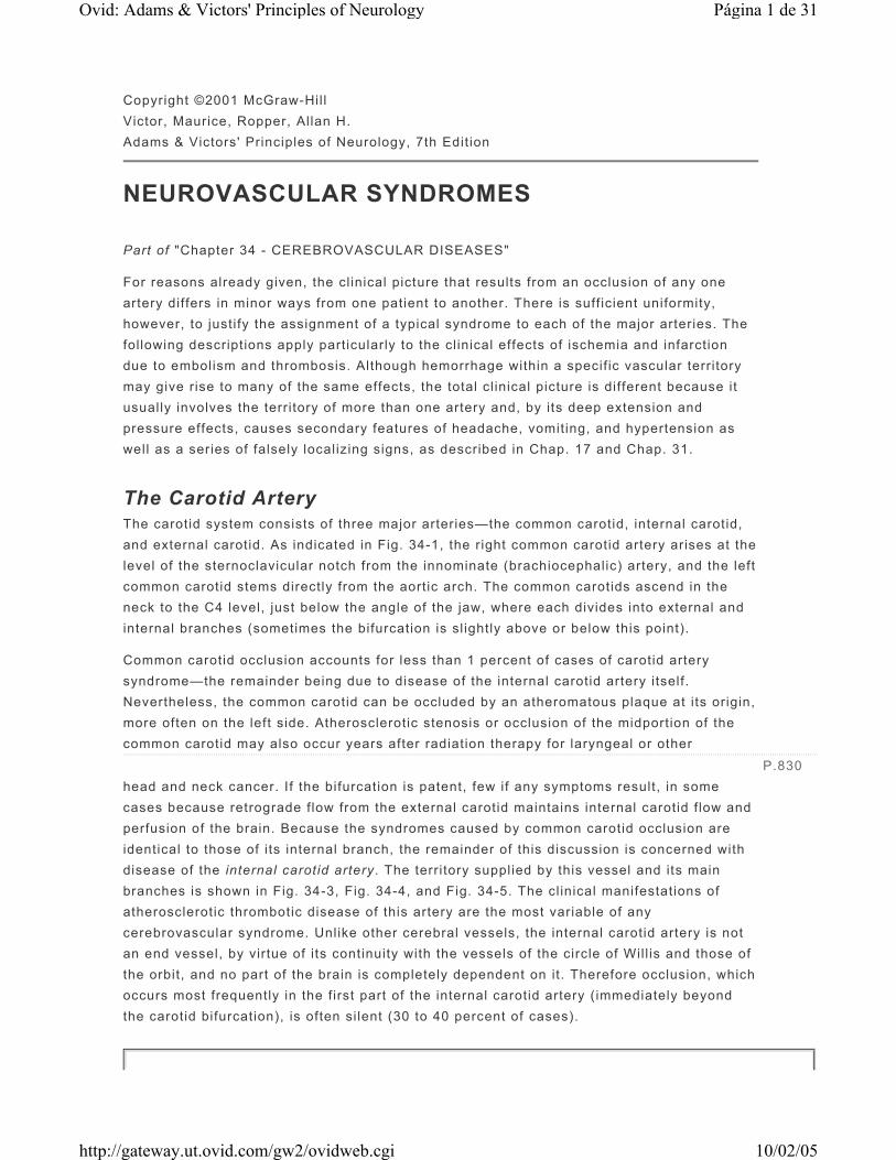
Figure 34-3 Diagram of a cerebral hemisphere, lateral aspect, showing the branches and distribution of the middle cerebral artery and the principal regions of cerebral localization. Below is a list of the clinical manifestations of infarction in the territory of this artery and the corresponding regions of cerebral damage.
Signs and symptoms Structures involved
Paralysis of the contralateral face, arm, and leg Somatic motor area for face and arm and the fibers descending from the leg area to enter the corona radiata
Sensory impairment over the contralateral face, arm, and leg (pinprick, cotton touch, vibration, position, two-point discrimination, stereognosis, tactile localization, baragnosis, cutaneographia)
Somatosensory area for face and arm and thalamoparietal projections
Página 2 de 31Ovid: Adams & Victors' Principles of Neurology
10/02/05http://gateway.ut.ovid.com/gw2/ovidweb.cgi
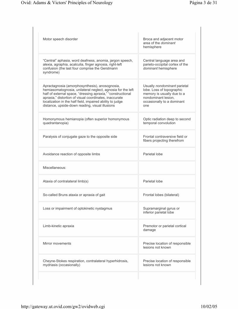
Motor speech disorder Broca and adjacent motor area of the dominant hemisphere
“Central” aphasia, word deafness, anomia, jargon speech, alexia, agraphia, acalculia, finger agnosia, right-left confusion (the last four comprise the Gerstmann syndrome)
Central language area and parieto-occipital cortex of the dominant hemisphere
Apractagnosia (amorphosynthesis), anosognosia, hemiasomatognosia, unilateral neglect, agnosia for the left half of external space, “dressing apraxia,” “constructional apraxia,” distortion of visual coordinates, inaccurate localization in the half field, impaired ability to judge distance, upside-down reading, visual illusions
Usually nondominant parietal lobe. Loss of topographic memory is usually due to a nondominant lesion, occasionally to a dominant one
Homonymous hemianopia (often superior homonymous quadrantanopia)
Optic radiation deep to second temporal convolution
Paralysis of conjugate gaze to the opposite side Frontal contraversive field or fibers projecting therefrom
Avoidance reaction of opposite limbs Parietal lobe
Miscellaneous:
Ataxia of contralateral limb(s) Parietal lobe
So-called Bruns ataxia or apraxia of gait Frontal lobes (bilateral)
Loss or impairment of optokinetic nystagmus Supramarginal gyrus or inferior parietal lobe
Limb-kinetic apraxia Premotor or parietal cortical damage
Mirror movements Precise location of responsible lesions not known
Cheyne-Stokes respiration, contralateral hyperhidrosis, mydriasis (occasionally)
Precise location of responsible lesions not known
Página 3 de 31Ovid: Adams & Victors' Principles of Neurology
10/02/05http://gateway.ut.ovid.com/gw2/ovidweb.cgi
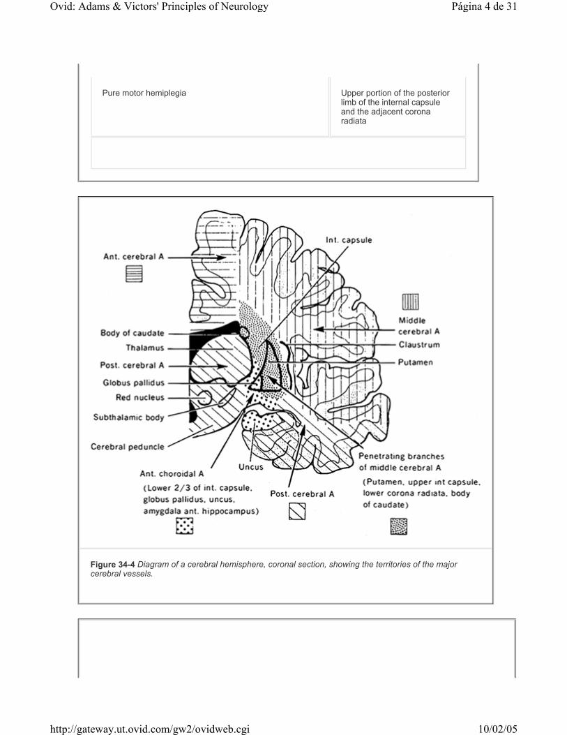
Pure motor hemiplegia Upper portion of the posterior limb of the internal capsule and the adjacent corona radiata
Figure 34-4 Diagram of a cerebral hemisphere, coronal section, showing the territories of the major cerebral vessels.
Página 4 de 31Ovid: Adams & Victors' Principles of Neurology
10/02/05http://gateway.ut.ovid.com/gw2/ovidweb.cgi
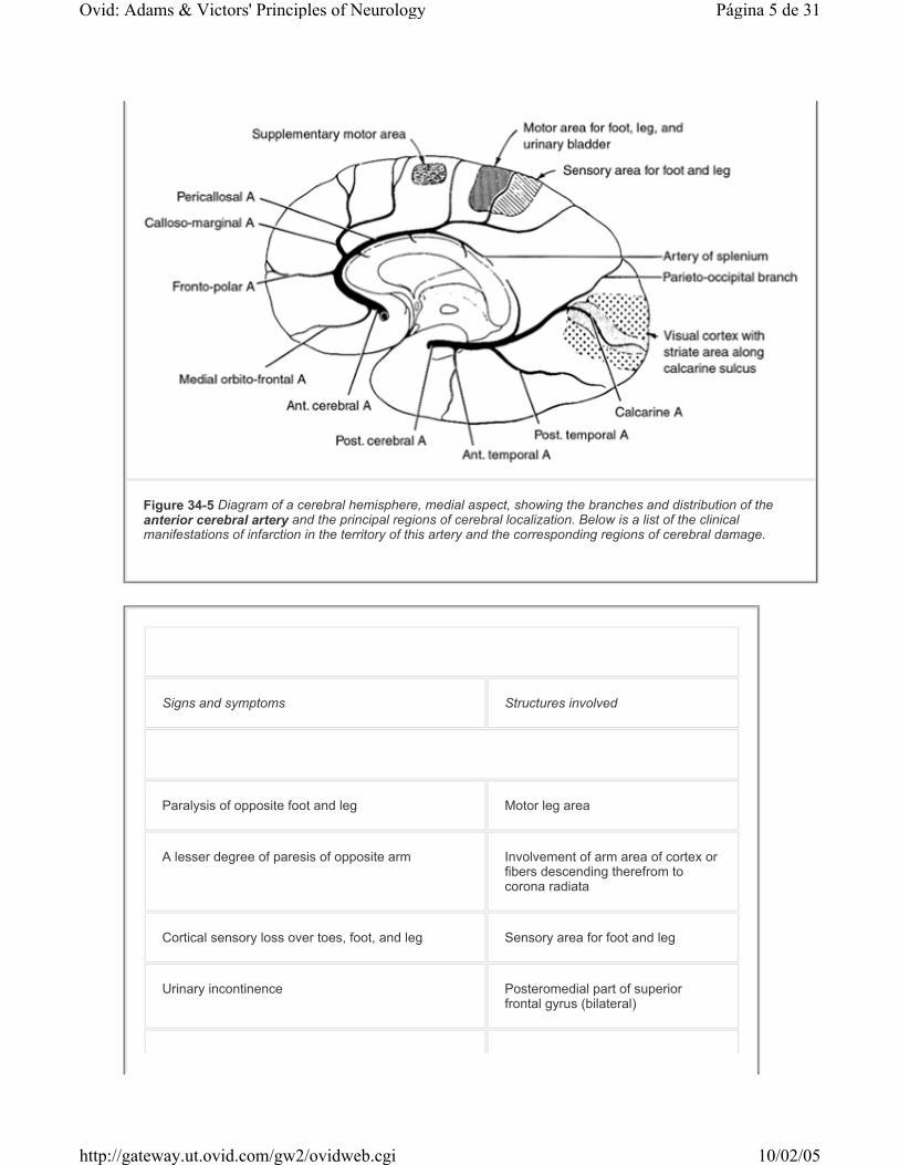
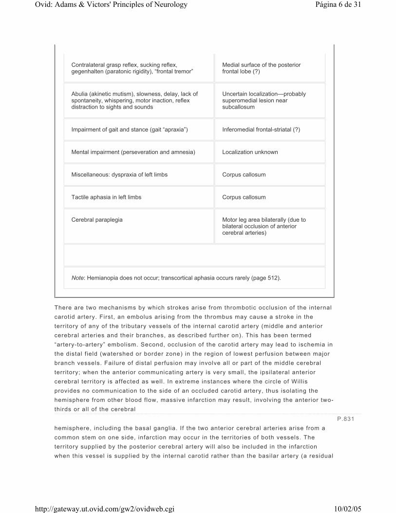
Figure 34-5 Diagram of a cerebral hemisphere, medial aspect, showing the branches and distribution of the anterior cerebral artery and the principal regions of cerebral localization. Below is a list of the clinical manifestations of infarction in the territory of this artery and the corresponding regions of cerebral damage.
Signs and symptoms Structures involved
Paralysis of opposite foot and leg Motor leg area
A lesser degree of paresis of opposite arm Involvement of arm area of cortex or fibers descending therefrom to corona radiata
Cortical sensory loss over toes, foot, and leg Sensory area for foot and leg
Urinary incontinence Posteromedial part of superior frontal gyrus (bilateral)
Página 5 de 31Ovid: Adams & Victors' Principles of Neurology
10/02/05http://gateway.ut.ovid.com/gw2/ovidweb.cgi
There are two mechanisms by which strokes arise from thrombotic occlusion of the internal carotid artery. First, an embolus arising from the thrombus may cause a stroke in the territory of any of the tr ibutary vessels of the internal carotid artery (middle and anterior cerebral arteries and their branches, as described further on). This has been termed “artery-to-artery” embolism. Second, occlusion of the carotid artery may lead to ischemia in the distal f ield (watershed or border zone) in the region of lowest perfusion between major branch vessels. Failure of distal perfusion may involve all or part of the middle cerebral territory; when the anterior communicating artery is very small, the ipsilateral anterior cerebral territory is affected as well. In extreme instances where the circle of Wil l is provides no communication to the side of an occluded carotid artery, thus isolating the hemisphere from other blood flow, massive infarction may result, involving the anterior two-thirds or al l of the cerebral hemisphere, including the basal ganglia. If the two anterior cerebral arteries arise from a common stem on one side, infarction may occur in the territories of both vessels. The territory supplied by the posterior cerebral artery wil l also be included in the infarction when this vessel is supplied by the internal carotid rather than the basilar artery (a residual
Contralateral grasp reflex, sucking reflex, gegenhalten (paratonic rigidity), “frontal tremor”
Medial surface of the posterior frontal lobe (?)
Abulia (akinetic mutism), slowness, delay, lack of spontaneity, whispering, motor inaction, reflex distraction to sights and sounds
Uncertain localization—probably superomedial lesion near subcallosum
Impairment of gait and stance (gait “apraxia”) Inferomedial frontal-striatal (?)
Mental impairment (perseveration and amnesia) Localization unknown
Miscellaneous: dyspraxia of left limbs Corpus callosum
Tactile aphasia in left limbs Corpus callosum
Cerebral paraplegia Motor leg area bilaterally (due to bilateral occlusion of anterior cerebral arteries)
Note: Hemianopia does not occur; transcortical aphasia occurs rarely (page 512).
P.831
Página 6 de 31Ovid: Adams & Victors' Principles of Neurology
10/02/05http://gateway.ut.ovid.com/gw2/ovidweb.cgi
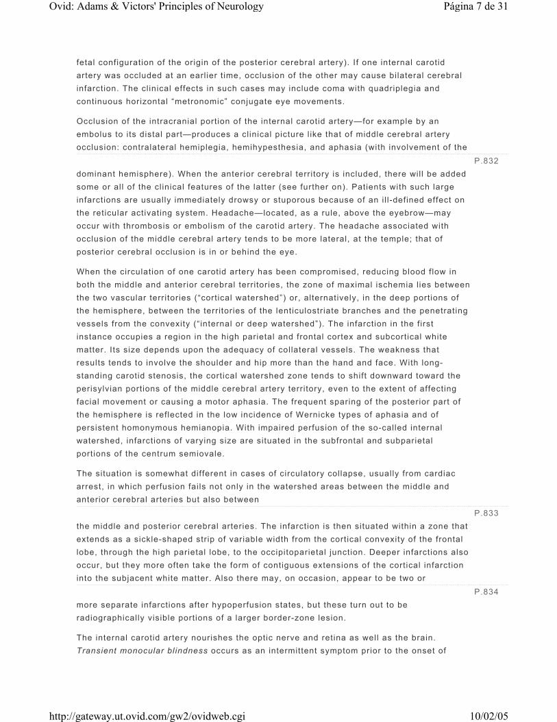
fetal configuration of the origin of the posterior cerebral artery). If one internal carotid artery was occluded at an earl ier t ime, occlusion of the other may cause bilateral cerebral infarction. The clinical effects in such cases may include coma with quadriplegia and continuous horizontal “metronomic” conjugate eye movements.
Occlusion of the intracranial portion of the internal carotid artery—for example by an embolus to its distal part—produces a clinical picture l ike that of middle cerebral artery occlusion: contralateral hemiplegia, hemihypesthesia, and aphasia (with involvement of the dominant hemisphere). When the anterior cerebral territory is included, there wil l be added some or all of the cl inical features of the latter (see further on). Patients with such large infarctions are usually immediately drowsy or stuporous because of an i l l-defined effect on the reticular activating system. Headache—located, as a rule, above the eyebrow—may occur with thrombosis or embolism of the carotid artery. The headache associated with occlusion of the middle cerebral artery tends to be more lateral, at the temple; that of posterior cerebral occlusion is in or behind the eye.
When the circulation of one carotid artery has been compromised, reducing blood flow in both the middle and anterior cerebral territories, the zone of maximal ischemia lies between the two vascular territories (“cortical watershed”) or, alternatively, in the deep portions of the hemisphere, between the territories of the lenticulostriate branches and the penetrating vessels from the convexity (“internal or deep watershed”). The infarction in the f irst instance occupies a region in the high parietal and frontal cortex and subcortical white matter. Its size depends upon the adequacy of collateral vessels. The weakness that results tends to involve the shoulder and hip more than the hand and face. With long-standing carotid stenosis, the cortical watershed zone tends to shift downward toward the perisylvian portions of the middle cerebral artery territory, even to the extent of affecting facial movement or causing a motor aphasia. The frequent sparing of the posterior part of the hemisphere is reflected in the low incidence of Wernicke types of aphasia and of persistent homonymous hemianopia. With impaired perfusion of the so-called internal watershed, infarctions of varying size are situated in the subfrontal and subparietal portions of the centrum semiovale.
The situation is somewhat different in cases of circulatory collapse, usually from cardiac arrest, in which perfusion fai ls not only in the watershed areas between the middle and anterior cerebral arteries but also between the middle and posterior cerebral arteries. The infarction is then situated within a zone that extends as a sickle-shaped strip of variable width from the cortical convexity of the frontal lobe, through the high parietal lobe, to the occipitoparietal junction. Deeper infarctions also occur, but they more often take the form of contiguous extensions of the cortical infarction into the subjacent white matter. Also there may, on occasion, appear to be two or more separate infarctions after hypoperfusion states, but these turn out to be radiographically visible portions of a larger border-zone lesion.
The internal carotid artery nourishes the optic nerve and retina as well as the brain. Transient monocular bl indness occurs as an intermittent symptom prior to the onset of
P.832
P.833
P.834
Página 7 de 31Ovid: Adams & Victors' Principles of Neurology
10/02/05http://gateway.ut.ovid.com/gw2/ovidweb.cgi
stroke in 10 to 25 percent of cases of symptomatic carotid occlusion. Yet central retinal artery ischemia is a relatively rare manifestation of carotid artery occlusion, presumably because of eff icient collateral supply.
Whereas most cerebral arteries can be evaluated only indirectly, by analysis of the cl inical effects of occlusion, more direct means are available for the evaluation of the common and internal carotid arteries in the neck. With severe atherosclerotic stenosis at the level of the carotid sinus, with or without a superimposed thrombus, auscultation frequently discloses a bruit, best heard with the bell of the stethoscope. Occasionally the bruit is due to stenosis at the origin of the external carotid artery or is a radiated murmur from the aortic valve and can then be misleading. If the bruit is heard at the angle of the jaw, the stenosis usually l ies in the carotid sinus; i f heard lower in the neck, i t is in the common carotid or subclavian artery. The duration and quality of the bruit are important—bruits that extend into diastole and are high-pitched are almost invariably associated with a t ight stenosis (lumen < l.5 mm). An addit ional though infrequent sign of carotid occlusion is the presence of a bruit over the opposite carotid artery, heard best by placing the bell of the stethoscope over the eyeball; presumably this murmur is due to augmented circulation through the patent vessel. Pulsation may be palpably reduced or absent in the common carotid artery in the neck, in the external carotid artery in front of the ear, and in the internal carotid artery in the lateral wall of the pharynx, but these are among the least dependable signs of carotid disease. In the presence of a unilateral internal carotid occlusion, compression of the normal common carotid should be avoided because it may precipitate neurologic symptoms. Central retinal artery pressure is reduced on the side of a carotid occlusion or severe stenosis, and this can be appreciated by gentle pressure on the globe while viewing the vessels emanating from the optic nerve head. A diastolic retinal pressure (determined by ophthalmic dynamometry) of less than 20 mmHg usually means that the common or internal carotid artery is occluded. These and other tests for measuring carotid f low have been largely supplanted by direct insonation or MRI imaging of the carotid artery.
The occurrence of emboli within retinal arteries, either shiny white or reddish in appearance, is another important sign of carotid disease (crystall ine cholesterol may be sloughed from an atheromatous ulcer).
Other signs of carotid occlusion include pulseless arms (as in Takayasu disease, see page 908); faintness on arising from the horizontal posit ion or recurrent loss of consciousness when walking; headache and sometimes ocular, retro-orbital, and neck pain; transient bl indness, either unilateral or bi lateral; unilateral visual loss or dimness of vision with exercise, after exposure to bright l ight, or on assuming an upright posit ion; retinal atrophy and pigmentation; atrophy of the ir is; heterochromia of the ir is (differing coloration on the two sides), which is a sign of carotid occlusion early in l i fe; leukomas (corneal scars); peripapil lary arteriovenous anastomoses in the retinae; optic atrophy; and claudication of jaw muscles.
Stenoses, ulcerations, and dissections of the internal carotid artery near its origin from the common carotid artery (the bulb, or sinus) may be a source of f ibrin platelet emboli or may cause a reduction in blood flow, result ing in transient ischemic attacks (TIAs). These are fully discussed further on, but here it can be stated that severe stenosis of the vessel
Página 8 de 31Ovid: Adams & Victors' Principles of Neurology
10/02/05http://gateway.ut.ovid.com/gw2/ovidweb.cgi
represents an important risk factor for stroke.
Middle Cerebral Artery This artery, through its cortical branches , supplies the lateral part of the cerebral hemisphere (Fig. 34-3). Its territory encompasses (1) cortex and white matter of the lateral and inferior parts of the frontal lobe—including motor areas 4 and 6, contraversive centers for lateral gaze—and motor speech area of Broca (dominant hemisphere); (2) cortex and white matter of the parietal lobe, including the sensory cortex and the angular and supramarginal convolutions; and (3) superior parts of the temporal lobe and insula, including the sensory language areas of Wernicke. The penetrating branches of the middle cerebral artery supply the putamen, part of the head and body of the caudate nucleus, the outer globus pall idus, the posterior l imb of the internal capsule, and the corona radiata (Fig. 34-4). The size of both the middle cerebral artery and the territory that i t supplies is larger than those of the anterior and posterior cerebral arteries.
The middle cerebral artery may be occluded in its stem, blocking the f low in deep penetrating as well as superficial cortical branches, or the two divisions in the sylvian sulcus and their major branches may be occluded separately. The classic picture of total occlusion of the stem is contralateral hemiplegia (face, arm, and leg), hemianesthesia, and homonymous hemianopia (due to infarction of the lateral geniculate body), with deviation of the head and eyes toward the side of the lesion; in addit ion, there is a global aphasia with left hemispheric lesions and anosognosia and amorphosynthesis with right-sided ones (see page 484). In the beginning the patient is dull or stuporous because of an i l l-defined effect of widespread paralysis of function. Once ful ly established, the motor, sensory, and language deficits remain static or improve very l i t t le as months and years pass. If the patient is globally aphasic for a prolonged period of t ime, he seldom ever again communicates effectively. Occlusion of branches of the middle cerebral artery gives rise to only parts of the symptom complex.
Occlusion of the stem of the middle cerebral artery by a thrombus, contrary to conventional teaching, is relatively infrequent (2 to 5 percent of middle cerebral artery occlusions). Pathologic studies have shown that most carotid occlusions are thrombotic, whereas most middle cerebral occlusions are embolic (Fisher, 1975; Caplan, 1989). The emboli tend to drift into superficial cortical branches; not more than 1 in 20 wil l enter deep penetrating branches. As indicated earl ier, the distal territory of the middle cerebral artery may also be rendered ischemic by fai lure of the systemic circulation, especially i f the carotid artery is stenotic; this may simulate embolic branch occlusions.
An embolus entering the middle cerebral artery most often lodges in one of its two main divisions, the superior division (supplying the rolandic and prerolandic areas) or the inferior division (supplying the lateral temporal and inferior parietal lobes). Major infarction in the territory of the superior division causes a dense sensorimotor deficit in the contralateral face, arm, and, to a lesser extent, leg as well as ipsilateral deviation of the head and eyes; i.e., i t mimics the syndrome of stem occlusion except that the foot is spared and the leg is less involved than the arm and face (“brachiofacial paralysis”); there is no impairment of alertness. If the occlusion is lasting (not merely transient ischemia with disintegration of
P.835
Página 9 de 31Ovid: Adams & Victors' Principles of Neurology
10/02/05http://gateway.ut.ovid.com/gw2/ovidweb.cgi
the embolus), there wil l be slow improvement; after a few months, the patient wil l be able to walk with a spastic leg, while the motor deficits of the arm and face remain. The sensory deficit may be profound, resembling that of a thalamic infarct (pseudothalamic syndrome of Foix), but more often it is less severe than the motor deficit, taking the form of stereoanesthesia, agraphesthesia, impaired posit ion sense, tacti le localization, and two-point discrimination as well as variable changes in touch, pain, and temperature sense. A rare pseudoradicular pattern of sensory loss in the hand and forearm (radial or ulnar half) from a parietal infarct was originally described by Déjerine. With left-sided lesions there may init ial ly be a global aphasia, which changes gradually to a predominantly motor (Broca) aphasia, with improvement in comprehension of spoken and written words and the emergence of an effortful, hesitant, grammatically simplif ied, and dysmelodic speech (Chap. 23).
Embolic occlusion l imited to one of the branches of the superior division produces a more circumscribed infarct that further fractionates the syndrome. With occlusion of the ascending frontal branch, the motor deficit is l imited to the face and arm with l i t t le affection of the leg, and the latter, if weakened at al l , soon improves; with left-sided lesions, an init ial mutism and mild comprehension defect give way, within days to weeks, to sl ightly dysfluent and agrammatic speech, with normal comprehension (page 505). Embolic occlusion of the rolandic branches results in sensorimotor paresis with severe dysarthria but l it t le evidence of aphasia. A cortical-subcortical branch occlusion may give rise to a brachial monoplegia. Embolic occlusion of ascending parietal and other posterior branches of the superior division may cause no sensorimotor deficit but only a conduction aphasia (page 510) and bilateral ideomotor apraxia. There are many other l imited stroke syndromes or combinations of the aforementioned syndromes relating to small regions of damage in the frontal, parietal, or temporal lobes. Most of these are discussed in Chap. 22, on lesions in particular parts of the cerebrum. Improvement can be expected within a few weeks to months.
Occlusion of the inferior division of the middle cerebral artery is less frequent than occlusion of the superior one, but again is nearly always due to cardiogenic embolism. The usual result in left-sided lesions is a Wernicke aphasia which generally remains static for weeks or a month or two, after which some improvement can be expected (page 509). In less extensive infarcts from branch occlusions (superior parietal, angular, or posterior temporal), the deficit in comprehension of spoken and writ ten language may be especially severe. Again, after a few months, the deficits usually improve, often to the point where they are evident only in self-generated efforts to read and copy visually presented words or phrases. With either right- or left-hemispheric lesions, there is usually a superior quadrantanopia or homonymous hemianopia and, with right-sided ones, a left visual neglect and other signs of amorphosynthesis; rarely, an agitated confusional state, presumably from temporal lobe damage, may be a prominent feature of dominant hemispheral lesions and sometimes of nondominant ones as well.
Deep (Striatocapsular) Infarctions A number of interesting syndromes occur with deep lesions in the territory of the
P.836
Página 10 de 31Ovid: Adams & Victors' Principles of Neurology
10/02/05http://gateway.ut.ovid.com/gw2/ovidweb.cgi
penetrating vessels of the middle cerebral artery. Most are attr ibutable to emboli from stem occlusions of the middle cerebral artery, although imaging studies show a patent vessel in half of the cases; a few are presumably atherothrombotic, but these judgments have been made from an analysis of imaging, not pathologic studies. Although centered in the deep white matter, most of these lesions are fragments of the cortical-subcortical stroke patterns described above. The most common type in our experience has been a large striatocapsular infarction , similar to that described by Weller and colleagues. Of their 29 patients, al l had some degree of hemiparesis and 6 had aphasia or hemineglect. Aphasia, when it occurs, tends to be a l imited form of the Broca type or anomia and, in our experience, has been short-l ived. We have most often encountered incomplete pure motor syndromes affecting only the arm and hand without language disturbance or neglect; these are quite diff icult to differentiate from small embolic cortical strokes. The lesions in the corona radiata are larger than typical lacunes but probably have a similar pathophysiology.
Foix and Levy, who described the clinical effects of deep capsular-basal ganglionic lesions and of superficial cortical-subcortical ones, found few important differences in the degree and pattern of the hemiplegia and sensory disorder. Homonymous hemianopia may occur with posterior capsular lesions, but i t must be distinguished from visual hemineglect of contralateral space. Bilateral cerebral infarctions involving mainly the insular-perisylvian (anterior opercular) regions manifest themselves by a facio-glosso-pharyngo-masticatory diplegia (anarthria without aphasia; see Mao et al).
The middle cerebral artery may become stenotic rather than occluded. In several series of such cases, some of the permanent deficiencies were preceded by TIAs, producing a picture resembling that of carotid stenosis (Day; Caplan).
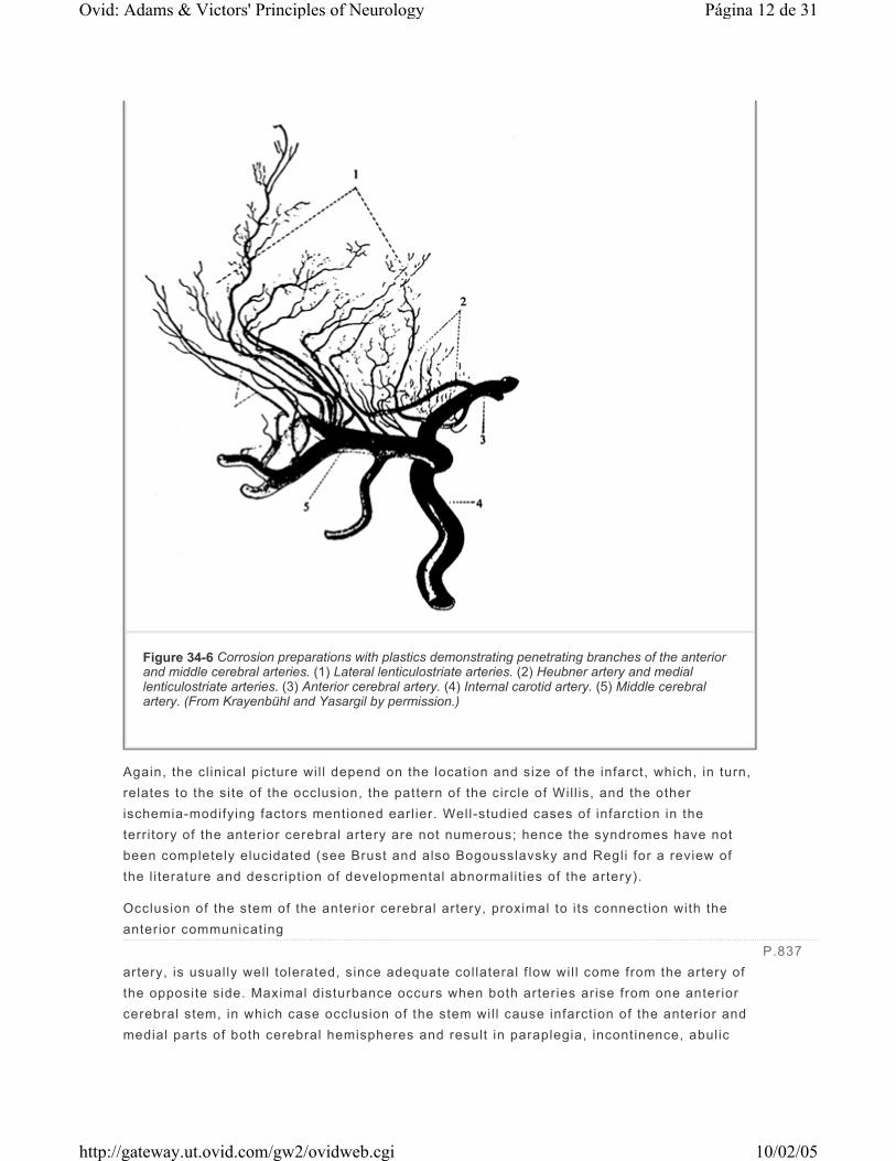
Anterior Cerebral Artery This artery, through its cortical branches, supplies the anterior three-quarters of the medial surface of the cerebral hemisphere, including the medial-orbital surface of the frontal lobe, the frontal pole, a strip of the lateral surface of the cerebral hemisphere along the superior border, and the anterior four-f i f ths of the corpus callosum. Deep branches, arising near the circle of Wil l is (proximal or distal to the anterior communicating artery), supply the anterior l imb of the internal capsule, the inferior part of the head of the caudate nucleus, and the anterior part of the globus pall idus (Fig. 34-4, Fig. 34-5, and Fig. 34-6). The largest of these deep branches is the artery of Heubner.
Página 11 de 31Ovid: Adams & Victors' Principles of Neurology
10/02/05http://gateway.ut.ovid.com/gw2/ovidweb.cgi
Again, the cl inical picture wil l depend on the location and size of the infarct, which, in turn, relates to the site of the occlusion, the pattern of the circle of Will is, and the other ischemia-modifying factors mentioned earl ier. Well-studied cases of infarction in the territory of the anterior cerebral artery are not numerous; hence the syndromes have not been completely elucidated (see Brust and also Bogousslavsky and Regli for a review of the l iterature and description of developmental abnormalit ies of the artery).
Occlusion of the stem of the anterior cerebral artery, proximal to its connection with the anterior communicating artery, is usually well tolerated, since adequate collateral f low wil l come from the artery of the opposite side. Maximal disturbance occurs when both arteries arise from one anterior cerebral stem, in which case occlusion of the stem wil l cause infarction of the anterior and medial parts of both cerebral hemispheres and result in paraplegia, incontinence, abulic
Figure 34-6 Corrosion preparations with plastics demonstrating penetrating branches of the anterior and middle cerebral arteries. (1) Lateral lenticulostriate arteries. (2) Heubner artery and medial lenticulostriate arteries. (3) Anterior cerebral artery. (4) Internal carotid artery. (5) Middle cerebral artery. (From Krayenbühl and Yasargil by permission.)
P.837
Página 12 de 31Ovid: Adams & Victors' Principles of Neurology
10/02/05http://gateway.ut.ovid.com/gw2/ovidweb.cgi

and motor aphasic symptoms, and frontal lobe personality changes (Chap. 22). Occlusion of the anterior cerebral arteries may also be embolic or surgical (fol lowing operations on anterior communicating aneurysms).
Complete infarction due to occlusion of one anterior cerebral artery distal to the anterior communicating artery results in a sensorimotor deficit of the opposite foot and leg and, to a lesser degree, of the shoulder and arm, with sparing of the hand and face. The motor disorder is more in the foot and leg than in the thigh. Sensory loss is mainly of the discriminative modalit ies and is mild or absent in some cases. The head and eyes may deviate to the side of the lesion. Urinary incontinence and a contralateral grasp reflex and paratonic rigidity (gegenhalten) may be evident. With a left-sided occlusion, there may be a sympathetic apraxia of the left arm and leg or involuntary misdirected movements of the left arm (alien arm or hand). Also, transcortical motor aphasia may occur with occlusions of Heubner's branch of the left anterior cerebral artery. Alexander and Schmitt cite cases in which a right hemiplegia (predominant in the leg) with grasping and groping responses of the right hand and buccofacial apraxia are accompanied by a diminution or absence of spontaneous speech, agraphia, labored telegraphic speech, and a limited abil ity to name objects and compose word lists but with a striking preservation of the abil i ty to repeat spoken and written sentences (transcortical motor aphasia). Disorders of behavior that may be overlooked are abulia, presenting as slowness and lack of spontaneity in all reactions; muteness or a tendency to speak in whispers; and distractibil i ty. Branch occlusions of the anterior cerebral artery produce only fragments of the total syndrome, usually a spastic weakness or cortical sensory loss in the opposite foot and leg.
With occlusion of penetrating branches of the anterior cerebral artery on one or both sides, the anterior l imb of the internal capsule is usually involved as well. In a series of 18 unilateral cases of caudate region infarcts collected by Caplan and associates, a transient hemiparesis was present in 13. Dysarthria and either abulia or agitation and hyperactivity were also common. Stuttering and language diff iculty occurred with two of the left-sided lesions and visuospatial neglect with three of the right-sided ones. To what extent these symptoms were due to disorder of neighboring structures is diff icult to decide. With bilateral caudate infarctions, a syndrome of inattentiveness, abulia, forgetfulness, and sometimes agitat ion and psychosis was observed. Transitory choreoathetosis and other dyskinesias have also been attr ibuted to ischemia of basal ganglia occurring sometimes under condit ions of prolonged standing and exercise (Caplan and Sergay; Margolin and Marsden).
Anterior Choroidal Artery This is a long, narrow artery that springs from the internal carotid, just above the origin of the posterior communicating artery. It supplies the internal segment of the globus pall idus and posterior l imb of the internal capsule and various contiguous structures including (in some patients) the optic tract; then it penetrates the temporal horn of the lateral ventricle, where it supplies the choroid plexus and anastomoses with the posterior choroidal artery.
Only a few complete cl inicopathologic studies have been made of the syndrome caused by occlusion of this artery. It was found by Foix and colleagues to consist of contralateral hemiplegia, hemihypesthesia, and homonymous hemianopia due to involvement of the
Página 13 de 31Ovid: Adams & Victors' Principles of Neurology
10/02/05http://gateway.ut.ovid.com/gw2/ovidweb.cgi
posterior l imb of the internal capsule and white matter posterolateral to it, through which the geniculocalcarine tract passes. Cognit ive function is notably spared. Decroix and colleagues reported 16 cases (identif ied by CT) in which the lesion appeared to l ie in the vascular territory of this artery. In most of their cases the cl inical syndrome fell short of what was expected on anatomic grounds. With right-sided lesions there may be left spatial neglect and constructional apraxia; sl ight disorders of speech and language may accompany left-sided lesions. Hupperts and colleagues have discussed the controversy regarding the effects of occlusion of the anterior choroidal artery and in particular the variabil i ty of i ts supply to the posterior paraventricular area of the corona radiata and adjacent regions. They concluded, also from a CT survey, that there was no uniform syndrome attr ibutable to occlusion of this vessel and that in most cases its territory of supply was overlapped by small surrounding vessels. Of course, in both these studies, the lesions may have extended beyond the territory of this artery, since postmortem confirmation was lacking. It should be remembered that for a t ime the anterior choroidal artery was being surgically l igated in order to abolish the tremor and rigidity of unilateral Parkinson disease, without these ischemic effects having been produced.
Vertebrobasilar and Posterior Cerebral Arteries
Posterior Cerebral Artery In about 70 percent of persons, both posterior cerebral arteries are formed by the bifurcation of the basilar artery, and only thin posterior communicating arteries join this system to the internal carotids. In 20 to 25 percent, one posterior cerebral artery arises from the basilar in the usual way, but the other arises from the internal carotid; in the remainder, both arise from the corresponding carotids.
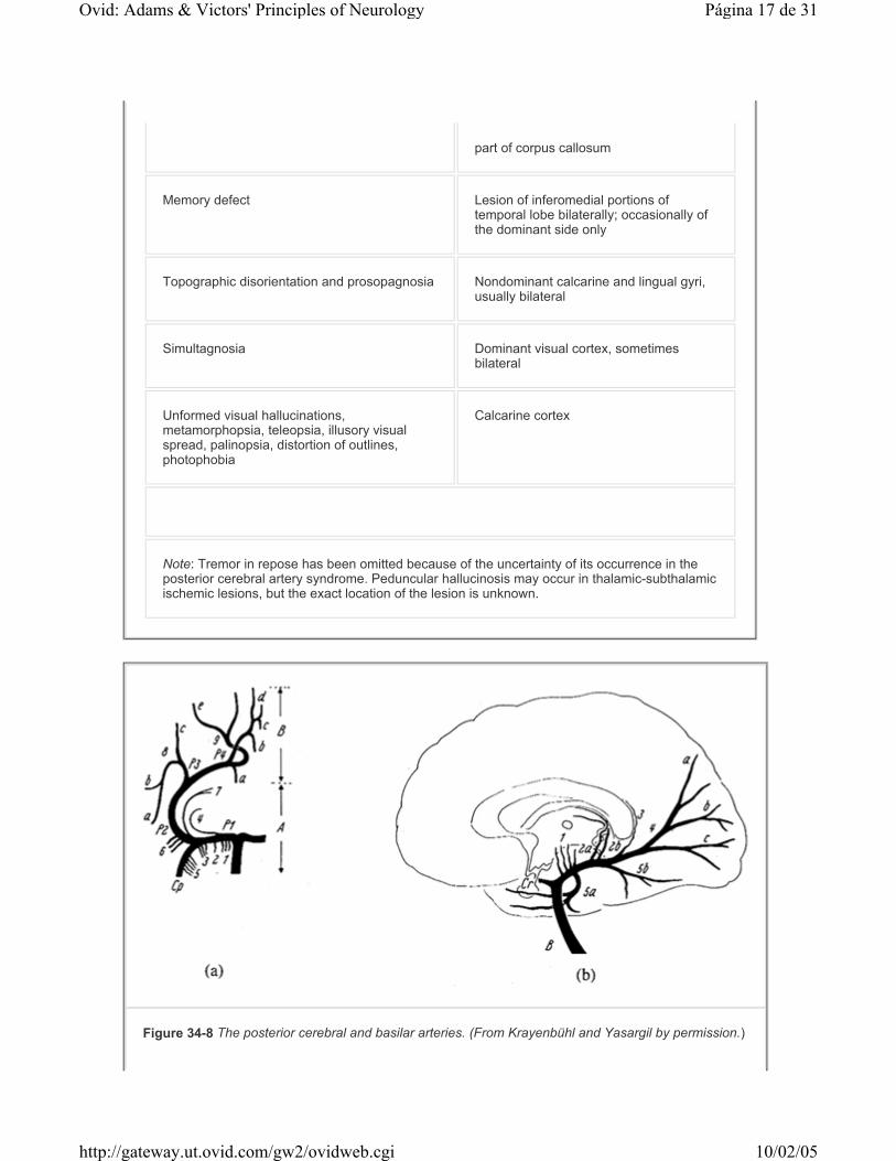
The configuration and branches of the proximal segment of the posterior cerebral artery are il lustrated in Fig. 34-7 and Fig. 34-8. The interpeduncular branches , which arise just above the basilar bifurcation, supply the red nuclei, the substantia nigra bilaterally, medial parts of the cerebral peduncles, oculomotor and trochlear nuclei and nerves, reticular substance of the upper brainstem, decussation of the brachia conjunctivae (superior cerebellar peduncles), medial longitudinal fasciculi, and medial lemnisci. The portion of the posterior cerebral artery giving rise to the interpeduncular branches (the portion between the bifurcation of the basilar artery and the ostium of the posterior communicating artery) is also referred to as the mesencephalic artery or the basilar communicating artery .
P.838
Página 14 de 31Ovid: Adams & Victors' Principles of Neurology
10/02/05http://gateway.ut.ovid.com/gw2/ovidweb.cgi
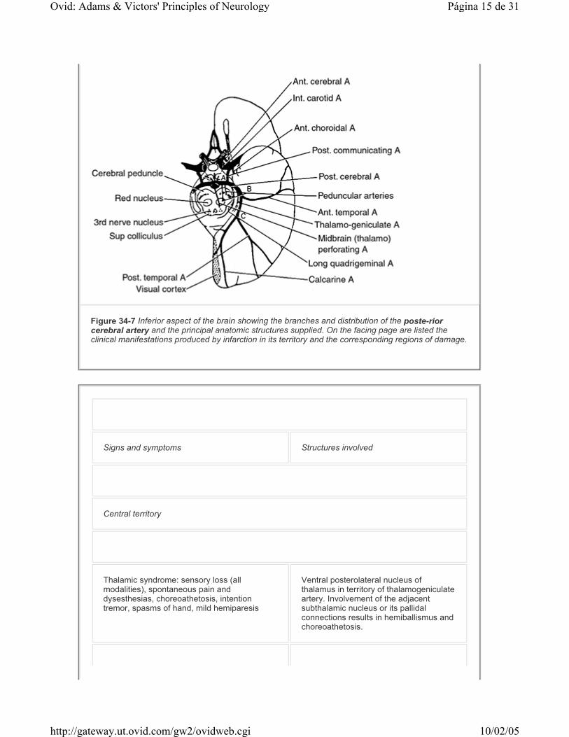
Figure 34-7 Inferior aspect of the brain showing the branches and distribution of the poste-rior cerebral artery and the principal anatomic structures supplied. On the facing page are listed the clinical manifestations produced by infarction in its territory and the corresponding regions of damage.
Signs and symptoms Structures involved
Central territory
Thalamic syndrome: sensory loss (all modalities), spontaneous pain and dysesthesias, choreoathetosis, intention tremor, spasms of hand, mild hemiparesis
Ventral posterolateral nucleus of thalamus in territory of thalamogeniculate artery. Involvement of the adjacent subthalamic nucleus or its pallidal connections results in hemiballismus and choreoathetosis.
Página 15 de 31Ovid: Adams & Victors' Principles of Neurology
10/02/05http://gateway.ut.ovid.com/gw2/ovidweb.cgi
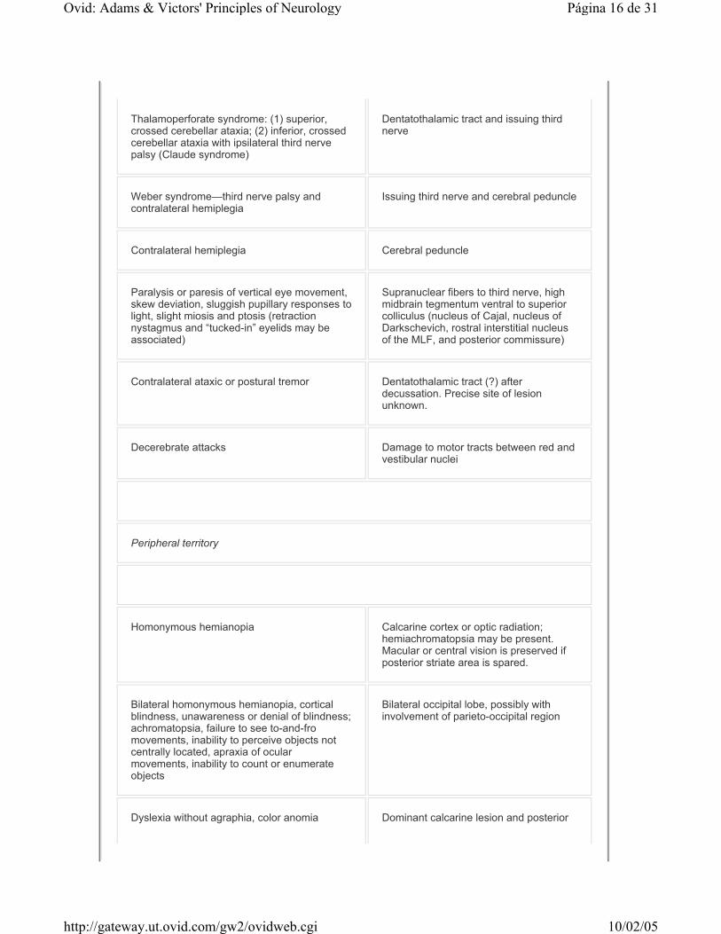
Thalamoperforate syndrome: (1) superior, crossed cerebellar ataxia; (2) inferior, crossed cerebellar ataxia with ipsilateral third nerve palsy (Claude syndrome)
Dentatothalamic tract and issuing third nerve
Weber syndrome—third nerve palsy and contralateral hemiplegia
Issuing third nerve and cerebral peduncle
Contralateral hemiplegia Cerebral peduncle
Paralysis or paresis of vertical eye movement, skew deviation, sluggish pupillary responses to light, slight miosis and ptosis (retraction nystagmus and “tucked-in” eyelids may be associated)
Supranuclear fibers to third nerve, high midbrain tegmentum ventral to superior colliculus (nucleus of Cajal, nucleus of Darkschevich, rostral interstitial nucleus of the MLF, and posterior commissure)
Contralateral ataxic or postural tremor Dentatothalamic tract (?) after decussation. Precise site of lesion unknown.
Decerebrate attacks Damage to motor tracts between red and vestibular nuclei
Peripheral territory
Homonymous hemianopia Calcarine cortex or optic radiation; hemiachromatopsia may be present. Macular or central vision is preserved if posterior striate area is spared.
Bilateral homonymous hemianopia, cortical blindness, unawareness or denial of blindness; achromatopsia, failure to see to-and-fro movements, inability to perceive objects not centrally located, apraxia of ocular movements, inability to count or enumerate objects
Bilateral occipital lobe, possibly with involvement of parieto-occipital region
Dyslexia without agraphia, color anomia Dominant calcarine lesion and posterior
Página 16 de 31Ovid: Adams & Victors' Principles of Neurology
10/02/05http://gateway.ut.ovid.com/gw2/ovidweb.cgi
part of corpus callosum
Memory defect Lesion of inferomedial portions of temporal lobe bilaterally; occasionally of the dominant side only
Topographic disorientation and prosopagnosia Nondominant calcarine and lingual gyri, usually bilateral
Simultagnosia Dominant visual cortex, sometimes bilateral
Unformed visual hallucinations, metamorphopsia, teleopsia, illusory visual spread, palinopsia, distortion of outlines, photophobia
Calcarine cortex
Note: Tremor in repose has been omitted because of the uncertainty of its occurrence in the posterior cerebral artery syndrome. Peduncular hallucinosis may occur in thalamic-subthalamic ischemic lesions, but the exact location of the lesion is unknown.
Figure 34-8 The posterior cerebral and basilar arteries. (From Krayenbühl and Yasargil by permission.)
Página 17 de 31Ovid: Adams & Victors' Principles of Neurology
10/02/05http://gateway.ut.ovid.com/gw2/ovidweb.cgi
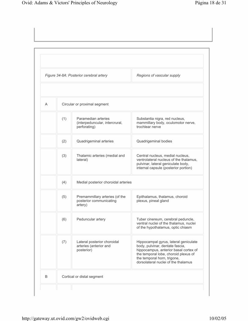
Figure 34-8A: Posterior cerebral artery Regions of vascular supply
A Circular or proximal segment
(1) Paramedian arteries
(interpeduncular, intercrural, perforating)
Substantia nigra, red nucleus, mammillary body, oculomotor nerve, trochlear nerve
(2) Quadrigeminal arteries Quadrigeminal bodies
(3) Thalamic arteries (medial and
lateral)Central nucleus, medial nucleus, ventrolateral nucleus of the thalamus, pulvinar, lateral geniculate body, internal capsule (posterior portion)
(4) Medial posterior choroidal arteries
(5) Premammillary arteries (of the
posterior communicating artery)
Epithalamus, thalamus, choroid plexus, pineal gland
(6) Peduncular artery Tuber cinereum, cerebral peduncle,
ventral nuclei of the thalamus, nuclei of the hypothalamus, optic chiasm
(7) Lateral posterior choroidal arteries (anterior and posterior)
Hippocampal gyrus, lateral geniculate body, pulvinar, dentate fascia, hippocampus, anterior basal cortex of the temporal lobe, choroid plexus of the temporal horn, trigone, dorsolateral nuclei of the thalamus
B Cortical or distal segment
Página 18 de 31Ovid: Adams & Victors' Principles of Neurology
10/02/05http://gateway.ut.ovid.com/gw2/ovidweb.cgi

(8) Lateral occipital artery
(a) Anterior temporal arteries
Laterobasal aspects of the temporal and occipital lobe
(b) Middle temporal arteries
(c) Posterior temporal arteries
(9) Medial occipital artery
(a) Dorsal callosal artery
Splenium
(b) Posterior parietal artery
Cuneus, precuneus
(c) Occipitoparietal artery
(d) Calcarine arteries Calcarine gyrus, occipital pole
(e) Occipitotemporal artery
Laterobasal occipital lobe
Figure 34-8B: Basilar artery
B Basilar artery
Cr Posterior communicating artery
Página 19 de 31Ovid: Adams & Victors' Principles of Neurology
10/02/05http://gateway.ut.ovid.com/gw2/ovidweb.cgi
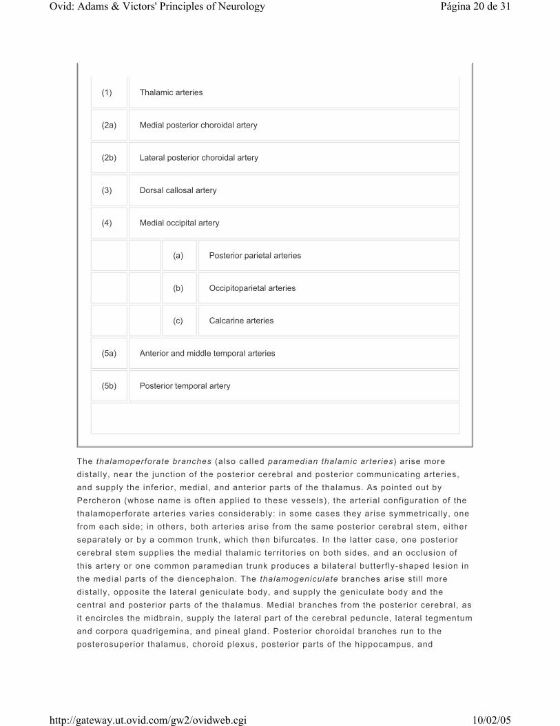
The thalamoperforate branches (also called paramedian thalamic arteries) arise more distally, near the junction of the posterior cerebral and posterior communicating arteries, and supply the inferior, medial, and anterior parts of the thalamus. As pointed out by Percheron (whose name is often applied to these vessels), the arterial configuration of the thalamoperforate arteries varies considerably: in some cases they arise symmetrically, one from each side; in others, both arteries arise from the same posterior cerebral stem, either separately or by a common trunk, which then bifurcates. In the latter case, one posterior cerebral stem supplies the medial thalamic territories on both sides, and an occlusion of this artery or one common paramedian trunk produces a bilateral butterf ly-shaped lesion in the medial parts of the diencephalon. The thalamogeniculate branches arise sti l l more distally, opposite the lateral geniculate body, and supply the geniculate body and the central and posterior parts of the thalamus. Medial branches from the posterior cerebral, as it encircles the midbrain, supply the lateral part of the cerebral peduncle, lateral tegmentum and corpora quadrigemina, and pineal gland. Posterior choroidal branches run to the posterosuperior thalamus, choroid plexus, posterior parts of the hippocampus, and
(1) Thalamic arteries
(2a) Medial posterior choroidal artery
(2b) Lateral posterior choroidal artery
(3) Dorsal callosal artery
(4) Medial occipital artery
(a) Posterior parietal arteries
(b) Occipitoparietal arteries
(c) Calcarine arteries
(5a) Anterior and middle temporal arteries
(5b) Posterior temporal artery
Página 20 de 31Ovid: Adams & Victors' Principles of Neurology
10/02/05http://gateway.ut.ovid.com/gw2/ovidweb.cgi
psalterium (decussation of fornices).
The terminal or cortical branches of the posterior cerebral artery supply the inferomedial part of the temporal lobe and the medial occipital lobe, including the lingula, cuneus, precuneus, and visual areas 17, 18, and 19 (see Fig. 34-5, Fig. 34-7, and Fig. 34-8).
Occlusion of the posterior cerebral artery can produce a greater variety of clinical effects than occlusion of any other artery, because both the upper brainstem, which is crowded with important structures, and the inferomedial parts of the temporal and occipital lobes l ie within its domain. Obviously the site of the occlusion and the arrangement of the circle of Wil l is wil l in large measure determine the location and extent of the result ing infarct. For example, occlusion proximal to the posterior communicating artery may be asymptomatic or have only transitory effects if the collateral f low is adequate (Fig. 34-7; see also Fig. 34-8). Even distal to the posterior communicating artery, an occlusion may cause relatively l i t t le damage if the collateral f low through border-zone collaterals from anterior and middle cerebral arteries is suff icient.
In the series of posterior cerebral artery strokes studied by Milandre and coworkers, the causes were in general similar to those of strokes in other vascular territories except that there was a surprisingly high incidence of atherosclerotic occlusion (35 patients) in contrast to cardioembolic types (15 patients). Two of their stroke cases were attr ibuted to migraine. Our experience has differed in that the proportion of presumed embolic occlusions has been greater.
For convenience of exposit ion, i t is helpful to divide the various posterior cerebral artery syndromes into three groups: (1) anterior and proximal ( involving interpeduncular, thalamic perforant, and thalamogeniculate branches), (2) cortical ( inferior temporal and medial occipital), and (3) bi lateral.
Anterior and Proximal Syndromes (See Fig. 34-8 and Fig. 34-9.) The thalamic syndrome of Déjerine and Roussy (page 172) follows infarction of the sensory relay nuclei in the thalamus, the result of occlusion of thalamogeniculate branches. There is both a deep and cutaneous sensory loss, usually severe in degree, of the opposite side of the body, accompanied by a transitory hemiparesis. A homonymous hemianopia may be conjoined. In some instances there is a dissociated sensory loss—pain and thermal sensation being more affected than touch, vibration, and posit ion—or only one part of the body may be anesthetic. After an interval, sensation begins to return, and the patient may then develop pain, paresthesia, and hyperpathia in the affected parts. The painful paresthetic syndrome may persist for years. There may also be distort ion of taste, athetotic posturing of the hand, and alteration of mood. Mania and depression have occasionally been observed with infarction of the diencephalon and adjacent structures, but the data are usually incomplete. The patient may be left with a severe amnesia, even with a unilateral lesion (see below).
P.839
P.840
Página 21 de 31Ovid: Adams & Victors' Principles of Neurology
10/02/05http://gateway.ut.ovid.com/gw2/ovidweb.cgi
Central midbrain and subthalamic syndromes are due to occlusion of the interpeduncular branches. The clinical syndromes include oculomotor palsy with contralateral hemiplegia (Weber syndrome), palsies of vertical gaze, stupor or coma, and movement disorders, most often an ataxic tremor that may be contralateral, i .e., on the side of the hemiparesis (see below). A persistent hemiplegia from infarction of the cerebral peduncle is rare.
Anteromedial-inferior thalamic syndromes fol low occlusion of the thalamoperforant branches. Here the most common effect is an extrapyramidal movement disorder (hemiball ismus or hemichoreoathetosis). Deep sensory loss, hemiataxia, or tremor may be added in various combinations. Hemiball ismus is usually due to occlusion of a small branch to the subthalamic nucleus (of Luys) or i ts connections with the pall idum. Occlusion of the paramedian thalamic branch(es) to the mediodorsal nuclei or to the dominant ( left) mediodorsal nucleus is the recognized substrate of the vascular amnesic (Korsakoff) syndrome.
Cortical Syndromes Occlusion of branches to the temporal and occipital lobes gives rise to a homonymous hemianopia because of involvement of the primary visual receptive area (calcarine or striate cortex) or of the converging geniculocalcarine f ibers. It may be incomplete and then involves the upper quadrants of the visual f ields more than the lower ones (see Chap. 13). Macular or central vision may be spared because of collateralization of the occipital pole from distal branches of the middle (or anterior) cerebral arteries. There may be visual hallucinations in the blind parts of the visual f ields (Cogan) or metamorphopsia and
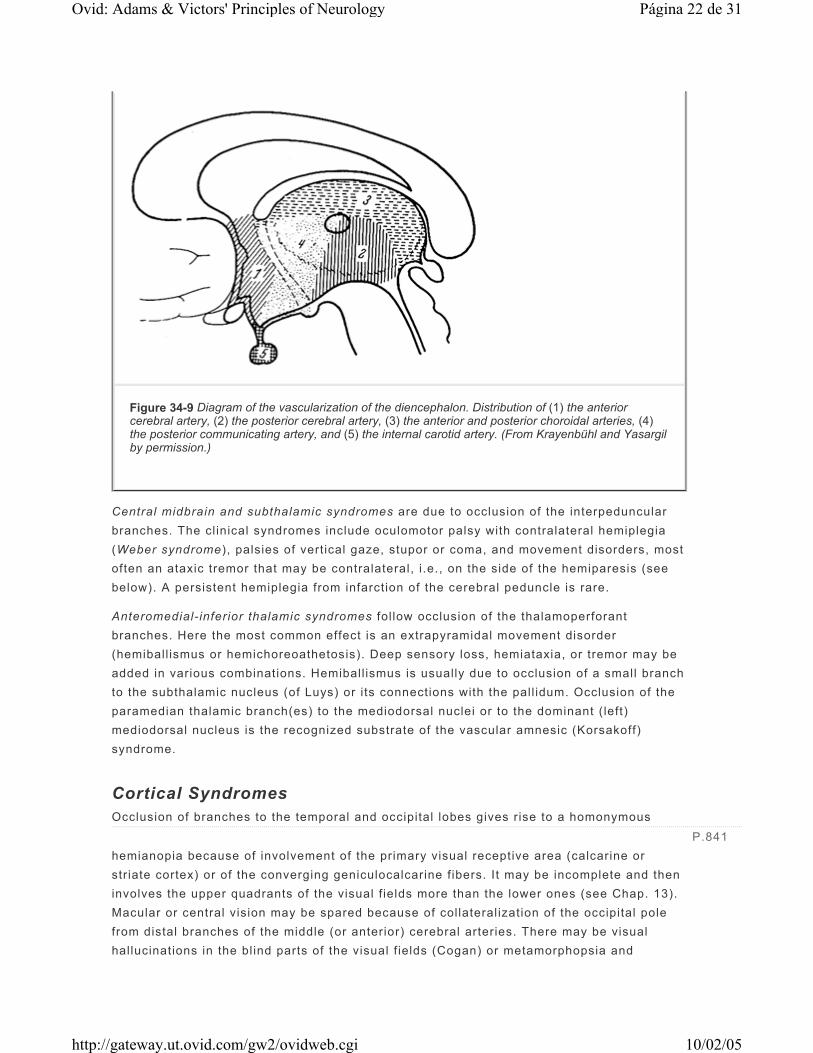
Figure 34-9 Diagram of the vascularization of the diencephalon. Distribution of (1) the anterior cerebral artery, (2) the posterior cerebral artery, (3) the anterior and posterior choroidal arteries, (4) the posterior communicating artery, and (5) the internal carotid artery. (From Krayenbühl and Yasargil by permission.)
P.841
Página 22 de 31Ovid: Adams & Victors' Principles of Neurology
10/02/05http://gateway.ut.ovid.com/gw2/ovidweb.cgi
palinopsia (Brust and Behrens). Posterior cort ical infarcts of the dominant hemisphere cause alexia (with or without agraphia), anomia (amnesic aphasia), a variety of visual agnosias, and rarely some degree of impaired memory. The anomias (dysnomias) are most severe for colors, but the naming of other visually presented material such as pictures, musical notes, mathematical symbols, and manipulable objects may also be impaired. The patient may treat objects as famil iar—that is, describe their functions and use them correctly—but be unable to name them. Color dysnomia and amnesic aphasia are more often present in this syndrome than is alexia. The defect in retentive memory is of varying severity and may or may not improve with the passage of t ime.
A complete proximal arterial occlusion leads to a syndrome that combines cortical and anterior-proximal syndromes in part or totally. The vascular lesion may be either an embolus or an atherosclerotic thrombus.
Bilateral Cortical Syndromes These may occur as a result of successive infarctions or from a single embolic or thrombotic occlusion of the upper basilar artery, especially i f the posterior communicating arteries are unusually small.
Bilateral lesions of the occipital lobes, i f extensive, cause total bl indness of the cort ical type, i.e., a bilateral homonymous hemianopia, sometimes accompanied by unformed visual hallucinations. The pupil lary reflexes are preserved, and the optic discs appear normal. Sometimes the patient is unaware of being blind and may deny it even when it is pointed out to him (Anton syndrome). More frequently the lesions are incomplete, and a sector of the visual f ields, usually on one side, remains intact. When the intact remnant is small, vision may fluctuate from moment to moment as the patient attempts to capture the image in the island of intact vision, in which case hysteria may be suspected. The Balint syndrome (page 491) is another effect of bi lateral occipitoparietal border-zone lesions. In bilateral lesions confined to the occipital poles, there may be a loss of central vision only (homonymous central scotomas). With more anteriorly placed lesions of the occipital pole, there may be homonymous paracentral scotomas, or the occipital poles may be spared, leaving the patient with only central vision. Horizontal or alt i tudinal f ield defects are usually due to lesions affecting the upper or lower banks of the calcarine sulci.
With bilateral lesions that involve the inferomedial portions of the temporal lobes, the impairment of memory may be severe, simulating the Korsakoff amnesic state. In several of our patients, a solely left-sided infarction of the inferomedial temporal lobe impaired retentive memory. The amnesic state and its accompaniments are fully described in Chap. 21 and Chap. 22. Bilateral mesiotemporal-occipital lesions may be accompanied by a lack of recognit ion of faces (prosopagnosia). These and other effects of temporal and occipital lesions are discussed in Chap. 13 and Chap. 22.
Vertebral Artery The vertebral arteries are the chief arteries of the medulla; each supplies the lower three-
P.842
Página 23 de 31Ovid: Adams & Victors' Principles of Neurology
10/02/05http://gateway.ut.ovid.com/gw2/ovidweb.cgi
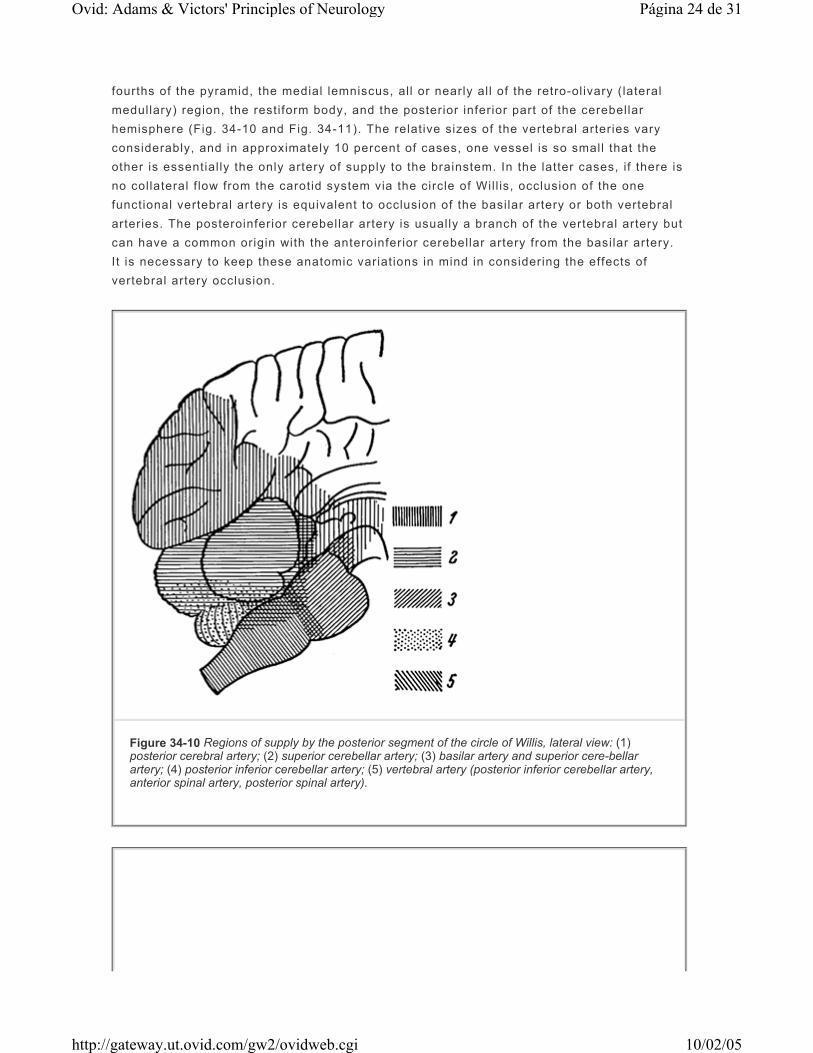
fourths of the pyramid, the medial lemniscus, all or nearly al l of the retro-olivary (lateral medullary) region, the restiform body, and the posterior inferior part of the cerebellar hemisphere (Fig. 34-10 and Fig. 34-11). The relative sizes of the vertebral arteries vary considerably, and in approximately 10 percent of cases, one vessel is so small that the other is essential ly the only artery of supply to the brainstem. In the latter cases, if there is no collateral f low from the carotid system via the circle of Wil l is, occlusion of the one functional vertebral artery is equivalent to occlusion of the basilar artery or both vertebral arteries. The posteroinferior cerebellar artery is usually a branch of the vertebral artery but can have a common origin with the anteroinferior cerebellar artery from the basilar artery. It is necessary to keep these anatomic variations in mind in considering the effects of vertebral artery occlusion.
Figure 34-10 Regions of supply by the posterior segment of the circle of Willis, lateral view: (1) posterior cerebral artery; (2) superior cerebellar artery; (3) basilar artery and superior cere-bellar artery; (4) posterior inferior cerebellar artery; (5) vertebral artery (posterior inferior cerebellar artery, anterior spinal artery, posterior spinal artery).
Página 24 de 31Ovid: Adams & Victors' Principles of Neurology
10/02/05http://gateway.ut.ovid.com/gw2/ovidweb.cgi
Since the vertebral arteries have a long extracranial course and pass through the transverse processes of C6 to C1 vertebrae before entering the cranial cavity, one might expect them to be subject to trauma, spondylotic compression, and a variety of other vertebral diseases. In our experience this happens only infrequently. We have not seen convincing examples of spondylotic occlusion. Neck rotation (chiropractic or other) of extreme degree has been reported as a cause of brainstem ischemia in more than 50 cases (Caplan). Extreme extension of the neck, as occurs in women having their hair washed in beauty parlors, may give rise to transient symptoms in the territories of the vertebral arteries. Dissection of the vertebral artery is well documented; i t declares itself by cervico-occipital pain and deficits of brainstem function, usually bilateral. It may be precipitated by vigorous and protracted bouts of coughing or by trauma to the neck or head. Examples of “posterior circulation” stroke in children have been reported in association with odontoid hypoplasia and other atlantoaxial dislocations, causing the vertebral arteries to be stretched or kinked in their course through the transverse processes of C1-C2 (Phil l ips et al).
The results of vertebral artery occlusion are quite variable. When there are two good-sized arteries, occlusion of one may cause no recognizable symptoms and signs or pathologic changes. If the subclavian artery is blocked proximal to the origin of the left vertebral artery, exercise of the arm on that side may draw blood from the right vertebral and basilar arteries, down the left vertebral and into the distal left subclavian artery—sometimes result ing in the symptoms of basilar insuff iciency. This phenomenon, described in 1961 by
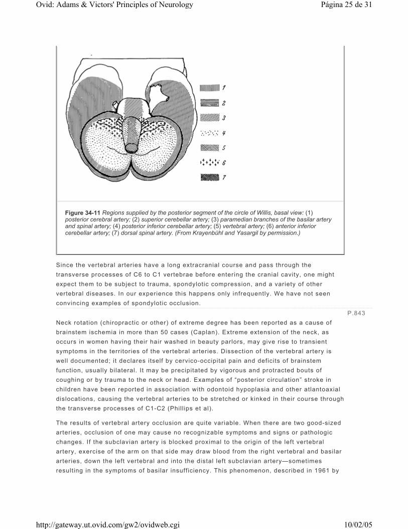
Figure 34-11 Regions supplied by the posterior segment of the circle of Willis, basal view: (1) posterior cerebral artery; (2) superior cerebellar artery; (3) paramedian branches of the basilar artery and spinal artery; (4) posterior inferior cerebellar artery; (5) vertebral artery; (6) anterior inferior cerebellar artery; (7) dorsal spinal artery. (From Krayenbühl and Yasargil by permission.)
P.843
Página 25 de 31Ovid: Adams & Victors' Principles of Neurology
10/02/05http://gateway.ut.ovid.com/gw2/ovidweb.cgi
Reivich and colleagues, was referred to by Fisher as the “subclavian steal .” Its most notable feature is transient weakness of the left arm on exercise. There may also be headache and claudication or pain of the arm. If the occlusion of the vertebral artery is so situated as to block the branches supplying the lateral medulla, a characteristic syndrome may result; in this configuration, vertigo may be a prominent symptom (see below). When the vertebral branch to the anterior spinal artery is blocked, f low from the other (corresponding) branch is usually suff icient to prevent infarction of the cervical cord. If the branch to the pyramid is occluded, part of the pyramidal tract may be infarcted, depending on the adequacy of collateral f low. Any of these branches may become occluded in its course as well as at i ts origin from the vertebral artery, with similar effects.
Rarely, occlusion of the vertebral artery or one of i ts medial branches produces an infarct that involves the medullary pyramid, the medial lemniscus, and the emergent hypoglossal f ibers; the resultant syndrome consists of a contralateral paralysis of arm and leg (with sparing of the face), contralateral loss of posit ion and vibration sense, and ipsilateral paralysis and later atrophy of the tongue. A more l imited lesion, from occlusion of one spinal artery arising from the vertebral artery, gives rise to a contralateral hemiplegia (rarely a quadriplegia) that spares the face. This is a fragment of the medial medullary syndrome (Fig. 34-12). Occlusion of a vertebral artery low in the neck is usually compensated by anastomotic f low to the upper part of the artery via the thyrocervical, deep cervical, and occipital arteries or reflux from the circle of Wil l is.
Lateral Medullary Syndrome Known also as the syndrome of Wallenberg (who described a case in 1895), this common syndrome (Fig. 34-12) is produced by infarction of a wedge of lateral medulla lying posterior to the inferior ol ivary nucleus. The complete syndrome, as outl ined by Fisher and colleagues (1961), reflects the involvement of the vestibular nuclei (nystagmus, oscil lopsia, vertigo, nausea, vomiting); spinothalamic tract (contralateral impairment of pain and thermal sense over half the body); descending sympathetic tract ( ipsilateral Horner syndrome—miosis, ptosis, decreased sweating); issuing f ibers of the ninth and tenth nerves (hoarseness, dysphagia, ipsilateral paralysis of the palate and vocal cord, diminished gag reflex); otolithic nucleus (vertical diplopia and i l lusion of t i lt ing of vision); ol ivocerebellar and/or spinocerebellar f ibers and, sometimes, restiform body and inferior cerebellum ( ipsi lateral ataxia of l imbs, fal l ing or toppling to the ipsilateral side, or lateropulsion); descending tract and nucleus of the f if th nerve (pain, burning, and impaired sensation over ipsilateral half of the face); nucleus and tractus solitarius (loss of taste); and rarely cuneate and gracile nuclei (numbness of ipsi lateral l imbs). Fragmentary syndromes occur frequently at the onset of the stroke: vertigo and ptosis, toppling and vertical diplopia, hoarseness and disequilibrium, etc. Vertigo alone, however, is not an indication of lateral medullary infarction. The smallest infarction we have studied gave rise only to symptoms of lateropulsion and mild ipsilateral l imb ataxia.
The eye signs of lateral medullary infarction may be dif f icult to interpret. Often there is a fragment of an internuclear ophthalmoplegia or a skew deviation (the globe on the affected
P.844
Página 26 de 31Ovid: Adams & Victors' Principles of Neurology
10/02/05http://gateway.ut.ovid.com/gw2/ovidweb.cgi
side usually higher). Direction changing nystagmus (with different head posit ions) is said to be the most useful feature that distinguishes vestibular nucleus infarction from labyrinthine disease, but some variants of benign posit ional vertigo also show this sign (see also Chap. 15).
The entire lateral medullary syndrome, one of the most striking in neurology, is almost always due to infarction, with only a small number of cases being the result of hemorrhage or tumor. Although it was tradit ionally attr ibuted to occlusion of the posterior inferior cerebellar artery (PICA), careful studies have shown that in 8 out of 10 cases it is the vertebral artery that is occluded; in the remainder, either the posterior inferior cerebellar artery or one of the lateral medullary arteries is occluded.
In recent years, we have become aware from our own cases that although most of those with lateral medullary infarction do well and make a considerable recovery, sudden and unexpected death may occur from respiratory or cardiac arrest in the absence of cerebellar swell ing or basilar artery thrombosis. Cases of this nature are reviewed by Norving and Cronqvist. The related and important issue of recognizing cerebellar swell ing after vertebral or PICA occlusion and the subsequent need for surgery is discussed under “Treatment of Cerebral Edema and Raised Intracranial Pressure” later in the chapter.
Basilar Artery The branches of the basilar artery may be conveniently grouped as follows: (1) paramedian, 7 to 10 in number, supplying a wedge of pons on either side of the midline; (2) short circumferential, 5 to 7 in number, supplying the lateral two-thirds of the pons and the middle and superior cerebellar peduncles; (3) long circumferential, 2 on each side (the superior and anterior inferior cerebellar arteries), which run laterally around the pons to reach the cerebellar hemispheres (Fig. 34-10 and Fig. 34-11); and (4) several paramedian (interpeduncular) branches at the bifurcation of the basilar artery supplying the high midbrain and medial subthalamic regions. The interpeduncular and other branches of the posterior cerebral artery have been described above.
The picture of basilar occlusion due to thrombosis may arise in several ways: (1) occlusion of the basilar artery itself, usually in the lower third at the site of an atherosclerotic plaque; (2) occlusion of both vertebral arteries; and (3) occlusion of a single vertebral artery when it is the only one of adequate size. It must be emphasized that thrombosis more frequently involves a branch of the basilar artery than the trunk (basilar branch occlusion). When the obstruction is embolic, the embolus usually lodges at the upper bifurcation of the basilar or in one of the posterior cerebral arteries, since the embolus, i f i t is small enough to pass through the vertebral artery, easily traverses the length of the basilar artery, which is of greater diameter than either vertebral artery.
The syndrome of basilar artery occlusion , as delineated by Kubik and Adams, reflects the involvement of a large number of structures: corticospinal and corticobulbar tracts, cerebellum, middle and superior cerebellar peduncles, medial and lateral lemnisci, spinothalamic tracts, medial longitudinal fasciculi, pontine nuclei, vestibular and cochlear
P.845P.846
Página 27 de 31Ovid: Adams & Victors' Principles of Neurology
10/02/05http://gateway.ut.ovid.com/gw2/ovidweb.cgi
nuclei, descending hypothalamospinal sympathetic f ibers, and the third through eighth cranial nerves (the nuclei and their segments within the brainstem).
The complete basilar syndrome comprises bilateral long tract signs (sensory and motor) with variable cerebellar, cranial nerve, and other segmental abnormalit ies of the brainstem. Often the patient is comatose because of ischemia of the high midbrain reticular activating system. Others are mute and quadriplegic but conscious, reflecting interruption of motor pathways but sparing of the reticular activating system (“locked-in” syndrome; see page 370). In the presence of the full syndrome, it is usually not diff icult to make the correct diagnosis. The aim should be, however, to recognize basilar insuff iciency long before the stage of total deficit has been reached. The early manifestations (some in the form of transient ischemic attacks) occur in many combinations, described in detail further on (page 860).
Occlusion of branches at the bifurcation (top) of the basilar artery results in a remarkable number of complex syndromes that include, in various combinations, somnolence, memory defects, akinetic mutism, visual hallucinations, ptosis, disorders of ocular movement (convergence spasm, paralysis of vertical gaze, retraction nystagmus, pseudoabducens palsy, retraction of upper eyelids, skew deviation of the eyes), an agitated confusional state, and visual defects. These have been reviewed by Petit and coworkers and Castaigne and associates as paramedian thalamic, subthalamic, and midbrain infarctions and by Caplan as “top of the basilar” syndromes.
The main signs of occlusion of the superior cerebellar artery are ipsilateral cerebellar ataxia (middle and/or superior cerebellar peduncles); nausea and vomiting; slurred (pseudobulbar) speech; and loss of pain and thermal sensation over the opposite side of the body (spinothalamic tract). Partial deafness, static tremor of the ipsilateral upper extremity, an ipsilateral Horner syndrome, and palatal myoclonus have also been reported.
With occlusion of the anteroinferior cerebellar artery , the extent of the infarct is extremely variable, since the size of this artery and the territory it supplies vary inversely with the size and territory of supply of the posteroinferior cerebellar artery. The principal f indings are vertigo; nausea; vomiting; nystagmus; t innitus and sometimes unilateral deafness; facial weakness; ipsilateral cerebellar ataxia (inferior or middle cerebellar peduncle); an ipsilateral Horner syndrome and paresis of conjugate lateral gaze; and contralateral loss of pain and temperature sense of the arm, trunk, and leg (lateral spinothalamic tract). If the occlusion is close to the origin of the artery, the corticospinal f ibers may also be involved, producing a hemiplegia; i f distal, there may be cochlear and labyrinthine infarction. Cerebellar swell ing herniation has not been seen in our cases or in the 20 cases collected by Amarenco and Hauw.
The most important manifestation of al l these brainstem infarcts is the “crossed” cranial nerve and long tract sensory or motor deficit. These crossed syndromes, which may involve cranial nerves III through XII, are l isted in Table 47-2. Although the f inding of bi lateral neurologic signs strongly suggests brainstem involvement, it must be emphasized that in many instances of infarction within the basilar territory, the signs are l imited to one side of the body, with or without cranial nerve involvement, indicating occlusion of a branch of the
P.847
Página 28 de 31Ovid: Adams & Victors' Principles of Neurology
10/02/05http://gateway.ut.ovid.com/gw2/ovidweb.cgi
basilar artery, not of the main trunk.
It is impossible to distinguish a hemiplegia of pontine origin from one of cerebral origin on the basis of motor signs alone. With brainstem lesions as with cerebral ones, a f laccid paralysis gives way to spasticity after a few days or weeks, and there is no satisfactory explanation for the variabil i ty in this period of delay or for the occurrence in some cases of spasticity from the onset of the stroke. There may also be a combined hemiparesis and ataxia of the l imbs on the same side. Localization of the level of the brainstem lesion depends upon coexisting neurologic signs. With a hemiplegia of pontine origin, the eyes may deviate to the side of the paralysis, i .e., the opposite of what occurs with supratentorial lesions. The pattern of sensory disturbance may be helpful. A dissociated sensory deficit over the ipsilateral face and contralateral half of the body usually indicates a lesion in the lower brainstem, while a hemisensory loss including the face and involving all modalit ies indicates a lesion in the upper brainstem, in the thalamus, or deep in the white matter of the parietal lobe. When posit ion sense, two-point discrimination, and tacti le localization are affected relatively more than pain or thermal and tacti le sense, a cerebral lesion is suggested; the converse indicates a brainstem localization. Bilaterality of both motor and sensory signs is almost certain evidence that the lesion lies infratentorial ly. When hemiplegia or hemiparesis and sensory loss are coextensive, the lesion usually l ies supratentorial ly. Addit ional manifestations that point unequivocally to a brainstem site are rotational dizziness, diplopia, cerebellar ataxia, a Horner syndrome, and deafness. The several brainstem syndromes i l lustrate the important point that the cerebellar pathways, spinothalamic tract, tr igeminal nucleus, and sympathetic f ibers can be involved at different levels, and “neighboring” phenomena must be used to identify the exact site.
A myriad of proper names have been applied to the brainstem syndromes, as noted in Table 47-2. Most of them were originally described in relation to tumors and other nonvascular diseases. The diagnosis of vascular disorders in this region of the brain is not greatly facil i tated by a knowledge of these eponymic syndromes; it is much more profitable to memorize the anatomy of the brainstem. The principal syndromes to be recognized are the ful l basilar, vertebral, posteroinferior cerebellar, anteroinferior cerebellar, superior cerebellar, pontomedullary, and medial medullary. Figure 34-12 to Figure 34-15 (supplied by Dr. C. M. Fisher and used in all previous edit ions of this book) present both medial and lateral syndromes at four levels of the medulla and pons. Other syndromes can usually be identif ied as fragments or combinations of the major ones.
Lacunar Syndromes As one might surmise, small penetrating branches of the cerebral arteries may become occluded, and the result- ing infarcts may be so small or so situated as to cause no symptoms whatsoever. As the softened tissue is removed, it leaves a small cavity, or lacune. Early in the twentieth century, Pierre Marie confirmed the occurrence of mult iple deep small cavit ies of this type, f irst described by Durant-Fardel in 1843. Marie referred to the condit ion as état lacunaire . Marie distinguished these lesions from a f ine loosening of t issue around thickened vessels that enter the anterior and posterior perforated spaces, a change to which he gave the name état criblé. Pathologists have not always agreed on these distinctions, but Fisher and Adams have taken the posit ion that the lacunar state is
Página 29 de 31Ovid: Adams & Victors' Principles of Neurology
10/02/05http://gateway.ut.ovid.com/gw2/ovidweb.cgi
always due to occlusion of small arteries, 50 to 200 mm in diameter, and the cribriform state to mere thickening of vessels and sl ight fraying of the surrounding t issue.
In our cl inical and pathologic material, there has always been a strong correlation of the lacunar state with a combination of hypertension and atherosclerosis and, to a lesser degree, with diabetes. Sacco and colleagues, in a population-based study in Rochester, Minnesota, found hypertension in 81 percent of patients with lacunar infarctions. One may hypothesize that the basis of the lacunar state is unusually severe atherosclerosis that has not just involved the large arteries, as it usually does, but has extended into their f inest branches.
When Fisher examined a series of such lesions in serial sections, from a basal parent artery up to and through the lacune, he found atheroma and thrombosis and less often embolic occlusion of small vessels to be the basic abnormality in some (usually the larger) lacunes, and a l ipohyalin degeneration and occlusion of small vessels in the smaller ones. In some instances the latter changes had resulted in false aneurysm formation, resembling the Charcot-Bouchard aneurysms that underl ie brain hemorrhage (see further on). Usually 4 to 6 and sometimes up to 10 to 15 lacunes are found in any given brain specimen. They are situated, in descending order of frequency, in the putamen and caudate nuclei, thalamus, basis pontis, internal capsule, and convolutional white matter. The cavit ies range from 3 to 15 mm in diameter, and whether they cause symptoms depends entirely on their location. In a series of 1042 consecutive adults whose brains were examined postmortem, Fisher observed one or more lacunes in 11 percent.
Fisher has delineated some of the more frequent symptomatic forms. If the lacune lies in the territory of a lenticulostriate artery, i .e., in the internal capsule or adjacent corona radiata, i t may cause a pure motor hemiplegia . Symptoms may be abrupt in onset or evolve over several hours, but in some instances (10 of 34 lacunar syndromes, according to Weisberg) the neurologic deficit evolves stepwise and relatively slowly, over as long a period as 2 to 3 days, simulating a small hemorrhage. CT scanning shows the lesions in about two-thirds of cases, and MRl, in probably a higher proportion. The weakness in pure motor hemiplegia involves the face, arm, and leg. Sometimes the motor disorder takes the form of a dysarthria, with or without hemiplegia. Recovery, which may begin within hours, days, or weeks, is often nearly complete.
Similarly, a lacune of the lateral thalamus or parietal white matter presents as a pure hemisensory defect (pure sensory stroke). The incidence, course, and outcome are much the same as in a pure hemiplegia. In the basis pontis, the syndrome may be one of pure motor hemiplegia, mimicking that of internal capsular infarction except for relative sparing of the face and the presence of an ipsilateral paresis of conjugate gaze in some cases; or there may be a combination of dysarthria and clumsiness of one hand. Occasionally a lacunar infarction of the pons, midbrain, internal capsule, or parietal white matter gives rise to a hemiparesis with ataxia on the same side as the weakness (Fisher; Sage and Lepore).
P.848P.849
P.850P.851
Página 30 de 31Ovid: Adams & Victors' Principles of Neurology
10/02/05http://gateway.ut.ovid.com/gw2/ovidweb.cgi
Some of the brainstem syndromes may blend with basilar branch syndromes. There are many other, less frequent syndromes, too numerous to tabulate here. Mult iple lacunar infarcts, involving the corticospinal and corticobulbar tracts, are by far the most common cause of pseudobulbar palsy.
In all these cases of lacunar infarction, the diagnosis depends essential ly on the occurrence of the aforementioned certain unique stroke syndromes of l imited proportions. As mentioned above, CT scanning is less reliable than MRI in demonstrating the lacunes. The EEG may be helpful in a negative sense; in the case of lacunes in the pons or the internal capsule, there is a notable discrepancy between the unilateral paralysis or sensory loss and the negligible electrical changes over the affected hemisphere.
Página 31 de 31Ovid: Adams & Victors' Principles of Neurology
10/02/05http://gateway.ut.ovid.com/gw2/ovidweb.cgi


![Neurovascular Devices and Clinical ApplicationsNEW [Autosaved]](https://img.pdfslide.net/doc/110x75/58a02b871a28ab4e768b65d7/neurovascular-devices-and-clinical-applicationsnew-autosaved.jpg)