Embed Size (px)
Citation preview
6
Neutron Influence in Charged Particle Therapy
Su Youwu, Li Wuyuan, Xu Junkui, Mao Wang and Li Zongqiang
Institute of Modern Physics, Chinese Academy of Sciences,
P. R. China
1. Introduction
In charged particle therapy, neutron will emit due to beam loss on accelerator components, beam shaping devices, treatment nozzle, and especially in the deposition of beam interactions in the patient. Those neutrons usually have high energy and strong penetrating power, so it will cause additional dose on the tumor and health tissue of the patient, which may influence the treatment effect. Thus it is essential to know the neutron influence in charged particle therapy.
Different to conventional rays, charged particle (proton and heavy ion) with certain energy has a determined “range” in matters and they loss maximum energy at the end of the “range”, which is the so-called Bragg peak. During the process of cancer therapy, charged particles can be stopped at tumor site by adjusting the energy range to achieve maximum tumor destruction using the characteristics of Bragg peak. Moreover, the normal tissue surrounding a tumor will not receive much irradiation due to Bragg peak.
Based on the characteristics of charged particles in cancer therapy, many institutions develop this technique and many facilities for cancer therapy are under construction such as German Heavy Ion Research Center (GSI, Germany), National Institution of Radiological Sciences (NIRS, Japan). However secondary neutron produced due to beam loss in accelerator component may cause damage to patient body which is not well known. Thus it is important to investigate the information about neutron radiation field in charged particle cancer therapy.
2. Neutron production mechanism of ion reactions
The process of charged particles interact with matters is very complicated. The interaction of particle with matters results in the production of different types of radiation, including gamma ray and neutrons with complex energy distribution. Heavy ions loss energy mainly through ionization and excitation of atoms as it traverses matter, which is called electromagnetic cascade process. Except at low velocities, it loses a negligible amount of energy in nuclear collisions. No neutrons will be produced in electromagnetic cascade process, but it determines the energy loss of the particle in matter, and especially when the
www.intechopen.com
Modern Practices in Radiation Therapy
86
material is the patient tissue, it determines the heavy ion dose. The interaction between ions and atomic electrons can be well described by a term “stopping power” - (dE/dx).
2 4 2 22
2 2 2
4 Z 2ln
(1 )
dE e n mc
dx mc I
(2-1)
Where Z is the atomic number of the heavy particle, e is the electron charge, n is the number of electrons per unit volume in the medium, and I is the mean excitation energy of the
medium, = v/c is the relative velocity of the particle.
The distance traveled per unit energy loss is given by the reciprocal of the stopping power. Thus, the range R(T) of a particle with kinetic energy(T) is the integral of the reciprocal of the stopping power down to zero energy and can be written in the following form:
)(
Z)(
2fM
TR
(2-2)
It is important to note that the mean range of particles of a given speed is proportional to the mass and varies as the inverse square of their charge. The dependence of the Bethe formula on Z2 implies that particles with the same mass and energy, but with inverse charge will have same range in matter.
Though it may not produce neutrons in the process of electromagnetic interactions with charged particles, it is the dominant way of charged particles to loss energy. A large part of heavy charged particle loss energy through interaction with extranuclear electron. Thus at different depth heavy charged particle has different energy at different depths, so the neutron produced by heavy charged particle on thick target can be regard as the results of interactions of heavy charged particle with matter in different energy region particle in different energy region and nucleus.
2.1 Nuclear interactions
Nuclear interactions include compound interactions and direct interactions.
In the process of compound interactions, the emission of the particles can be described by an evaporation process similar to the evaporation of a molecule from the surface of a liquid. The spectrum of the emitted neutrons may be described by a Maxwellian distribution:
)/exp( TEBEdE
dNnn
n
(2-3)
Where En is the energy of neutron, B is a constant, and T is the nuclear temperature. The nuclear temperature is characteristic of the target residual nucleus and its excitation energy, and has dimensions of energy. Its value lies between 2 and 8 MeV.
2.2 Heavy ion-nucleus interactions
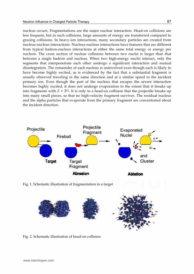
Nuclear interactions of heavy ions as they pass through matter arise from grazing or head-
on collisions. In grazing collisions, fragmentation of either the incident ion or the target
www.intechopen.com
Neutron Influence in Charged Particle Therapy
87
nucleus occurs. Fragmentations are the major nuclear interaction. Head-on collisions are
less frequent, but in such collisions, large amounts of energy are transferred compared to
grazing collisions. In heavy-ion interactions, many secondary particles are created from
nucleus-nucleus interactions. Nucleus-nucleus interactions have features that are different
from typical hadron-nucleus interactions at either the same total energy or energy per
nucleon. The cross section of nuclear collisions between two nuclei is larger than that
between a single hadron and nucleus. When two high-energy nuclei interact, only the
segments that interpenetrate each other undergo a significant interaction and mutual
disintegration. The remainder of each nucleus is uninvolved even though each is likely to
have become highly excited, as is evidenced by the fact that a substantial fragment is
usually observed traveling in the same direction and at a similar speed to the incident
primary ion. Even though the part of the nucleus that escapes the severe interaction
becomes highly excited, it does not undergo evaporation to the extent that it breaks up
into fragments with Z < 3[1]. It is only in a head-on collision that the projectile breaks up
into many small pieces, so that no high-velocity fragment survives. The residual nucleus
and the alpha particles that evaporate from the primary fragment are concentrated about
the incident direction.
Fig. 1. Schematic illustration of fragmentation in a target
Fig. 2. Schematic illustration of head-on collision
www.intechopen.com
Modern Practices in Radiation Therapy
88
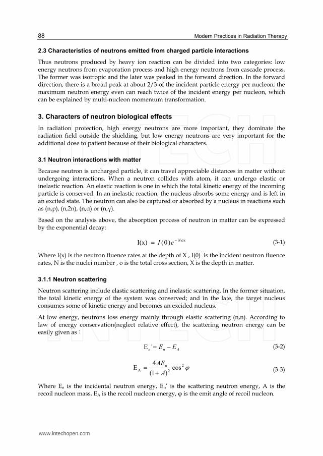
2.3 Characteristics of neutrons emitted from charged particle interactions
Thus neutrons produced by heavy ion reaction can be divided into two categories: low energy neutrons from evaporation process and high energy neutrons from cascade process. The former was isotropic and the later was peaked in the forward direction. In the forward direction, there is a broad peak at about 2/3 of the incident particle energy per nucleon; the maximum neutron energy even can reach twice of the incident energy per nucleon, which can be explained by multi-nucleon momentum transformation.
3. Characters of neutron biological effects
In radiation protection, high energy neutrons are more important, they dominate the radiation field outside the shielding, but low energy neutrons are very important for the additional dose to patient because of their biological characters.
3.1 Neutron interactions with matter
Because neutron is uncharged particle, it can travel appreciable distances in matter without undergoing interactions. When a neutron collides with atom, it can undergo elastic or inelastic reaction. An elastic reaction is one in which the total kinetic energy of the incoming particle is conserved. In an inelastic reaction, the nucleus absorbs some energy and is left in an excited state. The neutron can also be captured or absorbed by a nucleus in reactions such as (n,p), (n,2n), (n,α) or (n,γ).
Based on the analysis above, the absorption process of neutron in matter can be expressed by the exponential decay:
xNeI )0(I(x)
(3-1)
Where I(x) is the neutron fluence rates at the depth of X , I(0) is the incident neutron fluence rates, N is the nuclei number , ┫ is the total cross section, X is the depth in matter.
3.1.1 Neutron scattering
Neutron scattering include elastic scattering and inelastic scattering. In the former situation, the total kinetic energy of the system was conserved; and in the late, the target nucleus consumes some of kinetic energy and becomes an excided nucleus.
At low energy, neutrons loss energy mainly through elastic scattering (n,n). According to law of energy conservation(neglect relative effect), the scattering neutron energy can be easily given as:
Ann EE 'E
(3-2)
2
2A cos)1(
4E
A
AEn
(3-3)
Where En is the incidental neutron energy, En’ is the scattering neutron energy, A is the recoil nucleon mass, EA is the recoil nucleon energy, φ is the emit angle of recoil nucleon.
www.intechopen.com
Neutron Influence in Charged Particle Therapy
89
In elastic collision, Neutron will loss energy from 0 to nEA
A2)1(
4
; neutron loss more energy
on light nucleon than on heavy nucleon, it implies that light material have higher stopping
power than heavy material, for heavy nucleon such as uranium or lead target, neutron can
hardly lose energy by elastic scattering. But for hydrogen nucleon, neutron can almost lose
all kinetic energy at one elastic collision, so the hydrogen contents of material are of great
sense to neutron moderation and measurement.
At high energy region, inelastic scattering becomes more and more important. “Elastic
scattering can’t absorb high energy neutrons effectively. The higher the incident energy the
more forward peaked is the elastic scattering, and beyond 150MeV it is almost always in the
forward direction. Since the incident particle remain at least a fraction [(A-1)/(A+1)] of the
incident particle energy, it is clear that in most material with moderate mass, litter change in
either energy or direction results from elastic scattering”[2].
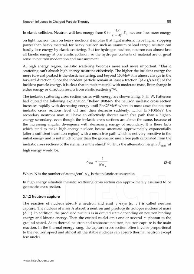
The inelastic scattering cross section varies with energy are shown in fig. 3. H. W. Patterson
had quoted the following explanation “Below 100MeV the neutron inelastic cross section
increases rapidly with decreasing energy until En<25MeV where in most cases the neutron
inelastic cross sections level off and then decrease suddenly……For En>100MeV the
secondary neutrons may still have an effectively shorter mean free path than a higher-
energy secondary, even though the inelastic cross sections are about the same, because of
the increasing angular divergence with decreasing energy of secondary. It is these facts
which tend to make high-energy nucleon beams attenuate approximately exponentially
(after a sufficient transition region) with a mean free path which is not very sensitive to the
initial energy and is not much longer than the geometric mean free path calculated from the
inelastic cross sections of the elements in the shield” [3]. Thus the attenuation length atten at
high energy would be:
inN 1atten
(3-4)
Where N is the number of atoms/cm3 in is the inelastic cross section.
In high energy situation inelastic scattering cross section can approximately assumed to be geometric cross section.
3.1.2 Neutron capture
The reaction of nucleus absorb a neutron and emit γ-rays (n, γ) is called neutron
capture. The nucleus of mass A absorb a neutron and produce its isotopes nucleus of mass
(A+1). In addition, the produced nucleus is in excited state depending on neutron binding
energy and kinetic energy. Then the excited nuclei emit one or several γ photon to the
ground stated. As to thermal neutron and resonance neutron, neutron capture is the main
reaction. In the thermal energy rang, the capture cross section often inverse proportional
to the neutron speed and almost all the stable nuclides can absorb thermal neutron except
few nuclei.
www.intechopen.com
Modern Practices in Radiation Therapy
90
The neutron capture reaction are quiet different with different nuclides at thermal and
resonance energy region, but at higher energy region the neutron capture cross section
variations become very gentle, fig.3 shows the neutron absorb cross section varies with
target mass number. The figure can be well expressed by the following equation:
69.0
a A43 (mb) (3-5)
The formula should be valid up to several hundred GeV[2], but it does not valid for some
special nuclides include hydrogen. Note that the nuclides which can not apply to the
formula above are those of great importance to neutron radiation protection such as H, 10B
Cd etc. Because hydrogen which is most widely distributed nature nuclide plays an
important part in nuclear reactor moderation and cooling. Meanwhile, 10B, Gd etc also very
significant in neutron protection and measurement.
Experiment indicate the ratio of the neutron elastic cross section to the absorption cross
section almost unchanged for mostly nuclides and energy it is 0.57 [2] .
Fig. 3. Inelastic scattering cross section of neutron on different targets [3]
www.intechopen.com
Neutron Influence in Charged Particle Therapy
91
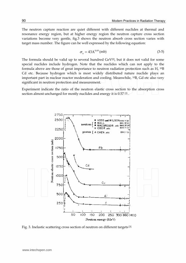
Fig. 4. Mean free path and atomic cross section as a function of mass number [2]
3.2 Human body response to neutron
3.2.1 Basic concepts of radiation dosimetry
The biological impact of radiation to human body is caused by energy deposition, thus the
energy deposited per unit mass in material (absorbed dose) is the fundamental concept of
radiation dosimetry. The absorbed dose unit is joule per kilogram and dedicated unit is gray
(Gy). However, same absorbed dose may not always cause same biological effect for
different radiation type and energy. To distinguish this difference, the International
Commission on Radiological Protection (ICRP) introduced the concept of equivalent dose [4],
which is defined as product of absorbed dose and radiation weighting factor:
RTRRT DwH ,,
(3-6)
Where DT,R is the absorbed dose of R type radiation in tissue or organ; WR is the radiation weighting factor.
The radiation weighting factor is a quantity which is related to relative biological
effectiveness. Furthermore, the relative biological effectiveness is defined as the ratio of the
doses required by two different types of radiation to cause the same level for a specified end
point. And the radiation weighting factor represents damage severity. Table 1 gives the
radiation weighting factor recommended by ICRP.
www.intechopen.com
Modern Practices in Radiation Therapy
92
Radiation type and energy region radiation weighting factor
Photon of all energy 1
Electron and μ particle:all energy region 1
Neutron energy :
<10keV 5
10~100keV 10
100keV~2MeV 20
2MeV ~20MeV 10
>20MeV 5
proton(except recoil proton),E>2MeV 5
α particle,fission fragment,heavy nuclei 20
Table 1. Radiation weighting factor (ICRP 60 publication)
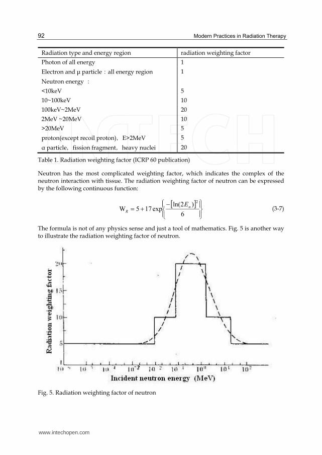
Neutron has the most complicated weighting factor, which indicates the complex of the neutron interaction with tissue. The radiation weighting factor of neutron can be expressed by the following continuous function:
6
)2ln(exp175W
2
n
R
E
(3-7)
The formula is not of any physics sense and just a tool of mathematics. Fig. 5 is another way to illustrate the radiation weighting factor of neutron.
Fig. 5. Radiation weighting factor of neutron
www.intechopen.com
Neutron Influence in Charged Particle Therapy
93
3.2.2 Radiation response of human body
The effect of radiation on the human body can be divided into deterministic effect and stochastic effect. Deterministic effect is bounded to a certain dose; it is caused by a large number of cells death. It must have a certain threshold, below this threshold the effect will not occur. Above this threshold the severity aggravating with dose is increasing, such as skin damage, cataract and temporary or permanent infertility etc. Stochastic effect may happen in any level of radiation exposure, as it is caused by a single irradiated cell mutation. The severity degree is not related to dose, but its occurrence probability will increase with dose level,such as cancer, genetic effect and so on.
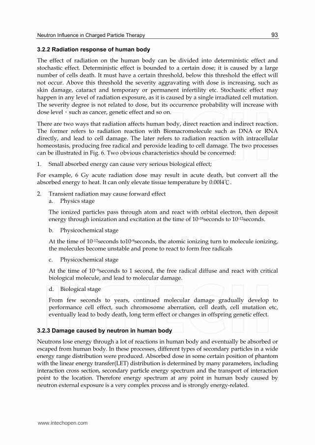
There are two ways that radiation affects human body, direct reaction and indirect reaction. The former refers to radiation reaction with Biomacromolecule such as DNA or RNA directly, and lead to cell damage. The later refers to radiation reaction with intracellular homeostasis, producing free radical and peroxide leading to cell damage. The two processes can be illustrated in Fig. 6. Two obvious characteristics should be concerned:
1. Small absorbed energy can cause very serious biological effect;
For example, 6 Gy acute radiation dose may result in acute death, but convert all the absorbed energy to heat. It can only elevate tissue temperature by 0.00l4℃.
2. Transient radiation may cause forward effect a. Physics stage
The ionized particles pass through atom and react with orbital electron, then deposit energy through ionization and excitation at the time of 10-18seconds to 10-12seconds.
b. Physicochemical stage
At the time of 10-12seconds to10-9seconds, the atomic ionizing turn to molecule ionizing, the molecules become unstable and prone to react to form free radicals
c. Physicochemical stage
At the time of 10~9seconds to 1 second, the free radical diffuse and react with critical biological molecule, and lead to molecular damage.
d. Biological stage
From few seconds to years, continued molecular damage gradually develop to performance cell effect, such chromosome aberration, cell death, cell mutation etc, eventually lead to body death, long term effect or changes in offspring genetic effect.
3.2.3 Damage caused by neutron in human body
Neutrons lose energy through a lot of reactions in human body and eventually be absorbed or escaped from human body. In these processes, different types of secondary particles in a wide energy range distribution were produced. Absorbed dose in some certain position of phantom with the linear energy transfer(LET) distribution is determined by many parameters, including interaction cross section, secondary particle energy spectrum and the transport of interaction point to the location. Therefore energy spectrum at any point in human body caused by neutron external exposure is a very complex process and is strongly energy-related.
www.intechopen.com
Modern Practices in Radiation Therapy
94
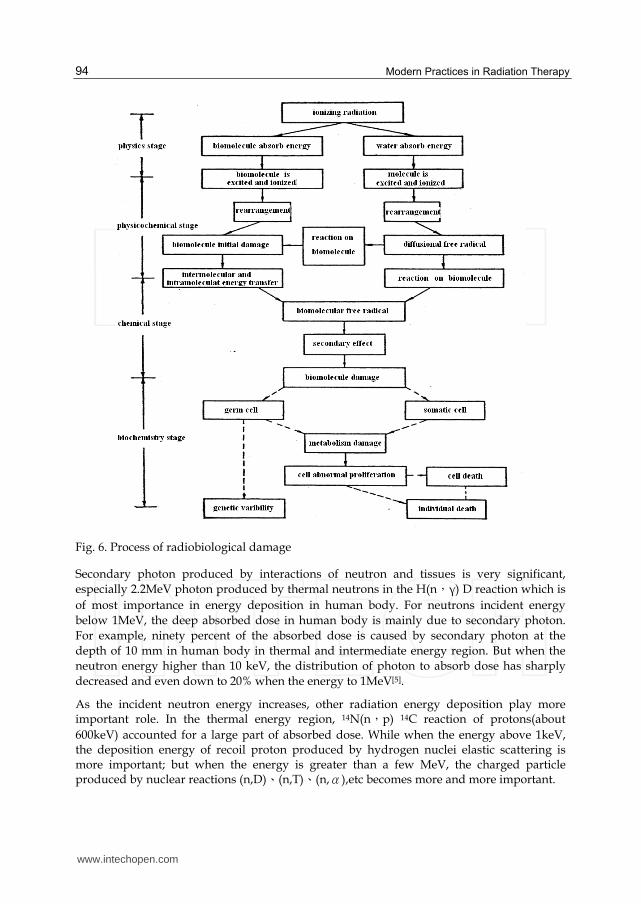
Fig. 6. Process of radiobiological damage
Secondary photon produced by interactions of neutron and tissues is very significant,
especially 2.2MeV photon produced by thermal neutrons in the H(n,γ) D reaction which is
of most importance in energy deposition in human body. For neutrons incident energy
below 1MeV, the deep absorbed dose in human body is mainly due to secondary photon.
For example, ninety percent of the absorbed dose is caused by secondary photon at the
depth of 10 mm in human body in thermal and intermediate energy region. But when the
neutron energy higher than 10 keV, the distribution of photon to absorb dose has sharply
decreased and even down to 20% when the energy to 1MeV[5].
As the incident neutron energy increases, other radiation energy deposition play more important role. In the thermal energy region, 14N(n,p) 14C reaction of protons(about
600keV) accounted for a large part of absorbed dose. While when the energy above 1keV, the deposition energy of recoil proton produced by hydrogen nuclei elastic scattering is more important; but when the energy is greater than a few MeV, the charged particle produced by nuclear reactions (n,D)、(n,T)、(n,α),etc becomes more and more important.
www.intechopen.com
Neutron Influence in Charged Particle Therapy
95
3.3 Neutron conversion factor
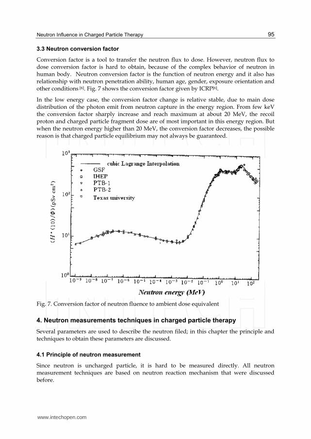
Conversion factor is a tool to transfer the neutron flux to dose. However, neutron flux to dose conversion factor is hard to obtain, because of the complex behavior of neutron in human body. Neutron conversion factor is the function of neutron energy and it also has relationship with neutron penetration ability, human age, gender, exposure orientation and other conditions [6]. Fig. 7 shows the conversion factor given by ICRP[6].
In the low energy case, the conversion factor change is relative stable, due to main dose distribution of the photon emit from neutron capture in the energy region. From few keV the conversion factor sharply increase and reach maximum at about 20 MeV, the recoil proton and charged particle fragment dose are of most important in this energy region. But when the neutron energy higher than 20 MeV, the conversion factor decreases, the possible reason is that charged particle equilibrium may not always be guaranteed.
Fig. 7. Conversion factor of neutron fluence to ambient dose equivalent
4. Neutron measurements techniques in charged particle therapy
Several parameters are used to describe the neutron filed; in this chapter the principle and techniques to obtain these parameters are discussed.
4.1 Principle of neutron measurement
Since neutron is uncharged particle, it is hard to be measured directly. All neutron measurement techniques are based on neutron reaction mechanism that were discussed before.
www.intechopen.com
Modern Practices in Radiation Therapy
96
4.1.1 Nucleus reaction method
This method is mainly based on reactions between neutron and some certain nuclide and then detecting products of these reactions to obtain the information of neutron radiation field. Usually, the reactions which can be used to detect neutron may have the following characteristics:
1. Have large neutron cross section to ensure the sensitivity of the detectors;
2. Target nucleus of high abundance in natural elements or easy to collection;
3. With large Q value which can help to discriminator the γray pulse amplitude
Based on this principle, the mostly used reactions are the following:
Neutron reaction with 10B
Reaction equation:
MeVLiHe
MeVLiHeBn
31.2*
792.2
7
3
4
2
7
3
4
2
10
5
1
0
(I)
73 480Li keV (II)
The reaction product 7Li can either be in ground state (reaction channel I) or in excited state
(reaction channel II). Experiments show that, the branch ratio of reaction channel I only
account 6.3%. Because the reaction energy is much greater than the kinetic energy of slow
neutron, kinetic energy of reaction products mainly comes from reaction energy. Moreover
the incident neutron momentum is small, the momentum of reaction products also becomes
approximately equal to zero. Thus the emit direction of two products is opposite and the
distribution proportion of reaction energy is determined. Therefore the kinetic energy of
αparticle will be 1.77 MeV and 1.47 MeV for the 7Li in ground and excited state
respectively.
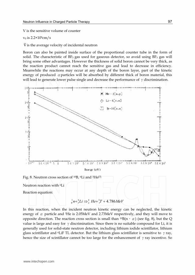
Neutron cross section in a wide energy range (from thermal to 1keV) is proportional to 1/v
and the thermal neutron cross section is about 3840×10-24cm2(see Fig.8 )[7]. The abundance of
nature boron is about 19% and it is easy to enrich.
BF3 proportional counter tube is most commonly used as gas ionization detector. In the
center of the tube there is an anode wire which is used to collect ion. The reaction products 7Li and αcause BF3 ionization and these ions are amplified by the center electric field then
collected by anode wire to form pulse signal. For gas detector, the detector efficiency is a
crucial factor. Since the inflation pressure can be very high and 90% of the tube must be
filled with BF3. Sometimes argon gas is filled in the tube in order to improve the detector
performance. The sensitivity of a BF3 counter (neutron energy less than 100keV) is:
v
vNV 0
0s
(4-1)
Where, N is the number of 10B contained in a unit volume;
www.intechopen.com
Neutron Influence in Charged Particle Therapy
97
V is the sensitive volume of counter
v0 is 2.2×105cm/s
v is the average velocity of incidental neutron
Boron can also be painted inside surface of the proportional counter tube in the form of solid. The characteristic of BF3 gas used for gaseous detector, so avoid using BF3 gas will bring some other advantages. However the thickness of solid boron cannot be very thick, as the reaction product cannot reach the sensitive gas and lead to decrease in efficiency. Meanwhile the reactions may occur at any depth of the boron layer, part of the kinetic energy of produced αparticles will be absorbed by different thick of boron material, this
will lead to generate lower pulse single and decrease the performance of γdiscrimination.
Fig. 8. Neutron cross section of 10B, 6Li and 3He[7]
Neutron reaction with 6Li
Reaction equation:
MeVTHeLin 786.43
1
4
2
6
3
1
0
In this reaction, when the incident neutron kinetic energy can be neglected, the kinetic energy of α particle and 3He is 2.05MeV and 2.73MeV respectively, and they will move to
opposite direction. The reaction cross section is small than 10B(n,α) (see fig. 8), but the Q
value is large and easy for γdiscrimination. Since there is no suitable compound for Li, it is
generally used for solid-state neutron detector, including lithium iodide scintillator, lithium glass scintillator and 6LiF TL detector. But the lithium glass scintillator is sensitive to γray,
hence the size of scintillator cannot be too large for the enhancement of γray incentive. So
www.intechopen.com
Modern Practices in Radiation Therapy
98
the electron produced by γray will escape from the sensitive volume before it is absorbed.
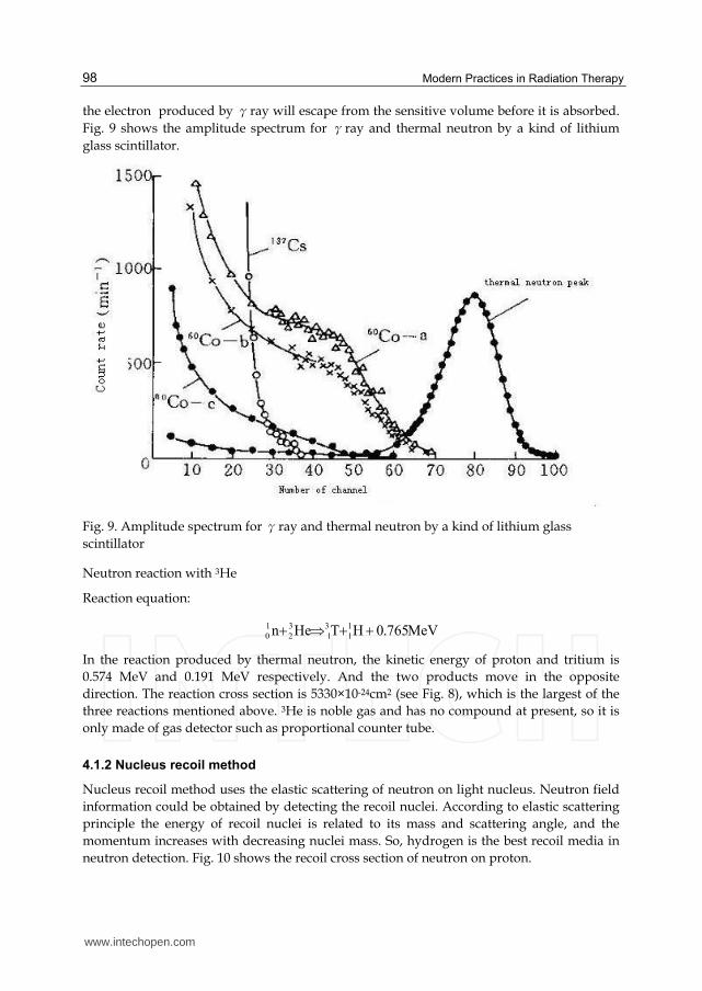
Fig. 9 shows the amplitude spectrum for γray and thermal neutron by a kind of lithium
glass scintillator.
Fig. 9. Amplitude spectrum for γray and thermal neutron by a kind of lithium glass
scintillator
Neutron reaction with 3He
Reaction equation:
MeV765.0HTHen 1
1
3
1
3
2
1
0
In the reaction produced by thermal neutron, the kinetic energy of proton and tritium is
0.574 MeV and 0.191 MeV respectively. And the two products move in the opposite
direction. The reaction cross section is 5330×10-24cm2 (see Fig. 8), which is the largest of the
three reactions mentioned above. 3He is noble gas and has no compound at present, so it is
only made of gas detector such as proportional counter tube.
4.1.2 Nucleus recoil method
Nucleus recoil method uses the elastic scattering of neutron on light nucleus. Neutron field
information could be obtained by detecting the recoil nuclei. According to elastic scattering
principle the energy of recoil nuclei is related to its mass and scattering angle, and the
momentum increases with decreasing nuclei mass. So, hydrogen is the best recoil media in
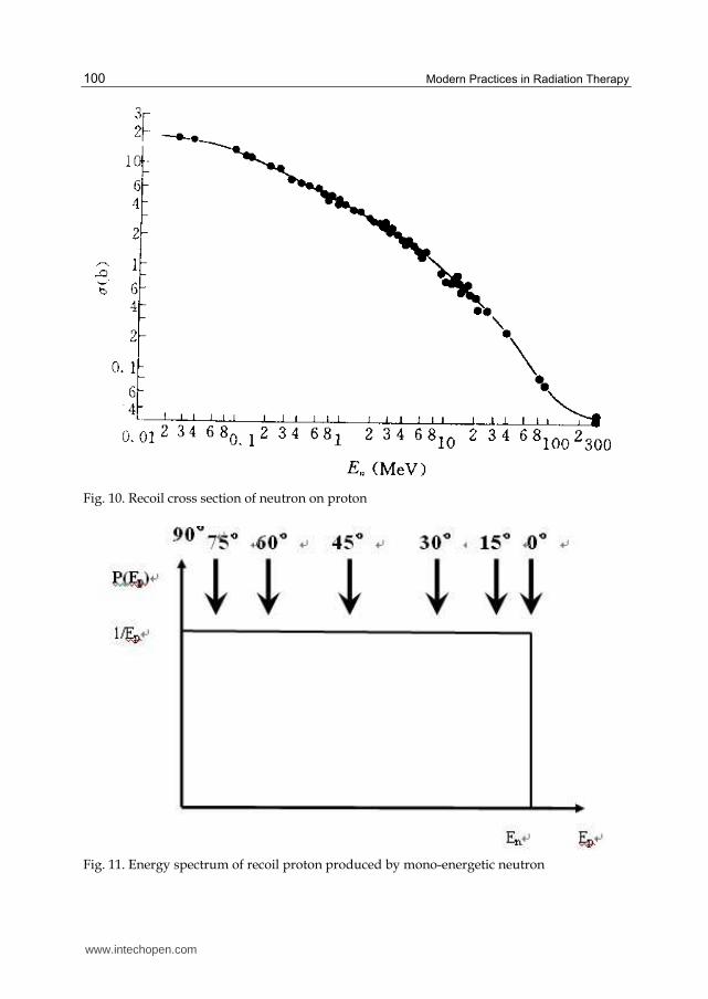
neutron detection. Fig. 10 shows the recoil cross section of neutron on proton.
www.intechopen.com
Neutron Influence in Charged Particle Therapy
99
If the incident direction of neutron is known, the neutron energy information could be obtained by measuring the spectrum of recoil proton from a certain angle. The energy information can be expressed by:
)cos(E
CE 2
aEW
″′
″′
(4-2)
Here C is the normalized constant.
Thus, the conversion of recoil nucleus spectra to neutron spectra attribute to the recalibrate
of energy scale(αEcos2φreplace EA). This method is called differential measurement. But
the weakness of this method is the low efficieny, for the thickness of recoil material and the
small spanned solid angle of detector.
When the neutron energy is not too high, the scattered proton in all direction could be detected in the hydrogen-filled ionization chamber or counter, the angular distribution of the scattered proton in the center of mass is isotropic, thus the ratio of scattering cross
section in certain angular() and the total scattering cross section should be ┫()/┫=1/4π.
The probability of recoiled proton scattering in angle with energy EA could be given:
aEnEA
1)(W
(4-3)
Where En is the incidental neutron energy, 2)1(
4a
A
A
The formula shows that the recoiled proton energy could be any value in the range of 0 to
aEn with equal probability when the incident neutron energy is En. So if the detector is
exposed to monoenergetic neutron, the shape of recoil spectrum could be a rectangle (see
Fig. 11).
If the incident neutron energy is a function of (E) , the recoil nucleus of energy EA could be
produced by any neutron with energy greater than EA/a . So the producing probability of
nucleus with EA could be given as:
aEA
E
dEEEC
/
A )()()E(W
(4-4)
Applying differential to the formula, the measured neutron spectrum can be expressed by:
aEAA
dE
dW
E
EcE
E)(
)(
(4-5)
This method is called integration method, which is used for neutron With energy not too high and the recoil proton range is not too large. However, due to the shielding effect of the detector surface, the recoil spectrum is not a rectangle as Fig. 11 shown even the detector was exposed in a monoenergy neutron field.
www.intechopen.com
Modern Practices in Radiation Therapy
100
Fig. 10. Recoil cross section of neutron on proton
Fig. 11. Energy spectrum of recoil proton produced by mono-energetic neutron
www.intechopen.com
Neutron Influence in Charged Particle Therapy
101
When neutron is detected by scintillator, its efficiency is higher than gas detector. Meanwhile, due to its high stopping power, it can be used for higher energy neutron up to 20MeV measurement. But other effect of scintillator such as multiple scattering and γ ray
etc will influence the shape of the neutron spectrum. Its spectrum is also not a rectangle. At the same time, the spectrometer energy resolution will become worse with the increasing volume of scintillator (the corresponding neutron energy will also increase).
4.1.3 Nucleus fission method
Nucleus fission method usually refers to detect the fission fragments which are produced by neutron captured by heavy nucleus to obtain neutron field information. Slow neutron and fast neutron can lead to heavy nucleus fission and release about 150~170MeV for each fusion, dividing into two fragments with masses nearly equal to each other. The energy released in these reactions almost equal distribute to the two fragments and each with 40~110MeV. The most advantages based on fission method is the high output pulse amplitude and is unaffected by other radiations.
4.1.4 Activation method
The information of the neutron field can be obtained by measure the neutron induced radioactivity. Some neutron reaction channel has a certain threshold, so only those neutrons with the energy higher than that threshold can induce reaction and produce certain instable nuclide. So the neutrons of some certain energy region can be obtained by measuring the products of different channel with different threshold. If we neglect the sample absorption to neutron, the radiation intensity of sample can be given as:
)1(A / TeN (4-6)
Where N is the number of nuclei in the sample, ┫ is activation cross section, φ is neutron fluence, T is the total irradiation time, ┬ is the mean life time of radioactive nuclei
The induced radioactivity is a function of radiation nuclide life. Generally, if the irradiation time is long enough (about 5~6 half-life), the radionuclide in sample will reach saturation (Amax=N┫φ). This value has no relationship with irradiation time, only with the number of target nucleon and the neutron flux.
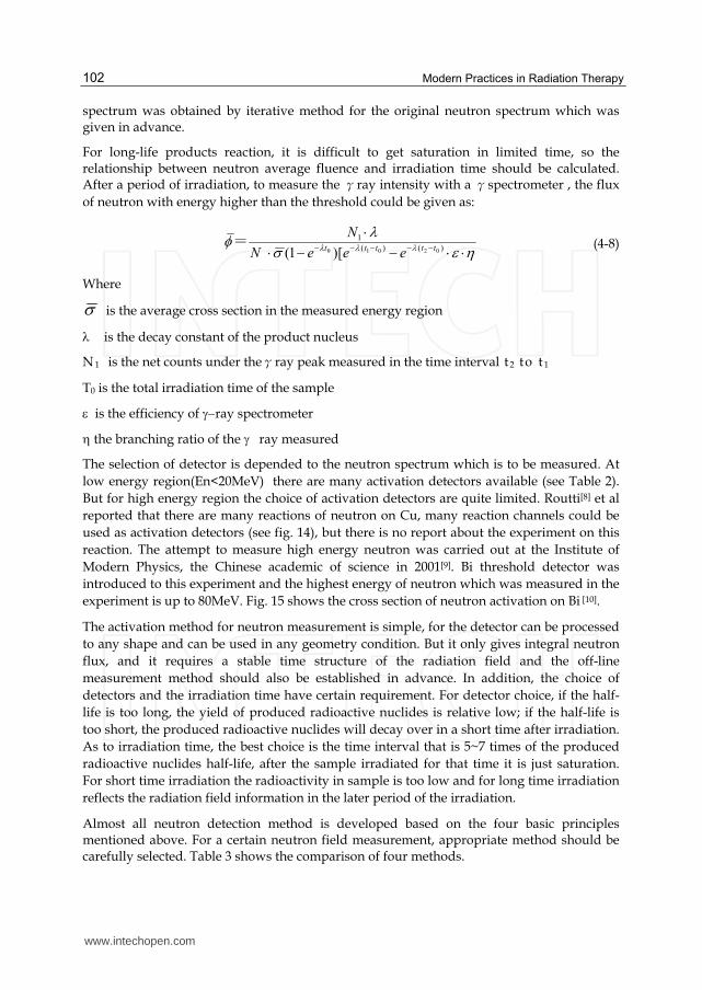
For the neutron activation measurement, the ideal cross section shape of threshold detector is rectangle, below the threshold energy the cross section is 0 and above the threshold energy the cross section is a constant. However, it is difficult to find the ideal reaction channel of the characteristic mentioned above, Fig. 13 shows the excite function of some threshold detector. From Fig. 13 almost all the activation reaction cross sectionσis the
function of neutron energy En. So in actual measurement, the concept of average cross section is introduced.
dE
E)(
E)(E)(
(4-7)
It can be seen from (4-7) that the average cross section is related to neutron spectrum. Yet the neutron spectrum is the parameter which is needed to be measured, so final neutron
www.intechopen.com
Modern Practices in Radiation Therapy
102
spectrum was obtained by iterative method for the original neutron spectrum which was given in advance.
For long-life products reaction, it is difficult to get saturation in limited time, so the relationship between neutron average fluence and irradiation time should be calculated. After a period of irradiation, to measure the γray intensity with a γspectrometer , the flux
of neutron with energy higher than the threshold could be given as:
)()(
1
02010 )[1(ttttt
eeeN
N=
(4-8)
Where
is the average cross section in the measured energy region
is the decay constant of the product nucleus
N 1 is the net counts under theγray peak measured in the time interval t 2 to t 1
T0 is the total irradiation time of the sample
is the efficiency of ray spectrometer
the branching ratio of the �ray measured
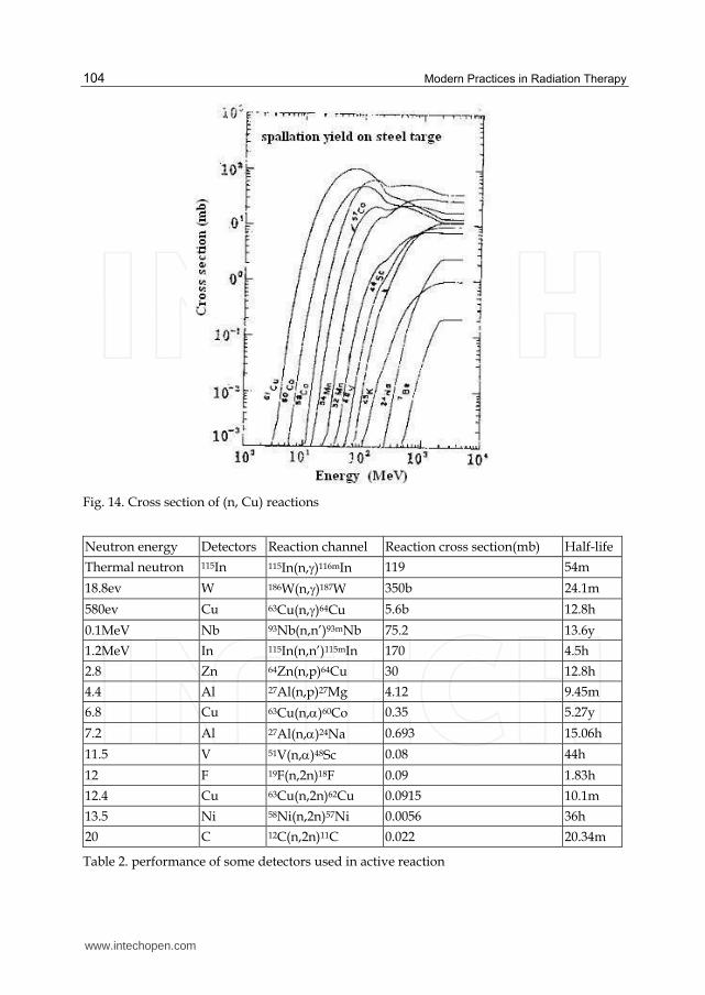
The selection of detector is depended to the neutron spectrum which is to be measured. At
low energy region(En<20MeV) there are many activation detectors available (see Table 2).
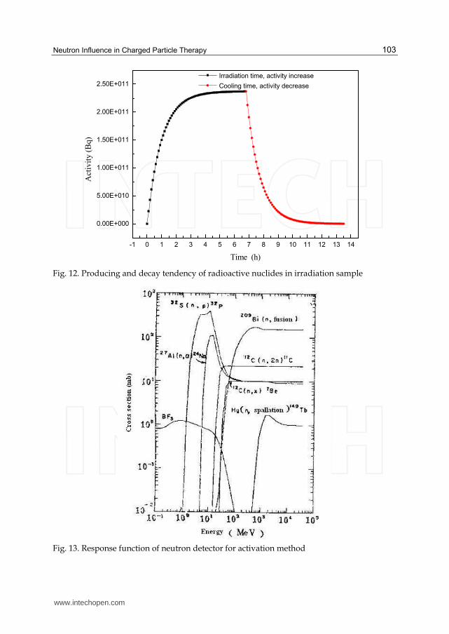
But for high energy region the choice of activation detectors are quite limited. Routti[8] et al
reported that there are many reactions of neutron on Cu, many reaction channels could be
used as activation detectors (see fig. 14), but there is no report about the experiment on this
reaction. The attempt to measure high energy neutron was carried out at the Institute of
Modern Physics, the Chinese academic of science in 2001[9]. Bi threshold detector was
introduced to this experiment and the highest energy of neutron which was measured in the
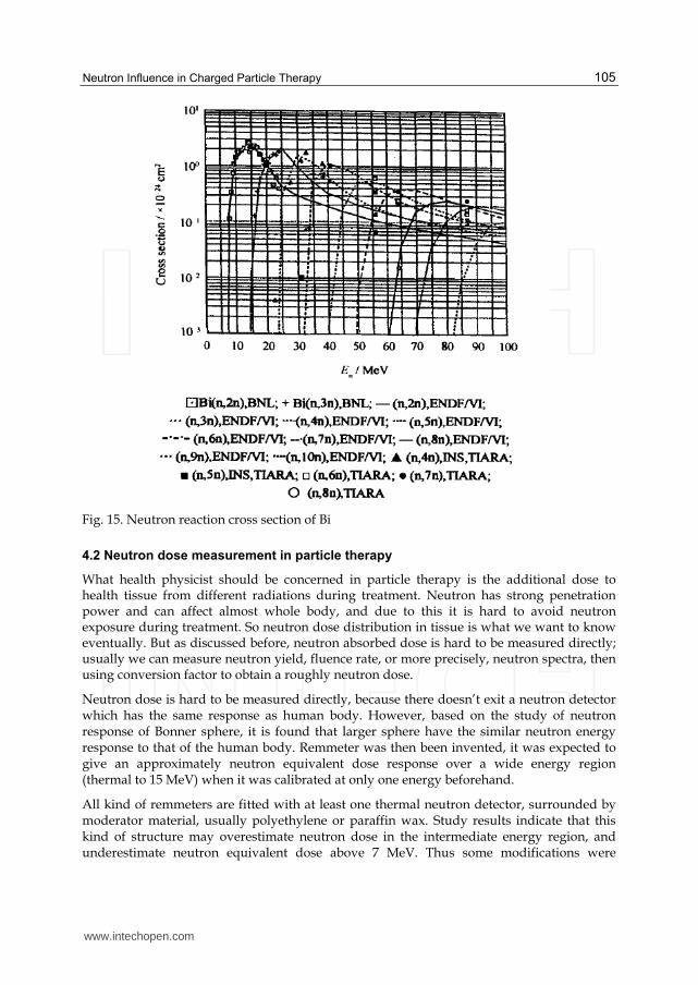
experiment is up to 80MeV. Fig. 15 shows the cross section of neutron activation on Bi [10].
The activation method for neutron measurement is simple, for the detector can be processed
to any shape and can be used in any geometry condition. But it only gives integral neutron
flux, and it requires a stable time structure of the radiation field and the off-line
measurement method should also be established in advance. In addition, the choice of
detectors and the irradiation time have certain requirement. For detector choice, if the half-
life is too long, the yield of produced radioactive nuclides is relative low; if the half-life is
too short, the produced radioactive nuclides will decay over in a short time after irradiation.
As to irradiation time, the best choice is the time interval that is 5~7 times of the produced
radioactive nuclides half-life, after the sample irradiated for that time it is just saturation.
For short time irradiation the radioactivity in sample is too low and for long time irradiation
reflects the radiation field information in the later period of the irradiation.
Almost all neutron detection method is developed based on the four basic principles mentioned above. For a certain neutron field measurement, appropriate method should be carefully selected. Table 3 shows the comparison of four methods.
www.intechopen.com
Neutron Influence in Charged Particle Therapy
103
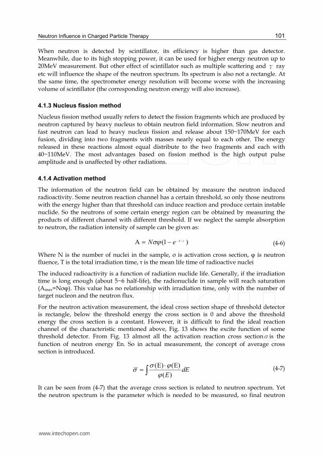
-1 0 1 2 3 4 5 6 7 8 9 10 11 12 13 14
0.00E+000
5.00E+010
1.00E+011
1.50E+011
2.00E+011
2.50E+011A
ctiv
ity (
Bq)
Time (h)
Irradiation time, activity increase
Cooling time, activity decrease
Fig. 12. Producing and decay tendency of radioactive nuclides in irradiation sample
Fig. 13. Response function of neutron detector for activation method
www.intechopen.com
Modern Practices in Radiation Therapy
104
Fig. 14. Cross section of (n, Cu) reactions
Neutron energy Detectors Reaction channel Reaction cross section(mb) Half-life
Thermal neutron 115In 115In(n,)116mIn 119 54m
18.8ev W 186W(n,)187W 350b 24.1m
580ev Cu 63Cu(n,)64Cu 5.6b 12.8h
0.1MeV Nb 93Nb(n,n’)93mNb 75.2 13.6y
1.2MeV In 115In(n,n’)115mIn 170 4.5h
2.8 Zn 64Zn(n,p)64Cu 30 12.8h
4.4 Al 27Al(n,p)27Mg 4.12 9.45m
6.8 Cu 63Cu(n,)60Co 0.35 5.27y
7.2 Al 27Al(n,)24Na 0.693 15.06h
11.5 V 51V(n,)48Sc 0.08 44h
12 F 19F(n,2n)18F 0.09 1.83h
12.4 Cu 63Cu(n,2n)62Cu 0.0915 10.1m
13.5 Ni 58Ni(n,2n)57Ni 0.0056 36h
20 C 12C(n,2n)11C 0.022 20.34m
Table 2. performance of some detectors used in active reaction
www.intechopen.com
Neutron Influence in Charged Particle Therapy
105
Fig. 15. Neutron reaction cross section of Bi
4.2 Neutron dose measurement in particle therapy
What health physicist should be concerned in particle therapy is the additional dose to health tissue from different radiations during treatment. Neutron has strong penetration power and can affect almost whole body, and due to this it is hard to avoid neutron exposure during treatment. So neutron dose distribution in tissue is what we want to know eventually. But as discussed before, neutron absorbed dose is hard to be measured directly; usually we can measure neutron yield, fluence rate, or more precisely, neutron spectra, then using conversion factor to obtain a roughly neutron dose.
Neutron dose is hard to be measured directly, because there doesn’t exit a neutron detector which has the same response as human body. However, based on the study of neutron response of Bonner sphere, it is found that larger sphere have the similar neutron energy response to that of the human body. Remmeter was then been invented, it was expected to give an approximately neutron equivalent dose response over a wide energy region (thermal to 15 MeV) when it was calibrated at only one energy beforehand.
All kind of remmeters are fitted with at least one thermal neutron detector, surrounded by moderator material, usually polyethylene or paraffin wax. Study results indicate that this kind of structure may overestimate neutron dose in the intermediate energy region, and underestimate neutron equivalent dose above 7 MeV. Thus some modifications were
www.intechopen.com
Modern Practices in Radiation Therapy
106
performed to improve the response of the remmeter to intermediate energy region. Undoubtedly A-B remmeter is the mostly successful one. In the structure of the moderator of a A-B remmeter, it contains a thermal neutron absorb layer in which holes have been drilled to allow some slow neutron pass. This improved the neutron response effectively.
For the purpose of measure higher-energy neutron dose, improvement of the moderator of
the remmeter continued. Birattari[11] reported an Improved remmeters which consist of
high-atomic number inserts such as lead or tungsten in the moderator. The interaction of
high-energy neutrons with this inserted material causes neutron multiplication and energy
Method Principle Characteristics examples
Nucleus excite
Measuring the radioactive nuclide produced by neutron reaction with some materials
With small volume and easy operation;
With strong Anti-γ ability;
Only obtain the average fluence;
No online measurement result;
Complex technology of accumulating radioactivity measurement;
Limitation choice in detector.
115 In (n, g) 116 mIn ; 19F(n,2n)18F; 27Al(n,a)24Na; 27Al(n,p)27Mg; 63Cu(n,2n)62Cu ……
Nucleus reaction
Measuring the producing charged particles in neutron reaction
With strong Anti-γ ability;
Applied to thermal neutron measurement;
BF3 count tube; 3He count tube; Boron painted counter tube; Li glass scintillator
Nucleus Recoil
Measuring the emit proton by neutron scattering on proton
Neutron-proton scattering cross section are well known;
Neutron energy information can be obtained;
γ ray influence.
Recoil proton ionizing; Organ crystal scintillator; Plastic scintillator; Recoil proton nuclear emulsion
Nucleus fission
Measuring the fusion fragments of fission reaction of neutron on 235U、238U、239Pu etc
With grate Q value in fusion and a obvious distinction of other; competitive reaction;
Suitable for thermal neutron measurement.
Fusion ionizing chamber
Table 3. Comparison of four methods for neutron measurement
www.intechopen.com
Neutron Influence in Charged Particle Therapy
107
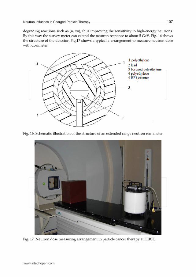
degrading reactions such as (n, xn), thus improving the sensitivity to high-energy neutrons.
By this way the survey meter can extend the neutron response to about 5 GeV. Fig. 16 shows
the structure of the detector, Fig.17 shows a typical a arrangement to measure neutron dose
with dosimeter.
Fig. 16. Schematic illustration of the structure of an extended range neutron rem meter
Fig. 17. Neutron dose measuring arrangement in particle cancer therapy at HIRFL
www.intechopen.com
Modern Practices in Radiation Therapy
108
4.3 Neutron spectrum measurement
For a certain neutron field, knowing neutron dose distribution is not enough. As we described before, many factors may influence neutron dose in tissue, such as neutron energy, produce point, irradiation direction etc. thus neutron spectra measurement is required. Neutron dose distribution can be calculated by using conversion factor if neutron spectrum is known.
Activation method is an old way to measure neutron spectra, by using different threshold
detectors neutron fluence with energy higher than certain energy can be measured. But at
higher energy region, appropriate threshold detectors are inadequate, hence it confined the
usage of this method in particle therapy.
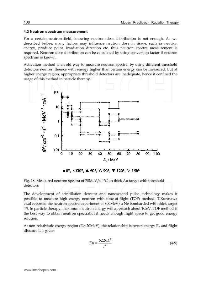
Fig. 18. Measured neutron spectra of 78MeV/u 12C on thick Au target with threshold detectors
The development of scintillation detector and nanosecond pulse technology makes it
possible to measure high energy neutron with time-of-flight (TOF) method. T.Kurosawa
et.al reported the neutron spectra experiment of 800MeV/u Ne bombarded with thick target
[12]. In particle therapy, maximum neutron energy will approach about 1GeV. TOF method is
the best way to obtain neutron spectrabut it needs enough flight space to get good energy
solution.
At non-relativistic energy region (En<20MeV), the relationship between energy En and flight
distance L is given:
2
25226En
t
L
(4-9)
www.intechopen.com
Neutron Influence in Charged Particle Therapy
109
Where t unit is ns, L unit is m, En unit is MeV.
At relativistic energy region, the relationship between neutron energy and its velocity is
given:
2
0
2
0 )m
c
v1
mEn c
″′
′
(4-10)
Flight distance L is easy to determine, but to obtain the flight time is more difficult, which
needs to know the start time and the end time. Usually the flight time is determined by the
associate particle method and the pulse bunch method, and the end single of neutron flight
time is determined by the neutron detector which is located in the terminal of flying
distance. Fast organic scintillator are often used as end signal detector, combined with fast
photomultiplier tube due to this the system time resolution is better than 1ns.
4.4 Result and discussions
Some experimental and theoretical results were reported in recent years. Nakamura and
Korusowa[13-15] had performed a systematic experiment with TOF method in 1998. In their
experiment, neutron yield, energy spectrum and angular distribution for 100~800MeV/u He、C、Ne bombarded with carbon, aluminum, copper and lead target were obtained.
Although the aim of these experiments are not for particle therapy, but the information
obtained were quite valuable to estimate neutron dose during treatment. With the rapid
progress of cancer therapy research, more attention is focused on the neutron yield in the
energy region for cancer therapy. For proton it is about 250MeV and for carbon ion is about
400MeV/u. Meanwhile, the target materials more inclined to tissue equivalent such as
water, polyethylene and carbon. K.Gunzert-Marx et al[16] measured the neutron distribution
of 200MeV/u 12C on thick water target in 2004, and D. Schardt et al[17] measured nuclear
fragments and fast neutron spectrum of 200MeV/u and 400MeV/u 12C on various thickness
water target in 2007.
Monte-Carlo calculation is also widely used in estimating neutron field in particle therapy.
A., Agosteo[18] simulated the double differential cross section(see fig 4-7) for neutron yield
in particle therapy in 1996, the good agreement with experimental results provide us a good
tool to evaluate neutron dose before treatment.
At the same time, neutron dose distributions are investigated in different conditions. H.
Iwase et.al. measured neutron dose angular distribution in a particle therapy facility with
extended neutron rem meter Wendi-II in 2007[19], pencil-like 12C beam was used in their
experiment.
The experimental and calculated results show that secondary neutron dose in cancer
therapy is about 1% of heavy ion dose. Since neutron can affect a large volume, to obtain the
same treatment dose, different tumor dimension may cause different neutron dose.
Furthermore, different treatment geometry arrangement and different irradiation direction
may affect neutron dose too, so it is essential to evaluate neutron dose for a certain therapy
facility.
www.intechopen.com
Modern Practices in Radiation Therapy
110
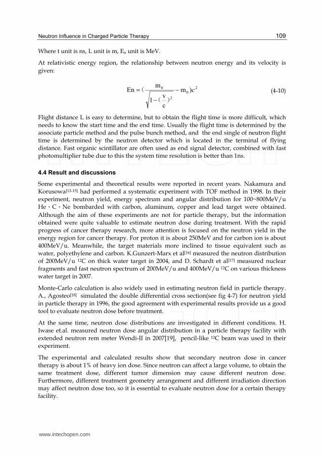
Fig. 19. Neutron spectra from 180 MeV/nucleon C ions incident on a C target
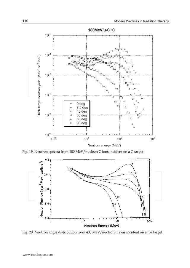
Fig. 20. Neutron angle distribution from 400 MeV/nucleon C ions incident on a Cu target
www.intechopen.com
Neutron Influence in Charged Particle Therapy
111
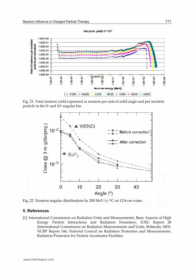
Fig. 21. Total neutron yield expressed as neutron per unit of solid angle and per incident particle in the 0o and 10o angular bin
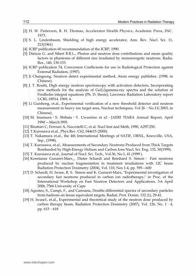
Fig. 22. Neutron angular distributions by 200 MeV/u 12C on 12.8-cm water.
5. References
[1] International Commission on Radiation Units and Measurements, Basic Aspects of High Energy Particle Interactions and Radiation Dosimtery, ICRU Report 28 (International Commission on Radiation Measurements and Units, Bethesda, MD). NCRP Report 144, National Council on Radiation Protection and Measurements, Radiation Protection for Particle Accelerator Facilities.
www.intechopen.com
Modern Practices in Radiation Therapy
112
[2] H. W. Patterson, R. H. Thomas, Accelerator Health Physics, Academic Press, INC. 1973.
[3] S. L. Lindenbaum, Shielding of high energy accelerator, Ann. Rev. Nucl. Sci. 11, 213(1961)
[4] ICRP publication 60 recommendation of the ICRP, 1990. [5] Dietzze G. and Sibert B.R.L., Photon and neutron dose contributions and mean quality
factors in phantoms of different size irradiated by monoerergetic neutrons. Radia. Res., 140, 130-133.
[6] ICRP publication 74, Conversion Coefficients for use in Radiological Protection against External Radiation, (1997).
[7] Ji Changsong, Neutron detect experimental method, Atom energy publisher, (1998, in Chinese).
[8] J. T. Routti, High energy neutron spectroscopy with activation detectors, Incorporating new methods for the analysis of Ge(Li)gamma-ray spectra and the solution of Fredholm integral equations (Ph. D. thesis), Lawrence Radiation Laboratory report UCRL-18514. 1969, 4.
[9] Li Guisheng, et.al., Experimental verification of a new threshold detector and neutron measurement in heavy ion target area, Nuclear techniques, Vol 26,No.11( 2003, in
Chinese). [10] M. Imamura,S. Shibata,Y. Uwamino et a1.JAERI TIARA Annual Report, April
1994'—March l995. [11] Birattari C, Ferrrari A, Nuccetelli C, et al. Nucl Inst and Meth, 1990, A297:250. [12] T.Kurosawa et.al., Phys.Rev. C62, 044615 (2000). [13] T. Nakamura et.al., the 4th International Meetings of SATIF, ORNL, Knoxville, USA,
Sep., (1998). [14] T. Kurosawa, et.al., Measurements of Secondary Neutrons Produced from Thick Targets
Bombarded by High-Energy Helium and Carbon Ions Nucl. Sci. Eng. 132, 30(1999). [15] T. Kurosawa et.al., Journal of Nucl. Sci. Tech., Vol.36, No.1, 41 (1999 ). [16] Konstanze Gunzert-Marx_, Dieter Schardt and Reinhard S. Simon, Fast neutrons
produced by nuclear fragmentation in treatment irradiations with 12C beam Radiation Protection Dosimetry (2004), Vol. 110, Nos 1-4, pp. 595---600
[17] D. Schardt, H. Iwase, R. S. Simon and K. Gunzert-Marx, "Experimental investigation of secondary fast neutrons produced in carbon ion radiotherapy," in Proc. of the International Workshop on Fast Neutron Detectors and Applications, 3-6 April 2006, 7566 University of Cape
[18] Agosteo, S., Campi, F., and Caresana, Double-differential spectra of secondary particles from hadrons on tissue equivalent targets, Radiat. Prot. Dosim. 132 (1), 29-41.
[19] H. Iwase1, et.al., Experimental and theoretical study of the neutron dose produced by carbon therapy beam, Radiation Protection Dosimetry (2007), Vol. 126, No. 1–4,
pp. 615–618
www.intechopen.com
Modern Practices in Radiation TherapyEdited by Dr. Gopishankar Natanasabapathi
ISBN 978-953-51-0427-8Hard cover, 370 pagesPublisher InTechPublished online 30, March, 2012Published in print edition March, 2012
InTech EuropeUniversity Campus STeP Ri Slavka Krautzeka 83/A 51000 Rijeka, Croatia Phone: +385 (51) 770 447 Fax: +385 (51) 686 166www.intechopen.com
InTech ChinaUnit 405, Office Block, Hotel Equatorial Shanghai No.65, Yan An Road (West), Shanghai, 200040, China
Phone: +86-21-62489820 Fax: +86-21-62489821
Cancer is the leading cause of death in economically developed countries and the second leading cause ofdeath in developing countries. It is an enormous global health encumbrance, growing at an alarming pace.Global statistics show that in 2030 alone, about 21.4 million new cancer cases and 13.2 million cancer deathsare expected to occur, simply due to the growth, aging of the population, adoption of new lifestyles andbehaviors. Amongst the several modes of treatment for cancer available, Radiation treatment has a majorimpact due to technological advancement in recent times. This book discusses the pros and cons of thistreatment modality. This book "Modern Practices in Radiation Therapy" has collaged topics contributed by topnotch professionals and researchers all around the world.
How to referenceIn order to correctly reference this scholarly work, feel free to copy and paste the following:
Su Youwu, Li Wuyuan, Xu Junkui, Mao Wang and Li Zongqiang (2012). Neutron Influence in Charged ParticleTherapy, Modern Practices in Radiation Therapy, Dr. Gopishankar Natanasabapathi (Ed.), ISBN: 978-953-51-0427-8, InTech, Available from: http://www.intechopen.com/books/modern-practices-in-radiation-therapy/neutron-influence-in-charged-particle-therapy