Embed Size (px)
Citation preview
New insights into the tPA-Annexin A2 interaction: is Annexin A2 Cys8 the sole
requirement for this association?
Oriol Roda*#, M. Luz Valero*, Sandra Peiró*, David Andreu*✦ , Francisco X. Real*#✦
and Pilar Navarro#�
* Departament de Ciències Experimentales i de la Salut, Facultat de Ciències de la Salut i de la Vida,
Universitat Pompeu Fabra, 08003-Barcelona, Spain.
# Unitat de Biologia Cel· lular i Molecular, Institut Municipal d’Investigació Mèdica, 08003-Barcelona,
Spain.
✦ These authors contributed equally to this work.
� Address correspondence to: Pilar Navarro, Unitat de Biologia Cel· lular i Molecular, Institut Municipal
d’Investigació Mèdica, Dr. Aiguader, 80, 08003-Barcelona, Spain. Tel: 34-93-2211009; Fax: 34-93-
2213237; E-mail: [email protected].
Running title
Analysis of the tPA-Annexin A2 interaction domain
Copyright 2002 by The American Society for Biochemistry and Molecular Biology, Inc.
JBC Papers in Press. Published on December 4, 2002 as Manuscript M207605200 by guest on M
arch 31, 2018http://w
ww
.jbc.org/D
ownloaded from
Summary
Annexin A2 has been described as an important receptor for tissue-type plasminogen activator in
endothelium and other cell types. Interaction between tissue-type plasminogen activator and its
cellular receptor is critical for many of the functions of this protease. The annexin A2 motif that
mediates tissue plasminogen activator interaction has been assigned to the hexapeptide LCKLSL in
the amino terminal domain of the protein and it has been proposed that Cys8 of this sequence is
essential for tPA binding. In an attempt to identify other amino acids critical for tPA-annexin A2
interaction, we have analyzed a set of peptides containing several modifications of the original
hexapeptide, including glycine scans, alanine scans, D-amino acid scans, conservative mutations,
cysteine blocking, and enantiomer and retroenantiomer sequences. Using a non-radioactive
competitive binding assay, we have found that all cysteine-containing peptides, independently of their
sequence, compete the interaction between tPA and annexin A2. Cysteine-containing peptides also
inhibit tPA binding to the surface of cultured human umbilical vein endothelial cells (HUVEC). Mass
spectrometry demonstrates that the peptides bind through a disulfide bond to a cysteine residue of
annexin A2, the same mechanism that has been suggested for the inhibition mediated by
homocysteine. These data call to a revision of the role of the LCKLSL sequence as the sole annexin
A2 structural region required to bind tPA and indicate that further studies are necessary to better
define the annexin A2-tPA interaction.
by guest on March 31, 2018
http://ww
w.jbc.org/
Dow
nloaded from
Introduction
Tissue-type plasminogen activator (tPA)1 is a serine protease that converts the zymogen plasminogen
to the active enzyme plasmin which in turn degrades the fibrin network of thrombi and blood clots
(1;2). In addition to its important role in thrombolysis, plasmin participates in the extravascular
breakdown of matrix and basement membrane in events such as cell migration, tissue remodelling,
and invasive growth (3-5). tPA is mainly synthesized in vascular endothelial cells and secreted into the
circulating blood as a 527-residue single chain glycoprotein that can be further converted into the two-
chain form upon specific cleavage at the Arg275-Ile276 peptide bond (6).
Characterization of specific receptors for components of the fibrinolytic system has been a crucial
point of interest in this area. In addition to regulating the dynamics of clot lysis, these receptors may
contribute to numerous cellular functions that are dependent upon cell-surface proteolytic activity (7).
Endothelial cell receptors for tPA and plasminogen are particularly relevant due to the proximity of
these cells to vascular injury and fibrin deposition sites.
Annexin A2 (also termed annexin II, p36, calpactin 1 or lipocortin II) has been identified as a receptor
for tPA and plasminogen (8;9) in the surface of endothelial cells. The annexins (for a review see Refs.
10;11) are a family of proteins that bind to acidic phospholipids in the presence of Ca2+. All members
of the annexin family contain four or more units of a conserved structural element of approximately 70
amino acids, designated the annexin repeat, and a highly variable amino-terminal domain believed to
determine individual annexin functions. A variety of biological functions have been described for the
annexins, including regulation of membrane traffick (12-14), transmembrane ion channel (15;16),
inhibition of blood coagulation (17-20), signal transduction in mitogenesis or differentiation (21-24) and
regulation of cell-matrix or cell-cell interactions (25-29).
Extracellular AnxA2 has also been described as a membrane-bound receptor for a number of different
molecules, although its interaction with the plasminogen system elements on the endothelial cell
surface is the best characterized. Simultaneous binding of tPA and plasminogen to AnxA2 at the
endothelial cell surface results in a 60-fold increase in catalytic efficiency of plasmin generation (9;30).
Previous reports (31) have mapped the tPA binding site of AnxA2 to the hexapeptide LCKLSL,
corresponding to residues 7-12 of its amino-terminal domain. These results were obtained using a
competitive solid phase radioligand binding assay with synthetic peptides corresponding to the AnxA2
amino terminal domain sequence as competitors of either purified or recombinant AnxA2, and were
further confirmed in vivo using primary cultures of human umbilical vein endothelial cells (HUVEC).
by guest on March 31, 2018
http://ww
w.jbc.org/
Dow
nloaded from
The LCKLSL sequence contains a free thiol group (Cys8) (32) that is crucial for tPA interaction. Two
main evidences pointed to this conclusion: i) mutation of AnxA2 Cys8 to Gly resulted in loss of tPA
binding, while mutations in the other Cys residues of the protein (C133G, C262G and C335G) did not
(31); ii) homocysteine (hC), an amino acid with prothrombotic properties, competed the binding of tPA
to AnxA2 by forming a disulfide bond with Cys8 (31).
AnxA2 is expressed not only in endothelial cells but also in other cell types (33;34) including tumor
cells (35). Interestingly, pancreas cancer cells overexpress AnxA2 (36;37). Previous results from our
group have demonstrated that tPA is also overexpressed in human pancreas tumors and its inhibition
using neutralizing antibodies or chemicals results in decreased invasiveness and tumorigenicity
(37;38). Therefore, it is important to identify tPA receptors in pancreatic cells and their involvement in
tumorigenesis. The fact that AnxA2 has been shown to be overexpressed in pancreatic tumors
supports the hypothesis that it could act as a tPA receptor. Recent work indicate that AnxA2 may
mediate, at least in part, the effects of tPA on pancreas cancer cells (39), and therefore support the
notion that this protein could be used as a potential therapeutic target. To further probe this
hypothesis, we evaluated a panel of synthetic peptides as competitors of the tPA-AnxA2 interaction.
On the basis of the original LCKLSL motif, we generated a small library of peptides with alterations in
sequence, chain length, configuration/conformation and/or availability of the Cys free thiol group. Our
results demonstrate that all Cys-containing peptides, regardless of their sequence, are able to
compete the tPA-AnxA2 interaction. MALDI-TOF mass spectrometry was used to determine the nature
of the interaction between recombinant AnxA2 and Cys (LCKLSL)- or homocysteine [L(hC)KLSL]-
containing peptides, and with a Cys-lacking peptide of similar sequence (LAKLSL). In the first two
instances, an increase in molecular weight compatible with a Cys and HC residue, respectively,
strongly argued for the formation of the disulfide bond, while incubation of rAnxA2 with the peptide
lacking a thiol group had no effect. These results suggest that the LCKLSL hexapeptide, which has
been assumed to be a key feature in tPA-AnxA2 binding, plays a distinctive, though probably not
exclusive, role in such interaction, to the extend that blocking of the Cys residue will preclude the
association of both proteins. Further research thus appears to be necessary to identify additional
domains of AnxA2 involved in binding to tPA.
by guest on March 31, 2018
http://ww
w.jbc.org/
Dow
nloaded from
Experimental Procedures
Materials
All reagents were purchased from Sigma (St. Louis, MO), unless otherwise indicated.
Recombinant AnxA2 (rAnxA2) was prepared from BL21 Escherichia Coli transformed with the
pET21b(+) vector containing the human AnxA2 cDNA, kindly provided by Dr. K. A. Hajjar (Cornell
University Medical College, New York, NY), as previously described (40). Purification was performed
using a nickel nitrilotriacetic acid-agarose column after elution using a pH gradient from pH =8 to pH
=4.8. Two elution peaks were collected, one at pH =5.2, corresponding to the monomeric form of the
protein, and another one at pH =4.8, corresponding to the dimer. Both peaks were pooled and stored
at –80 ºC.
Peptides
Hexapeptides designed on the basis of the LCKLSL sequence (Table I) were synthesized on p-
methylbenzhydrylamine resin as C-terminal carboxamides by solid phase methods using Boc
chemistry. After HF cleavage, the peptides were purified to homogeneity (≥90% by analytical HPLC)
by preparative reverse phase HPLC (41). Peptides were satisfactorily characterized by amino acid
analysis and by MALDI-TOF mass spectrometry (Voyager DE-STR, Applied Biosystems, Foster City,
CA), which was also used to confirm their oxidative state (free thiol).
Cell Culture
Human umbilical vein endothelial cells (HUVEC, passage 2-6) were cultured in M199 medium with 10
% FCS supplemented with heparin and endothelial cell growth factors as previously described (42).
tPA Biotinylation
Recombinant tPA (Actilyse, Boehringer Ingelheim, Barcelona, Spain) (2 mg) was biotinylated using a
20-fold molar excess of Sulfo-NHS-LC-biotin (Pierce, Rockford, IL) for 2 h at 25ºC, according to the
manufacturer’s instructions. Excess unreacted biotin was removed by gel filtration using Sephadex
G25. The integrity of biotinylated tPA was examined by SDS-PAGE (43) and protein concentration
was determined by the Bradford method (44) using the BioRad Protein Assay (Bio-Rad, Munich,
Germany).
Non-radiaoactive Binding Assays
Binding experiments were performed essentially as previously described (31;40) except that
biotinylated tPA (btntPA) was used instead of 125I-tPA.
by guest on March 31, 2018
http://ww
w.jbc.org/
Dow
nloaded from
rAnxA2 binding assays: Ninety-six well Nunc Maxisorp plates (Nunc, Naperville, IL) were incubated
with rAnxA2 (50 µl/well at 10 µg/ml) overnight at 4 ºC. After three washes, plates were equilibrated (2
h, 37 ºC) with incubation buffer (11 mM Hepes, 137 mM NaCl, 4 mM KCl, 3 mM CaCl2, 2 mM MgCl2, 1
mM glucose, 0.5 % BSA, pH 7.2) and btntPA (100 nM, 2 h 37 ºC) was then added in the presence or in
the absence of peptides. Alternatively, peptides were incubated with rAnxA2 and btntPA was
subsequently added, either directly or after washing. Plates where washed 3 times with 0.02 % Tween
20, 0.5 % BSA in PBS and then incubated for 1 h at 37 ºC with alkaline phosphatase-coupled
streptavidin (Zymed, San Francisco, CA). Enzymatic activity was measured using 4-methylumbelliferyl
phosphate (1 mg/ml in triethanolamine buffer pH 9.5) for 20 min at room temperature. Quantification
was performed using a Cytofluor 235 instrument (Millipore, Bedford, MA).
HUVEC binding assays: confluent HUVEC, cultured in 96-well Nunclon plates (Nunc, Naperville, IL),
were equilibrated for 1 h at 37 ºC with incubation buffer (same as above) and 100 nM btntPA was then
added for 1 h at 37 ºC in the absence or presence of selected peptides (10-1000 µM). After three
washes with incubation buffer, cells were fixed with methanol (5 min at –20 ºC), washed 3 times with
0.02% Tween 20 in PBS, incubated with alkaline phosphatase-coupled streptavidin (1 h, 37 ºC) and
reactions were developed and quantified as described above. To determine specific binding, a 50-fold
excess of unlabeled tPA was added during the incubation with btntPA.
Mass Spectrometry
rAnxA2 (450µM in PBS) was incubated with a 5 mM solution of peptide [LCKLSL, L(hC)KLSL or
LAKLSL] and with 5 mM free homocysteine (hC) for 3 h at 37ºC. The pH was adjusted to 7.4 with
NH4CO3 before the incubation. The reaction was stopped with 1 µl of formic acid and diluted 1:5 with
50% methanol, 0.1% trifluoroacetic acid; one microliter of the resulting solution was mixed with
sinapinic acid (1:1) and analyzed by MALDI-TOF mass spectometry (Voyager DE-STR, Applied
Biosystems, Foster City, CA). The direct binding of LCKLSL, L(hC)KLSL, and LAKLSL peptides to
rtPA was analyzed under the same experimental conditions.
by guest on March 31, 2018
http://ww
w.jbc.org/
Dow
nloaded from
Results
Amino acid modifications of the LCKLSL AnxA2 sequence
Previous work led to the identification of the hexapeptide LCKLSL (residues 7-12) from the AnxA2
amino terminal domain as the minimum sequence required for the interaction between AnxA2 and tPA
(31). Replacement of the Cys8 of this sequence by Gly abolished the interaction with tPA therefore
indicating that this amino acid is critical for such interaction. In order to determine if additional amino
acids of this hexapeptide are required for the tPA-AnxA2 interaction and to identify peptides with
improved blocking properties, we designed a small hexapeptide library with the modifications
summarized on Table I. These modifications include glycine scans (peptides 2-7), alanine scans
(peptides 8-13), D-amino acid scans (peptides 14-19), conservative replacements (Leu/Ile, peptides
20-22; Leu/Val, peptides 23-25; Lys/Arg, peptide 26; Ser/Thr, peptide 27), Cys protection (peptide 28),
and enantiomer and retroenantiomer sequences (peptides 29 and 30). The competitive properties of
the peptides were then evaluated in the tPA-AnxA2 interaction assays described below.
Receptor binding properties of biotinylated tPA
We employed a non-radioactive modification of the competitive binding experiments already described
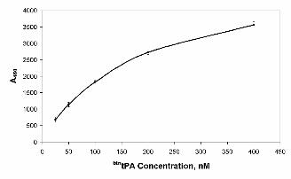
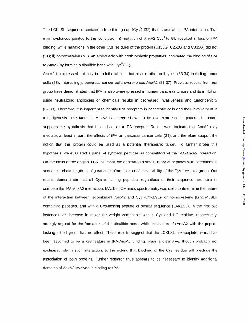
(31) using tPA labeled with biotin (see Experimental Procedures for details). Figure 1 shows that
soluble btntPA interacts in a dose-dependent fashion with immobilized rAnxA2, showing half-maximal
binding at a 200 nM concentration. This binding profile is in the same range as that already described
for 125I-tPA (8), indicating the comparability of both assays. Moreover, an excess of unlabeled tPA
completely inhibited the binding of btntPA to AnxA2 (not shown).
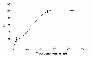
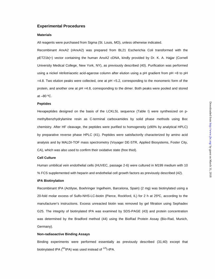
In addition, cell binding studies were performed by assaying the binding of btntPA to HUVEC cells as
AnxA2 is the main receptor for tPA in endothelial cells. As shown in Figure 2, btntPA binds to the
surface of HUVEC in a selective and dose-dependent manner, indicating that the labeled protein also
recognizes native AnxA2 on the cell membrane.
Competitive tPA-AnxA2 binding properties of LCKLSL and modified hexapeptides in vitro and
in vivo
The peptides described in Table I were tested for their ability to compete with the interaction between
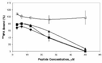
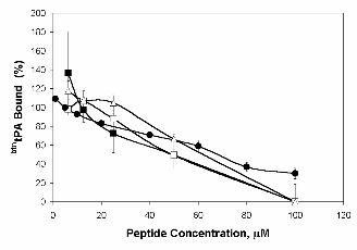
btntPA and rAnxA2. Figure 3 shows the results obtained with several representative peptides. As
expected, peptides lacking Cys8 (Figure 3, peptide 9, LAKLSL) did not compete for tPA binding: the
by guest on March 31, 2018
http://ww
w.jbc.org/
Dow
nloaded from
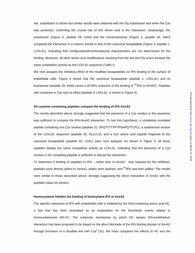
Ala substitution is shown but similar results were obtained with the Gly substitution and when the Cys
was protected, confirming the crucial role of this amino acid in the interaction. Surprisingly, the
enantiomer (Figure 3, peptide 29, lcklsl) and the retroenantiomer (Figure 3, peptide 30, lslkcl)
competed the interaction in a manner similar to that of the canonical hexapeptide (Figure 3, peptide 1,
LCKLSL), indicating that configurational/conformational characteristics are not determinant for the
binding. Moreover, all other amino acid modifications resulting from the Ala and Gly scans showed the
same competition activity as the LCKLSL sequence (Table I).
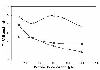
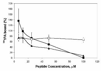
We next assayed the inhibitory effect of the modified hexapeptides on tPA binding to the surface of
endothelial cells. Figure 4 shows that the canonical hexapeptide (peptide 1, LCKLSL) and its
enantiomer (peptide 29, lcklsl) cause a 40-60% reduction of the binding of btntPA to HUVEC. Peptides
with mutations in Cys had no effect (peptide 9, LAKLSL, is shown in Figure 4).
All cysteine containing peptides compete the binding of tPA-AnxA2
The results described above strongly suggested that the presence of a Cys residue in the sequence
was sufficient to compete the tPA-AnxA2 interaction. To test this hypothesis, a completely unrelated
peptide containing one Cys residue (peptide 31, SFQTTTTYPTPSHPQTTLPC), a randomized version
of the LCKLSL sequence (peptide 32, KLLCLS), and a four amino acid peptide fragment of the
canonical hexapeptide (peptide 33, LCKL) were next assayed. As shown in Figure 5, all these
peptides display the same competitive activity as LCKLSL, indicating that the presence of a Cys
residue in the competing peptide is sufficient to disrupt the interaction.
To determine if binding of peptides to tPA – rather than to AnxA2 - was required for the inhibition,
peptides were directly added to rAnxA2, plates were washed, and btntPA was then added. The results
were similar to those described above, strongly suggesting the direct interaction of AnxA2 with the
peptides (data not shown).
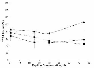
Homocysteine inhibits the binding of biotinylated tPA to AnxA2
The specific interaction of tPA with endothelial cells is inhibited by the thiol-containing amino acid HC,
a fact that has been postulated as an explanation for the thrombotic events related to
homocysteinemia (45-47). The molecular mechanism by which HC impairs tPA-endothelium
interaction has been proposed to be based on the direct blockade of the tPA binding domain of AnxA2
through formation of a disulfide link with Cys8 (31). We have compared the effects of HC and the
by guest on March 31, 2018
http://ww
w.jbc.org/
Dow
nloaded from
peptides used in this study on the binding of tPA to AnxA2 under the experimental conditions
described above. Figure 6 shows that HC competed less efficiently than the peptides. In contrast,
when an analogue of the canonical hexapeptide containing HC instead of Cys [peptide 34, L(hC)KLSL]
was assayed, it competed as efficiently as the consensus LCKLSL peptide. The lower effect of free
versus peptide-incorporated HC may be explained by differences in stability of the reduced state.
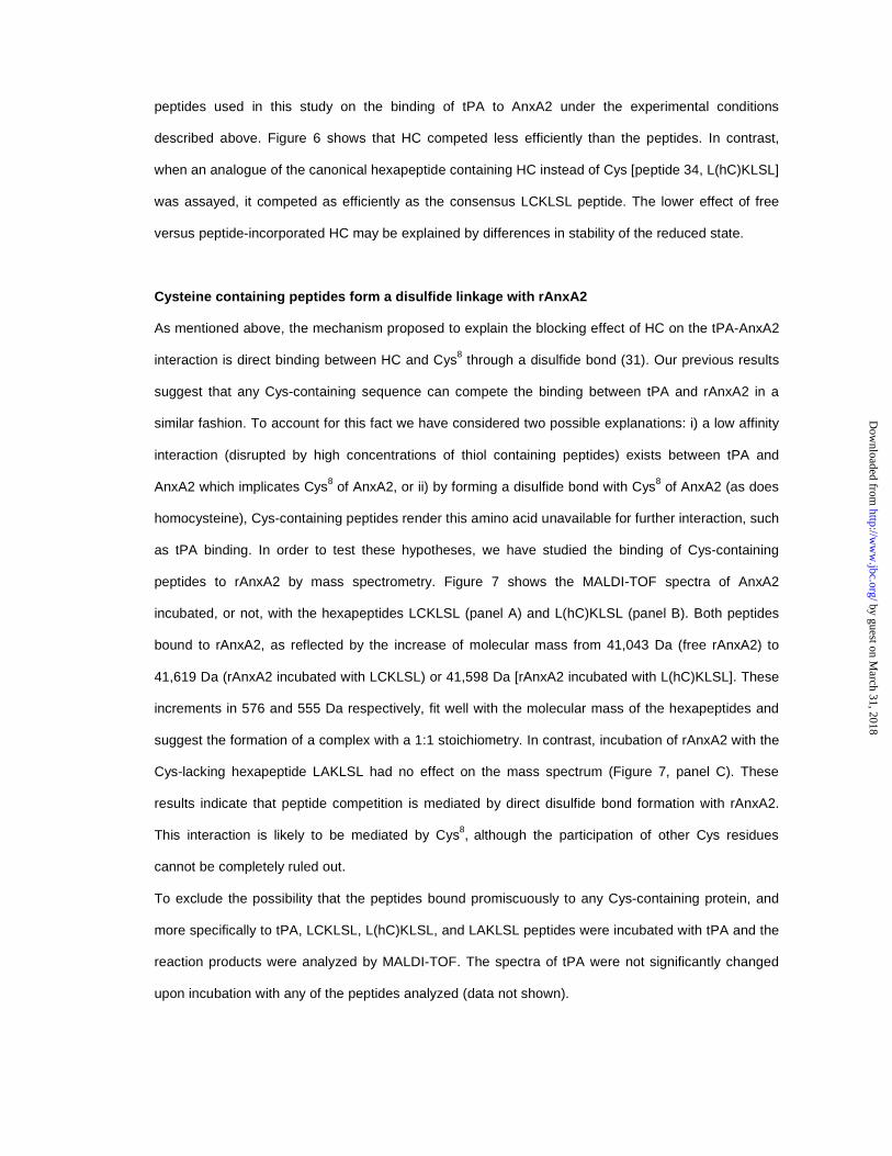
Cysteine containing peptides form a disulfide linkage with rAnxA2
As mentioned above, the mechanism proposed to explain the blocking effect of HC on the tPA-AnxA2
interaction is direct binding between HC and Cys8 through a disulfide bond (31). Our previous results
suggest that any Cys-containing sequence can compete the binding between tPA and rAnxA2 in a
similar fashion. To account for this fact we have considered two possible explanations: i) a low affinity
interaction (disrupted by high concentrations of thiol containing peptides) exists between tPA and
AnxA2 which implicates Cys8 of AnxA2, or ii) by forming a disulfide bond with Cys8 of AnxA2 (as does
homocysteine), Cys-containing peptides render this amino acid unavailable for further interaction, such
as tPA binding. In order to test these hypotheses, we have studied the binding of Cys-containing
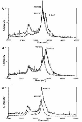
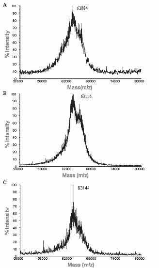
peptides to rAnxA2 by mass spectrometry. Figure 7 shows the MALDI-TOF spectra of AnxA2
incubated, or not, with the hexapeptides LCKLSL (panel A) and L(hC)KLSL (panel B). Both peptides
bound to rAnxA2, as reflected by the increase of molecular mass from 41,043 Da (free rAnxA2) to
41,619 Da (rAnxA2 incubated with LCKLSL) or 41,598 Da [rAnxA2 incubated with L(hC)KLSL]. These
increments in 576 and 555 Da respectively, fit well with the molecular mass of the hexapeptides and
suggest the formation of a complex with a 1:1 stoichiometry. In contrast, incubation of rAnxA2 with the
Cys-lacking hexapeptide LAKLSL had no effect on the mass spectrum (Figure 7, panel C). These
results indicate that peptide competition is mediated by direct disulfide bond formation with rAnxA2.
This interaction is likely to be mediated by Cys8, although the participation of other Cys residues
cannot be completely ruled out.
To exclude the possibility that the peptides bound promiscuously to any Cys-containing protein, and
more specifically to tPA, LCKLSL, L(hC)KLSL, and LAKLSL peptides were incubated with tPA and the
reaction products were analyzed by MALDI-TOF. The spectra of tPA were not significantly changed
upon incubation with any of the peptides analyzed (data not shown).
by guest on March 31, 2018
http://ww
w.jbc.org/
Dow
nloaded from
Discussion
The dramatic increase in tPA catalytic activity resulting from its binding to cellular membranes provides
an interesting target for the modulation of its biological functions related to the proteolytic degradation
of fibrin as well as other substrates. We have recently shown that tPA is overexpressed in pancreas
cancer cells and the blockade of tPA is associated with reduced in vitro invasiveness and
tumorigenicity (37;38). Therefore, the tPA system constitutes an attractive target for the development
of novel therapies. Because cell surface receptors allow focalization of the proteolytic activity at the
cell membrane and may also participate in signal transduction, their identification is of major
importance.
AnxA2 has been shown to be a major tPA receptor in endothelial cells, among others, and preliminary
data indicate that this protein is also overexpressed in pancreas cancers (36;37;39). On the basis of
these findings, it can be postulated that AnxA2 might play a role in stimulating tPA activity, and
plasmin generation, at the membrane of cancer cells potentially leading to increased tumorigenic
properties. Nevertheless, our data do not exclude the possibility that some of the effects of tPA and
it(s) receptor(s) take place independently of its proteolytic activity, and/or that of plasmin, as is the
case of some effects mediated by the receptors for urokinase-type plasminogen activator (48). The
blockade of the interaction between tPA and AnxA2 might provide clues about the precise role of this
molecule as a tPA cell surface receptor in cancer cells.
Hajjar et al. have recently reported (31) on the critical role of a Cys residue in the LCKLSL (residues
7-12) sequence at the N-terminus of AnxA2 for tPA and HC binding, and have proposed that the
interaction between the latter and AnxA2 leads to a reduction of tPA binding, thus providing an
explanation for the prothrombotic effects of HC (45-47). This conclusion was based on the fact that: 1)
an AnxA2 33 kDa chymotryptic peptide lacking the N-terminal peptide did not compete tPA-AnxA2
binding, 2) the hexapeptide abolished the binding of 125I-tPA to immobilized purified AnxA2 and to
HUVEC cells, 3) synthetic peptides lacking Cys8 did not inhibit binding, and 4) a systematic mutation
of all Cys residues of AnxA2 showed that only Cys8 was required for the interaction. Therefore, we
initiated a search for variant peptides able to better compete AnxA2-tPA binding having more suitable
pharmacological properties. These peptides were tested using the experimental procedures previously
described (31) except that tPA was labeled with biotin instead of 125I. Our findings confirm the
requirement of Cys in the LCKLSL sequence but also suggest that other regions of the molecule must
be necessary in order to confer specificity to the interaction.
by guest on March 31, 2018
http://ww
w.jbc.org/
Dow
nloaded from
We found that all peptides containing a Cys residue, regardless of the rest of their sequence and of
the position in which the Cys was located, were able to compete the interaction between tPA and
AnxA2 due to their capacity to bind AnxA2. The fact that the consensus peptide, as well as its
enantiomer and retroenantiomer versions, showed similar properties in these assays and the small
size of the peptides used, argue in favor of mechanisms independent of a strong secondary structure.
Because HC has been shown to block AnxA2-tPA binding through the formation of a disulfide bond
with the thiol group of Cys8, we considered the possibility that the effects of synthetic peptides used in
the assays performed here and in the work of Hajjar et al might act through similar mechanisms. Our
results indicate that the peptides compete with AnxA2-tPA binding more efficiently than HC and that
peptide LCKLSL and its enantiomer bind covalently to AnxA2 and induce an increase in molecular
mass, as determined by mass spectrometry, whereas a peptide with a Cys-Ala substitution does not
bind. While we cannot exclude the covalent binding of the peptides to other Cys residues, all available
evidence points to an interaction with Cys8, since it has been shown that it is the only residue to which
HC binds (31). Similar analyses using mass spectrometry showed that the same peptides do not bind
tPA.
AnxA2 has been described to exist as at least three different forms (49): monomer, heterodimer
(composed of one molecule of AnxA2 and one molecule of 3-phosphoglycerate kinase), and
heterotetramer (two AnxA2 subunits and two 11kDa regulatory subunits called p11 or S100A10).
These different forms seem to be present in distinct subcellular compartments: AnxA2 monomer is
mainly cytosolic (49-51) whereas the heterodimer has been described in the nucleus (52-54), and the
heterotetramer is associated with the plasma membrane (55). The AnxA2 heterotetramer is the most
abundant form of the protein, representing 90-95% of the total AnxA2 in endothelial, epithelial, and
MDCK cells (34;56). The p11 light chain regulates many of the activities of AnxA2 and confers to the
heterotetramer biochemical properties distinct from those of the monomer (49;57;58). In particular, the
tetramer is an extremely potent activator of plasminogen, stimulating the rate of activation of
[Glu]plasminogen about 341-fold, compared with an approximate 6-fold stimulation by the monomer,
and inducing a 90-fold increase in the catalytic efficiency of tPA for [Glu]plasminogen (59). Binding of
p11 to AnxA2 also decreases the Kd (Ca2+) for the binding of AnxA2 to biological membranes. In
addition, the monomer bundles F-actin to a much lesser extent than the tetramer and actin binding is
important for regulation of intracellular AnxA2-membrane mediated functions. Overall, the data
suggest that p11 acts as a modulator of properties displayed by the p36 core protein. Interestingly, the
by guest on March 31, 2018
http://ww
w.jbc.org/
Dow
nloaded from
interaction domain for p11 has been located, using fluorescence spectroscopy, within the first 9 amino
acids of the AnxA2 tail (STVHEILCK) (32). This sequence partially overlaps with the LCKLSL peptide,
suggesting that both p11 and tPA could compete for binding to AnxA2. However, the fact that tPA can
bind to the tetramer, hereby leading to enhanced plasmin generation activity, supports the notion that
non-overlapping domains must also be involved in binding of AnxA2 to p11 and to tPA. Our studies do
not modify the interpretation of the mechanism of interaction of AnxA2 with p11 as the latter has been
shown to be dependent on the N-terminal acetyl group of Ser1 (60,61) and unaffected by substitution
of Cys8 with a fluorophore used in fluorescence spectroscopy studies (32). Because the AnxA2
tetramer is overexpressed at the extracellular side of the membrane in tumor cells (35) and its
expression has been associated to cellular transformation and metastasis (35, 28), elucidating the
precise structure of the complex reamins an important task.
Regarding the binding site in the tPA molecule, Beebe et al. (62) have proposed that residues 7-17 of
its finger domain (RDEKTQMIYQQ) are involved in binding to AnxA2. This sequence mimics partially
the sequence of p11 responsible for AnxA2 binding (CRDGK, residues 61-65), suggesting again the
possibility of competition between both molecules for AnxA2 binding. However, the high
concentrations (mM) of tPA-derived peptide required for HUVEC binding suggest that other regions of
the molecule may also play a role. For instance, tPA deletion mutants lacking the finger domain, or
both the finger and growth factor domains, are still able, although to a much lesser extent, to bind to
endothelial cells (63) and stimulate plasminogen activation (64). However, in these assays the cellular
receptors involved in the interaction with tPA or tPA-derived fragments were not molecularly
characterized. Altogether, the available evidence indicates that a reassessment of the domains
involved in the binding of AnxA2, tPA, and p11 is necessary and that it is important to take into
account that different domains may be involved in different cellular processes given the broad range of
functions ascribed to the molecules involved in this complex.
The precise mechanisms through which the overexpression of tPA and AnxA2 may contribute to tumor
progression in pancreas cancer, as well as in other tumor types, are not clear and may be severalfold.
To tackle more effectively such processes and to develop therapeutic strategies a better
understanding of the molecular interactions between these two proteins is necessary.
by guest on March 31, 2018
http://ww
w.jbc.org/
Dow
nloaded from
Acknowledgements
We are very grateful to Dr. C. Castellernau and Dr. M.L. Lampugnani for kindly providing the HUVEC
cells and Dr. A. García de Herreros and Dr. J. Domínguez for helpful discussions and critical reading
of the manuscript. This work was supported by grants from Instituto de Salud Carlos III (00/0462),
Biomed Program (BMH4-CT98.3085), Dirección General de Enseñanza Superior e Investigación
Científica (PM97-0077) and CIRIT (Generalitat de Catalunya) (SGR-00245 and SGR-00410). O. R. is
supported by a Fellowship from the Ministerio de Educación, Cultura y Deporte.
by guest on March 31, 2018
http://ww
w.jbc.org/
Dow
nloaded from
References
1. Collen, D. and Lijnen, H. R. (1995) Thromb.Haemost. 74, 167-171
2. Madison, E. L. (1994) Fibrinolysis 8, 221-236
3. Dano, K., Andreasen, P. A., Grondahl-Hansen, J., Kristensen, P., Nielsen, L. S., and Skriver, L. (1985) Adv.Cancer Res. 44, 139-266
4. Liotta, L. A., Rao, C. N., and Barsky, S. H. (1983) Lab Invest 49, 636-649
5. DeClerck, Y. A., Imren, S., Montgomery, A. M., Mueller, B. M., Reisfeld, R. A., and Laug, W. E. (1997) Adv.Exp.Med.Biol. 425, 89-97
6. Lamba, D., Bauer, M., Huber, R., Fischer, S., Rudolph, R., Kohnert, U., and Bode, W. (1996) J Mol.Biol. 258, 117-135
7. Redlitz, A. and Plow, E. F. (1995) Baillieres Clin.Haematol. 8, 313-327
8. Hajjar, K. A., Jacovina, A. T., and Chacko, J. (1994) J Biol.Chem. 269, 21191-21197
9. Cesarman, G. M., Guevara, C. A., and Hajjar, K. A. (1994) J Biol.Chem. 269, 21198-21203
10. Gerke, V. and Moss, S. E. (2002) Physiol Rev 82, 331-371
11. Gerke, V. and Moss, S. E. (1997) Biochim.Biophys.Acta 1357, 129-154
12. Emans, N., Gorvel, J. P., Walter, C., Gerke, V., Kellner, R., Griffiths, G., and Gruenberg, J. (1993) J Cell Biol. 120, 1357-1369
13. Burgoyne, R. D., Morgan, A., and Roth, D. (1994) Ann.N.Y.Acad.Sci. 710, 333-346
14. Morgan, A., Roth, D., Martin, H., Aitken, A., and Burgoyne, R. D. (1993) Biochem.Soc.Trans. 21, 401-405
15. Rojas, E., Arispe, N., Haigler, H. T., Burns, A. L., and Pollard, H. B. (1992) Bone Miner. 17, 214-218
16. Pollard, H. B., Guy, H. R., Arispe, N., de la, F. M., Lee, G., Rojas, E. M., Pollard, J. R., Srivastava, M., Zhang-Keck, Z. Y., Merezhinskaya, N., and . (1992) Biophys.J 62, 15-18
17. Sun, J., Bird, P., and Salem, H. H. (1993) Thromb.Res. 69, 279-287
18. Kondo, S., Noguchi, M., Funakoshi, T., Fujikawa, K., and Kisiel, W. (1987) Thromb.Res. 48, 449-459
19. Andree, H. A., Stuart, M. C., Hermens, W. T., Reutelingsperger, C. P., Hemker, H. C., Frederik, P. M., and Willems, G. M. (1992) J Biol.Chem. 267, 17907-17912
20. Romisch, J., Schorlemmer, U., Fickenscher, K., Paques, E. P., and Heimburger, N. (1990) Thromb.Res. 60, 355-366
21. Keutzer, J. C. and Hirschhorn, R. R. (1990) Exp.Cell Res. 188, 153-159
22. Masiakowski, P. and Shooter, E. M. (1988) Proc.Natl.Acad.Sci.U.S.A 85, 1277-1281
23. Harder, T., Thiel, C., and Gerke, V. (1993) J Cell Sci. 104 ( Pt 4), 1109-1117
24. Leung, M. F., Lin, T. S., and Sartorelli, A. C. (1992) Cancer Res. 52, 3063-3066
by guest on March 31, 2018
http://ww
w.jbc.org/
Dow
nloaded from
25. Pfaffle, M., Ruggiero, F., Hofmann, H., Fernandez, M. P., Selmin, O., Yamada, Y., Garrone, R., and von der, M. K. (1988) EMBO J 7, 2335-2342
26. Kirsch, T. and Pfaffle, M. (1992) FEBS Lett. 310, 143-147
27. Wu, L. N., Genge, B. R., Lloyd, G. C., and Wuthier, R. E. (1991) J Biol.Chem. 266, 1195-1203
28. Tressler, R. J., Updyke, T. V., Yeatman, T., and Nicolson, G. L. (1993) J Cell Biochem. 53, 265-276
29. Tressler, R. J. and Nicolson, G. L. (1992) J Cell Biochem. 48, 162-171
30. Hajjar, K. A. and Krishnan, S. (1999) Trends Cardiovasc.Med. 9, 128-138
31. Hajjar, K. A., Mauri, L., Jacovina, A. T., Zhong, F., Mirza, U. A., Padovan, J. C., and Chait, B. T. (1998) J Biol.Chem. 273, 9987-9993
32. Johnsson, N., Marriott, G., and Weber, K. (1988) EMBO J 7, 2435-2442
33. Ma, A. S., Bell, D. J., Mittal, A. A., and Harrison, H. H. (1994) J Cell Sci. 107, 1973-1984
34. Gerke, V. and Weber, K. (1984) EMBO J 3, 227-233
35. Yeatman, T. J., Updyke, T. V., Kaetzel, M. A., Dedman, J. R., and Nicolson, G. L. (1993) Clin.Exp.Metastasis 11, 37-44
36. Vishwanatha, J. K., Chiang, Y., Kumble, K. D., Hollingsworth, M. A., and Pour, P. M. (1993) Carcinogenesis 14, 2575-2579
37. Paciucci, R., Tora, M., Diaz, V. M., and Real, F. X. (1998) Oncogene 16, 625-633
38. Paciucci, R., Berrozpe, G., Tora, M., Navarro, E., Garcia, d. H., and Real, F. X. (1996) FEBS Lett. 385, 72-76
39. Peiró, S. et al., unpublished Work
40. Hajjar, K. A., Guevara, C. A., Lev, E., Dowling, K., and Chacko, J. (1996) J Biol.Chem. 271, 21652-21659
41. Carreño, C., Roig, X., Cairó, J., Camarero, J., Mateu, M. G., Domingo, E., Giralt, E., Andreu, D., and . (92 A.D.) Int.J.Peptide Protein Res. 39, 41-47
42. Lampugnani, M. G., Resnati, M., Raiteri, M., Pigott, R., Pisacane, A., Houen, G., Ruco, L. P., and Dejana, E. (1992) J Cell Biol. 118, 1511-1522
43. Laemmli, U. K. (1970) Nature 227, 680-685
44. Bradford, M. M. (1976) Anal.Biochem. 72, 248-254
45. Hajjar, K. A. (2001) J Clin.Invest 107, 663-664
46. Graham, I. M., Daly, L. E., Refsum, H. M., Robinson, K., Brattstrom, L. E., Ueland, P. M., Palma-Reis, R. J., Boers, G. H., Sheahan, R. G., Israelsson, B., Uiterwaal, C. S., Meleady, R., McMaster, D., Verhoef, P., Witteman, J., Rubba, P., Bellet, H., Wautrecht, J. C., de Valk, H. W., Sales Luis, A. C., Parrot-Rouland, F. M., Tan, K. S., Higgins, I., Garcon, D., Andria, G., and . (1997) JAMA 277, 1775-1781
47. D'Angelo, A. and Selhub, J. (1997) Blood 90, 1-11
48. Ossowski, L. and Aguirre-Ghiso, J. A. (2000) Curr.Opin.Cell Biol. 12, 613-620
by guest on March 31, 2018
http://ww
w.jbc.org/
Dow
nloaded from
49. Waisman, D. M. (1995) Mol.Cell Biochem. 149-150, 301-322
50. Osborn, M., Johnsson, N., Wehland, J., and Weber, K. (1988) Exp.Cell Res. 175, 81-96
51. Zokas, L. and Glenney, J. R., Jr. (1987) J Cell Biol. 105, 2111-2121
52. Vishwanatha, J. K., Jindal, H. K., and Davis, R. G. (1992) J Cell Sci. 101, 25-34
53. Jindal, H. K., Chaney, W. G., Anderson, C. W., Davis, R. G., and Vishwanatha, J. K. (1991) J Biol.Chem. 266, 5169-5176
54. Vishwanatha, J. K. and Kumble, S. (1993) J Cell Sci. 105, 533-540
55. Thiel, C., Osborn, M., and Gerke, V. (1992) J Cell Sci. 103, 733-742
56. Nilius, B., Gerke, V., Prenen, J., Szucs, G., Heinke, S., Weber, K., and Droogmans, G. (1996) J Biol.Chem. 271, 30631-30636
57. Drust, D. S. and Creutz, C. E. (1988) Nature 331, 88-91
58. Kang, H. M., Kassam, G., Jarvis, S. E., Fitzpatrick, S. L., and Waisman, D. M. (1997) Biochemistry 36, 2041-2050
59. Kassam, G., Choi, K. S., Ghuman, J., Kang, H. M., Fitzpatrick, S. L., Zackson, T., Zackson, S., Toba, M., Shinomiya, A., and Waisman, D. M. (1998) J Biol.Chem. 273, 4790-4799
60. Becker, T., Weber, K., and Johnsson, N. (1990) EMBO J. 9, 4207-4213
61. König, J., Prenen, J., Nilius, B., and Gerke, V. (1998) J. Biol. Chem. 273, 19679-19684
62. Beebe, D. P., Miles, L. A., and Plow, E. F. (1989) Blood 74, 2034-2037
63. Barnathan, E. S., Cines, D. B., Barone, K., Kuo, A., and Larsen, G. R. (1988) Fibrinolysis 2, 58
64. Sinniger, V., Merton, R. E., Fabregas, P., Felez, J., Longstaff, C. (1999) J. Biol. Chem. 274, 12414-12422
by guest on March 31, 2018
http://ww
w.jbc.org/
Dow
nloaded from
Footnotes
1 The abbreviations used are: tPA, tissue-type plasminogen activator; AnxA2, annexin A2; rAnxA2,
recombinant annexin A2; btntPA, biotinylated tPA; HC, homocysteine; p11, p11 light chain of annexin
A2 tetramer or S100A10; HUVEC, human umbilical vein endothelial cells.
by guest on March 31, 2018
http://ww
w.jbc.org/
Dow
nloaded from
Legends to the Figures
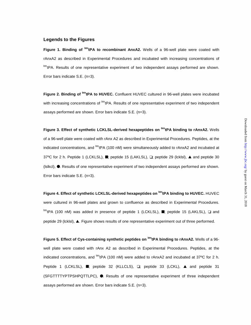
Figure 1. Binding of btntPA to recombinant AnxA2. Wells of a 96-well plate were coated with
rAnxA2 as described in Experimental Procedures and incubated with increasing concentrations of
btntPA. Results of one representative experiment of two independent assays performed are shown.
Error bars indicate S.E. (n=3).
Figure 2. Binding of btntPA to HUVEC. Confluent HUVEC cultured in 96-well plates were incubated
with increasing concentrations of btntPA. Results of one representative experiment of two independent
assays performed are shown. Error bars indicate S.E. (n=3).
Figure 3. Effect of synthetic LCKLSL-derived hexapeptides on btntPA binding to rAnxA2. Wells
of a 96-well plate were coated with rAnx A2 as described in Experimental Procedures. Peptides, at the
indicated concentrations, and btntPA (100 nM) were simultaneously added to rAnxA2 and incubated at
37ºC for 2 h. Peptide 1 (LCKLSL), ■ ; peptide 15 (LAKLSL), ❏ ; peptide 29 (lcklsl), ▲ and peptide 30
(lslkcl), ● . Results of one representative experiment of two independent assays performed are shown.
Error bars indicate S.E. (n=3).
Figure 4. Effect of synthetic LCKLSL-derived hexapeptides on btntPA binding to HUVEC. HUVEC
were cultured in 96-well plates and grown to confluence as described in Experimental Procedures.
btntPA (100 nM) was added in presence of peptide 1 (LCKLSL), ■ ; peptide 15 (LAKLSL), ❏ and
peptide 29 (lcklsl), ▲. Figure shows results of one representative experiment out of three performed.
Figure 5. Effect of Cys-containing synthetic peptides on btntPA binding to rAnxA2. Wells of a 96-
well plate were coated with rAnx A2 as described in Experimental Procedures. Peptides, at the
indicated concentrations, and btntPA (100 nM) were added to rAnxA2 and incubated at 37ºC for 2 h.
Peptide 1 (LCKLSL), ■ ; peptide 32 (KLLCLS), ❏ ; peptide 33 (LCKL), ▲ and peptide 31
(SFGTTTTYPTPSHPQTTLPC), ● . Results of one representative experiment of three independent
assays performed are shown. Error bars indicate S.E. (n=3).
by guest on March 31, 2018
http://ww
w.jbc.org/
Dow
nloaded from
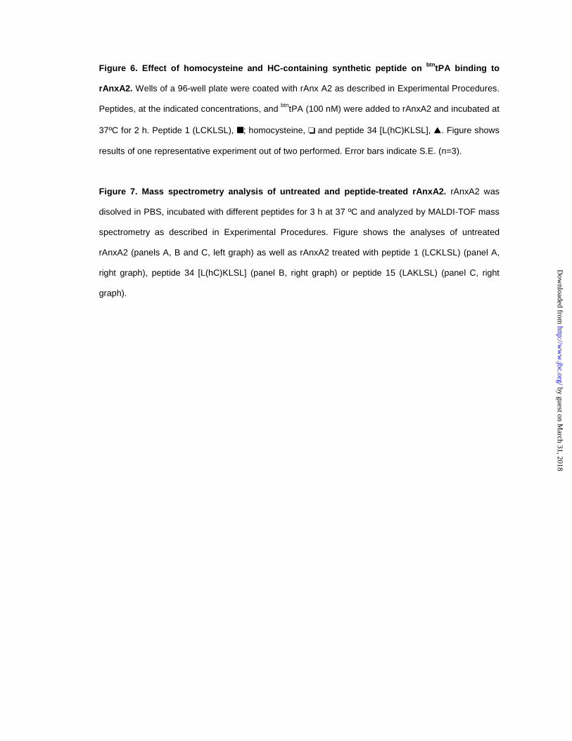
Figure 6. Effect of homocysteine and HC-containing synthetic peptide on btntPA binding to
rAnxA2. Wells of a 96-well plate were coated with rAnx A2 as described in Experimental Procedures.
Peptides, at the indicated concentrations, and btntPA (100 nM) were added to rAnxA2 and incubated at
37ºC for 2 h. Peptide 1 (LCKLSL), ■ ; homocysteine, ❏ and peptide 34 [L(hC)KLSL], ▲. Figure shows
results of one representative experiment out of two performed. Error bars indicate S.E. (n=3).
Figure 7. Mass spectrometry analysis of untreated and peptide-treated rAnxA2. rAnxA2 was
disolved in PBS, incubated with different peptides for 3 h at 37 ºC and analyzed by MALDI-TOF mass
spectrometry as described in Experimental Procedures. Figure shows the analyses of untreated
rAnxA2 (panels A, B and C, left graph) as well as rAnxA2 treated with peptide 1 (LCKLSL) (panel A,
right graph), peptide 34 [L(hC)KLSL] (panel B, right graph) or peptide 15 (LAKLSL) (panel C, right
graph).
by guest on March 31, 2018
http://ww
w.jbc.org/
Dow
nloaded from

Table I. Synthetic peptides used to compete the binding of tPA to AnxA2. Synthetic peptides containing the indicated modifications were obtained on the basis of the canonical hexapeptide 7-12 amino terminal motif of annexin A2. The ability of all peptides to compete the interaction between biotinylated tPA and AnxA2 was analyzed as described in Experimental Procedures.
Modification Sequence Peptide number
tPA binding inhibitiona
Canonical LCKLSL 1 +++ Gly scan GCKLSL 2 ++ LGKLSL 3 - LCGLSL 4 +++ LCKGSL 5 ++ LCKLGL 6 +++ LCKLSG 7 ++ Ala scan ACKLSL 8 ++ LAKLSL 9 - LCALSL 10 ++ LCKASL 11 +++ LCKLAL 12 +++ LCKLSA 13 +++ D-amino acid scan lCKLSL 14 +++ LcKLSL 15 ++ LCkLSL 16 +++ LCKlSL 17 ++ LCKLsL 18 +++ LCKLSl 19 +++ Conservative replacements ICKLSL 20 ++ (underlined) LCKISL 21 +++
LCKLSI 22 +++ VCKLSL 23 ++ LCKVSL 24 ++ LCKLSV 25 ++ LCRLSL 26 ++ LCKLTL 27 +++ Cys protection LC[Acm]KLSL 28 - Enantiomer lcklsl 29 +++ Retroenantiomer lslkcl 30 +++ Unrelated SFQTTTTYPTPSHPQTTLPC 31 +++ Random KLLCLS 32 +++ Tetrapeptide LCKL 33 +++ Cys/hC replacement L[hC]KLSL 34 +++
a tPA binding inhibition was considered as: +++, >60%; ++, 40-60% and –, <10% at 100 nM biotinylated tPA and 100 µM peptide concentration.
by guest on March 31, 2018
http://ww
w.jbc.org/
Dow
nloaded from
NavarroOriol Roda, María Luz Valero, Sandra Peiró, David Andreu, Francisco X. Real and Pilar
requirement for this association?New insights into the tPA-Annexin A2 interaction: is Annexin A2 Cys8 the sole
published online December 4, 2002J. Biol. Chem.
10.1074/jbc.M207605200Access the most updated version of this article at doi:
Alerts:
When a correction for this article is posted•
When this article is cited•
to choose from all of JBC's e-mail alertsClick here
by guest on March 31, 2018
http://ww
w.jbc.org/
Dow
nloaded from