Embed Size (px)
Citation preview
NMR Relaxometry in Complex systems
Friday 1st July 2016
ABSTRACTS
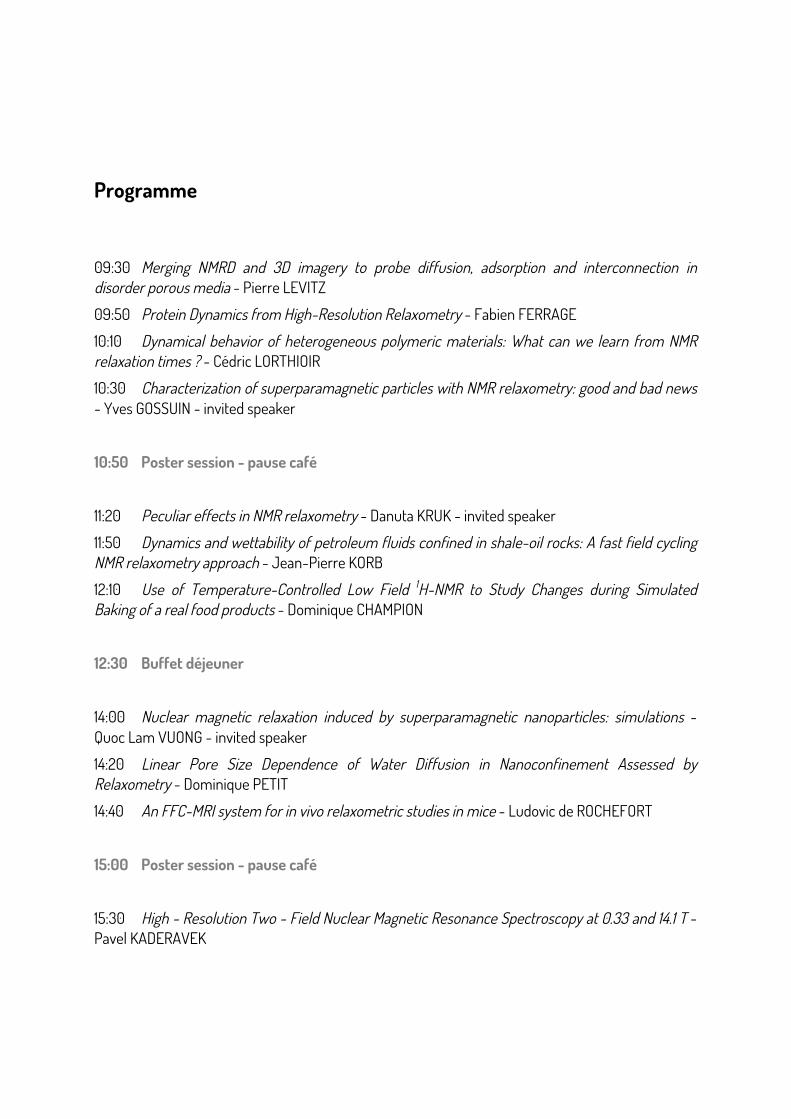
Programme
09:30 Merging NMRD and 3D imagery to probe diffusion, adsorption and interconnection in disorder porous media - Pierre LEVITZ
09:50 Protein Dynamics from High-Resolution Relaxometry - Fabien FERRAGE
10:10 Dynamical behavior of heterogeneous polymeric materials: What can we learn from NMR relaxation times ? - Cédric LORTHIOIR
10:30 Characterization of superparamagnetic particles with NMR relaxometry: good and bad news - Yves GOSSUIN - invited speaker
10:50 Poster session - pause café
11:20 Peculiar effects in NMR relaxometry - Danuta KRUK - invited speaker
11:50 Dynamics and wettability of petroleum fluids confined in shale-oil rocks: A fast field cycling NMR relaxometry approach - Jean-Pierre KORB
12:10 Use of Temperature-Controlled Low Field 1H-NMR to Study Changes during Simulated Baking of a real food products - Dominique CHAMPION
12:30 Buffet déjeuner
14:00 Nuclear magnetic relaxation induced by superparamagnetic nanoparticles: simulations - Quoc Lam VUONG - invited speaker
14:20 Linear Pore Size Dependence of Water Diffusion in Nanoconfinement Assessed by Relaxometry - Dominique PETIT
14:40 An FFC-MRI system for in vivo relaxometric studies in mice - Ludovic de ROCHEFORT
15:00 Poster session - pause café
15:30 High - Resolution Two - Field Nuclear Magnetic Resonance Spectroscopy at 0.33 and 14.1 T - Pavel KADERAVEK

Merging NMRD and 3D imagery to probe diffusion, adsorption and interconnection in disorder porous media
Pierre Levitz
PHENIX, Sorbonne Universités, UPMC Univ Paris 06 - CNRS, Paris, France.
Diffusion of a molecule inside a disordered porous material and its interaction with the walls can be nicely probed by NMRD. Even more, 2D experiments such as T2-evolution-T2 allow to investigate interconnectivity of different pore networks inside multiscale porous materials. However, these two series of complementary experiments are not ” q-dependent” in contrary to the pulsed gradient NMR spectroscopy or the spin-echo neutron scattering. In order to complement analytical models used to analyze NMRD and 2D experiments, it can be interesting to probe the potentiality of a multimodal approach involving the 3D imagery of the interfacial system under study. We first present the case of an intermittent regime involving molecular diffusion and adsorption in some porous materials and the corresponding NMRD experiments. Second, we briefly discuss the case of the intermittent exchange associated to the interconnectivity of different classes of pore networks in relation with 2D NMR experiments.
Protein Dynamics from High-Resolution Relaxometry
Fabien Ferrage Département de Chimie, Ecole Normale Supérieure, PSL Research University, UPMC Univ Paris 06, CNRS, Laboratoire des Biomolécules (LBM), 24 rue Lhomond, 75005 Paris, France ; and Sorbonne Universités, UPMC Univ Paris 06, Paris, France.
A proper understanding of the physics and chemistry that underlie the function of biological macromolecules requires an atomic-resolution description of their conformational space and the timescales of the motions in this space. Methodological and computational developments over the last three decades have made nuclear magnetic resonance a mainstream experimental technique to characterize dynamics of biomolecules, and particularly proteins. The measurement of nuclear spin relaxation has given access to motions on multiple timescales between the tens of picoseconds and nanoseconds. However, the analysis of motions has relied on limited experimental data (most often only three relaxation rates per residue in a protein) and used simple model-free approaches. High-resolution relaxometry, as introduced by Redfield [1], is a powerful technique to quantify nuclear spin relaxation over a broad range of magnetic fields and provides unprecedented sets of experimental data to quantify protein motions in the ps-ns range.
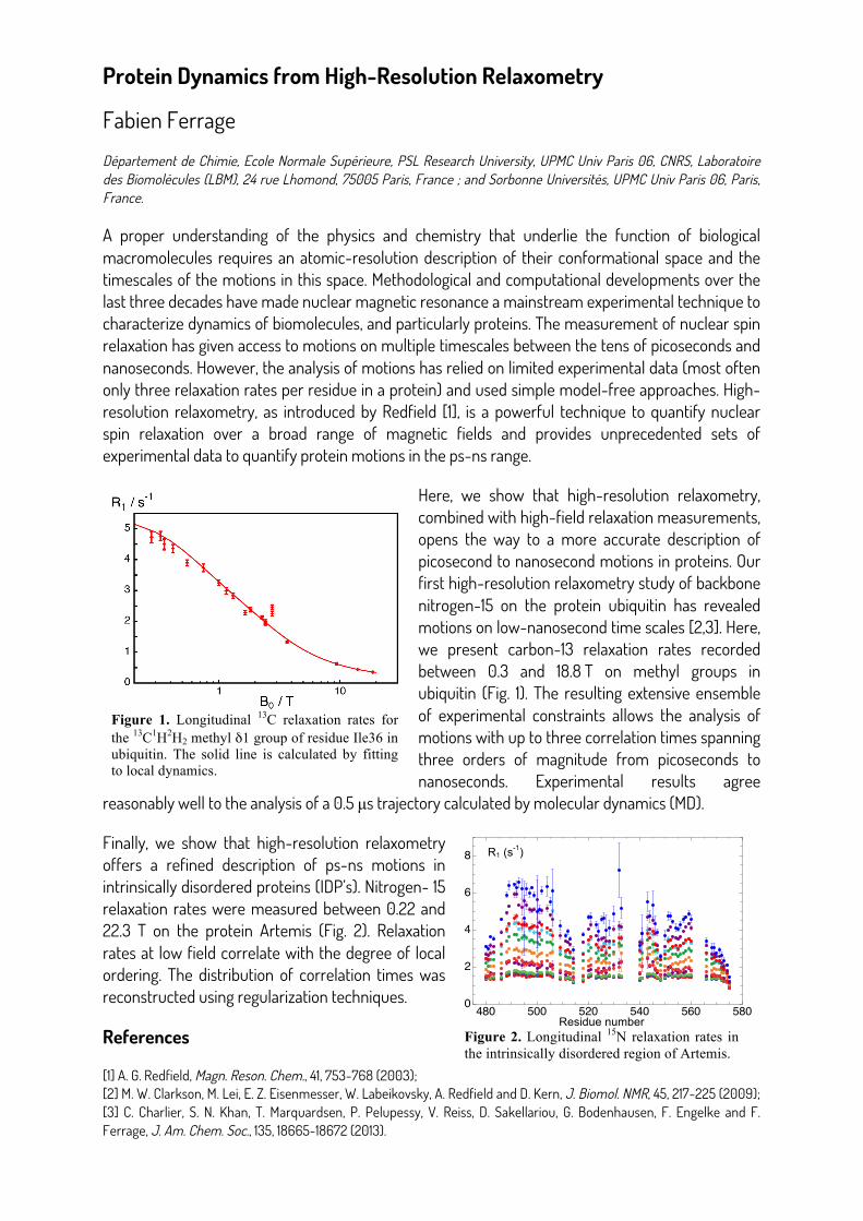
Here, we show that high-resolution relaxometry, combined with high-field relaxation measurements, opens the way to a more accurate description of picosecond to nanosecond motions in proteins. Our first high-resolution relaxometry study of backbone nitrogen-15 on the protein ubiquitin has revealed motions on low-nanosecond time scales [2,3]. Here, we present carbon-13 relaxation rates recorded between 0.3 and 18.8 T on methyl groups in ubiquitin (Fig. 1). The resulting extensive ensemble of experimental constraints allows the analysis of motions with up to three correlation times spanning three orders of magnitude from picoseconds to nanoseconds. Experimental results agree
reasonably well to the analysis of a 0.5 µs trajectory calculated by molecular dynamics (MD).
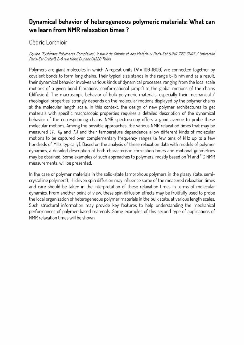
Finally, we show that high-resolution relaxometry offers a refined description of ps-ns motions in intrinsically disordered proteins (IDP’s). Nitrogen- 15 relaxation rates were measured between 0.22 and 22.3 T on the protein Artemis (Fig. 2). Relaxation rates at low field correlate with the degree of local ordering. The distribution of correlation times was reconstructed using regularization techniques.
References
[1] A. G. Redfield, Magn. Reson. Chem., 41, 753-768 (2003); [2] M. W. Clarkson, M. Lei, E. Z. Eisenmesser, W. Labeikovsky, A. Redfield and D. Kern, J. Biomol. NMR, 45, 217-225 (2009); [3] C. Charlier, S. N. Khan, T. Marquardsen, P. Pelupessy, V. Reiss, D. Sakellariou, G. Bodenhausen, F. Engelke and F. Ferrage, J. Am. Chem. Soc., 135, 18665-18672 (2013).
Protein Dynamics from High-Resolution Relaxometry
Fabien Ferrage
Département de Chimie, Ecole Normale Supérieure, PSL Research University, UPMC Univ Paris 06, CNRS, Laboratoire des Biomolécules (LBM), 24 rue Lhomond, 75005 Paris, France ; and Sorbonne Universités, UPMC Univ Paris 06, Paris, France. A proper understanding of the physics and chemistry that underlie the function of biological macromolecules requires an atomic-resolution description of their conformational space and the timescales of the motions in this space. Methodological and computational developments over the last three decades have made nuclear magnetic resonance a mainstream experimental technique to characterize dynamics of biomolecules, and particularly proteins. The measurement of nuclear spin relaxation has given access to motions on multiple timescales between the tens of picoseconds and nanoseconds. However, the analysis of motions has relied on limited experimental data (most often only three relaxation rates per residue in a protein) and used simple model-free approaches. High-resolution relaxometry, as introduced by Redfield [1], is a powerful technique to quantify nuclear spin relaxation over a broad range of magnetic fields and provides unprecedented sets of experimental data to quantify protein motions in the ps-ns range. Here, we show that high-resolution relaxometry, combined with high-field relaxation
measurements, opens the way to a more accurate description of picosecond to nanosecond motions in proteins. Our first high-resolution relaxometry study of backbone nitrogen-15 on the protein ubiquitin has revealed motions on low-nanosecond time scales [2,3]. Here, we present carbon-13 relaxation rates recorded between 0.3 and 18.8 T on methyl groups in ubiquitin (Fig. 1). The resulting extensive ensemble of experimental constraints allows the analysis of motions with up to three correlation times spanning three orders of magnitude from picoseconds to nanoseconds. Experimental results agree reasonably well to the analysis of a 0.5 µs trajectory calculated by molecular dynamics (MD).
Finally, we show that high-resolution relaxometry offers a refined description of ps-ns motions in intrinsically disordered proteins (IDP’s). Nitrogen-15 relaxation rates were measured between 0.22 and 22.3 T on the protein Artemis (Fig. 2). Relaxation rates at low field correlate with the degree of local ordering. The distribution of correlation times was reconstructed using regularization techniques. References [1] A. G. Redfield, Magn. Reson. Chem., 41, 753-768 (2003); [2] M. W. Clarkson, M. Lei, E. Z. Eisenmesser, W. Labeikovsky, A. Redfield and D. Kern, J. Biomol. NMR, 45, 217-225 (2009); [3] C. Charlier, S. N. Khan, T. Marquardsen, P. Pelupessy, V. Reiss, D. Sakellariou, G. Bodenhausen, F. Engelke and F. Ferrage, J. Am. Chem. Soc., 135, 18665-18672 (2013).
Figure 1. Longitudinal 13C relaxation rates for the 13C1H2H2 methyl δ1 group of residue Ile36 in ubiquitin. The solid line is calculated by fitting to local dynamics.
Residue number Figure 2. Longitudinal 15N relaxation rates in the intrinsically disordered region of Artemis.
480 500 520 540 560 5800
2
4
6
8 R1 (s-1)
Dynamical behavior of heterogeneous polymeric materials: What can we learn from NMR relaxation times ?
Cédric Lorthioir
Equipe "Syste ̀mes Polyme ̀res Complexes", Institut de Chimie et des Mate ́riaux Paris-Est (UMR 7182 CNRS / Universite ́ Paris-Est Cre ́teil), 2-8 rue Henri Dunant 94320 Thiais
Polymers are giant molecules in which N repeat units (N ≈ 100-1000) are connected together by covalent bonds to form long chains. Their typical size stands in the range 5-15 nm and as a result, their dynamical behavior involves various kinds of dynamical processes, ranging from the local scale motions of a given bond (librations, conformational jumps) to the global motions of the chains (diffusion). The macroscopic behavior of bulk polymeric materials, especially their mechanical / rheological properties, strongly depends on the molecular motions displayed by the polymer chains at the molecular length scale. In this context, the design of new polymer architectures to get materials with specific macroscopic properties requires a detailed description of the dynamical behavior of the corresponding chains. NMR spectroscopy offers a good avenue to probe these molecular motions. Among the possible approaches, the various NMR relaxation times that may be measured (T1, T1ρ and T2) and their temperature dependence allow different kinds of molecular motions to be captured over complementary frequency ranges (a few tens of kHz up to a few hundreds of MHz, typically). Based on the analysis of these relaxation data with models of polymer dynamics, a detailed description of both characteristic correlation times and motional geometries may be obtained. Some examples of such approaches to polymers, mostly based on 1H and 13C NMR measurements, will be presented.
In the case of polymer materials in the solid-state (amorphous polymers in the glassy state, semi-crystalline polymers), 1H-driven spin diffusion may influence some of the measured relaxation times and care should be taken in the interpretation of these relaxation times in terms of molecular dynamics. From another point of view, these spin diffusion effects may be fruitfully used to probe the local organization of heterogeneous polymer materials in the bulk state, at various length scales. Such structural information may provide key features to help understanding the mechanical performances of polymer-based materials. Some examples of this second type of applications of NMR relaxation times will be shown.
Characterization of superparamagnetic particles with NMR relaxometry: good and bad news
Yves Gossuin
Biomedical Physics Unit, UMONS, Mons, Belgium
Superparamagnetic iron oxide particles find their main application as contrast agents for cellular and molecular Magnetic Resonance Imaging (1). The contrast they bring is due to the shortening of the relaxation times of water protons T1 and T2 (2). In order to understand their influence on proton relaxation, different theoretical relaxation models have been developed, each of them presenting a certain validity domain, which depends on the particle characteristics and proton dynamics (3). In this work, relaxation properties of suspensions of iron oxide particles in different solvents and at different temperatures, corresponding to different proton diffusion properties, were evaluated thanks to the measurement of nuclear magnetic relaxation dispersion (NMRD) profiles (4). These latter represent the evolution of the relaxation times with the magnetic field. The fitting of T1 NMRD profiles by the suited theory constitutes an interesting tool of characterization of the nanoparticles. The Roch theory, developed in the Motional Averaging Regime, was successfully used to fit T1 NMRD profiles, even completely outside the MAR validity domain, and provided a good estimate of the particle size. In order to refine the characterization of the particles, we tried to perform a simultaneous fitting of T1 and T2 NMRD data, which was unfortunately impossible. This occurrence constitutes a clear limitation of the Roch model. Finally, the theory was shown to fit satisfactorily the deuterium T1 NMRD profile of superparamagnetic particles suspensions in heavy water, usually dominated by quadrupolar interactions, providing good estimates of the size and magnetization of the particles.
References
[1] Bulte J W M and Kraitchman D L 2004 Iron oxide MR contrast agents for molecular and cellular imaging NMR Biomed. 17 484–99 [2] Gossuin Y, Gillis P, Hocq A, Vuong Q L and Roch A 2009 Magnetic resonance relaxation properties of superparamagnetic particles Wiley Interdiscip. Rev. Nanomed. Nanobiotech- nol. 1 299–310 [3] Roch A, Muller R N and Gillis P 1999 Theory of proton relaxation induced by superparamagnetic particles J. Chem. Phys. 110 5403–11 [4] Gossuin Y, Orlando T, Basini M, Henrard D et al NMR relaxation induced by iron oxide particles: testing theoretical models Nanotechnology 27 155706
Peculiar effects in NMR relaxometry
Danuta Kruk1 University of Warmia & Mazury in Olsztyn, Faculty of Mathematics and Computer Science, Słoneczna 54, 10-710 Olsztyn, Poland, email: [email protected] Nuclear Magnetic Resonance (NMR) relaxometry is a unique experimental method probing mechanisms and characteristic time constants of dynamical processes in condensed matter on the molecular (atomistic) level. The remarkably broad range of magnetic fields covered by NMR relaxometry: from about 10 kHz to 40 MHz (referring to the 1H resonance frequency) implies that one can investigate, by a single experiment, motional processes across a huge range of time scales (from ms to ns). NMR relaxometry is widely applied to inquire into dynamical and structural properties of liquid and solid systems of various complexity: from simple liquids, via electrolytes and macromolecules to complex solids; the studies encompass diamagnetic, paramagnetic and superparamagnetic systems. The variety of dynamical features of the systems is reflected by their, often unusual relaxation properties. Interplay between spin interactions modulated by various motional processes leads to relaxation effects from which unique information about the dynamics can be revealed, provided appropriate theoretical models are available. I will discuss some of the relaxation phenomena, illustrating them by experimental examples; for instance:
• Relaxation in molecular liquids is a sum of intra-molecular and inter-molecular contributions. Intramolecular dipolar interactions fluctuate in time due rotation of the molecule, while inter-molecular couplings are modulated by translation diffusion of the interacting molecules. This implies that NMR relaxometry allows for probing rotational and translational dynamics by a single experiment, which is exceptional. Moreover, translation diffusion coefficients can be straightforwardly determined from the low frequency slope of the relaxation rate versus square root of the resonance frequency.
• Discussing relaxation processes for electrolytes (ionic liquids) one has to take into account that the system, in most cases, contains different types of NMR active nuclei (for instance 1H containing cations and 19F containing anions). In consequence, the shape of the relaxation dispersion data stems from several intra-es and inter-ionic relaxation pathways including interferences between 1H-1H, 1H-19F and 19F-19F relaxation pathways. Moreover, for liquids in confinement, the motion in restricted geometry is reflected by the shape of the relaxation profiles that show then unusual properties.
• For solids containing nuclei possessing quadrupole moments one can observe frequency specific enhancement of the spin 1/2 (1H, 19F) relaxation, referred to Quadrupole Relaxation Enhancement. The relaxation maxima (“quadrupole peaks”) can be seen for systems undergoing slow dynamics when the magnetic field is set to a value for which the Zeeman splitting of the spin 1/2 nucleus matches the energy splitting of the quadrupolar nucleus (determined by its residual quadrupolar coupling and Zeeman interaction).
• In paramagnetic solutions the nuclear relaxation originates from strong magnetic dipole- dipole interactions between the electron spin of the paramagnetic molecule and the nuclear spin of the solvent molecules. Moreover, the electron spin relaxation acts as an additional factor modulating the electron spin – nuclear spin interactions, considerably influencing the nuclear relaxation and leading to Paramagnetic Relaxation Enhancement. For superparamagnetic nanocrystals the dipolar interactions with the large electronic magnetic moments lead to specific relaxation effects.
!!!!!!!!!!!!!!!!!!!!!!!!!!!!!!!!!!!!!!!!!!!!!!!!!!!!!!!!1 Danuta Kruk is a visiting professor at UPMC thanks to the Labex MATISSE.
Dynamics and wettability of petroleum fluids confined in shale-oil rocks: A fast field cycling NMR relaxometry approach
Jean-Pierre Korba, Benjamin Nicotb, G. Ferrantec a Physique de la Matière Condensée, Ecole Polytechnique- CNRS, 91128 Palaiseau, France; b Centre Scientifique et Technique Jean Feger (CSTJF), TOTAL EP, 64018 Pau, France; c STELAR s.r.l., Via Enrico Fermi, 4-27035 Mede, Pavia (PV), Italy
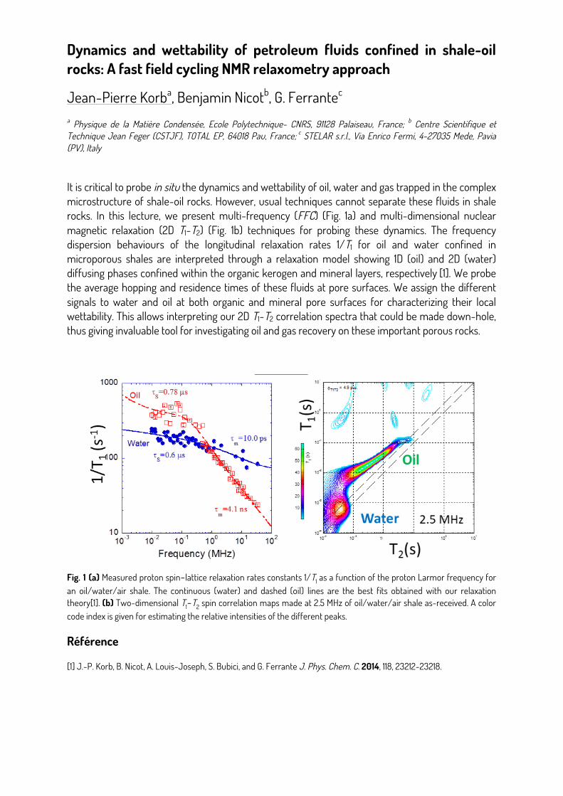
It is critical to probe in situ the dynamics and wettability of oil, water and gas trapped in the complex microstructure of shale-oil rocks. However, usual techniques cannot separate these fluids in shale rocks. In this lecture, we present multi-frequency (FFC) (Fig. 1a) and multi-dimensional nuclear magnetic relaxation (2D T1-T2) (Fig. 1b) techniques for probing these dynamics. The frequency dispersion behaviours of the longitudinal relaxation rates 1/T1 for oil and water confined in microporous shales are interpreted through a relaxation model showing 1D (oil) and 2D (water) diffusing phases confined within the organic kerogen and mineral layers, respectively [1]. We probe the average hopping and residence times of these fluids at pore surfaces. We assign the different signals to water and oil at both organic and mineral pore surfaces for characterizing their local wettability. This allows interpreting our 2D T1-T2 correlation spectra that could be made down-hole, thus giving invaluable tool for investigating oil and gas recovery on these important porous rocks.
Fig. 1 (a) Measured proton spin−lattice relaxation rates constants 1/T1 as a function of the proton Larmor frequency for an oil/water/air shale. The continuous (water) and dashed (oil) lines are the best fits obtained with our relaxation theory[1]. (b) Two-dimensional T1−T2 spin correlation maps made at 2.5 MHz of oil/water/air shale as-received. A color code index is given for estimating the relative intensities of the different peaks.
Référence
[1] J.-P. Korb, B. Nicot, A. Louis-Joseph, S. Bubici, and G. Ferrante J. Phys. Chem. C. 2014, 118, 23212-23218.
Use of Temperature-Controlled Low Field 1H-NMR to Study Changes during Simulated Baking of a real food products.
Dominique Champion, Ali Assifaoui UMR PAM. Agrosup Dijon, Université Bourgogne, Franche Comté
The quality of food products is governed by its organoleptic properties, which are the results of numerous physical-chemical transformations through the different steps of its process (mixing, kneading, cooling, or baking...). When water content is or is becoming the limiting parameter in food materials, the competitive hydration of the different components may explain evolutions in the products like crystallization, protein denaturation, non-enzymatic browning, lipid oxidation... The objectives of food technologists are to manage these changes in raw food materials in order to obtain either intermediary ingredients able to be transformed in an industrial way, mainly by a fast kinetic of rehydration, or stable food products for consumers, or even new food materials for the market development. Low field NMR relaxometry is now widely used in food domain as routine quality control test for the determination of solid/liquid or oil/water ratios in plant seeds, oil, margarine and other fat content products. In order to predict food stability or specificity, more developments are still needed to study molecular mobility, and not only the water one, in food materials that are mostly very heterogeneous in composition and could be at low water content.
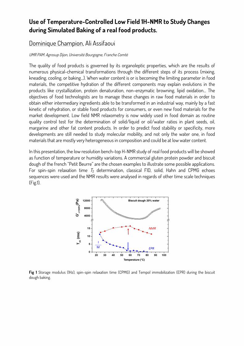
In this presentation, the low resolution bench-top H-NMR study of real food products will be showed as function of temperature or humidity variations. A commercial gluten protein powder and biscuit dough of the french “Petit Beurre” are the chosen examples to illustrate some possible applications. For spin-spin relaxation time T2 determination, classical FID, solid, Hahn and CPMG echoes sequences were used and the NMR results were analysed in regards of other time scale techniques (Fig.1).
!
Fig 1 Storage modulus (1Hz), spin-spin relaxation time (CPMG) and Tempol immobilization (EPR) during the biscuit dough baking.
Use of Temperature-Controlled Low Field 1H-NMR to Study Changes
during Simulated Baking of a real food products.
Dominique Champion. Ali Assifaoui UMR PAM. Agrosup Dijon, Université Bourgogne, Franche Comté
The quality of food products is governed by its organoleptic properties which are the results
of numerous physical-chemical transformations through the different steps of its process (mixing,
kneading, cooling, or baking…). When water content is or is becoming the limiting parameter in food
materials, the competitive hydration of the different components may explain evolutions in the
products like crystallization, protein denaturation, non-enzymatic browning, lipid oxidation… The objectives of food technologists are to manage these changes in raw food materials in order to
obtain either intermediary ingredients able to be transformed in an industrial way, mainly by a fast
kinetic of rehydration, or stable food products for consumers, or even new food materials for the
market development. Low field NMR relaxometry is now widely used in food domain as routine
quality control test for the determination of solid/liquid or oil/water ratios in plant seeds, oil,
margarine and other fat content products. In order to predict food stability or specificity, more
developments are still needed to study molecular mobility, and not only the water one, in food
materials that are mostly very heterogeneous in composition and could be at low water content.
In this presentation, the low resolution bench-top H-NMR study of real food products will be showed
as function of temperature or humidity variations. A commercial gluten protein powder and biscuit
dough of the french “Petit Beurre” are the chosen examples to illustrate some possible applications.
For spin-spin relaxation time T2 determination, classical FID, solid, Hahn and CPMG echoes
sequences were used and the NMR results were analysed in regards of other time scale techniques
(Fig.1).
Fig1: Storage modulus (1Hz), spin-spin relaxation time
(CPMG) and Tempol immobilization (EPR) during the biscuit
dough baking.
NMR
EPR
I
M
Temperature ( C)
Biscuit dough 20% water
Nuclear magnetic relaxation induced by superparamagnetic nanoparticles: simulations
Quoc Lam Vuong Biomedical Physics Unit, UMONS, Mons, Belgium
Superaparamagnetic nanoparticles are used as negative contrast agents in Magnetic Resonance Imaging (MRI). Their great magnetization produces magnetic inhomogeneities, which shorten the relaxation times. Their efficiency can be quantified by their relaxivities, i.e. their relaxation rates normalized by the iron concentration.
Nuclear magnetic relaxation relies on the interaction between the proton spins and their fluctuating magnetic environment. In the case of superparamagnetic nanoparticles, proton relaxation is induced by the dipolar magnetic field produced by these particles [1]. The Redfield formalism is usually used to predict the relaxation times and involves non intuitive and long stochastic quantum calculus.
Another approach for the understanding of the relaxation is to work with the equivalent classical equations of the magnetic moment. Similar mathematical expressions for the relaxation times can be analytically derived. This paradigm also leads to a simple simulation algorithm which has the advantage not to depend on approximations.
Simulation results of proton relaxation induced by superparamagnetic particles will be presented. Nuclear magnetic relaxation dispersion curves of aqueous samples containing superparamagnetic nanoparticles in and out of the Redfield condition were simulated [2-3]. More complex systems as NPs entrapped in cells are also studied.
References:
[1] Gossuin, Y., Gillis, P., Hocq, A., Vuong, Q. L., & Roch, A. (2009). Magnetic resonance relaxation properties of superparamagnetic particles. Wiley Interdisciplinary Reviews: Nanomedicine and Nanobiotechnology, 1(3), 299-310. Lévy M., Wilhelm C., Devaud M., Levitz P. and Gazeau F. Contrast Media & Molecular Imaging 7(4), 373-383, 2012. [2] Vuong, Q. L., Gossuin, Y., Gillis, P., & Delangre, S. (2012). New simulation approach using classical formalism to water nuclear magnetic relaxation dispersions in presence of superparamagnetic particles used as MRI contrast agents. The Journal of chemical physics, 137(11), 114505. [3] Vuong, Q. L., Berret, J. F., Fresnais, J., Gossuin, Y., & Sandre, O. (2012). A Universal Scaling Law to Predict the Efficiency of Magnetic Nanoparticles as MRI T2-Contrast Agents. Advanced healthcare materials, 1(4), 502-512.
Dépendance linéaire avec la taille des pores de la diffusion de l’eau nano-confinée mise en évidence par relaxométrie
H. Chemmi1, D. Petit1,2*, P. Levitz3, R. Denoyel4, A. Galarneau5, J.-P. Korb1 1Physique de la Matière Condensée, Ecole Polytechnique-CNRS UMR7643, Palaiseau 91128, France 2Lab. Charles Coulomb CNRS UMR5221, Campus Triolet, Université de Montpellier, 34095 Montpellier, France 3Physicochimie des Electrolytes et Nanosystèmes Interfaciaux, CNRS-UMR 8234, Université Pierre et Marie Curie, 4 place Jussieu, 72522 Paris Cedex 5, France 4MADIREL, Aix-Marseille Université, CNRS-UMR 7246, Centre de St Jérôme, 13397 Marseille Cedex 20, France 5Institut Charles Gerhardt Montpellier, UMR 5253 CNRS-UM-ENSCM, ENSCM, 8 rue de l’Ecole Normale, 34296, Montpellier Cedex 05, France Keywords: Water diffusion, Nano-pore, NMR Relaxation, Scaling, Confined dynamics. Nous montrons que les expériences de relaxation magnétique nucléaire à des champs magnétiques variables (NMRD) fournissent des moyens non invasifs pour sonder la dépendance spatiale de la diffusion de liquide près des interfaces liquides/solides [1]. Ces expériences effectuées sur des échantillons présentant des nano-pores de géométries cylindriques et sphériques démontrent que le coefficient de diffusion moyen paralléle à l’interface est proportionnel au rayon des pores dans différents régimes de dynamique. Un procédé de courbe maîtresse permet l’extraction de gradients de coefficients de diffusion à proximité de la surface des pores, indicatifs de l’efficacité de couplage entre les couches liquides. En raison de leur sélectivité en fréquence, les expériences NMRD sont capables de différencier les différents événements dynamiques de l’eau induits par des surfaces hétérogènes ou des processus dynamiques composées. Cette analyse du confinement, pertinente en Physique et Biologie, suggère un lien entre la description moléculaire et continue de la dynamique des fluides près des interfaces fluides/solides.
Reference:
[1] H. Chemmi, D. Petit, P. Levitz, R. Denoyel, A. Galarneau, J.-P. Korb, J. Phys. Chem. Lett., 7, 393-398 (2016).
An FFC-MRI system for in vivo relaxometric studies in mice
!L. de Rochefort, N. Chanet, G. Willoquet, R.M. Dubuisson, M. Poirier-Quinot, G. Guillot Univ. Paris-Sud, CNRS, UMR8081, IR4M, Université Paris-Saclay, Orsay, France;
The transfer of fast field-cycling relaxometric techniques to MRI (1) provides a valuable experimental tool for the characterization of relaxometric properties of biological tissues, potentially enabling finer in vivo validation of relaxometric models in complex tissue structures (2). The additional degree of freedom enabled by the rapid modification of the magnetic field also leads to innovative contrasts making use of the dispersive properties of tissues (3,4) or contrast agents (5,6). The combination of dispersive property characterization with the broad capability of MRI may offers a unique opportunity to improve specificity and evaluate new potential biomarkers in various diseases such as cancer or neurodegeneration.
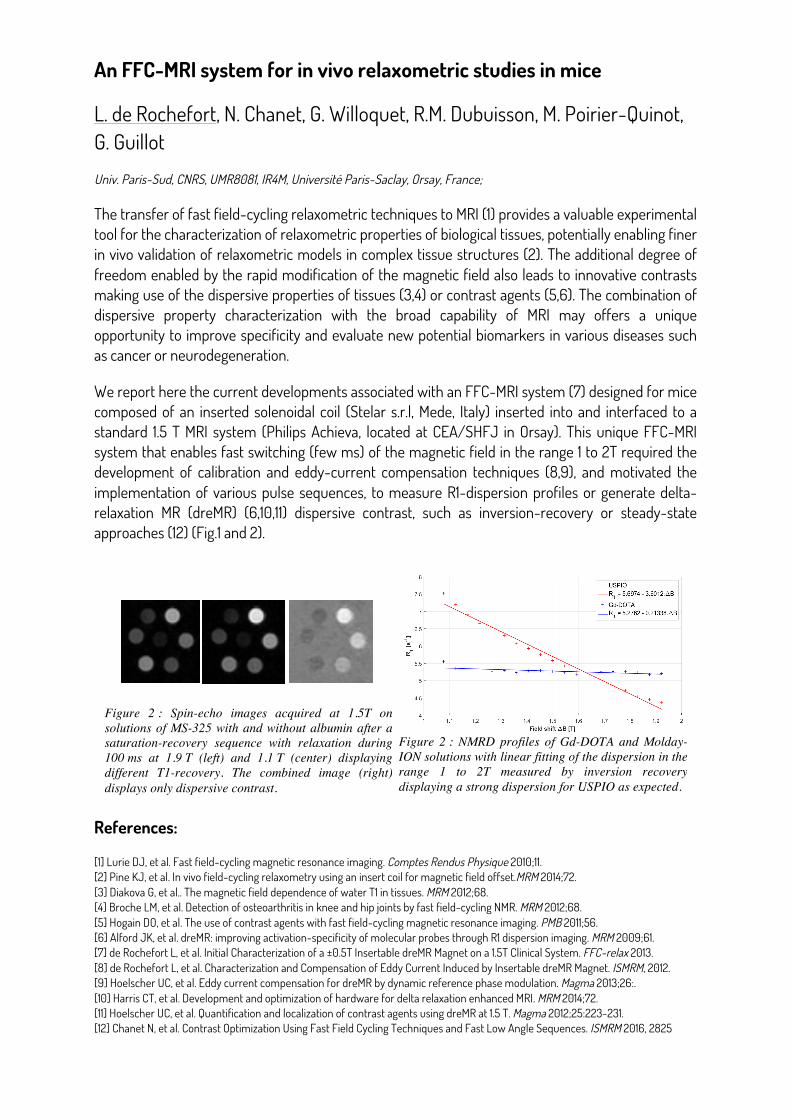
We report here the current developments associated with an FFC-MRI system (7) designed for mice composed of an inserted solenoidal coil (Stelar s.r.l, Mede, Italy) inserted into and interfaced to a standard 1.5 T MRI system (Philips Achieva, located at CEA/SHFJ in Orsay). This unique FFC-MRI system that enables fast switching (few ms) of the magnetic field in the range 1 to 2T required the development of calibration and eddy-current compensation techniques (8,9), and motivated the implementation of various pulse sequences, to measure R1-dispersion profiles or generate delta-relaxation MR (dreMR) (6,10,11) dispersive contrast, such as inversion-recovery or steady-state approaches (12) (Fig.1 and 2).
References:
[1] Lurie DJ, et al. Fast field-cycling magnetic resonance imaging. Comptes Rendus Physique 2010;11. [2] Pine KJ, et al. In vivo field-cycling relaxometry using an insert coil for magnetic field offset.MRM 2014;72. [3] Diakova G, et al.. The magnetic field dependence of water T1 in tissues. MRM 2012;68. [4] Broche LM, et al. Detection of osteoarthritis in knee and hip joints by fast field-cycling NMR. MRM 2012;68. [5] Hogain DO, et al. The use of contrast agents with fast field-cycling magnetic resonance imaging. PMB 2011;56. [6] Alford JK, et al. dreMR: improving activation-specificity of molecular probes through R1 dispersion imaging. MRM 2009;61. [7] de Rochefort L, et al. Initial Characterization of a ±0.5T Insertable dreMR Magnet on a 1.5T Clinical System. FFC-relax 2013. [8] de Rochefort L, et al. Characterization and Compensation of Eddy Current Induced by Insertable dreMR Magnet. ISMRM, 2012. [9] Hoelscher UC, et al. Eddy current compensation for dreMR by dynamic reference phase modulation. Magma 2013;26:. [10] Harris CT, et al. Development and optimization of hardware for delta relaxation enhanced MRI. MRM 2014;72. [11] Hoelscher UC, et al. Quantification and localization of contrast agents using dreMR at 1.5 T. Magma 2012;25:223-231. [12] Chanet N, et al. Contrast Optimization Using Fast Field Cycling Techniques and Fast Low Angle Sequences. ISMRM 2016, 2825
An FFC-MRI system for in vivo relaxometric studies in mice
L. de Rochefort, N. Chanet, G. Willoquet, R.M. Dubuisson, M. Poirier-Quinot, G. Guillot
Univ. Paris-Sud, CNRS, UMR8081, IR4M, Université Paris-Saclay, Orsay, France;
The transfer of fast field-cycling relaxometric techniques to MRI (1) provides a valuable experimental tool for the characterization of relaxometric properties of biological tissues, potentially enabling finer in vivo validation of relaxometric models in complex tissue structures (2). The additional degree of freedom enabled by the rapid modification of the magnetic field also leads to innovative contrasts making use of the dispersive properties of tissues (3,4) or contrast agents (5,6). The combination of dispersive property characterization with the broad capability of MRI may offers a unique opportunity to improve specificity and evaluate new potential biomarkers in various diseases such as cancer or neurodegeneration.
We report here the current developments associated with an FFC-MRI system (7) designed for mice composed of an inserted solenoidal coil (Stelar s.r.l, Mede, Italy) inserted into and interfaced to a standard 1.5 T MRI system (Philips Achieva, located at CEA/SHFJ in Orsay). This unique FFC-MRI system that enables fast switching (few ms) of the magnetic field in the range 1 to 2T required the development of calibration and eddy-current compensation techniques (8,9), and motivated the implementation of various pulse sequences, to measure R1-dispersion profiles or generate delta-relaxation MR (dreMR) (6,10,11) dispersive contrast, such as inversion-recovery or steady-state approaches (12) (Fig.1 and 2).
1. Lurie DJ, et al. Fast field-cycling magnetic resonance imaging. Comptes Rendus Physique 2010;11. 2. Pine KJ, et al. In vivo field-cycling relaxometry using an insert coil for magnetic field offset.MRM 2014;72. 3. Diakova G, et al.. The magnetic field dependence of water T1 in tissues. MRM 2012;68. 4. Broche LM, et al. Detection of osteoarthritis in knee and hip joints by fast field-cycling NMR. MRM 2012;68. 5. Hogain DO, et al. The use of contrast agents with fast field-cycling magnetic resonance imaging. PMB 2011;56. 6. Alford JK, et al. dreMR: improving activation-specificity of molecular probes through R1 dispersion imaging. MRM 2009;61. 7. de Rochefort L, et al. Initial Characterization of a ±0.5T Insertable dreMR Magnet on a 1.5T Clinical System. FFC-relax 2013. 8. de Rochefort L, et al. Characterization and Compensation of Eddy Current Induced by Insertable dreMR Magnet. ISMRM, 2012. 9. Hoelscher UC, et al. Eddy current compensation for dreMR by dynamic reference phase modulation. Magma 2013;26:. 10. Harris CT, et al. Development and optimization of hardware for delta relaxation enhanced MRI. MRM 2014;72. 11. Hoelscher UC, et al. Quantification and localization of contrast agents using dreMR at 1.5 T. Magma 2012;25:223-231. 12. Chanet N, et al. Contrast Optimization Using Fast Field Cycling Techniques and Fast Low Angle Sequences. ISMRM 2016, 2825
Figure 2 : NMRD profiles of Gd-DOTA and Molday-ION solutions with linear fitting of the dispersion in the range 1 to 2T measured by inversion recovery displaying a strong dispersion for USPIO as expected.
Figure 2 : Spin-echo images acquired at 1.5T on solutions of MS-325 with and without albumin after a saturation-recovery sequence with relaxation during100 ms at 1.9 T (left) and 1.1 T (center) displaying different T1-recovery. The combined image (right) displays only dispersive contrast.
High - Resolution Two - Field Nuclear Magnetic Resonance Spectroscopy at 0.33 and 14.1 T
P. Kader ̌ávek,1 S. Cousin,1 C. Charlier,1 B. Haddou,1 L. Strouk,1 T.Marquardsen,2 J.-M. Tyburn,3 P.-A. Bovier,4 F. Engelke,2 W. Maas,5 G. Bodenhausen,1 P. Pelupessy,1 F. Ferrage1 1École Normale Supérieure - PSL research University, Sorbonne Universités - UPMC Univ Paris 06, CNRS UMR 7203, LBM and CNRS UMR 8640 PASTEUR, 24 rue Lhomond, 75005 Paris, France; 2Bruker BioSpin GmbH, Silberstreifen 4, D 76287 Rheinstetten, Germany; 3Bruker BioSpin, 34 rue de l’Industrie BP 10002, 67166 Wissembourg Cedex, France; 4Bruker BioSpin AG, Industriestrasse 26, 8117 Fällanden, Switzerland; 5Bruker BioSpin, Billerica, Massachusetts 01821, USA. High magnetic fields enhance the sensitivity and resolution of Nuclear Magnetic Resonance experiments. Therefore, a significant effort is being focused on the development of NMR spectrometers with the highest magnetic field possible. However, some nuclear properties become less favorable at high fields than at low fields. (i) The range of NMR frequencies may become too large to obtain effective irradiation by limited radiofrequency fields, (ii) the transverse relaxation of nuclei with high chemical shift anisotropy can become too fast at high fields, (iii) chemical exchange may lead to severe signal broadening even beyond the detection limit.
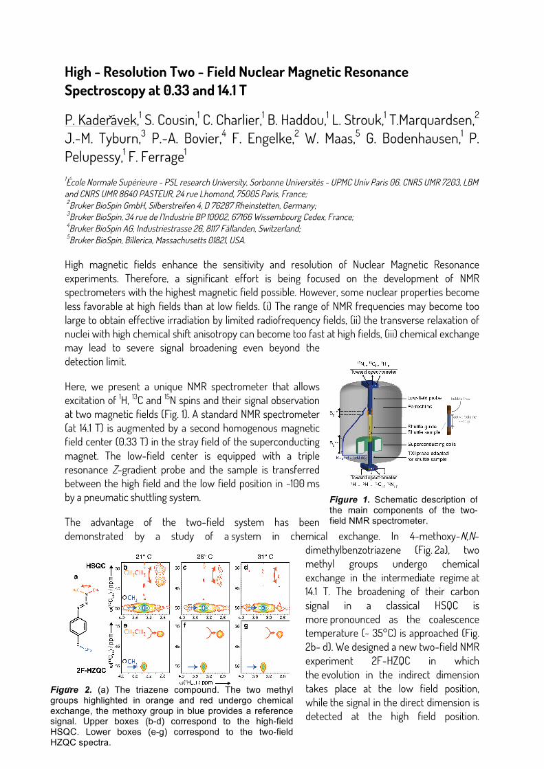
Here, we present a unique NMR spectrometer that allows excitation of 1H, 13C and 15N spins and their signal observation at two magnetic fields (Fig. 1). A standard NMR spectrometer (at 14.1 T) is augmented by a second homogenous magnetic field center (0.33 T) in the stray field of the superconducting magnet. The low-field center is equipped with a triple resonance Z-gradient probe and the sample is transferred between the high field and the low field position in ~100 ms by a pneumatic shuttling system.
The advantage of the two-field system has been demonstrated by a study of a system in chemical exchange. In 4-methoxy-N,N-
dimethylbenzotriazene (Fig. 2a), two methyl groups undergo chemical exchange in the intermediate regime at 14.1 T. The broadening of their carbon signal in a classical HSQC is more pronounced as the coalescence temperature (~ 35°C) is approached (Fig. 2b- d). We designed a new two-field NMR experiment 2F-HZQC in which the evolution in the indirect dimension takes place at the low field position, while the signal in the direct dimension is detected at the high field position.
High-Resolution Two-Field Nuclear Magnetic Resonance Spectroscopy at 0.33 and 14.1 T
P. Kadeřávek,1 S. Cousin,1 C. Charlier,1 B. Haddou,1 L. Strouk,1 T. Marquardsen,2 J.-M. Tyburn,3 P.-A. Bovier,4 F. Engelke,2 W. Maas,5 G. Bodenhausen,1 P. Pelupessy,1 F. Ferrage1
1École Normale Supérieure - PSL research University, Sorbonne Universités - UPMC Univ Paris 06, CNRS UMR 7203, LBM and CNRS UMR 8640 PASTEUR, 24 rue Lhomond, 75005 Paris, France; 2Bruker BioSpin GmbH,
Silberstreifen 4, D 76287 Rheinstetten, Germany; 3Bruker BioSpin, 34 rue de l’Industrie BP 10002, 67166 Wissembourg Cedex, France; 4Bruker BioSpin AG, Industriestrasse 26, 8117 Fällanden, Switzerland; 5Bruker
BioSpin, Billerica, Massachusetts 01821, USA. High magnetic fields enhance the sensitivity and resolution of Nuclear Magnetic Resonance experiments. Therefore, a significant effort is being focused on the development of NMR spectrometers with the highest magnetic field possible. However, some nuclear properties become less favorable at high fields than at low fields. (i) The range of NMR frequencies may become too large to obtain effective irradiation by limited radiofrequency fields, (ii) the transverse relaxation of nuclei with high chemical shift anisotropy can become too fast at high fields, (iii) chemical exchange may lead to severe signal broadening even beyond the detection limit. Here, we present a unique NMR spectrometer that allows excitation of 1H, 13C and 15N spins and their signal observation at two magnetic fields (Fig. 1). A standard NMR spectrometer (at 14.1 T) is augmented by a second homogenous magnetic field center (0.33 T) in the stray field of the superconducting magnet. The low-field center is equipped with a triple resonance Z-gradient probe and the sample is transferred between the high field and the low field position in ~100 ms by a pneumatic shuttling system. The advantage of the two-field system has been demonstrated by a study of a system in chemical exchange. In 4-methoxy-N,N-dimethylbenzotriazene (Fig. 2a), two methyl groups undergo chemical exchange in the intermediate regime at 14.1 T. The broadening of their carbon signal in a classical HSQC is more pronounced as the coalescence temperature (~ 35º C) is approached (Fig. 2b-d). We designed a new two- field NMR experiment 2F-HZQC in which the evolution in the indirect dimension takes place at the low field position, while the signal in the direct dimension is detected at the high field position. The evolution of zero-quantum coherence in the indirect dimension is used in order to suppress the effect of remaining
inhomogenities of the magnetic field. Then, the carbon chemical shift is re-introduced using a shearing transformation during the processing. The signal of the exchanging carbons is obtained in 2F-HZQC as the exchange at low field is in the fast exchange regime. Figure 2. (a) The triazene compound. The two methyl groups highlighted in orange and red undergo chemical exchange, the methoxy group in blue provides a reference signal. Upper boxes (b-d) correspond to the high-field HSQC. Lower boxes (e-g) correspond to the two-field HZQC spectra.
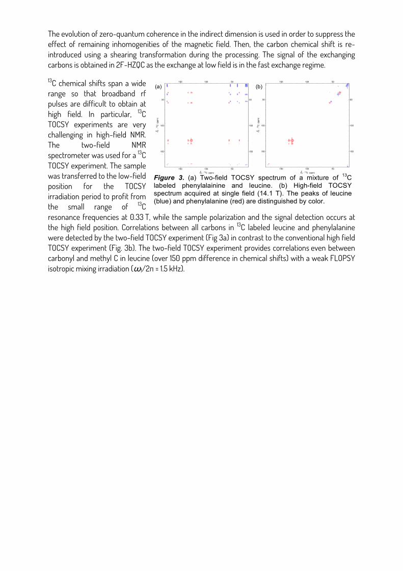
13C chemical shifts span a wide range so that broadband rf pulses are difficult to obtain at high field. In particular, 13C TOCSY experiments are very challenging in high-field NMR. The two-field NMR spectrometer was used for a 13C TOCSY experiment. The sample was transferred to the low-field position for the TOCSY irradiation period to profit from the small range of 13C resonance frequencies at 0.33 T, while the sample polarization and the signal detection occurs at the high field position. Correlations between all carbons in 13C labeled leucine and phenylalanine were detected by the two-field TOCSY experiment (Fig 3a) in contrast to the conventional high field TOCSY experiment (Fig. 3b). The two-field TOCSY experiment provides correlations even between carbonyl and methyl 13C in leucine (over 150 ppm difference in chemical shifts) with a weak FLOPSY isotropic mixing irradiation (ω1/2π = 1.5 kHz).
Figure 1. Schematic description of the main components of the two-field NMR spectrometer.
2 - 13C (ppm)
1-
13C
(p
pm
)
150
150
100
100
50
50
150 150
100 100
50 50
150
150
100
100
50
50
2 - 13C (ppm)
150 150
100 100
50 50
1-
13C
(p
pm
)
Figure 3. (a) Two-field TOCSY spectrum of a mixture of 13C labeled phenylalainine and leucine. (b) High-field TOCSY spectrum acquired at single field (14.1 T). The peaks of leucine (blue) and phenylalanine (red) are distinguished by color.
(a) (b)
High-Resolution Two-Field Nuclear Magnetic Resonance Spectroscopy at 0.33 and 14.1 T
P. Kadeřávek,1 S. Cousin,1 C. Charlier,1 B. Haddou,1 L. Strouk,1 T. Marquardsen,2 J.-M. Tyburn,3 P.-A. Bovier,4 F. Engelke,2 W. Maas,5 G. Bodenhausen,1 P. Pelupessy,1 F. Ferrage1
1École Normale Supérieure - PSL research University, Sorbonne Universités - UPMC Univ Paris 06, CNRS UMR 7203, LBM and CNRS UMR 8640 PASTEUR, 24 rue Lhomond, 75005 Paris, France; 2Bruker BioSpin GmbH,
Silberstreifen 4, D 76287 Rheinstetten, Germany; 3Bruker BioSpin, 34 rue de l’Industrie BP 10002, 67166 Wissembourg Cedex, France; 4Bruker BioSpin AG, Industriestrasse 26, 8117 Fällanden, Switzerland; 5Bruker
BioSpin, Billerica, Massachusetts 01821, USA. High magnetic fields enhance the sensitivity and resolution of Nuclear Magnetic Resonance experiments. Therefore, a significant effort is being focused on the development of NMR spectrometers with the highest magnetic field possible. However, some nuclear properties become less favorable at high fields than at low fields. (i) The range of NMR frequencies may become too large to obtain effective irradiation by limited radiofrequency fields, (ii) the transverse relaxation of nuclei with high chemical shift anisotropy can become too fast at high fields, (iii) chemical exchange may lead to severe signal broadening even beyond the detection limit. Here, we present a unique NMR spectrometer that allows excitation of 1H, 13C and 15N spins and their signal observation at two magnetic fields (Fig. 1). A standard NMR spectrometer (at 14.1 T) is augmented by a second homogenous magnetic field center (0.33 T) in the stray field of the superconducting magnet. The low-field center is equipped with a triple resonance Z-gradient probe and the sample is transferred between the high field and the low field position in ~100 ms by a pneumatic shuttling system. The advantage of the two-field system has been demonstrated by a study of a system in chemical exchange. In 4-methoxy-N,N-dimethylbenzotriazene (Fig. 2a), two methyl groups undergo chemical exchange in the intermediate regime at 14.1 T. The broadening of their carbon signal in a classical HSQC is more pronounced as the coalescence temperature (~ 35º C) is approached (Fig. 2b-d). We designed a new two- field NMR experiment 2F-HZQC in which the evolution in the indirect dimension takes place at the low field position, while the signal in the direct dimension is detected at the high field position. The evolution of zero-quantum coherence in the indirect dimension is used in order to suppress the effect of remaining
inhomogenities of the magnetic field. Then, the carbon chemical shift is re-introduced using a shearing transformation during the processing. The signal of the exchanging carbons is obtained in 2F-HZQC as the exchange at low field is in the fast exchange regime. Figure 2. (a) The triazene compound. The two methyl groups highlighted in orange and red undergo chemical exchange, the methoxy group in blue provides a reference signal. Upper boxes (b-d) correspond to the high-field HSQC. Lower boxes (e-g) correspond to the two-field HZQC spectra.
13C chemical shifts span a wide range so that broadband rf pulses are difficult to obtain at high field. In particular, 13C TOCSY experiments are very challenging in high-field NMR. The two-field NMR spectrometer was used for a 13C TOCSY experiment. The sample was transferred to the low-field position for the TOCSY irradiation period to profit from the small range of 13C resonance frequencies at 0.33 T, while the sample polarization and the signal detection occurs at the high field position. Correlations between all carbons in 13C labeled leucine and phenylalanine were detected by the two-field TOCSY experiment (Fig 3a) in contrast to the conventional high field TOCSY experiment (Fig. 3b). The two-field TOCSY experiment provides correlations even between carbonyl and methyl 13C in leucine (over 150 ppm difference in chemical shifts) with a weak FLOPSY isotropic mixing irradiation (ω1/2π = 1.5 kHz).
Figure 1. Schematic description of the main components of the two-field NMR spectrometer.
2 - 13C (ppm)
1-
13C
(p
pm
)
150
150
100
100
50
50
150 150
100 100
50 50
150
150
100
100
50
50
2 - 13C (ppm)
150 150
100 100
50 50
1-
13C
(p
pm
)
Figure 3. (a) Two-field TOCSY spectrum of a mixture of 13C labeled phenylalainine and leucine. (b) High-field TOCSY spectrum acquired at single field (14.1 T). The peaks of leucine (blue) and phenylalanine (red) are distinguished by color.
(a) (b)
The evolution of zero-quantum coherence in the indirect dimension is used in order to suppress the effect of remaining inhomogenities of the magnetic field. Then, the carbon chemical shift is re-introduced using a shearing transformation during the processing. The signal of the exchanging carbons is obtained in 2F-HZQC as the exchange at low field is in the fast exchange regime.
13C chemical shifts span a wide range so that broadband rf pulses are difficult to obtain at high field. In particular, 13C TOCSY experiments are very challenging in high-field NMR. The two-field NMR spectrometer was used for a 13C TOCSY experiment. The sample was transferred to the low-field position for the TOCSY irradiation period to profit from the small range of 13C resonance frequencies at 0.33 T, while the sample polarization and the signal detection occurs at the high field position. Correlations between all carbons in 13C labeled leucine and phenylalanine were detected by the two-field TOCSY experiment (Fig 3a) in contrast to the conventional high field TOCSY experiment (Fig. 3b). The two-field TOCSY experiment provides correlations even between carbonyl and methyl C in leucine (over 150 ppm difference in chemical shifts) with a weak FLOPSY isotropic mixing irradiation (ω1/2π = 1.5 kHz).
High-Resolution Two-Field Nuclear Magnetic Resonance Spectroscopy at 0.33 and 14.1 T
P. Kadeřávek,1 S. Cousin,1 C. Charlier,1 B. Haddou,1 L. Strouk,1 T. Marquardsen,2 J.-M. Tyburn,3 P.-A. Bovier,4 F. Engelke,2 W. Maas,5 G. Bodenhausen,1 P. Pelupessy,1 F. Ferrage1
1École Normale Supérieure - PSL research University, Sorbonne Universités - UPMC Univ Paris 06, CNRS UMR 7203, LBM and CNRS UMR 8640 PASTEUR, 24 rue Lhomond, 75005 Paris, France; 2Bruker BioSpin GmbH,
Silberstreifen 4, D 76287 Rheinstetten, Germany; 3Bruker BioSpin, 34 rue de l’Industrie BP 10002, 67166 Wissembourg Cedex, France; 4Bruker BioSpin AG, Industriestrasse 26, 8117 Fällanden, Switzerland; 5Bruker
BioSpin, Billerica, Massachusetts 01821, USA. High magnetic fields enhance the sensitivity and resolution of Nuclear Magnetic Resonance experiments. Therefore, a significant effort is being focused on the development of NMR spectrometers with the highest magnetic field possible. However, some nuclear properties become less favorable at high fields than at low fields. (i) The range of NMR frequencies may become too large to obtain effective irradiation by limited radiofrequency fields, (ii) the transverse relaxation of nuclei with high chemical shift anisotropy can become too fast at high fields, (iii) chemical exchange may lead to severe signal broadening even beyond the detection limit. Here, we present a unique NMR spectrometer that allows excitation of 1H, 13C and 15N spins and their signal observation at two magnetic fields (Fig. 1). A standard NMR spectrometer (at 14.1 T) is augmented by a second homogenous magnetic field center (0.33 T) in the stray field of the superconducting magnet. The low-field center is equipped with a triple resonance Z-gradient probe and the sample is transferred between the high field and the low field position in ~100 ms by a pneumatic shuttling system. The advantage of the two-field system has been demonstrated by a study of a system in chemical exchange. In 4-methoxy-N,N-dimethylbenzotriazene (Fig. 2a), two methyl groups undergo chemical exchange in the intermediate regime at 14.1 T. The broadening of their carbon signal in a classical HSQC is more pronounced as the coalescence temperature (~ 35º C) is approached (Fig. 2b-d). We designed a new two- field NMR experiment 2F-HZQC in which the evolution in the indirect dimension takes place at the low field position, while the signal in the direct dimension is detected at the high field position. The evolution of zero-quantum coherence in the indirect dimension is used in order to suppress the effect of remaining
inhomogenities of the magnetic field. Then, the carbon chemical shift is re-introduced using a shearing transformation during the processing. The signal of the exchanging carbons is obtained in 2F-HZQC as the exchange at low field is in the fast exchange regime. Figure 2. (a) The triazene compound. The two methyl groups highlighted in orange and red undergo chemical exchange, the methoxy group in blue provides a reference signal. Upper boxes (b-d) correspond to the high-field HSQC. Lower boxes (e-g) correspond to the two-field HZQC spectra.
13C chemical shifts span a wide range so that broadband rf pulses are difficult to obtain at high field. In particular, 13C TOCSY experiments are very challenging in high-field NMR. The two-field NMR spectrometer was used for a 13C TOCSY experiment. The sample was transferred to the low-field position for the TOCSY irradiation period to profit from the small range of 13C resonance frequencies at 0.33 T, while the sample polarization and the signal detection occurs at the high field position. Correlations between all carbons in 13C labeled leucine and phenylalanine were detected by the two-field TOCSY experiment (Fig 3a) in contrast to the conventional high field TOCSY experiment (Fig. 3b). The two-field TOCSY experiment provides correlations even between carbonyl and methyl 13C in leucine (over 150 ppm difference in chemical shifts) with a weak FLOPSY isotropic mixing irradiation (ω1/2π = 1.5 kHz).
Figure 1. Schematic description of the main components of the two-field NMR spectrometer.
2 - 13C (ppm)
1-
13C
(p
pm
)
150
150
100
100
50
50
150 150
100 100
50 50
150
150
100
100
50
50
2 - 13C (ppm)
150 150
100 100
50 50
1-
13C
(p
pm
)
Figure 3. (a) Two-field TOCSY spectrum of a mixture of 13C labeled phenylalainine and leucine. (b) High-field TOCSY spectrum acquired at single field (14.1 T). The peaks of leucine (blue) and phenylalanine (red) are distinguished by color.
(a) (b)
Posters
• Measurements of T1 dispersion profiles over the range of 1 to 2 teslas using a small animal
Fast Field Cycling MRI system Nicolas Chanet, Georges Willoquet, Laurène Jourdain, Geneviève Guillot, Ludovic de Rochefort
• Embedded hardware and software description for Mobile Low Field NMR spectrometers Alain Louis-Joseph, Denis Coupvent-Desgravier, Alexis Nauton, Jean-Pierre Korb
• Dynamical monitoring of water status in plants : low fields NMR investigations Rahima Sidi-Boulenouar, Olivier Yzebe, Eric Nativel, Christophe Coillot, Jean-Luc Verdeil, Fréderic Gatineau, Eric Alibert, Nadia Bertin, Christophe Goze-Bac
• Inversion de Laplace en relaxation magnétique nucléaire Désirée Gomis, Anne-Laure Rollet, Jean-Pierre Korb
Measurements of T1 dispersion profiles over the range of 1 to 2 teslas using a small animal Fast Field Cycling MRI system
Nicolas Chanet, Georges Willoquet, Laurène Jourdain, Geneviève Guillot, Ludovic De Rochefort Imagerie par Résonance Magnétique Médicale et Multi-Modalités (IR4M) CNRS : UMR8081, Hôpital Bicêtre, Université Paris XI - Paris Sud
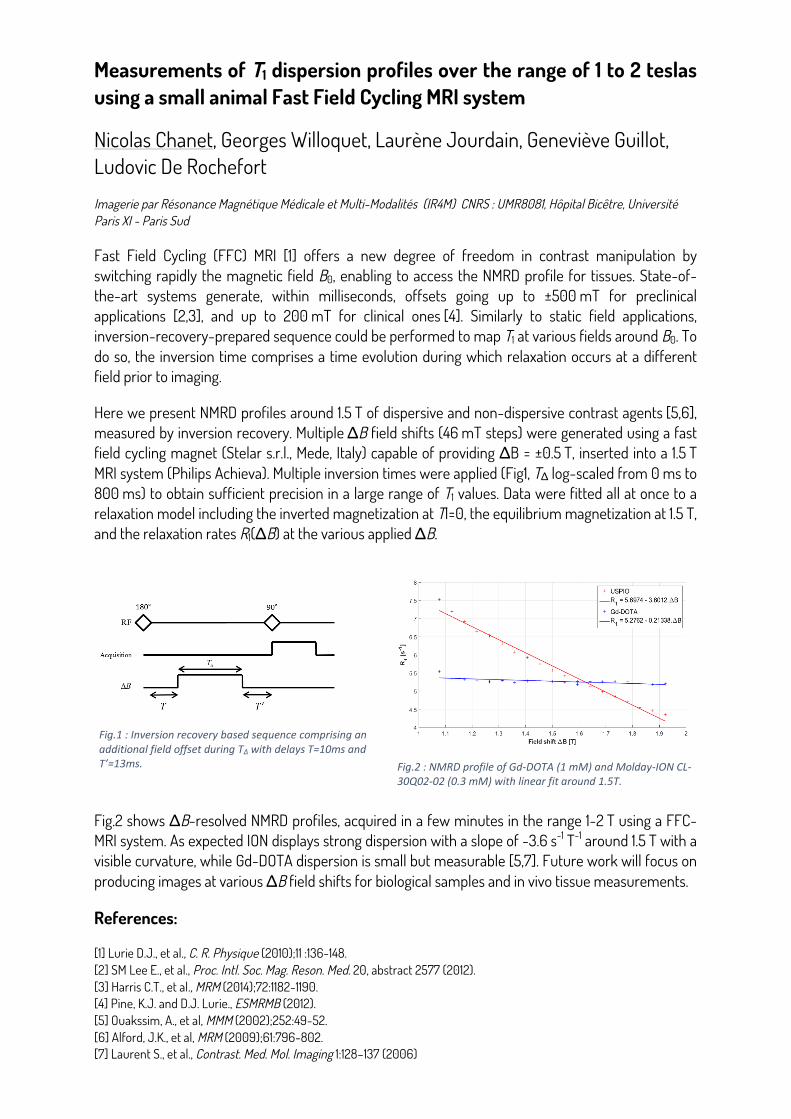
Fast Field Cycling (FFC) MRI [1] offers a new degree of freedom in contrast manipulation by switching rapidly the magnetic field B0, enabling to access the NMRD profile for tissues. State-of-the-art systems generate, within milliseconds, offsets going up to ±500 mT for preclinical applications [2,3], and up to 200 mT for clinical ones [4]. Similarly to static field applications, inversion-recovery-prepared sequence could be performed to map T1 at various fields around B0. To do so, the inversion time comprises a time evolution during which relaxation occurs at a different field prior to imaging.
Here we present NMRD profiles around 1.5 T of dispersive and non-dispersive contrast agents [5,6], measured by inversion recovery. Multiple ΔB field shifts (46 mT steps) were generated using a fast field cycling magnet (Stelar s.r.l., Mede, Italy) capable of providing ΔB = ±0.5 T, inserted into a 1.5 T MRI system (Philips Achieva). Multiple inversion times were applied (Fig1, TΔ log-scaled from 0 ms to 800 ms) to obtain sufficient precision in a large range of T1 values. Data were fitted all at once to a relaxation model including the inverted magnetization at TI=0, the equilibrium magnetization at 1.5 T, and the relaxation rates R1(ΔB) at the various applied ΔB.
Fig.2 shows ΔB-resolved NMRD profiles, acquired in a few minutes in the range 1-2 T using a FFC-MRI system. As expected ION displays strong dispersion with a slope of -3.6 s-1 T-1 around 1.5 T with a visible curvature, while Gd-DOTA dispersion is small but measurable [5,7]. Future work will focus on producing images at various ΔB field shifts for biological samples and in vivo tissue measurements.
References:
[1] Lurie D.J., et al., C. R. Physique (2010);11 :136-148. [2] SM Lee E., et al., Proc. Intl. Soc. Mag. Reson. Med. 20, abstract 2577 (2012). [3] Harris C.T., et al., MRM (2014);72:1182-1190. [4] Pine, K.J. and D.J. Lurie., ESMRMB (2012). [5] Ouakssim, A., et al, MMM (2002);252:49-52. [6] Alford, J.K., et al, MRM (2009);61:796-802. [7] Laurent S., et al., Contrast. Med. Mol. Imaging 1:128–137 (2006)
Measurements of T1 dispersion profiles over the range of 1 to 2 teslas using a small animal Fast Field Cycling MRI system
CHANET Nicolas1, WILLOQUET Georges1, JOURDAIN Laurène1, GUILLOT Geneviève1 and DE ROCHEFORT Ludovic1
1IR4M (Imagerie par Résonance Magnétique Médicale et Multi-modalités), Univ. Paris-Sud, CNRS, UMR8081, Université Paris-Saclay, Orsay,
France
Fast Field Cycling (FFC) MRI 1 offers a new degree of freedom in contrast manipulation by switching rapidly the magnetic field B0, enabling to access the NMRD profile for tissues. State-of-the-art systems generate, within milliseconds, offsets going up to ±500 mT for preclinical applications 2,3, and up to 200 mT for clinical ones 4. Similarly to static field applications, inversion-recovery-prepared sequence could be performed to map T1 at various fields around B0. To do so, the inversion time comprises a time evolution during which relaxation occurs at a different field prior to imaging.
Here we present NMRD profiles around 1.5 T of dispersive and non-dispersive contrast agents 5,6, measured by inversion recovery. Multiple ΔB field shifts (46mT steps) were generated using a fast field cycling magnet (Stelar s.r.l., Mede, Italy) capable of providing ΔB = ±0.5T, inserted into a 1.5T MRI system (Philips Achieva). Multiple inversion times were applied (Fig1, TΔ log-scaled from 0ms to 800ms) to obtain sufficient precision in a large range of T1 values. Data were fitted all at once to a relaxation model including the inverted magnetization at TI=0, the equilibrium magnetization at 1.5T, and the relaxation rates R1(ΔB) at the various applied ΔB.
Fig.1 : Inversion recovery based sequence comprising an additional field offset during TΔ with delays T=10ms and T’=13ms.
Fig.2 : NMRD profile of Gd-DOTA (1 mM) and Molday-ION CL-30Q02-02 (0.3 mM) with linear fit around 1.5T.
Fig.2 shows ΔB-resolved NMRD profiles, acquired in a few minutes in the range 1-2 T using a FFC-MRI system. As expected ION displays strong dispersion with a slope of -3.6 s-1.T-1 around 1.5T with a visible curvature, while Gd-DOTA dispersion is small but measurable 5,7. Future work will focus on producing images at various ΔB field shifts for biological samples and in vivo tissue measurements.
References:
[1] Lurie D.J., et al., C. R. Physique (2010);11 :136-148. [2] SM Lee E., et al., Proc. Intl. Soc. Mag. Reson. Med. 20, abstract 2577 (2012). [3] Harris C.T., et al., MRM (2014);72:1182-1190. [4] Pine, K.J. and D.J. Lurie., ESMRMB (2012). [5] Ouakssim, A., et al, MMM (2002);252:49-52. [6] Alford, J.K., et al, MRM (2009);61:796-802. [7] Laurent S., et al., Contrast. Med. Mol. Imaging 1:128–137 (2006)
Embedded hardware and software description for Mobile Low Field NMR spectrometers.!
Alain Louis-Joseph , Denis Coupvent-Desgravier, Alexis Nauton, Jean-Pierre Korb Laboratoire de Physique de la Matière Condensée (CNRS UMR7643) CNRS : UMR7643, Ecole Polytechnique Uni. PARIS-SACLAY Palaiseau; École Polytechnique, Paris-Saclay Route de Saclay, 91128 Palaiseau - France
!Thanks to great improvements in electronic hardware, numerous compact NMR spectrometers have been designed for an easy measure of proton NMR spectra. High sensitivity and resolution can be reached even with low field spectrometers (LFNMR) (i.e.: 60MHz), wich opens up a wide field of analytical quantification and relaxation applications. One specificity of Low field NMR spectrometer is the use of a permanent and cryogen free magnet technology, avoiding the need for weekly and expensive cryogenic services.
We present and describe a low field NMR spectrometer fabricated in our laboratory. This spectrometer operates at a basic frequency of 6 MHz, with a standard sample diameter (5-10 mm). All the embedded hardware is compact and requires only a 24 V DC power supply, so this spectrometer is portable, easy to install. This spectrometer is dedicated to education and quantification, and enables low-field NMR research. It may be coupled with scientific experiments not requiring high magnetic fields, for example when nuclear spins are dynamically polarized by coupling with other nuclei or with spin-polarized electrons.
The spectrometer includes all the embedded electronic hardware and software either for the transmission or for the receiver channel. A powerful Field Programmable Gate Area (FPGA) is used for the pulse programmer sequencer (Garay, Janvier, Geerebaert, & Louis-Joseph, 2014) and a recent ARM microprocessor devices controls the spectrometer. Direct Digital Signal technic is used for frequency synthesis. A radio frequency low noise preamplifier and quadrature demodulation design are implemented to get an improved sensitivity. The connection to a PC is via only one USB port for the control and processing of experiments. A general purpose software interface has been developed and is easy to use, allowing access by different IDE (Interface Development Environment) for processing and development. Users can either design their own pulse programming NMR sequences or load/customize ones.
Reference
Garay, P. I., Janvier, A., Geerebaert, Y., & Louis-Joseph, A. (2014, Février 6). Pilotage programmable de spectromètre RMN. Rapport d'EA PHY573A, Conception expérimentale micro et nanoélectronique, Département de Physique, X2013. Palaiseau, Essonnes, France: Ecole Polytechnique.
!! !
Dynamical monitoring of water status in plants : low fields NMR investigations
Rahima Sidi-Boulenouar1,2, Olivier Yzebe1, Eric Nativel3 , Christophe Coillot1, Jean-Luc Verdeil2, Fréderic Gatineau2 , Eric Alibert1, Nadia Bertin4, Christophe Goze-Bac1 1Charles Coulomb Laboratory (L2C-BioNanoNMRI team), UMR 5221 Centre National de la Recherche Scientifique -University, Montpellier, France ; 2 Centre internationale pour le développement durable des régions tropicales et méditerranéennes (CIRAD), UMR AGAP, Montpellier, France ; 3Institut d’Electronique et des systèmes (IES), UMR5214 Centre National de la Recherche Scientifique -University, Montpellier, France, 4 Institut National de la Recherche Agronomique (INRA), PSH (UR 1115), Avignon, France Keywords : RF Antennas, Climatic chamber, NMR rates.
Understand how plants respond to water stress is essential today to meet the challenge of developing new cultivars and new management cultures compatible with maintaining the productivity of plants, despite the global warming. Understanding water/plant relation is of central interest but paradoxically, there is no direct and non- invasive method to quantify and measure the flow of water in plants. In this study, we extract from NMR relaxation rates measurements at low fields some ecophysiological markers like water content and its mobility for different sorghum genotypes in normal and water stress conditions. The results are confronted with other methods: stomatal conductance, chlorophyll fluorescence, infrared thermography. For this purpose, a dedicated apparatus is developed with various antennas designed from an analytical model. A comparative study of the Signal to Noise Ratio (SNR) at different frequencies will be presented. The ultimate goal is to perform the experiments in the fields with a portable NMR device.
References
[1] NMR probeheads for biophysical and biomedical experiments, Mispelter J, Lupu M, Briguet A, Theorical principles and practical guidelines., London: Imperial College Press, 2006. [2] MRI of plants and foods, Henk Van As and John van Duynhoven, Journal of Magnetic Resonance 229 , 25–34., 2013. [3] Signal Modelling of MRI Ribbon Solenoid Coil Dedicated to Spinal Cord Injuries Studies, C. Coillot, R.Sidi- Boulenouar, E. Nativel, E. Alibert, M. Zanca, G. Saintmartin, H. Noristani, N. Lonjon, F. Perrin and C. Goze- Bac, J. Sens. Sens. Syst., Accepted, http://www.j-sens-sens-syst.net/5/137/2016/, 2016. !
This work has been carried out with financial aid of the LabEx NUMEV, CIRAD and INRA.
Laplace inversion in nuclear magnetic relaxation
Désirée Gomis1, Anne-Laure Rollet1, Jean-Pierre Korb1,2 1Laboratoire PHENIX, Sorbonne Universités, UPMC Univ Paris 06 - CNRS, Paris, France. 2Physique de la Matière Condensée, Ecole Polytechnique-CNRS, 91128 Palaiseau
An efficient method is presented for estimating solutions of the inverse problem in one- and two-dimensional nuclear magnetic relaxation techniques. The algorithm is divided in different successive steps: discretization, compression, optimization and regularization of the input data. We checked the efficiency of the method in presence of noisy data. The method has been applied for interpreting nuclear magnetic relaxation dispersion (NMRD) data of petroleum fluids in bulk and in confinement. This has been useful for characterizing the saturation, dynamics and wettabilities of these fluids (oil and brine) confined in petroleum rocks.