Embed Size (px)
Citation preview
No. 12No. 12
1. Introduction of the Genital System1. Introduction of the Genital System
2. Male Genital System2. Male Genital System
Chapter 5 The Genital SystemChapter 5 The Genital System
The introduction of the genital systemThe introduction of the genital system:: FunctionsFunctions: The organs of the male and : The organs of the male and
female reproductive system ensure the female reproductive system ensure the continuance of the species. They do this continuance of the species. They do this by producing gametes, or germ cells, and by producing gametes, or germ cells, and by providing a method by which the by providing a method by which the gametes of the male (sperm) can be gametes of the male (sperm) can be introduced into the body of the female, introduced into the body of the female, where one of them joins with a gamete where one of them joins with a gamete (ovum) of the female.(ovum) of the female.
ConstitutionsConstitutions: The : The genitalgenital (or (or reproductivereproductive) ) ssystemystem includes the male and the female genit includes the male and the female genital organs. Both systems are composed of interal organs. Both systems are composed of internal and external genital organs.nal and external genital organs.
The internal genital organs consist of gonad, rThe internal genital organs consist of gonad, reproductive canals and accessory glands.eproductive canals and accessory glands.
The external genital organs are mainly copulatThe external genital organs are mainly copulatory organs.ory organs.
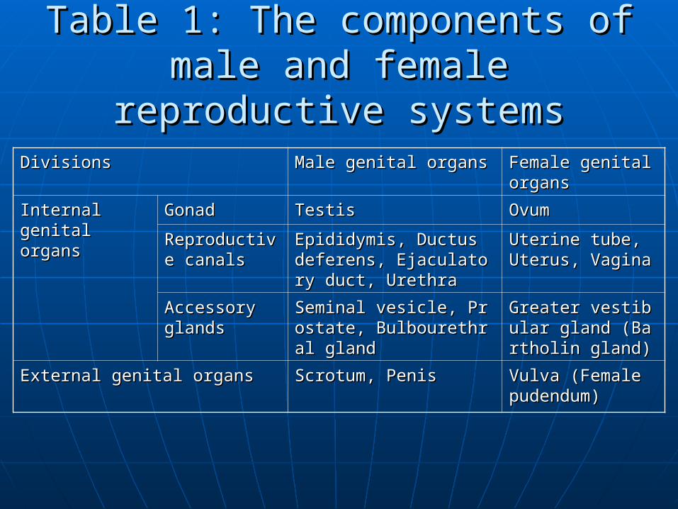
Table 1: The components of male Table 1: The components of male and female reproductive systemsand female reproductive systems
Divisions Divisions Male genital organs Male genital organs Female genital Female genital organs organs
Internal genital Internal genital organs organs
Gonad Gonad Testis Testis Ovum Ovum
Reproductive Reproductive canals canals
Epididymis, Ductus deferEpididymis, Ductus deferens, Ejaculatory duct, Urens, Ejaculatory duct, Urethra ethra
Uterine tube, Uterine tube, Uterus, Vagina Uterus, Vagina
Accessory Accessory glands glands
Seminal vesicle, Prostate,Seminal vesicle, Prostate, Bulbourethral gland Bulbourethral gland
Greater vestibular Greater vestibular gland (Bartholin glgland (Bartholin gland) and)
External genital organs External genital organs Scrotum, Penis Scrotum, Penis Vulva (Female Vulva (Female pudendum) pudendum)
Section 1 The Male Genital Section 1 The Male Genital OrgansOrgans
CompositionComposition:: The The male genital organsmale genital organs are composed of int are composed of int
ernal and external genital organs.ernal and external genital organs. 1. 1. The The internal male genital organsinternal male genital organs consist of consist of:: GonadGonad—testes—testes Reproductive canalsReproductive canals (epididymis, ductus defer (epididymis, ductus defer
ens, ejaculatory duct, urethra)ens, ejaculatory duct, urethra) Accessory glandsAccessory glands (seminal vesicle, prostate gla (seminal vesicle, prostate gla
nd, bulbourethral gland).nd, bulbourethral gland).
The testes produce sperms, which are stored iThe testes produce sperms, which are stored in the epididymides. During ejaculation the spen the epididymides. During ejaculation the sperms pass through the ductus deferens, ejacularms pass through the ductus deferens, ejaculatory ducts and urethra to be expelled from the tory ducts and urethra to be expelled from the body.body.
The accessory glands secrete the fluid to be adThe accessory glands secrete the fluid to be added to the seminal fluid to maintain the nutritided to the seminal fluid to maintain the nutrition and activity of the sperms.on and activity of the sperms.
2. 2. The The external male genital organsexternal male genital organs are are the scrotum and penisthe scrotum and penis..
The penis is the copulatory organ by whiThe penis is the copulatory organ by which the sperms are introduced into the fech the sperms are introduced into the female genital tract.male genital tract.
The scrotum houses the testes and epidiThe scrotum houses the testes and epididymides.dymides.
ⅠⅠ. The Internal Genital Organs. The Internal Genital Organs
Ⅰ Ⅰ) The Testes) The Testes The testes are male gonads, The testes are male gonads,
producing gametes of the male producing gametes of the male (sperms) and male hormones. They (sperms) and male hormones. They average 4-5 cm in length and weight average 4-5 cm in length and weight 20-30 g. 20-30 g.
1. The location of testes1. The location of testes
The testes are paired oval-shaped The testes are paired oval-shaped organs, being housed in the scrotum.organs, being housed in the scrotum.
2. The features of testes2. The features of testes
Each testis has the superior and inferior Each testis has the superior and inferior extremities, the lateral and medial surfaextremities, the lateral and medial surfaces, and the anterior and posterior bordces, and the anterior and posterior borders.ers.
The blood vessels, nerves and lymphaticThe blood vessels, nerves and lymphatics pass through the posterior border to es pass through the posterior border to enter or leave the testis. The posterior bonter or leave the testis. The posterior border connect with the epididymis.rder connect with the epididymis.
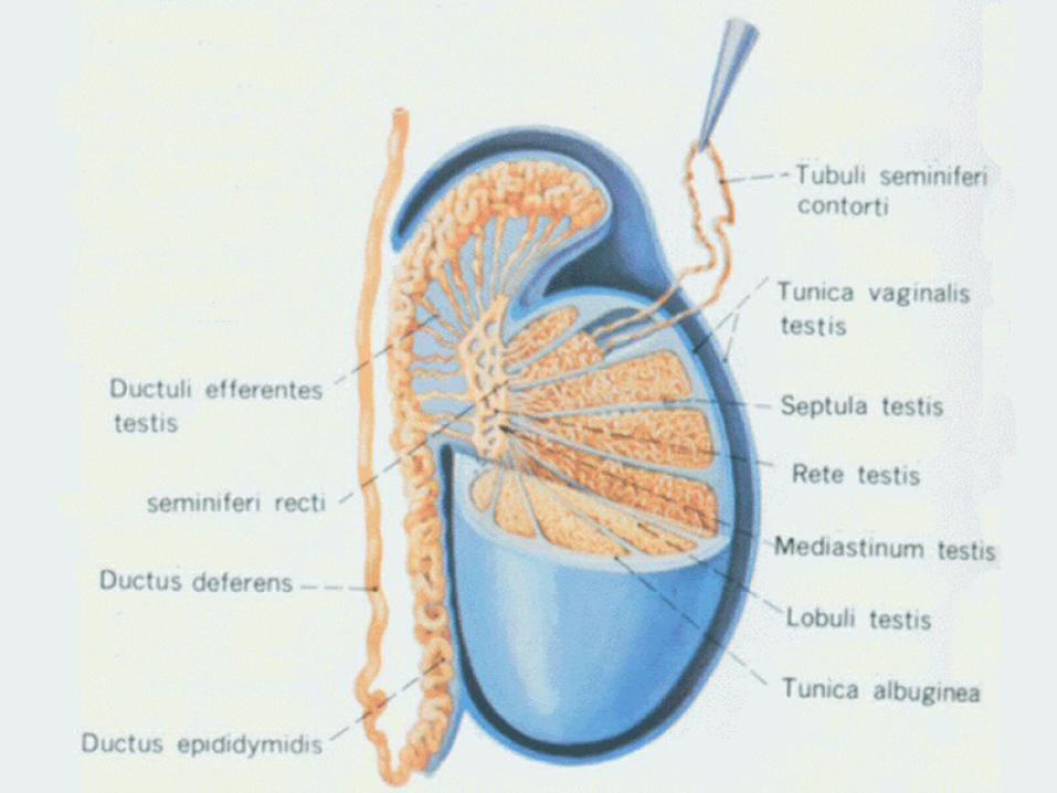
3. The structure of testes3. The structure of testes The testes are covered by a dense layer of fibrThe testes are covered by a dense layer of fibr
ous membrane, the ous membrane, the tunica albugineatunica albuginea. It is thic. It is thicker on the posterior border of the testis and foker on the posterior border of the testis and form the rm the mediastinum testismediastinum testis, which gives off th, which gives off the e septula testisseptula testis extending inward to divide ea extending inward to divide each testis into a series of compartments (100-20ch testis into a series of compartments (100-200) called lobules of testis.0) called lobules of testis.
Each lobule contains two to four tiny coiled tuEach lobule contains two to four tiny coiled tubules, the bules, the convoluted seminiferous tubulesconvoluted seminiferous tubules, , that produce the sperms by a process called that produce the sperms by a process called sspermatogenesispermatogenesis..

In the mediastinum testis the convoluted semiIn the mediastinum testis the convoluted seminiferous tubules join to form the niferous tubules join to form the straight semistraight seminiferous tubulesniferous tubules which enter the fibrous tissu which enter the fibrous tissue of the mediastinum testis. They then form the of the mediastinum testis. They then form the e rete testisrete testis. At the upper end of the mediasti. At the upper end of the mediastinum testis the rete testis terminate in 12num testis the rete testis terminate in 12 ~~ 15 15 efferent ductules of testis, passing from the teefferent ductules of testis, passing from the testis to the epididymis.stis to the epididymis.
4. The function of testes4. The function of testes
After puberty the testes produce After puberty the testes produce sperms and secrete male hormone.sperms and secrete male hormone.
ⅡⅡ) The Epididymis) The Epididymis
1. Location of epididymis1. Location of epididymis It is attached to the superior extremity aIt is attached to the superior extremity a
nd the posterolateral surface of the testind the posterolateral surface of the testis.s.

2. Morphological feature of epididymis2. Morphological feature of epididymis The epididymis is comma—shaped and is divided into The epididymis is comma—shaped and is divided into
the head, body and tail.the head, body and tail. HeadHead: The enlarged superior portion of the epididymi: The enlarged superior portion of the epididymi
s is known as the head.s is known as the head. BodyBody: The portion posterior to the testis is called the : The portion posterior to the testis is called the
body.body. TailTail: The inferior portion is referred to as the tail.: The inferior portion is referred to as the tail. The head of the epididymis contains the efferent ductThe head of the epididymis contains the efferent duct
ules which terminate in a coiled duct of epididymis wiules which terminate in a coiled duct of epididymis within the body and tail. The end of the duct of epididymthin the body and tail. The end of the duct of epididymis passes upward to continue with the ductus deferens.is passes upward to continue with the ductus deferens.
3. Function of epididymis3. Function of epididymis The epididymis is the principal store—hThe epididymis is the principal store—h
ouse for sperms. It also adds an essentiaouse for sperms. It also adds an essential secretion to the seminal fluid in which tl secretion to the seminal fluid in which the sperms are developed and activated.he sperms are developed and activated.
ⅢⅢ) The ) The Ductus DeferensDuctus Deferens
It is a continuation of the duct of epididyIt is a continuation of the duct of epididymis, about 50 cm long. The ductus is divimis, about 50 cm long. The ductus is divided into four parts:ded into four parts:
1. The 1. The testicular parttesticular part It begins at the tail of epididymis, and asIt begins at the tail of epididymis, and as
cends on the medial side of the epididycends on the medial side of the epididymis, posterior to the testis.mis, posterior to the testis.

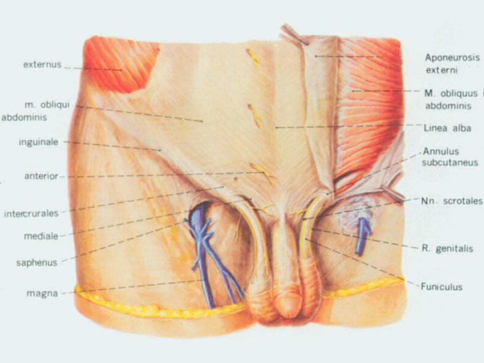
2. The 2. The funicular (spermatic) partfunicular (spermatic) part It extends from the superior It extends from the superior
extremity of the testis to the extremity of the testis to the superficial inguinal ring.superficial inguinal ring.
It lies in medial and posterior portion It lies in medial and posterior portion of the spermatic cord and can be of the spermatic cord and can be palpated easily in the living body.palpated easily in the living body.
This part is the place where the This part is the place where the vasectomy is performed.vasectomy is performed.
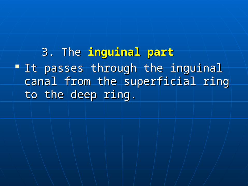
3. The3. The inguinal partinguinal part It passes through the inguinal canal It passes through the inguinal canal
from the superficial ring to the deep from the superficial ring to the deep ring.ring.

4. The 4. The pelvic partpelvic part It enters the abdominal cavity from the deep ring of thIt enters the abdominal cavity from the deep ring of th
e inguinal canal. Then passes downward and medially e inguinal canal. Then passes downward and medially along the lateral wall of the pelvis. It crosses over the talong the lateral wall of the pelvis. It crosses over the terminal portion of the ureter anterosuperioly to its meerminal portion of the ureter anterosuperioly to its medial side.dial side.
The terminal portion of the ductus dererens is dilated, The terminal portion of the ductus dererens is dilated, and called the and called the ampulla ductus deferentisampulla ductus deferentis as it desce as it descends behind the fundus of the bladder. Its end is thin ands behind the fundus of the bladder. Its end is thin and joins the excretory duct of the seminal vesicle to fond joins the excretory duct of the seminal vesicle to form the rm the ejaculatory ductejaculatory duct..
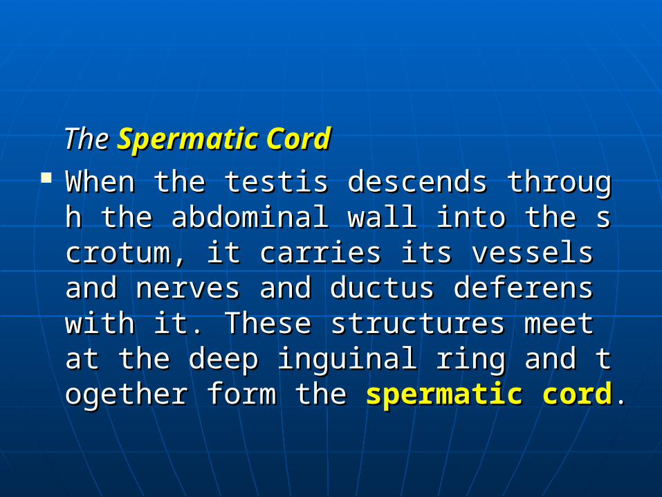
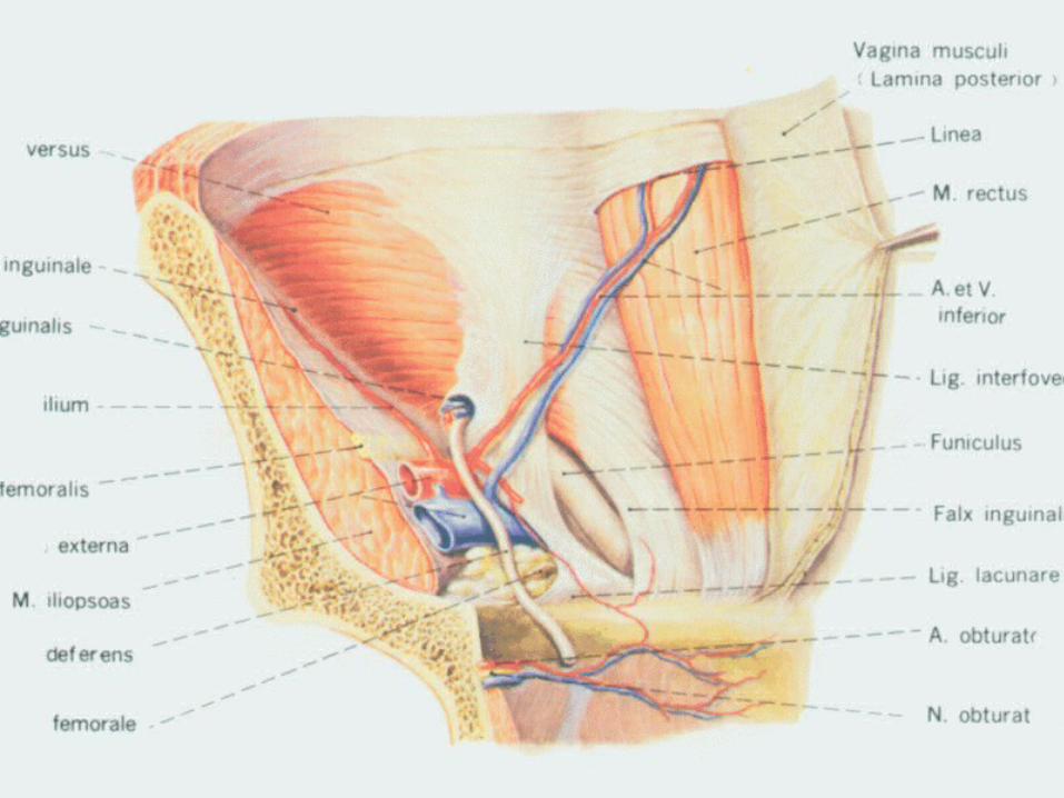
The The Spermatic CordSpermatic Cord When the testis descends through the aWhen the testis descends through the a
bdominal wall into the scrotum, it carriebdominal wall into the scrotum, it carries its vessels and nerves and ductus defers its vessels and nerves and ductus deferens with it. These structures meet at the ens with it. These structures meet at the deep inguinal ring and together form the deep inguinal ring and together form the spermatic cordspermatic cord..
It extends from superior extremity of the It extends from superior extremity of the testis to the deep inguinal ring, 11testis to the deep inguinal ring, 11 ~~ 15 15 cm in length.cm in length.
The main structures of spermatic cord arThe main structures of spermatic cord are the ductus deferens, testicular artery, e the ductus deferens, testicular artery, pampiniform plexus of vein, nervous plepampiniform plexus of vein, nervous plexus, lymphatic vessels and the vestige of xus, lymphatic vessels and the vestige of vaginal process.vaginal process.
ⅣⅣ) The ) The Ejaculatory DuctEjaculatory Duct
It is formed by the union of the terminal It is formed by the union of the terminal part of the ductus deferens and the excrpart of the ductus deferens and the excretory duct of seminal vesicle.etory duct of seminal vesicle.
It commences at the base of prostate, aIt commences at the base of prostate, and passes downward and forward to opnd passes downward and forward to open into the prostatic portion of the urethen into the prostatic portion of the urethra.ra.

ⅤⅤ) The ) The Seminal VesiclesSeminal Vesicles
They are two oval sacculated organs placed pThey are two oval sacculated organs placed posterior to the fundus of urinary bladder, laterosterior to the fundus of urinary bladder, lateral to the ampulla ductus dererentis and in fronal to the ampulla ductus dererentis and in front of the rectum.t of the rectum.
Its excretory duct joins the corresponding ductIts excretory duct joins the corresponding ductus deferens to form the ejaculatory duct.us deferens to form the ejaculatory duct.
The secretion of the seminal vesicles is a compThe secretion of the seminal vesicles is a component of seminal fluid.onent of seminal fluid.
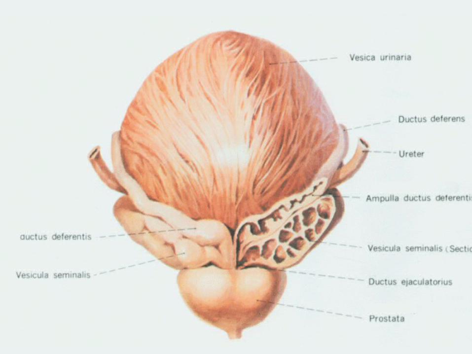
ⅥⅥ) The ) The ProstateProstate
It is a single, firm gland consisting of muIt is a single, firm gland consisting of muscular and glandular tissues.scular and glandular tissues.
FunctionFunction:: Its secretion, the important component Its secretion, the important component
of the seminal fluid, is conveyed into the of the seminal fluid, is conveyed into the prostatic portion of urethra through 1prostatic portion of urethra through 166 ~~ 32 excretory ducts.32 excretory ducts.
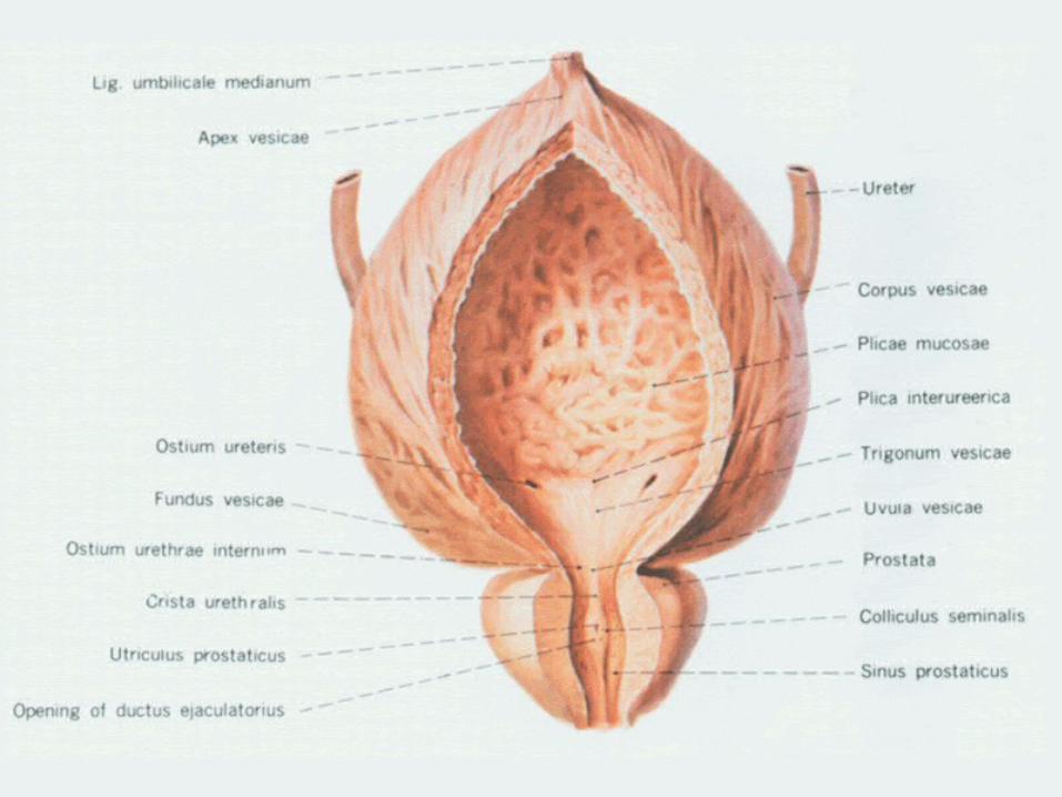
1. Location1. Location
The prostate lies in the lesser pelvic caviThe prostate lies in the lesser pelvic cavity, below the urinary bladder and arounty, below the urinary bladder and around the superior part of the urethra, betwed the superior part of the urethra, between the bladder and the urogenital diaphen the bladder and the urogenital diaphragm. It is in front of the rectum, posteriragm. It is in front of the rectum, posterior to the pubic symphysis. or to the pubic symphysis.
2. External Features:2. External Features: The prostate is chestnut-shaped and has an apex, a baThe prostate is chestnut-shaped and has an apex, a ba
se.se. The The apexapex of prostate is inferior and rests on the urogeof prostate is inferior and rests on the uroge
nital diaphragm.nital diaphragm. The base is superior and is against the neck of bladder.The base is superior and is against the neck of bladder. The urethra passes through the prostate.The urethra passes through the prostate. The anterior surface of prostate is posterior to the pubThe anterior surface of prostate is posterior to the pub
ic symphysis.ic symphysis. There is a shallow sulcus, called There is a shallow sulcus, called prostatic sulcusprostatic sulcus, alo, alo
ng the midline of the posterior surface. This sulcus, apng the midline of the posterior surface. This sulcus, apmula ductus deferentis and seminal vesicles can be pamula ductus deferentis and seminal vesicles can be palpated through the anterior wall of the rectum in the lilpated through the anterior wall of the rectum in the living body.ving body.

The lobes of prostateThe lobes of prostate:: The prostate may be divided into five The prostate may be divided into five
lobes:lobes: anterior, median, posterior and two lateral anterior, median, posterior and two lateral
lobes.lobes. The median lobe (isthmus of prostate) lies The median lobe (isthmus of prostate) lies
behind the urethra, between the right and behind the urethra, between the right and left lobes, in front of the posterior lobe.left lobes, in front of the posterior lobe.
In old men, the prostate sometimes In old men, the prostate sometimes enlarges and bulges into the lumen of enlarges and bulges into the lumen of urethra causing obstruction of it.urethra causing obstruction of it.
ⅦⅦ) The Bulbourethral Glands (of C) The Bulbourethral Glands (of Cowper)owper)
They are about the size of pea, one They are about the size of pea, one on each side of the membranous part on each side of the membranous part of urethra, embedded in the deep of urethra, embedded in the deep transverse muscle of perineum. Their transverse muscle of perineum. Their excretory ducts open into the upper excretory ducts open into the upper part of the spongy part of urethra.part of the spongy part of urethra.

TheThe seminal fluid seminal fluid is the mixture of the sp is the mixture of the sperms and secretion of accessory glands. erms and secretion of accessory glands. It is white milk like fluid, with weak alkalIt is white milk like fluid, with weak alkalinity. In every ejaculation about 2inity. In every ejaculation about 2 ~~ 5 ml 5 ml of seminal fluid is expelled, which contaiof seminal fluid is expelled, which contains 300ns 300 ~~ 500 million of sperms500 million of sperms
ⅡⅡ. The External Genital Organs. The External Genital Organs
Ⅰ Ⅰ) The Scrotum) The Scrotum It is a pendulous pouch of skin and supeIt is a pendulous pouch of skin and supe
rficial fascia evaginated from the lowed rficial fascia evaginated from the lowed part of the abdominal wall.part of the abdominal wall.
The scrotal wall is composed of skin and The scrotal wall is composed of skin and dartos. dartos.
① ① The The skinskin: The skin is thin and extensible, which con: The skin is thin and extensible, which contains few hairs, but many sebaceous and sweat glands,tains few hairs, but many sebaceous and sweat glands, pigment cells and nerves. pigment cells and nerves.
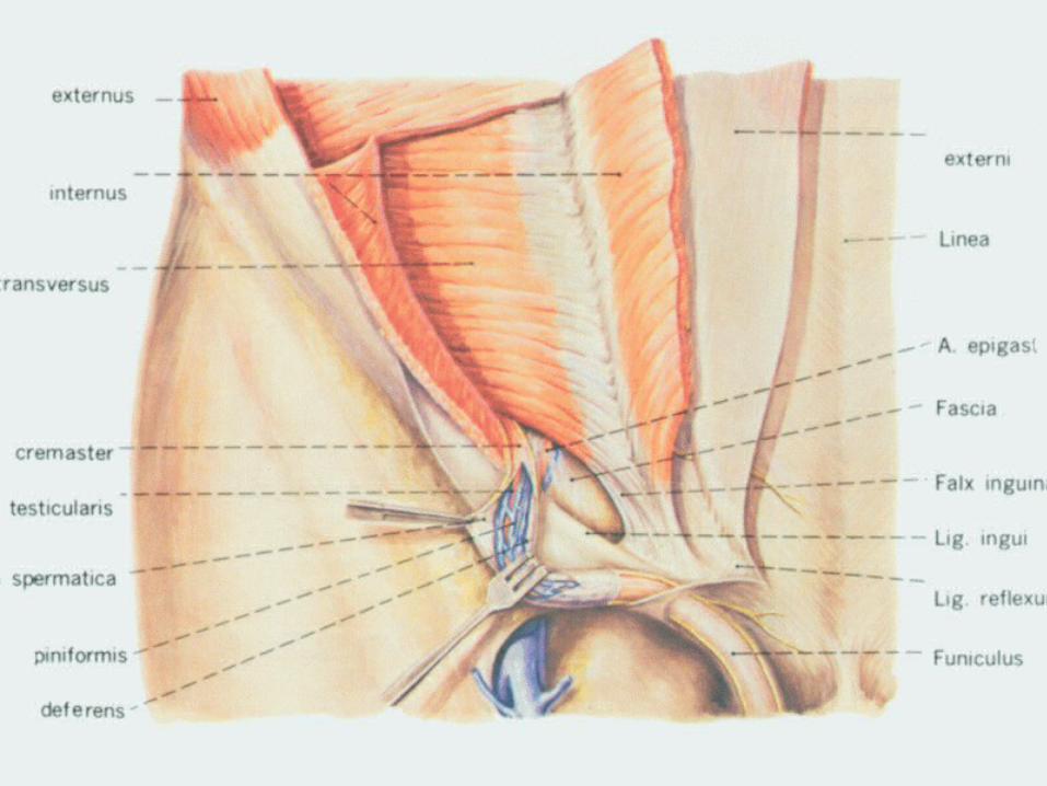
② ② The The dartosdartos: Involuntary muscle fibers lie within the : Involuntary muscle fibers lie within the superficial fascia of the scrotum to form the dartos. Thsuperficial fascia of the scrotum to form the dartos. The contraction of dartos causes the scrotal skin wrinkle e contraction of dartos causes the scrotal skin wrinkle when cold. Beneath the dartos there are three layers owhen cold. Beneath the dartos there are three layers of tunics to enclose the testis and spermatic cord, that if tunics to enclose the testis and spermatic cord, that is the external spermatic fascia, the cremaster and the s the external spermatic fascia, the cremaster and the internal spermatic fascia.internal spermatic fascia.
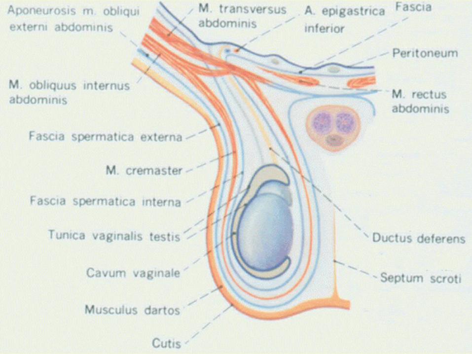
③ ③ The The external spermatic fasciaexternal spermatic fascia: It is the con: It is the continuation of the aponeurosis of the obliquus extinuation of the aponeurosis of the obliquus externus abdominis.ternus abdominis.
④ ④ The The cremastercremaster: It is the middle layer comin: It is the middle layer coming from the obliquus internus abdominis and trg from the obliquus internus abdominis and transversus abdominis.ansversus abdominis.
⑤ ⑤ TheThe internal spermatic fasciainternal spermatic fascia: It is the cont: It is the continuation of the transverse fascia.inuation of the transverse fascia.
⑥ ⑥ The The tunica vaginalis of testistunica vaginalis of testis:: It is from the peritonIt is from the peritoneum. The tunica vaginalis is the remnant of the lower eum. The tunica vaginalis is the remnant of the lower end of the vaginal process, the upper part of which is end of the vaginal process, the upper part of which is obliterated. The tunica vaginalis consists of visceral laobliterated. The tunica vaginalis consists of visceral layer and perietal layer. Between these two layers there yer and perietal layer. Between these two layers there is a serous cavity with few fluid in it. If the upper part ois a serous cavity with few fluid in it. If the upper part of the vaginal process is not obliterated, the serous cavif the vaginal process is not obliterated, the serous cavity of testis communicates with the abdominal cavity, ty of testis communicates with the abdominal cavity, and the herniated abdominal content, such as loop of and the herniated abdominal content, such as loop of ileum will enter the serous cavity close to the testis. Tileum will enter the serous cavity close to the testis. This case is called the his case is called the congenital indirect inguinal hernicongenital indirect inguinal herniaa..
ⅡⅡ) The Penis) The Penis It is the male organ of copulation and urination.It is the male organ of copulation and urination. It comprises a root, a body and a head.It comprises a root, a body and a head. The The rootroot of penis is the posterior portion and attaches of penis is the posterior portion and attaches
to the pubic arch.to the pubic arch. The The bodybody continuing with the root is the movable part, continuing with the root is the movable part,
and is covered with skin and fascia. and is covered with skin and fascia. The The headhead of penis is a slightly enlarged portion called of penis is a slightly enlarged portion called
the glans penis, which is separated from the body by a the glans penis, which is separated from the body by a constriction, the constriction, the neckneck of penis. A median slit at the tip of penis. A median slit at the tip of the glans is the external orifice of urethra.of the glans is the external orifice of urethra.
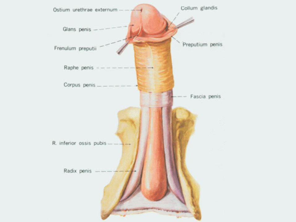
At the neck, the skin is folded upon itself to form the pAt the neck, the skin is folded upon itself to form the prepuce. The free border of the prepuce is called the repuce. The free border of the prepuce is called the ororifice of prepuceifice of prepuce. The prepuce in the child is longer th. The prepuce in the child is longer than in the adult, and the diameter of the orifice of prepan in the adult, and the diameter of the orifice of prepuce is smaller than in adult. In some cases, the prepucuce is smaller than in adult. In some cases, the prepuce is longer than normal (e is longer than normal (redundant prepuceredundant prepuce) or the ori) or the orifice of prepuce is smaller than normal and the prepucfice of prepuce is smaller than normal and the prepuce cannot be retracted over the glans (e cannot be retracted over the glans (phimosisphimosis). This ). This may permit accumulation of the secretions beneath tmay permit accumulation of the secretions beneath the prepuce, leading to inflammation. And it is now gehe prepuce, leading to inflammation. And it is now generally believed that chronic inflammation of the prepnerally believed that chronic inflammation of the prepuce predisposes to carcinoma of the glans penis. For tuce predisposes to carcinoma of the glans penis. For these reasons prophylactic circumcision is commonly hese reasons prophylactic circumcision is commonly practiced.practiced.
The The frenulum of prepucefrenulum of prepuce is a median fold passing fro is a median fold passing from the deep surface of the prepuce to a point near the m the deep surface of the prepuce to a point near the external orifice of urethra ventrally. It must be protectexternal orifice of urethra ventrally. It must be protected during circumcision.ed during circumcision.
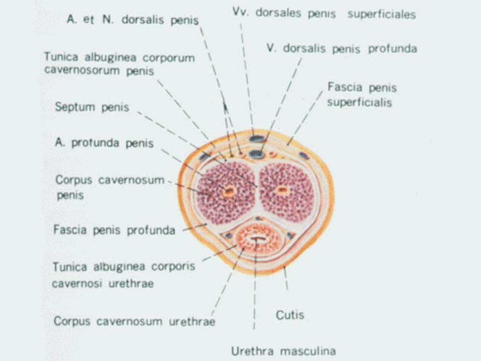
Structure of the penis:Structure of the penis: Structurally the penis is composed of three cylindrical Structurally the penis is composed of three cylindrical
masses of erectile tissue.masses of erectile tissue. Cavernous body of penisCavernous body of penis: The two dorsally located m: The two dorsally located m
asses are called the cavernous body of penis.asses are called the cavernous body of penis. Cavernous body of urethraCavernous body of urethra: The single smaller ventr: The single smaller ventr
al mass, the cavernous body of urethra, contains the sal mass, the cavernous body of urethra, contains the spongy part of the urethra.pongy part of the urethra.
ⅢⅢ) The Male Urethra) The Male Urethra
It extends from the internal orifice of It extends from the internal orifice of urethra in the urinary bladder to urethra in the urinary bladder to external orifice at the end of the external orifice at the end of the penis. It is 17—20 cm long and may penis. It is 17—20 cm long and may be divided into three parts:be divided into three parts:
1. The 1. The prostatic partprostatic part It is about 2.5 cm in length with the largest caliIt is about 2.5 cm in length with the largest cali
ber.ber. On the midline of posterior wall there is a longiOn the midline of posterior wall there is a longi
tudinal ridge, tudinal ridge, urethral cresturethral crest. At the middle po. At the middle portion of the urethral crest there is a small promrtion of the urethral crest there is a small prominence, seminal colliculus, lateral to which is tinence, seminal colliculus, lateral to which is the opening of ejaculatory duct. The prostatic dhe opening of ejaculatory duct. The prostatic ductules open into the urethra near the crest.uctules open into the urethra near the crest.

2. The 2. The membranous partmembranous part It is 1—2 cm in length, and is the shortest, least dilataIt is 1—2 cm in length, and is the shortest, least dilata
ble and narrowest part of the urethra. It is within the uble and narrowest part of the urethra. It is within the urogenital diaphragm, and surrounded by the sphincterogenital diaphragm, and surrounded by the sphincter of urethra.r of urethra.
This striate muscle is under voluntary control after earThis striate muscle is under voluntary control after early infancy.ly infancy.
Anterior ruethra and posterior urethra:Anterior ruethra and posterior urethra: In the clinic the first and second parts of urethra are cIn the clinic the first and second parts of urethra are c
alled the posterior urethra, and the last spongy part thalled the posterior urethra, and the last spongy part the anterior urethra.e anterior urethra.
3. The 3. The cavernous (spongy) partcavernous (spongy) part It is contained in the cavernous body of urethrIt is contained in the cavernous body of urethr
a, about 15 cm in length. It extends from the ea, about 15 cm in length. It extends from the end of the membranous part to the external orifnd of the membranous part to the external orifice of urethra.ice of urethra.
In this part there are two dilated places. One is In this part there are two dilated places. One is at the proximal portion within the bulb of peniat the proximal portion within the bulb of penis, called the bulbous portion of urethra, which s, called the bulbous portion of urethra, which is the common site of rupture in straddle injuriis the common site of rupture in straddle injuries. The other is near the external orifice of uretes. The other is near the external orifice of urethra, the navicular fassa.hra, the navicular fassa.
The male urethra presents three The male urethra presents three strictures, three dilatations and two strictures, three dilatations and two curvatures.curvatures.
Three stricturesThree strictures::
The three strictures are situated in:The three strictures are situated in: ① ① The internal urethral orifice,The internal urethral orifice, ② ② The membranous portionThe membranous portion ③ ③ The external orifice of urethra.The external orifice of urethra.

Three dilatations:Three dilatations: The three dilatations are situated in:The three dilatations are situated in: ① ① The prostatic part of urethra.The prostatic part of urethra. ② ② The bulbous portion of urethraThe bulbous portion of urethra ③ ③ Navicular fossa of urethra.Navicular fossa of urethra.
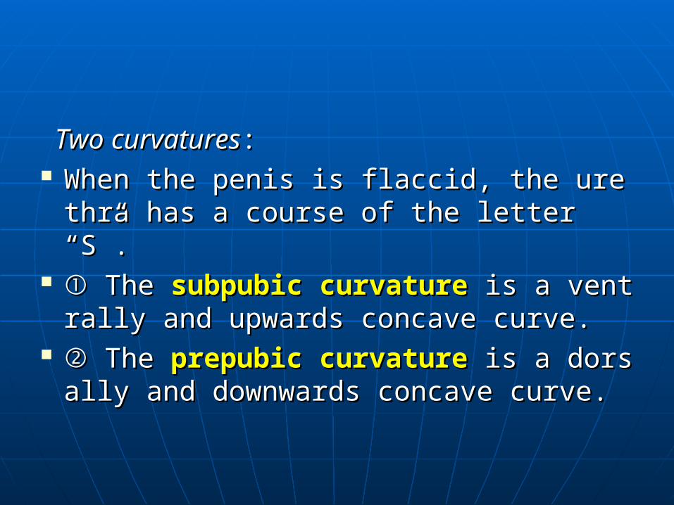
Two curvaturesTwo curvatures:: When the penis is flaccid, the urethra haWhen the penis is flaccid, the urethra ha
s a course of the letter “S”.s a course of the letter “S”. ① ① The The subpubic curvaturesubpubic curvature is a ventrally is a ventrally
and upwards concave curve.and upwards concave curve. ② ② The The prepubic curvatureprepubic curvature is a dorsally is a dorsally
and downwards concave curve.and downwards concave curve.