Embed Size (px)
Citation preview
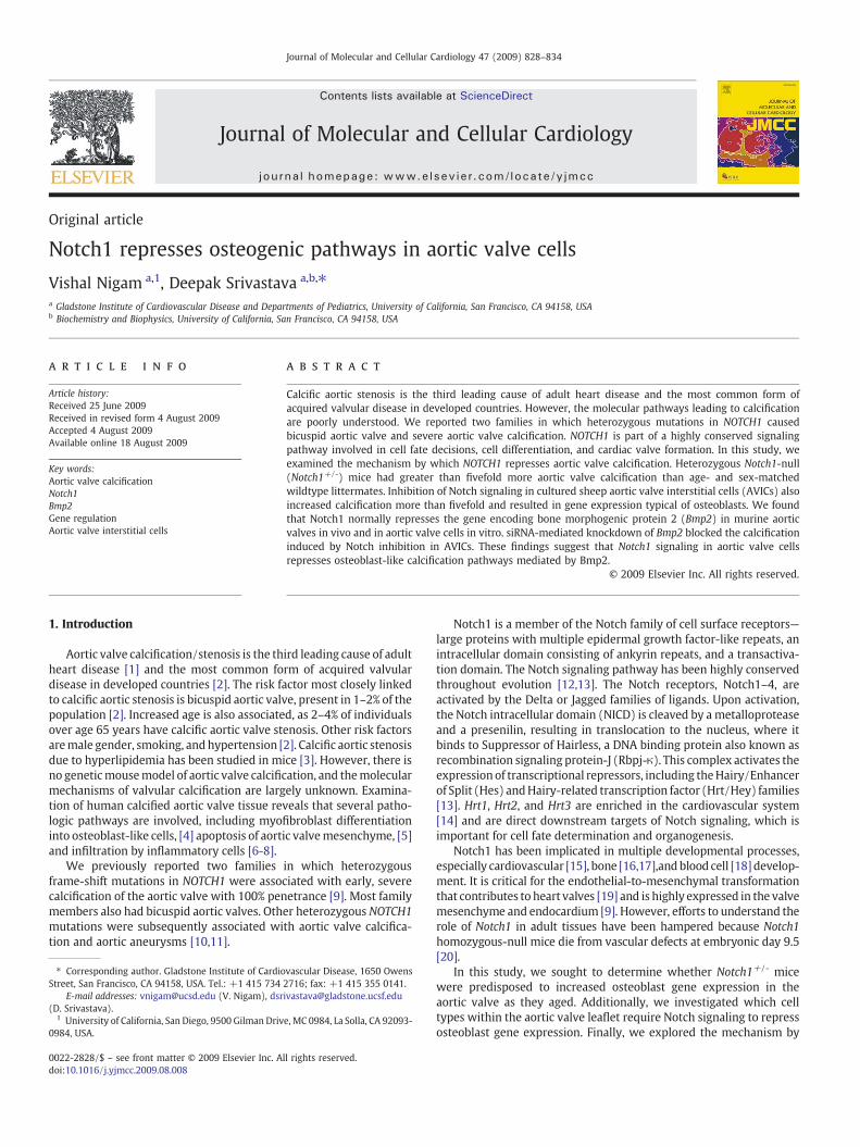
Journal of Molecular and Cellular Cardiology 47 (2009) 828–834
Contents lists available at ScienceDirect
Journal of Molecular and Cellular Cardiology
j ourna l homepage: www.e lsev ie r.com/ locate /y jmcc
Original article
Notch1 represses osteogenic pathways in aortic valve cells
Vishal Nigam a,1, Deepak Srivastava a,b,⁎a Gladstone Institute of Cardiovascular Disease and Departments of Pediatrics, University of California, San Francisco, CA 94158, USAb Biochemistry and Biophysics, University of California, San Francisco, CA 94158, USA
⁎ Corresponding author. Gladstone Institute of CardioStreet, San Francisco, CA 94158, USA. Tel.: +1 415 734
E-mail addresses: [email protected] (V. Nigam), dsr(D. Srivastava).
1 University of California, San Diego, 9500 Gilman Driv0984, USA.
0022-2828/$ – see front matter © 2009 Elsevier Inc. Adoi:10.1016/j.yjmcc.2009.08.008
a b s t r a c t
a r t i c l e i n f oArticle history:Received 25 June 2009Received in revised form 4 August 2009Accepted 4 August 2009Available online 18 August 2009
Key words:Aortic valve calcificationNotch1Bmp2Gene regulationAortic valve interstitial cells
Calcific aortic stenosis is the third leading cause of adult heart disease and the most common form ofacquired valvular disease in developed countries. However, the molecular pathways leading to calcificationare poorly understood. We reported two families in which heterozygous mutations in NOTCH1 causedbicuspid aortic valve and severe aortic valve calcification. NOTCH1 is part of a highly conserved signalingpathway involved in cell fate decisions, cell differentiation, and cardiac valve formation. In this study, weexamined the mechanism by which NOTCH1 represses aortic valve calcification. Heterozygous Notch1-null(Notch1+/-) mice had greater than fivefold more aortic valve calcification than age- and sex-matchedwildtype littermates. Inhibition of Notch signaling in cultured sheep aortic valve interstitial cells (AVICs) alsoincreased calcification more than fivefold and resulted in gene expression typical of osteoblasts. We foundthat Notch1 normally represses the gene encoding bone morphogenic protein 2 (Bmp2) in murine aorticvalves in vivo and in aortic valve cells in vitro. siRNA-mediated knockdown of Bmp2 blocked the calcificationinduced by Notch inhibition in AVICs. These findings suggest that Notch1 signaling in aortic valve cellsrepresses osteoblast-like calcification pathways mediated by Bmp2.
© 2009 Elsevier Inc. All rights reserved.
1. Introduction
Aortic valve calcification/stenosis is the third leading cause of adultheart disease [1] and the most common form of acquired valvulardisease in developed countries [2]. The risk factor most closely linkedto calcific aortic stenosis is bicuspid aortic valve, present in 1–2% of thepopulation [2]. Increased age is also associated, as 2–4% of individualsover age 65 years have calcific aortic valve stenosis. Other risk factorsaremale gender, smoking, andhypertension [2]. Calcific aortic stenosisdue to hyperlipidemia has been studied in mice [3]. However, there isno geneticmousemodel of aortic valve calcification, and themolecularmechanisms of valvular calcification are largely unknown. Examina-tion of human calcified aortic valve tissue reveals that several patho-logic pathways are involved, including myofibroblast differentiationinto osteoblast-like cells, [4] apoptosis of aortic valvemesenchyme, [5]and infiltration by inflammatory cells [6-8].
We previously reported two families in which heterozygousframe-shift mutations in NOTCH1 were associated with early, severecalcification of the aortic valve with 100% penetrance [9]. Most familymembers also had bicuspid aortic valves. Other heterozygous NOTCH1mutations were subsequently associated with aortic valve calcifica-tion and aortic aneurysms [10,11].
vascular Disease, 1650 Owens2716; fax: +1 415 355 [email protected]
e, MC 0984, La Solla, CA 92093-
ll rights reserved.
Notch1 is a member of the Notch family of cell surface receptors—large proteins with multiple epidermal growth factor-like repeats, anintracellular domain consisting of ankyrin repeats, and a transactiva-tion domain. The Notch signaling pathway has been highly conservedthroughout evolution [12,13]. The Notch receptors, Notch1–4, areactivated by the Delta or Jagged families of ligands. Upon activation,the Notch intracellular domain (NICD) is cleaved by ametalloproteaseand a presenilin, resulting in translocation to the nucleus, where itbinds to Suppressor of Hairless, a DNA binding protein also known asrecombination signaling protein-J (Rbpj-κ). This complex activates theexpressionof transcriptional repressors, including theHairy/Enhancerof Split (Hes) andHairy-related transcription factor (Hrt/Hey) families[13]. Hrt1, Hrt2, and Hrt3 are enriched in the cardiovascular system[14] and are direct downstream targets of Notch signaling, which isimportant for cell fate determination and organogenesis.
Notch1 has been implicated in multiple developmental processes,especially cardiovascular [15], bone [16,17],andblood cell [18] develop-ment. It is critical for the endothelial-to-mesenchymal transformationthat contributes to heart valves [19] and is highly expressed in the valvemesenchyme and endocardium [9]. However, efforts to understand therole of Notch1 in adult tissues have been hampered because Notch1homozygous-null mice die from vascular defects at embryonic day 9.5[20].
In this study, we sought to determine whether Notch1+/- micewere predisposed to increased osteoblast gene expression in theaortic valve as they aged. Additionally, we investigated which celltypes within the aortic valve leaflet require Notch signaling to repressosteoblast gene expression. Finally, we explored the mechanism by
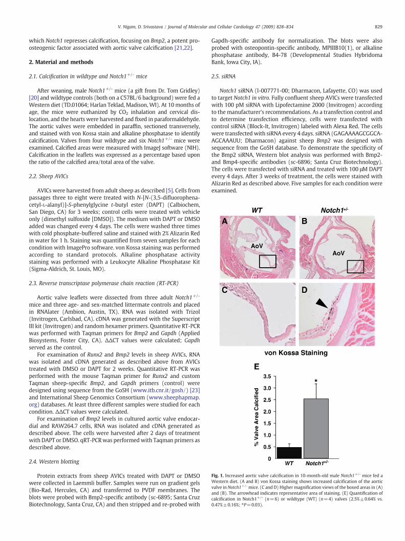
Fig. 1. Increased aortic valve calcification in 10-month-old male Notch1+/- mice fed aWestern diet. (A and B) von Kossa staining shows increased calcification of the aorticvalve in Notch1+/- mice. (C and D) Higher magnification views of the boxed areas in (A)and (B). The arrowhead indicates representative area of staining. (E) Quantification ofcalcification in Notch1+/- (n=6) or wildtype (WT) (n=4) valves (2.5%±0.64% vs.0.47%±0.16%; ⁎P=0.03).
829V. Nigam, D. Srivastava / Journal of Molecular and Cellular Cardiology 47 (2009) 828–834
which Notch1 represses calcification, focusing on Bmp2, a potent pro-osteogenic factor associated with aortic valve calcification [21,22].
2. Material and methods
2.1. Calcification in wildtype and Notch1+/- mice
After weaning, male Notch1+/- mice (a gift from Dr. Tom Gridley)[20] and wildtype controls (both on a C57BL/6 background) were fed aWestern diet (TD.01064; Harlan Teklad, Madison, WI). At 10months ofage, the mice were euthanized by CO2 inhalation and cervical dis-location, and the hearts were harvested and fixed in paraformaldehyde.The aortic valves were embedded in paraffin, sectioned transversely,and stained with von Kossa stain and alkaline phosphatase to identifycalcification. Valves from four wildtype and six Notch1+/- mice wereexamined. Calcified areas were measured with ImageJ software (NIH).Calcification in the leaflets was expressed as a percentage based uponthe ratio of the calcified area/total area of the valve.
2.2. Sheep AVICs
AVICs were harvested from adult sheep as described [5]. Cells frompassages three to eight were treated with N-[N-(3,5-difluorophena-cetyl-L-alanyl)]-S-phenylglycine t-butyl ester (DAPT) (Calbiochem,San Diego, CA) for 3 weeks; control cells were treated with vehicleonly (dimethyl sulfoxide [DMSO]). The medium with DAPT or DMSOadded was changed every 4 days. The cells were washed three timeswith cold phosphate-buffered saline and stained with 2% Alizarin Redin water for 1 h. Staining was quantified from seven samples for eachcondition with ImagePro software. von Kossa staining was performedaccording to standard protocols. Alkaline phosphatase activitystaining was performed with a Leukocyte Alkaline Phosphatase Kit(Sigma-Aldrich, St. Louis, MO).
2.3. Reverse transcriptase polymerase chain reaction (RT-PCR)
Aortic valve leaflets were dissected from three adult Notch1+/-
mice and three age- and sex-matched littermate controls and placedin RNAlater (Ambion, Austin, TX). RNA was isolated with Trizol(Invitrogen, Carlsbad, CA). cDNA was generated with the SuperscriptIII kit (Invitrogen) and randomhexamer primers. Quantitative RT-PCRwas performed with Taqman primers for Bmp2 and Gapdh (AppliedBiosystems, Foster City, CA). ΔΔCT values were calculated; Gapdhserved as the control.
For examination of Runx2 and Bmp2 levels in sheep AVICs, RNAwas isolated and cDNA generated as described above from AVICstreated with DMSO or DAPT for 2 weeks. Quantitative RT-PCR wasperformed with the mouse Taqman primer for Runx2 and customTaqman sheep-specific Bmp2, and Gapdh primers (control) weredesigned using sequence from the GoSH (www.itb.cnr.it/gosh/) [23]and International Sheep Genomics Consortium (www.sheephapmap.org) databases. At least three different samples were studied for eachcondition. ΔΔCT values were calculated.
For examination of Bmp2 levels in cultured aortic valve endocar-dial and RAW264.7 cells, RNA was isolated and cDNA generated asdescribed above. The cells were harvested after 2 days of treatmentwith DAPT or DMSO. qRT-PCRwas performedwith Taqman primers asdescribed above.
2.4. Western blotting
Protein extracts from sheep AVICs treated with DAPT or DMSOwere collected in Laemmli buffer. Samples were run on gradient gels(Bio-Rad, Hercules, CA) and transferred to PVDF membranes. Theblots were probed with Bmp2-specific antibody (sc-6895; Santa CruzBiotechnology, Santa Cruz, CA) and then stripped and re-probed with
Gapdh-specific antibody for normalization. The blots were alsoprobed with osteopontin-specific antibody, MPIIIB10(1), or alkalinephosphatase antibody, B4-78 (Developmental Studies HybridomaBank, Iowa City, IA).
2.5. siRNA
Notch1 siRNA (l-007771-00; Dharmacon, Lafayette, CO) was usedto target Notch1 in vitro. Fully confluent sheep AVICs were transfectedwith 100 pM siRNA with Lipofectamine 2000 (Invitrogen) accordingto the manufacturer's recommendations. As a transfection control andto determine transfection efficiency, cells were transfected withcontrol siRNA (Block-It, Invitrogen) labeled with Alexa Red. The cellswere transfected with siRNA every 4 days. siRNA (GAGAAAAGCGGCA-AGCAAAUU; Dharmacon) against sheep Bmp2 was designed withsequence from the GoSH database. To demonstrate the specificity ofthe Bmp2 siRNA, Western blot analysis was performed with Bmp2-and Bmp4-specific antibodies (sc-6896; Santa Cruz Biotechnology).The cells were transfected with siRNA and treated with 100 μM DAPTevery 4 days. After 3 weeks of treatment, the cells were stained withAlizarin Red as described above. Five samples for each condition wereexamined.
830 V. Nigam, D. Srivastava / Journal of Molecular and Cellular Cardiology 47 (2009) 828–834
2.6. Culturing of aortic valve endocardial cells
The aortic valve leaflets from adult mice were dissected and placedin wells coated with 0.1% gelatin in a small amount of medium (20%
fetal bovine serum, 40%Ham'smedium, 40%Dulbecco'smodified Eaglemedium, 100 mg/ml endothelial growth supplement factor, and 100units/ml heparin). Over several days, endocardial cellsmigrated out ofthe explant. No beating cells were seen. The endocardial identity of the
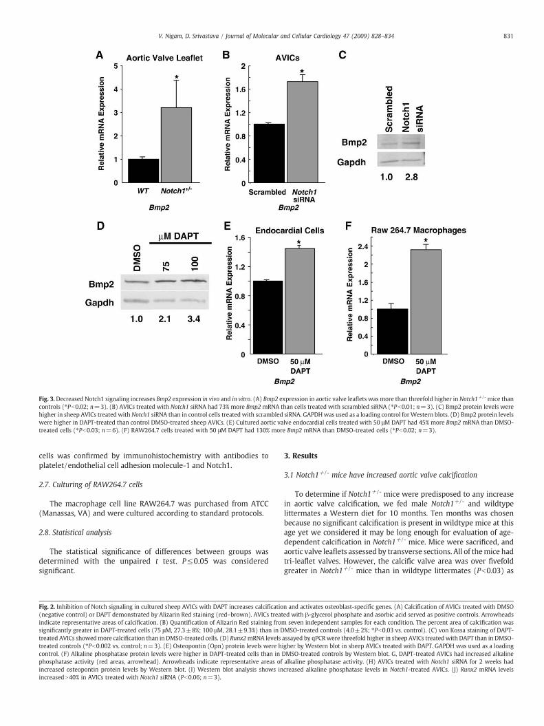
Fig. 3. Decreased Notch1 signaling increases Bmp2 expression in vivo and in vitro. (A) Bmp2 expression in aortic valve leaflets was more than threefold higher in Notch1+/- mice thancontrols (⁎Pb0.02; n=3). (B) AVICs treated with Notch1 siRNA had 73% more Bmp2mRNA than cells treated with scrambled siRNA (⁎Pb0.01; n=3). (C) Bmp2 protein levels werehigher in sheep AVICs treated with Notch1 siRNA than in control cells treated with scrambled siRNA. GAPDHwas used as a loading control for Western blots. (D) Bmp2 protein levelswere higher in DAPT-treated than control DMSO-treated sheep AVICs. (E) Cultured aortic valve endocardial cells treated with 50 μM DAPT had 45% more Bmp2 mRNA than DMSO-treated cells (⁎Pb0.03; n=6). (F) RAW264.7 cells treated with 50 μM DAPT had 130% more Bmp2 mRNA than DMSO-treated cells (⁎Pb0.02; n=3).
831V. Nigam, D. Srivastava / Journal of Molecular and Cellular Cardiology 47 (2009) 828–834
cells was confirmed by immunohistochemistry with antibodies toplatelet/endothelial cell adhesion molecule-1 and Notch1.
2.7. Culturing of RAW264.7 cells
The macrophage cell line RAW264.7 was purchased from ATCC(Manassas, VA) and were cultured according to standard protocols.
2.8. Statistical analysis
The statistical significance of differences between groups wasdetermined with the unpaired t test. P≤0.05 was consideredsignificant.
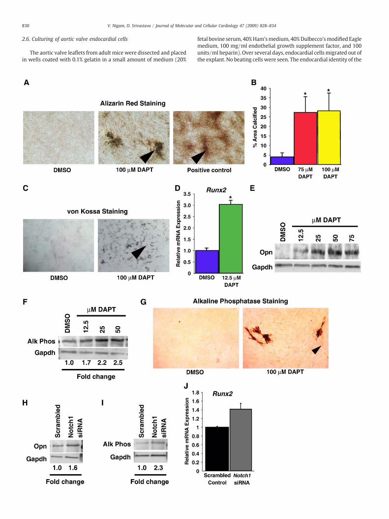
Fig. 2. Inhibition of Notch signaling in cultured sheep AVICs with DAPT increases calcificatio(negative control) or DAPT demonstrated by Alizarin Red staining (red–brown). AVICs treatindicate representative areas of calcification. (B) Quantification of Alizarin Red staining fromsignificantly greater in DAPT-treated cells (75 μM, 27.3±8%; 100 μM, 28.1±9.3%) than in Dtreated AVICs showedmore calcification than in DMSO-treated cells. (D) Runx2mRNA levelstreated controls (⁎Pb0.002 vs. control; n=3). (E) Osteopontin (Opn) protein levels were hcontrol. (F) Alkaline phosphatase protein levels were higher in DAPT-treated cells than in Dphosphatase activity (red areas, arrowhead). Arrowheads indicate representative areas ofincreased osteopontin protein levels by Western blot. (I) Western blot analysis shows inincreasedN40% in AVICs treated with Notch1 siRNA (Pb0.06; n=3).
3. Results
3.1 Notch1+/- mice have increased aortic valve calcification
To determine if Notch1+/- mice were predisposed to any increasein aortic valve calcification, we fed male Notch1+/- and wildtypelittermates a Western diet for 10 months. Ten months was chosenbecause no significant calcification is present in wildtype mice at thisage yet we considered it may be long enough for evaluation of age-dependent calcification in Notch1+/- mice. Mice were sacrificed, andaortic valve leaflets assessed by transverse sections. All of themice hadtri-leaflet valves. However, the calcific valve area was over fivefoldgreater in Notch1+/- mice than in wildtype littermates (Pb0.03) as
n and activates osteoblast-specific genes. (A) Calcification of AVICs treated with DMSOed with β-glycerol phosphate and asorbic acid served as positive controls. Arrowheadsseven independent samples for each condition. The percent area of calcification was
MSO-treated controls (4.0±2%; ⁎Pb0.03 vs. control). (C) von Kossa staining of DAPT-assayed by qPCRwere threefold higher in sheep AVICs treated with DAPT than in DMSO-igher by Western blot in sheep AVICs treated with DAPT. GAPDH was used as a loadingMSO-treated controls by Western blot. G, DAPT-treated AVICs had increased alkalinealkaline phosphatase activity. (H) AVICs treated with Notch1 siRNA for 2 weeks hadcreased alkaline phosphatase levels in Notch1-treated AVICs. (J) Runx2 mRNA levels
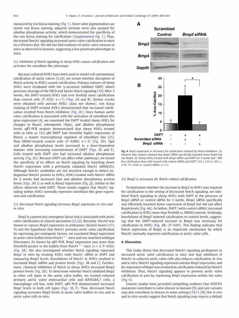
Fig. 4. Bmp2 expression is necessary for calcification induced by Notch inhibition. (A)Western blot analysis showed that Bmp2 siRNA specifically knocked down Bmp2 butnot Bmp4. (B) Sheep AVICs treated with Bmp2 siRNA and DAPT for 3 weeks had N80%less calcification than cells treated with control siRNA and DAPT (3.4±1.5% vs. 20.1±5.7%, ⁎P=0.02 vs. control siRNA; n=5).
832 V. Nigam, D. Srivastava / Journal of Molecular and Cellular Cardiology 47 (2009) 828–834
measured by von Kossa staining (Fig. 1). Since valve pigmentation canmimic von Kossa staining, adjacent sections were also assayed foralkaline phosphatase activity, which demonstrated the specificity ofthe von Kossa staining for calcification (Supplemental Fig. 1). Thus,decreased Notch1 signaling increased aortic valve calcification inmiceon aWestern diet. We did not find evidence of aortic valve stenosis inmice as observed in humans, suggesting a less penetrant phenotype inmice.
3.2. Inhibition of Notch signaling in sheep AVICs causes calcification andactivates the osteoblast-like phenotype
Because cultured AVICs have been used to model cell-autonomouscalcification of aortic valves [5,24], we tested whether disruption ofNotch activity in AVICs caused calcification. Primary cultures of sheepAVICs were incubated with the γ-secretase inhibitor DAPT, whichprevents cleavage of the NICD and blocks Notch signaling [19]. After 3weeks, the DAPT-treated AVICs had over fivefold more calcificationthan control cells (Pb0.03; n=7) (Figs. 2A and B). Similar resultswere obtained with porcine AVICs (data not shown). von Kossastaining of DAPT-treated AVICs demonstrated that increased calcifi-cation resulted from Notch inhibition (Fig. 2C). Since human aorticvalve calcification is associated with the activation of osteoblast-likegene expression [4], we examined the DAPT treated sheep AVICs forchanges in Runx2, osteopontin (Opn), and alkaline phosphataselevels. qRT-PCR analysis demonstrated that sheep AVICs treatedwith as little as 12.5 μM DAPT had threefold higher expression ofRunx2, a master transcriptional regulator of osteoblast fate [25],than DMSO-treated control cells (Pb0.002; n=3) (Fig. 2D). Opnand alkaline phosphatase levels increased in a dose-dependentmanner with increasing concentrations of DAPT (Figs. 2E and F).Cells treated with DAPT also had increased alkaline phosphataseactivity (Fig. 2G). Because DAPT can affect other pathways, we testedthe specificity of its effects on Notch signaling by knocking downNotch1 expression with a previously validated Notch1 siRNA[26].Although Notch1 antibodies are not sensitive enough to detect en-dogenous Notch1 protein in AVICs, AVICs treated with Notch1 siRNAfor 2 weeks had increased Opn and alkaline phosphatase proteinlevels (Figs. 2H, I) as well as Runx2 expression (Fig. 2J), similar to theeffects observed with DAPT. These results suggest that Notch1 sig-naling within AVICs normally represses osteoblast-like gene expres-sion and calcification.
3.3. Decreased Notch signaling increases Bmp2 expression in vivo andin vitro
Bmp2 is a potent pro-osteogenic factor that is associatedwith aorticvalve calcification in clinical specimens [21,22]. Recently, Notch2 wasshown to repress Bmp2 expression in embryonic chicken hearts [27].To test the hypothesis that Notch1 prevents aortic valve calcificationby repressing pro-osteogenic factors, we examined Bmp2 expressionin aortic valve leaflets fromNotch1+/- mice and sex-matchedwildtypelittermates. As shown by qRT-PCR, Bmp2 expression was more thanthreefold greater in the leaflets from Notch1+/- mice (n=3, Pb0.02)(Fig. 3A). We also investigated whether Notch signaling repressedBmp2 in vitro by treating AVICs with Notch1 siRNA or DAPT andmeasuring Bmp2 levels. Knockdown of Notch1 in AVICs resulted inincreased Bmp2 mRNA and protein levels (Figs. 3B and C). Further-more, chemical inhibition of Notch in sheep AVICs increased Bmp2protein levels (Fig. 3D). To determine whether Notch inhibited Bmp2in other cell types in the aortic valve leaflet, we treated culturedprimary aortic valve endocardial cells and RAW264.7 cells, amacrophage cell line, with DAPT. qRT-PCR demonstrated increasedBmp2 levels in both cell types (Figs. 3E, F). Thus, decreased Notchsignaling increases Bmp2 levels in aortic valve leaflets in vivo and inaortic valve cells in vitro.
3.4. Bmp2 is necessary for Notch-related calcification
To determine whether the increase in Bmp2 in AVICs was requiredfor calcification in the setting of decreased Notch signaling, we inhi-bited Notch signaling in sheep AVICs with DAPT in the presence ofBmp2 siRNA or control siRNA for 3 weeks. Bmp2 siRNA specificallyand efficiently knocked down expression of Bmp2 but did not affectBmp4 levels (Fig. 4A). As before, DAPT (with control siRNA) increasedcalcification in AVICs more than fivefold vs. DMSO controls. Strikingly,knockdown of Bmp2 lowered calcification to control levels, suggest-ing that the DAPT-induced increase in Bmp2 was necessary forcalcification in AVICs (Fig. 4B) (Pb0.03). This finding indicates thatNotch repression of Bmp2 is an important mechanism by whichNotch1 normally represses calcification in aortic valve cells.
4. Discussion
This study shows that decreased Notch1 signaling predisposes toincreased aortic valve calcification in mice and that inhibition ofNotch1 in cultured aortic valve cells also induces calcification. In vivoand in vitro, Notch1 signaling repressed valvular Bmp2 expression, andde-repression of Bmp2was involved in calcification induced byNotch1inhibition. Thus, Notch1 signaling appears to prevent aortic valvecalcification in part by repressing Bmp2 expression within the valve(Fig. 5).
Genetic studies have provided compelling evidence that NOTCH1mutations contribute to valve disease in humans [9], and rare variantsmay also contribute to disease in some settings [10,11,28]. Our in vivoand in vitro results suggest that Notch signaling may repress a default
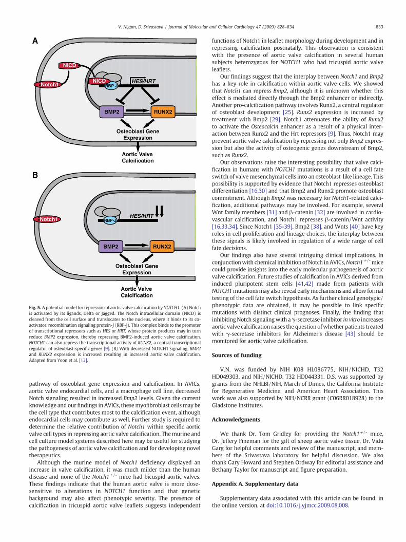
Fig. 5. A potential model for repression of aortic valve calcification byNOTCH1. (A) Notchis activated by its ligands, Delta or Jagged. The Notch intracellular domain (NICD) iscleaved from the cell surface and translocates to the nucleus, where it binds to its co-activator, recombination signaling protein-J (RBP-J). This complex binds to the promoterof transcriptional repressors such as HES or HRT, whose protein products may in turnreduce BMP2 expression, thereby repressing BMP2-induced aortic valve calcification.NOTCH1 can also repress the transcriptional activity of RUNX2, a central transcriptionalregulator of osteoblast-specific genes [9]. (B) With decreased NOTCH1 signaling, BMP2and RUNX2 expression is increased resulting in increased aortic valve calcification.Adapted from Yoon et al. [13].
833V. Nigam, D. Srivastava / Journal of Molecular and Cellular Cardiology 47 (2009) 828–834
pathway of osteoblast gene expression and calcification. In AVICs,aortic valve endocardial cells, and a macrophage cell line, decreasedNotch signaling resulted in increased Bmp2 levels. Given the currentknowledge and our findings in AVICs, thesemyofibroblast cellsmay bethe cell type that contributes most to the calcification event, althoughendocardial cells may contribute as well. Further study is required todetermine the relative contribution of Notch1 within specific aorticvalve cell types in repressing aortic valve calcification. Themurine andcell culture model systems described here may be useful for studyingthe pathogenesis of aortic valve calcification and for developing noveltherapeutics.
Although the murine model of Notch1 deficiency displayed anincrease in valve calcification, it was much milder than the humandisease and none of the Notch1+/- mice had bicuspid aortic valves.These findings indicate that the human aortic valve is more dose-sensitive to alterations in NOTCH1 function and that geneticbackground may also affect phenotypic severity. The presence ofcalcification in tricuspid aortic valve leaflets suggests independent
functions of Notch1 in leaflet morphology during development and inrepressing calcification postnatally. This observation is consistentwith the presence of aortic valve calcification in several humansubjects heterozygous for NOTCH1 who had tricuspid aortic valveleaflets.
Our findings suggest that the interplay between Notch1 and Bmp2has a key role in calcification within aortic valve cells. We showedthat Notch1 can repress Bmp2, although it is unknown whether thiseffect is mediated directly through the Bmp2 enhancer or indirectly.Another pro-calcification pathway involves Runx2, a central regulatorof osteoblast development [25]. Runx2 expression is increased bytreatment with Bmp2 [29]. Notch1 attenuates the ability of Runx2to activate the Osteocalcin enhancer as a result of a physical inter-action between Runx2 and the Hrt repressors [9]. Thus, Notch1 mayprevent aortic valve calcification by repressing not only Bmp2 expres-sion but also the activity of osteogenic genes downstream of Bmp2,such as Runx2.
Our observations raise the interesting possibility that valve calci-fication in humans with NOTCH1 mutations is a result of a cell fateswitch of valvemesenchymal cells into an osteoblast-like lineage. Thispossibility is supported by evidence that Notch1 represses osteoblastdifferentiation [16,30] and that Bmp2 and Runx2 promote osteoblastcommitment. Although Bmp2 was necessary for Notch1-related calci-fication, additional pathways may be involved. For example, severalWnt family members [31] and β-catenin [32] are involved in cardio-vascular calcification, and Notch1 represses β-catenin/Wnt activity[16,33,34]. Since Notch1 [35-39], Bmp2 [38], and Wnts [40] have keyroles in cell proliferation and lineage choices, the interplay betweenthese signals is likely involved in regulation of a wide range of cellfate decisions.
Our findings also have several intriguing clinical implications. Inconjunctionwith chemical inhibition of Notch in AVICs,Notch1+/- micecould provide insights into the early molecular pathogenesis of aorticvalve calcification. Future studies of calcification in AVICs derived frominduced pluripotent stem cells [41,42] made from patients withNOTCH1mutationsmay also reveal earlymechanisms and allow formaltesting of the cell fate switch hypothesis. As further clinical genotypic/phenotypic data are obtained, it may be possible to link specificmutations with distinct clinical prognoses. Finally, the finding thatinhibitingNotch signalingwith aγ-secretase inhibitor in vitro increasesaortic valve calcification raises the question ofwhether patients treatedwith γ-secretase inhibitors for Alzheimer's disease [43] should bemonitored for aortic valve calcification.
Sources of funding
V.N. was funded by NIH K08 HL086775, NIH/NICHD, T32HD049303, and NIH/NICHD, T32 HD044331. D.S. was supported bygrants from the NHLBI/NIH, March of Dimes, the California Institutefor Regenerative Medicine, and American Heart Association. Thiswork was also supported by NIH/NCRR grant (C06RR018928) to theGladstone Institutes.
Acknowledgments
We thank Dr. Tom Gridley for providing the Notch1+/- mice,Dr. Jeffery Fineman for the gift of sheep aortic valve tissue, Dr. ViduGarg for helpful comments and review of the manuscript, and mem-bers of the Srivastava laboratory for helpful discussion. We alsothank Gary Howard and Stephen Ordway for editorial assistance andBethany Taylor for manuscript and figure preparation.
Appendix A. Supplementary data
Supplementary data associated with this article can be found, inthe online version, at doi:10.1016/j.yjmcc.2009.08.008.
834 V. Nigam, D. Srivastava / Journal of Molecular and Cellular Cardiology 47 (2009) 828–834
References
[1] Thom T, Haase N, Rosamond W, Howard VJ, Rumsfeld J, Manolio T, et al. Heartdisease and stroke statistics-2006 update: a report from the American HeartAssociation Statistics Committee and Stroke Statistics Subcommittee. Circulation2006;113(6):e85–151.
[2] Freeman RV, Otto CM. Spectrum of calcific aortic valve disease: pathogenesis,disease progression, and treatment strategies. Circulation 2005;111(24):3316–26.
[3] Weiss RM, Ohashi M, Miller JD, Young SG, Heistad DD. Calcific aortic valve stenosisin old hypercholesterolemic mice. Circulation 2006;114(19):2065–9.
[4] Rajamannan NM, Subramaniam M, Rickard D, Stock SR, Donovan J, Springett M,et al. Human aortic valve calcification is associated with an osteoblast phenotype.Circulation 2003;107(17):2181–4.
[5] JianB,NarulaN, LiQY,Mohler 3rdER, LevyRJ. Progressionof aortic valve stenosis: TGF-beta1 ispresent incalcifiedaortic valvecuspsandpromotes aortic valve interstitial cellcalcification via apoptosis. Ann Thorac Surg 2003;75(2):457–65 discussion 65-6.
[6] Otto CM, Kuusisto J, Reichenbach DD, Gown AM, O'Brien KD. Characterization ofthe early lesion of ‘degenerative’ valvular aortic stenosis. Histological and immu-nohistochemical studies. Circulation 1994;90(2):844–53.
[7] Olsson M, Dalsgaard CJ, Haegerstrand A, Rosenqvist M, Ryden L, Nilsson J.Accumulation of T lymphocytes and expression of interleukin-2 receptors innonrheumatic stenotic aortic valves. J Am Coll Cardiol 1994;23(5):1162–70.
[8] Wallby L, Janerot-Sjoberg B, Steffensen T, Broqvist M. T lymphocyte infiltration innon-rheumatic aortic stenosis: a comparative descriptive study between tricuspidand bicuspid aortic valves. Heart 2002;88(4):348–51.
[9] Garg V, Muth AN, Ransom JF, Schluterman MK, Barnes R, King IN, et al. Mutationsin NOTCH1 cause aortic valve disease. Nature 2005;437(7056):270–4.
[10] Mohamed SA, Aherrahrou Z, Liptau H, Erasmi AW, Hagemann C, Wrobel S, et al.Novel missense mutations (p.T596M and p.P1797H) in NOTCH1 in patients withbicuspid aortic valve. Biochem Biophys Res Commun 2006;345(4):1460–5.
[11] McKellar SH, Tester DJ, Yagubyan M, Majumdar R, Ackerman MJ, Sundt 3rd TM.Novel NOTCH1 mutations in patients with bicuspid aortic valve disease andthoracic aortic aneurysms. J Thorac Cardiovasc Surg 2007;134(2):290–6.
[12] Artavanis-Tsakonas S, Rand MD, Lake RJ. Notch signaling: cell fate control andsignal integration in development. Science 1999;284(5415):770–6.
[13] Yoon K, Gaiano N. Notch signaling in the mammalian central nervous system:insights from mouse mutants. Nat Neurosci 2005;8(6):709–15.
[14] Nakagawa O, Nakagawa M, Richardson JA, Olson EN, Srivastava D. HRT1, HRT2,and HRT3: a new subclass of bHLH transcription factors marking specific cardiac,somitic, and pharyngeal arch segments. Dev Biol 1999;216(1):72–84.
[15] Krebs LT, Xue Y, Norton CR, Shutter JR, Maguire M, Sundberg JP, et al. Notchsignaling is essential for vascular morphogenesis inmice. Genes Dev 2000;14(11):1343–52.
[16] Sciaudone M, Gazzerro E, Priest L, Delany AM, Canalis E. Notch 1 impairs osteo-blastic cell differentiation. Endocrinology 2003;144(12):5631–9.
[17] Shindo K, Kawashima N, Sakamoto K, Yamaguchi A, Umezawa A, Takagi M, et al.Osteogenic differentiation of the mesenchymal progenitor cells, Kusa is sup-pressed by Notch signaling. Exp Cell Res 2003;290(2):370–80.
[18] Radtke F, Wilson A, Mancini SJ, MacDonald HR. Notch regulation of lymphocytedevelopment and function. Nat Immunol 2004;5(3):247–53.
[19] Timmerman LA, Grego-Bessa J, Raya A, Bertran E, Perez-Pomares JM, Diez J, et al.Notch promotes epithelial-mesenchymal transition during cardiac developmentand oncogenic transformation. Genes Dev 2004;18(1):99–115.
[20] Swiatek PJ, Lindsell CE, del Amo FF, Weinmaster G, Gridley T. Notch1 is essentialfor postimplantation development in mice. Genes Dev 1994;8(6):707–19.
[21] Mohler 3rd ER, Gannon F, Reynolds C, Zimmerman R, Keane MG, Kaplan FS. Boneformation and inflammation in cardiac valves. Circulation 2001;103(11):1522–8.
[22] Kaden JJ, Bickelhaupt S, Grobholz R, Vahl CF, Hagl S, Brueckmann M, et al.Expression of bone sialoprotein and bone morphogenetic protein-2 in calcificaortic stenosis. J Heart Valve Dis 2004;13(4):560–6.
[23] Caprera A, Lazzari B, Stella A, Merelli I, Caetano AR, Mariani P. GoSh: a web-based database for goat and sheep EST sequences. Bioinformatics 2007;23(8):1043–5.
[24] Osman L, Yacoub MH, Latif N, Amrani M, Chester AH. Role of human valve inter-stitial cells in valve calcification and their response to atorvastatin. Circulation2006;114(1 Suppl):I547–52.
[25] Ducy P, Zhang R, Geoffroy V, Ridall AL, Karsenty G. Osf2/Cbfa1: a transcriptionalactivator of osteoblast differentiation. Cell 1997;89(5):747–54.
[26] Akiyoshi T, Nakamura M, Yanai K, Nagai S, Wada J, Koga K, et al. Gamma-secretaseinhibitors enhance taxane-induced mitotic arrest and apoptosis in colon cancercells. Gastroenterology 2008;134(1):131–44.
[27] Rutenberg JB, Fischer A, Jia H, Gessler M, Zhong TP, Mercola M. Developmentalpatterning of the cardiac atrioventricular canal by Notch and Hairy-related trans-cription factors. Development 2006;133(21):4381–90.
[28] McBride KL, RileyMF, Zender GA, Fitzgerald-Butt SM, Towbin JA, Belmont JW, et al.NOTCH1 mutations in individuals with left ventricular outflow tract malforma-tions reduce ligand-induced signaling. Hum Mol Genet 2008 Sept 15;17(18):2886–93.
[29] de Jong DS, Vaes BL, Dechering KJ, Feijen A, Hendriks JM, Wehrens R, et al. Identi-fication of novel regulators associated with early-phase osteoblast differentiation.J Bone Miner Res 2004;19(6):947–58.
[30] Zanotti S, Smerdel-Ramoya A, Stadmeyer L, Durant D, Radtke F, Canalis E. Notchinhibits osteoblast differentiation and causes osteopenia. Endocrinology 2008;149(8):3890–9.
[31] Shao JS, Cheng SL, Pingsterhaus JM, Charlton-Kachigian N, Loewy AP, Towler DA.Msx2 promotes cardiovascular calcification by activating paracrine Wnt signals.J Clin Invest 2005;115(5):1210–20.
[32] Rajamannan NM, Subramaniam M, Caira F, Stock SR, Spelsberg TC. Atorvastatininhibits hypercholesterolemia-induced calcification in the aortic valves via theLrp5 receptor pathway. Circulation 2005;112(9 Suppl):I229–34.
[33] Nicolas M,Wolfer A, Raj K, Kummer JA, Mill P, van Noort M, et al. Notch1 functionsas a tumor suppressor in mouse skin. Nat Genet 2003;33(3):416–21.
[34] Siveke JT, Lubeseder-Martellato C, Lee M, Mazur PK, Nakhai H, Radtke F, et al.Notch signaling is required for exocrine regeneration after acute pancreatitis.Gastroenterology 2008;134(2):544–55.
[35] Fox V, Gokhale PJ, Walsh JR, Matin M, Jones M, Andrews PW. Cell-cell signalingthrough NOTCH regulates human embryonic stem cell proliferation. Stem Cells2008;26(3):715–23.
[36] Chen VC, Stull R, Joo D, Cheng X, Keller G. Notch signaling respecifies thehemangioblast to a cardiac fate. Nat Biotechnol 2008 Oct;26(10):1169–78.
[37] Nemir M, Croquelois A, Pedrazzini T, Radtke F. Induction of cardiogenesis inembryonic stem cells via downregulation of Notch1 signaling. Circ Res 2006;98(12):1471–8.
[38] Pera MF, Andrade J, Houssami S, Reubinoff B, Trounson A, Stanley EG, et al.Regulation of human embryonic stem cell differentiation by BMP-2 and itsantagonist noggin. J Cell Sci 2004;117(Pt 7):1269–80.
[39] Ivey KN, Muth A, Arnold J, King FW, Yeh RF, Fish JE, et al. MicroRNA regulation ofcell lineages in mouse and human embryonic stem cells. Cell Stem Cell 2008;2(3):219–29.
[40] Reya T, Duncan AW, Ailles L, Domen J, Scherer DC, Willert K, et al. A role for Wntsignalling in self-renewal of haematopoietic stem cells. Nature 2003;423(6938):409–14.
[41] Takahashi K, Tanabe K, Ohnuki M, Narita M, Ichisaka T, Tomoda K, et al. Inductionof pluripotent stem cells from adult human fibroblasts by defined factors. Cell2007;131(5):861–72.
[42] Yu J, Vodyanik MA, Smuga-Otto K, Antosiewicz-Bourget J, Frane JL, Tian S, et al.Induced pluripotent stem cell lines derived from human somatic cells. Science(New York, NY) 2007;318(5858):1917–20.
[43] Fleisher AS, Raman R, Siemers ER, Becerra L, Clark CM, Dean RA, et al. Phase 2safety trial targeting amyloid beta production with a gamma-secretase inhibitor inAlzheimer disease. Arch Neurol 2008;65(8):1031–8.