Embed Size (px)
Citation preview
Biosci. Rep. (2016) / 36 / art:e00282 / doi 10.1042/BSR20150226
Nucleotide-binding mechanisms inpseudokinasesHenrik M. Hammaren*1, Anniina T. Virtanen* and Olli Silvennoinen*†1
*School of Medicine, University of Tampere, Biokatu 8, FI-33014 Tampere, Finland†Clinical Hematology, Department of Internal Medicine, Tampere University Hospital, Medisiinarinkatu 3, FI-33520 Tampere, Finland
SynopsisPseudokinases are classified by the lack of one or several of the highly conserved motifs involved in nucleotide(nt) binding or catalytic activity of protein kinases (PKs). Pseudokinases represent ∼10% of the human kinomeand they are found in all evolutionary classes of kinases. It has become evident that pseudokinases, which wereinitially considered somewhat peculiar dead kinases, are important components in several signalling cascades.Furthermore, several pseudokinases have been linked to human diseases, particularly cancer, which is raising interestfor therapeutic approaches towards these proteins. The ATP-binding pocket is a well-established drug target andelucidation of the mechanism and properties of nt binding in pseudokinases is of significant interest and importance.Recent studies have demonstrated that members of the pseudokinase family are very diverse in structure as wellas in their ability and mechanism to bind nts or perform phosphoryl transfer reactions. This diversity also precludesprediction of pseudokinase function, or the importance of nt binding for said function, based on primary sequencealone. Currently available data indicate that ∼40% of pseudokinases are able to bind nts, whereas only few are ableto catalyse occasional phosphoryl transfer. Pseudokinases employ diverse mechanisms to bind nts, which usuallyoccurs at low, but physiological, affinity. ATP binding serves often a structural role but in most cases the functionalroles are not precisely known. In the present review, we discuss the various mechanisms that pseudokinases employfor nt binding and how this often low-affinity binding can be accurately analysed.
Key words: ATP, kinase activity, kinome, nucleotide binding, pseudokinase, signalling.
Cite this article as: Bioscience Reports (2016) 36, e00282, doi:10.1042/BSR20150226
INTRODUCTION
In their landmark paper in 2002, Manning and colleagues presen-ted for the first time a comprehensive catalogue of human proteinkinases (PKs) [1]. One of the salient findings of their analysiswas that ∼10 % of the 518 human PKs lack at least one of theclassical conserved catalytic residues described by Hanks et al.[2]: the β3 lysine (K in the ‘VAIK’ consensus motif, Figure 1),the catalytic aspartate (D in ‘HRD’) or the cation-binding as-partate (D in ‘DFG’). Some of these kinases, like the WNK(‘With no lysine’) family, were already known to have phos-phoryl transfer activity despite the lack of canonical catalytic
. . . . . . . . . . . . . . . . . . . . . . . . . . . . . . . . . . . . . . . . . . . . . . . . . . . . . . . . . . . . . . . . . . . . . . . . . . . . . . . . . . . . . . . . . . . . . . . . . . . . . . . . . . . . . . . . . . . . . . . . . . . . . . . . . . . . . . . . . . . . . . . . . . . . . . . . . . . . . . . . . . . . . . . . . . . . . . . . . . . . . . . . . . . . . . . . . . . . . . . . . . . . . . . . . . . . . . . . . . . . . . . . . . . . . . . . . . . . . . . . . . . . . . . . . . . . . . . . . . . . . . . . . . . . . . . . . . . . . . . . . . . . . . . . . . . . . . . . . . . .
Abbreviations: ADCK, aarF domain-containing protein kinase; AMP-PNP, adenylate-imidodiphosphate; ANK, ankyrin repeat domain (in RNase L); ANPa/ANPb, atrial natriuretic peptidereceptor type A/B, also known as atrial natriuretic peptide receptor 1 (NPR1) and NPR2, respectively. Also known as GC-A/GC-B; ATPγ S, adenosine 5′ -[γ -thio]triphosphate; BAK1,BCL2-antagonist/Killer 1; BIR2, BAK1-interacting receptor-like kinase 2; BSK8, brassinosteroid signalling kinase 8; CaM-kinase, Ca2 + /calmodulin-dependent protein kinase; CASK,calcium/calmodulin-dependent serine protein kinase; COP1, constitutive photomorphogenesis protein 1; EGFR, epidermal growth factor receptor; ePK, eukaryotic protein kinase; GC,guanylate cyclase; GUCY2, guanylate cyclase, e.g. GUCY2C, also known as GC-C, and heat stable enterotoxin receptor (HSER); HER3, human epidermal growth factor 3, also known asERBB3; IRAK, interleukin-1 receptor-associated kinase; ITC, isothermal titration calorimetry; JAK, Janus kinase; JH, JAK homology; LKB1, liver kinase B1; MLKL, mixed lineage kinasedomain-like; nt, nucleotide; NTE, N-terminal extension; PAN3, PAB1P-dependent poly(A)-nuclease; PK, protein kinase; PDZ, protein domain found in PSD95, Dlg1 and zo-1; PKA,cAMP-dependent protein kinase A; PKD, pseudokinase domain; PKL, protein kinase-like; RIP3, receptor-interacting serine-threonine kinase 3; RLK, receptor-like kinase; ROPK, rhoptrykinase; ROR, receptor tyrosine kinase-like orphan receptor; RTK, receptor tyrosine kinase; SH3, Src-homology 3 domain; SPR, surface plasmon resonance; STRAD, Ste20-relatedadaptor; TK, titin kinase; TLR, Toll-like receptor; TSA, thermal shift assay; VRK, vaccinia-related kinase; WNK, With no lysine.1 Correspondence may be addressed to either of these authors (email [email protected] or [email protected]).
sites. Yet, the other 50 identified proteins or protein domains weredeemed likely to be inactive and consequently dubbed ‘pseudok-inases’. Subsequently, also some of these pseudokinases (likeHaspin) were shown to be catalytically active, and thus couldbe reclassified as atypical PKs [3]. For these and some morerecent examples of catalytic activity like KSR2 [4], HER3 [5],JAK2 JH2 [6] or CASK [7] the distinction of pseudokinase andatypical kinase has become somewhat unclear. However, as pro-posed by Eyers and Murphy [8], the bioinformatic definition of apseudokinase (with the inclusion of kinases that have experi-mentally been found to be inactive) should be maintained forthe sake of clarity, and will be used in the present review aswell.
c© 2016 Authors. This is an open access article published by Portland Press Limited and distributed under the Creative Commons Attribution Licence 3.0. 1
H.M. Hammaren, A.T. Virtanen and O. Silvennoinen
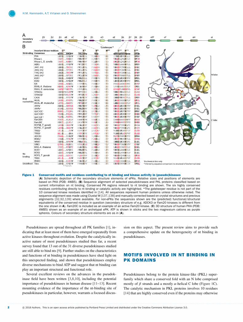
Figure 1 Conserved motifs and residues contributing to nt binding and kinase activity in (pseudo)kinases(A) Schematic depiction of the secondary structure elements of ePKs. Relative sizes and positions of elements arebased on PKA (PDB: 4WB5). (B) Sequence alignment of selected pseudokinases and PKL proteins classified based oncurrent information on nt binding. Conserved PK regions relevant to nt binding are shown. The six highly conservedresidues contributing directly to nt binding or catalytic activity are highlighted. *The gatekeeper residue is not part of the10 conserved kinase residues identified in [14]. All sequences represent human proteins unless otherwise noted. Thesequence alignment was made using Clustal W [137,138] and manually corrected based on crystal structures and previousalignments [32,92,139] where available. For non-ePKs the sequences shown are the (predicted) functional/structuralequivalents of the conserved residue in question (secondary structure of e.g. ADCK3 or Fam20 kinases is different fromthe one shown in A). Fam20C is included as an example of an active Fam20 kinase. (C) 3D structure of human PKA (PDB:4WB5) shown as an example of an archetypal ePK. ATP is shown in sticks and the two magnesium cations as purplespheres. Colours of secondary structure elements are as in (A).
Pseudokinases are spread throughout all PK families [1], in-dicating that at least most of them have emerged repeatedly fromactive kinases throughout evolution. Despite the catalytically in-active nature of most pseudokinases studied thus far, a recentsurvey found that 13 out of the 31 diverse pseudokinases studiedare still able to bind nts [9]. Further studies on the characteristicsand functions of nt binding in pseudokinases have shed light onthis unexpected finding, and shown that pseudokinases employdiverse mechanisms to bind ATP and suggest that nt binding canplay an important structural and functional role.
Several excellent reviews on the advances in the pseudok-inase field have been written [3,8,10], including the potentialimportance of pseudokinases in human disease [11–13]. Recentmounting evidence of the importance of the nt-binding site ofpseudokinases in particular, however, warrants a focused discus-
sion on this aspect. The present review aims to provide sucha comprehensive update on the heterogeneity of nt binding inpseudokinases.
MOTIFS INVOLVED IN NT BINDING INPK DOMAINS
Pseudokinases belong to the protein kinase-like (PKL) super-family which share a conserved fold with an N lobe comprisedmostly of β strands and a mostly α-helical C lobe (Figure 1C).The catalytic mechanism in PKL proteins involves 10 residues[14] that are highly conserved even if the proteins may otherwise
. . . . . . . . . . . . . . . . . . . . . . . . . . . . . . . . . . . . . . . . . . . . . . . . . . . . . . . . . . . . . . . . . . . . . . . . . . . . . . . . . . . . . . . . . . . . . . . . . . . . . . . . . . . . . . . . . . . . . . . . . . . . . . . . . . . . . . . . . . . . . . . . . . . . . . . . . . . . . . . . . . . . . . . . . . . . . . . . . . . . . . . . . . . . . . . . . . . . . . . . . . . . . . . . . . . . . . . . . . . . . . . . . . . . . . . . . . . . . . . . . . . . . . . . . . . . . . . . . . . . . . . . . . . . . . . . . . . . . . . . . . . . . . . . . . . . . . . . . . . . . . . . . . . . . . . . . . . . . . . . . . . . . . . . . . . . . . . . . . . . . . . . . . . . . . . . . . . . . . . . . .
2 c© 2016 Authors. This is an open access article published by Portland Press Limited and distributed under the Creative Commons Attribution Licence 3.0.
Nucleotide binding in pseudokinases
lack sequence conservation [14,15]. Six out of these 10 residuesare directly involved in nt or substrate binding or catalysis [14](highlighted in Figure 1B), whereas the function of the remain-ing four residues is still not completely understood [14], but isprobably mainly structural [16].
The first of the critical residues for nt binding is Gly52 (asthe de facto archetypal protein kinase, human cAMP-dependentprotein kinase A (PKA) is usually used for numbering of aminoacid residues in protein kinase motifs and will also be used inthe present review) in the glycine-rich loop (‘Gly-rich loop’,also known as the ‘phosphate-binding loop’ or ‘P-loop’) locatedbetween strands β1 and β2 (Figure 1). The function of the Gly-rich loop is best understood in PKA, in which glycine at thisposition enables the peptide backbone of the tip of the loop(Ser53) to bind the γ -phosphate of ATP. Mutation of the Gly-rich loop, and especially Gly52, lowers affinity towards ATP andaffects kinase activity [17] as the γ -phosphate can no longer beefficiently positioned for phosphoryl transfer between the tip ofthe Gly-rich loop and a basic residue (Lys168) from the so-called‘catalytic loop’ situated between β6 and β7 (Figure 1) [18].Although the function of the Gly-rich loop is well known forPKA, the motif has not been extensively studied in other kinasesand it is unclear how universal this function is.
The second residue is Lys72 of the ‘VAIK’ motif in β3, whichis the most conserved residue in all PKL proteins, and the onlyresidue not missing in any of the known families (Figure 1) [14].Despite its virtually universal conservation and its position nextto the α and β phosphates of ATP (Figure 2, PKA), the function ofLys72 in nt binding is not entirely clear, and it has been reported tobe dispensable for nt binding in multiple canonical kinases [19–22]. However, the lysine seems to be required for nt binding in thepseudokinases GUCY2C [23], HER3 [5], TRIB2 [24] and murine(and to a lesser extent also human) MLKL [25,26]. Lys72 (oranother lysine in its spatial position, like Lys223, WNK1 in the WNKfamily [27]) is absolutely required for catalytic activity both inmultiple canonical kinases [19,20,22], as well as in the low-activity pseudokinase JAK2 JH2 [6]. Even though its function inATP binding is somewhat unclear, Lys72 is critical in making asalt bridge to the conserved Glu91 in the C helix (αC) (Figure 2,PKA), thus linking αC to the nt-binding pocket and the helixin the ‘in’ position, which is a hallmark of the active kinaseconformation [16,28].
The last three of the six conserved residues are required forcatalysis: Asp166 (‘HRD’) and Asn171 in the catalytic loop, andAsp184 in the ‘DFG’ motif (Figure 1). Asn171 and Asp184 parti-cipate in the binding of the two divalent cations accompanyingATP in the canonical mode of ATP binding in kinases (Figure 2,PKA). Similarly to the β3 lysine, Asp166 and Asp184 are not ab-solutely required for ATP binding, but rather needed for efficientcatalysis [22], Asp166 being the catalytic base in the phosphoryltransfer reaction [29].
In addition to these conserved single residues, the purinepocket of the nt-binding site is lined with a group of hydro-phobic residues from β2 (Val57) and β3 (Ala70) from the N lobe,and β7 (Leu173) from the C lobe (Figure 2, PKA). These residuesare part of the so-called ‘catalytic spine’ (C spine), which is a
conserved hydrophobic structure typically found in active kinases[16,30,31], and which is completed upon binding of a nt’s purinering between the N and C lobes (shown in light blue in Figure 2).Additionally, PKs also have another nonlinear, conserved struc-tural element called the ‘regulatory spine’ (R spine) [16,30,31],made up of four residues from DFG, HRD, αC and the αC-β4loop (shown in beige in Figure 2). This structure is stabilizedby phosphorylation of the activation loop in canonical kinases,and a fully assembled R spine is usually a prerequisite for kinaseactivity [16,30,31].
Functional prediction based on sequences –exceptions to the ruleThe aforementioned conserved residues have been used to pre-dict nt-binding ability and/or catalytic activity of unknown PKs.The presence of an intact Gly-rich loop and VAIK motifs, for ex-ample, seem to have been the best predictors for catalytic activityin the past [32] – even in noncanonical active kinases like CASK,which lacks Asp184 but has an intact Gly-rich loop and a β3 lysine.However, nt-binding ability and mechanisms cannot be reliablypredicted from sequence data alone, as is shown by the innovativenon-canonical nt-binding modes employed by several pseudok-inases (see below). Thus, accurate biochemical and biophysicalmeasurements that allow careful differentiation between nt bind-ing, catalytic activity, and their possible physiological roles, are anecessity when analysing divergent proteins like pseudokinases.
METHODOLOGY OF MEASURING NTBINDING
The inherent properties of pseudokinases, i.e. low or absentkinase activity coupled to unknown nt binding, are often incom-patible with traditional kinase assays and often multiple methodsand rigorous controls need to be employed in order to obtain reli-able results. Current techniques, and their challenges with respectto pseudokinases have been recently described in the excellentreview by Lucet et al. [33]. In order to provide the backgroundfor the following inspection of nt-binding properties in pseudok-inases, we will highlight some important technical aspects in thepresent study as well.
Phosphoryl transfer or ATP hydrolysis activity of pseudok-inases is generally orders of magnitude lower compared withactive kinases, and therefore even tiny kinase contaminations canlead to false conclusion of pseudokinase catalytic activity [34].As an example, in vertebrate mitotic checkpoint protein BUBR1kinase-inactivating mutations and deletion studies demonstratedthat the initially observed kinase activity was actually derivedfrom contaminating kinases [35]. Furthermore, as with conven-tional kinases, choice of substrate can be critical, and the lackof phosphorylation of a priori likely substrate candidates or anartificial substrate does not preclude that the protein could have
. . . . . . . . . . . . . . . . . . . . . . . . . . . . . . . . . . . . . . . . . . . . . . . . . . . . . . . . . . . . . . . . . . . . . . . . . . . . . . . . . . . . . . . . . . . . . . . . . . . . . . . . . . . . . . . . . . . . . . . . . . . . . . . . . . . . . . . . . . . . . . . . . . . . . . . . . . . . . . . . . . . . . . . . . . . . . . . . . . . . . . . . . . . . . . . . . . . . . . . . . . . . . . . . . . . . . . . . . . . . . . . . . . . . . . . . . . . . . . . . . . . . . . . . . . . . . . . . . . . . . . . . . . . . . . . . . . . . . . . . . . . . . . . . . . . . . . . . . . . . . . . . . . . . . . . . . . . . . . . . . . . . . . . . . . . . . . . . . . . . . . . . . . . . . . . . . . . . . . . . . .
c© 2016 Authors. This is an open access article published by Portland Press Limited and distributed under the Creative Commons Attribution Licence 3.0. 3
H.M. Hammaren, A.T. Virtanen and O. Silvennoinen
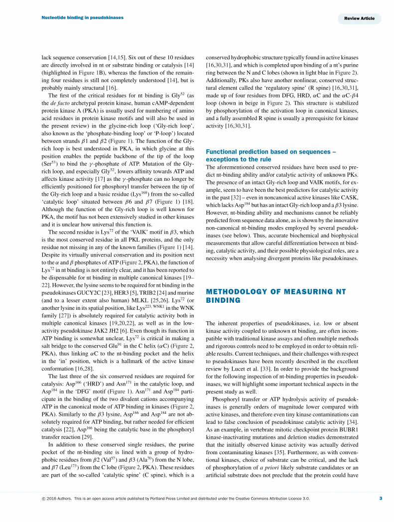
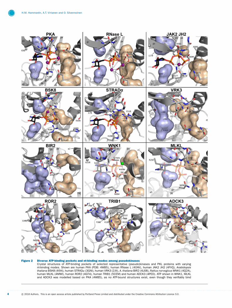
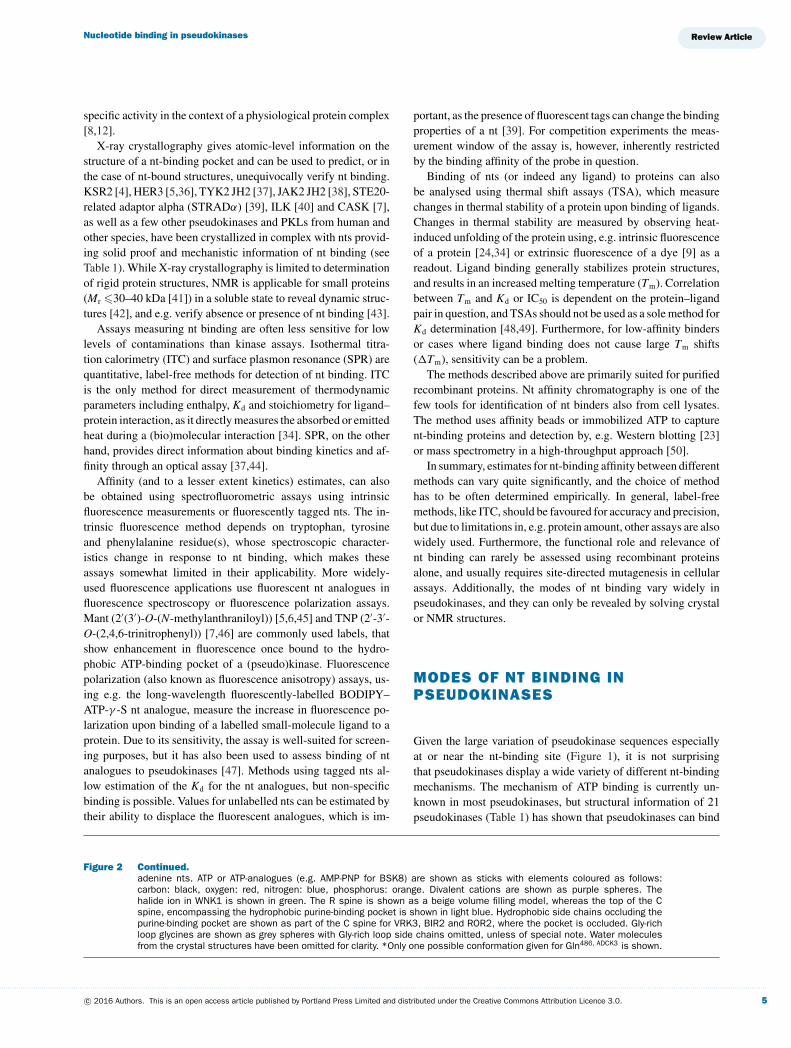
Figure 2 Diverse ATP-binding pockets and nt-binding modes among pseudokinasesCrystal structures of ATP-binding pockets of selected representative (pseudo)kinases and PKL proteins with varyingnt-binding modes. Shown are human PKA (PDB: 4WB5), human RNase L (4OAV), human JAK2 JH2 (4FVQ), Arabidopsisthaliana BSK8 (4I94), human STRADα (3GNI), human VRK3 (2JII), A. thaliana BIR2 (4L68), Rattus norvegicus WNK1 (4Q2A),human MLKL (4MWI), human ROR2 (4GT4), human TRIB1 (5CEM) and human ADCK3 (4PED). ATP shown in WNK1, MLKLand ADCK3 was modelled based on PKA (4WB5), as no ATP-bound structures exist, even though they verifiably bind
. . . . . . . . . . . . . . . . . . . . . . . . . . . . . . . . . . . . . . . . . . . . . . . . . . . . . . . . . . . . . . . . . . . . . . . . . . . . . . . . . . . . . . . . . . . . . . . . . . . . . . . . . . . . . . . . . . . . . . . . . . . . . . . . . . . . . . . . . . . . . . . . . . . . . . . . . . . . . . . . . . . . . . . . . . . . . . . . . . . . . . . . . . . . . . . . . . . . . . . . . . . . . . . . . . . . . . . . . . . . . . . . . . . . . . . . . . . . . . . . . . . . . . . . . . . . . . . . . . . . . . . . . . . . . . . . . . . . . . . . . . . . . . . . . . . . . . . . . . . . . . . . . . . . . . . . . . . . . . . . . . . . . . . . . . . . . . . . . . . . . . . . . . . . . . . . . . . . . . . . . .
4 c© 2016 Authors. This is an open access article published by Portland Press Limited and distributed under the Creative Commons Attribution Licence 3.0.
Nucleotide binding in pseudokinases
specific activity in the context of a physiological protein complex[8,12].
X-ray crystallography gives atomic-level information on thestructure of a nt-binding pocket and can be used to predict, or inthe case of nt-bound structures, unequivocally verify nt binding.KSR2 [4], HER3 [5,36], TYK2 JH2 [37], JAK2 JH2 [38], STE20-related adaptor alpha (STRADα) [39], ILK [40] and CASK [7],as well as a few other pseudokinases and PKLs from human andother species, have been crystallized in complex with nts provid-ing solid proof and mechanistic information of nt binding (seeTable 1). While X-ray crystallography is limited to determinationof rigid protein structures, NMR is applicable for small proteins(Mr �30–40 kDa [41]) in a soluble state to reveal dynamic struc-tures [42], and e.g. verify absence or presence of nt binding [43].
Assays measuring nt binding are often less sensitive for lowlevels of contaminations than kinase assays. Isothermal titra-tion calorimetry (ITC) and surface plasmon resonance (SPR) arequantitative, label-free methods for detection of nt binding. ITCis the only method for direct measurement of thermodynamicparameters including enthalpy, Kd and stoichiometry for ligand–protein interaction, as it directly measures the absorbed or emittedheat during a (bio)molecular interaction [34]. SPR, on the otherhand, provides direct information about binding kinetics and af-finity through an optical assay [37,44].
Affinity (and to a lesser extent kinetics) estimates, can alsobe obtained using spectrofluorometric assays using intrinsicfluorescence measurements or fluorescently tagged nts. The in-trinsic fluorescence method depends on tryptophan, tyrosineand phenylalanine residue(s), whose spectroscopic character-istics change in response to nt binding, which makes theseassays somewhat limited in their applicability. More widely-used fluorescence applications use fluorescent nt analogues influorescence spectroscopy or fluorescence polarization assays.Mant (2′(3′)-O-(N-methylanthraniloyl)) [5,6,45] and TNP (2′-3′-O-(2,4,6-trinitrophenyl)) [7,46] are commonly used labels, thatshow enhancement in fluorescence once bound to the hydro-phobic ATP-binding pocket of a (pseudo)kinase. Fluorescencepolarization (also known as fluorescence anisotropy) assays, us-ing e.g. the long-wavelength fluorescently-labelled BODIPY–ATP-γ -S nt analogue, measure the increase in fluorescence po-larization upon binding of a labelled small-molecule ligand to aprotein. Due to its sensitivity, the assay is well-suited for screen-ing purposes, but it has also been used to assess binding of ntanalogues to pseudokinases [47]. Methods using tagged nts al-low estimation of the Kd for the nt analogues, but non-specificbinding is possible. Values for unlabelled nts can be estimated bytheir ability to displace the fluorescent analogues, which is im-
portant, as the presence of fluorescent tags can change the bindingproperties of a nt [39]. For competition experiments the meas-urement window of the assay is, however, inherently restrictedby the binding affinity of the probe in question.
Binding of nts (or indeed any ligand) to proteins can alsobe analysed using thermal shift assays (TSA), which measurechanges in thermal stability of a protein upon binding of ligands.Changes in thermal stability are measured by observing heat-induced unfolding of the protein using, e.g. intrinsic fluorescenceof a protein [24,34] or extrinsic fluorescence of a dye [9] as areadout. Ligand binding generally stabilizes protein structures,and results in an increased melting temperature (Tm). Correlationbetween Tm and Kd or IC50 is dependent on the protein–ligandpair in question, and TSAs should not be used as a sole method forKd determination [48,49]. Furthermore, for low-affinity bindersor cases where ligand binding does not cause large Tm shifts(�Tm), sensitivity can be a problem.
The methods described above are primarily suited for purifiedrecombinant proteins. Nt affinity chromatography is one of thefew tools for identification of nt binders also from cell lysates.The method uses affinity beads or immobilized ATP to capturent-binding proteins and detection by, e.g. Western blotting [23]or mass spectrometry in a high-throughput approach [50].
In summary, estimates for nt-binding affinity between differentmethods can vary quite significantly, and the choice of methodhas to be often determined empirically. In general, label-freemethods, like ITC, should be favoured for accuracy and precision,but due to limitations in, e.g. protein amount, other assays are alsowidely used. Furthermore, the functional role and relevance ofnt binding can rarely be assessed using recombinant proteinsalone, and usually requires site-directed mutagenesis in cellularassays. Additionally, the modes of nt binding vary widely inpseudokinases, and they can only be revealed by solving crystalor NMR structures.
MODES OF NT BINDING INPSEUDOKINASES
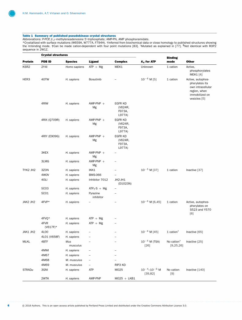
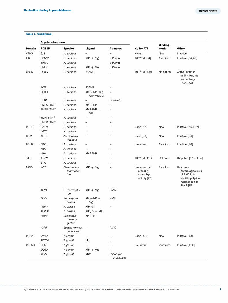
Given the large variation of pseudokinase sequences especiallyat or near the nt-binding site (Figure 1), it is not surprisingthat pseudokinases display a wide variety of different nt-bindingmechanisms. The mechanism of ATP binding is currently un-known in most pseudokinases, but structural information of 21pseudokinases (Table 1) has shown that pseudokinases can bind
Figure 2 Continued.adenine nts. ATP or ATP-analogues (e.g. AMP-PNP for BSK8) are shown as sticks with elements coloured as follows:carbon: black, oxygen: red, nitrogen: blue, phosphorus: orange. Divalent cations are shown as purple spheres. Thehalide ion in WNK1 is shown in green. The R spine is shown as a beige volume filling model, whereas the top of the Cspine, encompassing the hydrophobic purine-binding pocket is shown in light blue. Hydrophobic side chains occluding thepurine-binding pocket are shown as part of the C spine for VRK3, BIR2 and ROR2, where the pocket is occluded. Gly-richloop glycines are shown as grey spheres with Gly-rich loop side chains omitted, unless of special note. Water moleculesfrom the crystal structures have been omitted for clarity. *Only one possible conformation given for Gln486, ADCK3 is shown.
. . . . . . . . . . . . . . . . . . . . . . . . . . . . . . . . . . . . . . . . . . . . . . . . . . . . . . . . . . . . . . . . . . . . . . . . . . . . . . . . . . . . . . . . . . . . . . . . . . . . . . . . . . . . . . . . . . . . . . . . . . . . . . . . . . . . . . . . . . . . . . . . . . . . . . . . . . . . . . . . . . . . . . . . . . . . . . . . . . . . . . . . . . . . . . . . . . . . . . . . . . . . . . . . . . . . . . . . . . . . . . . . . . . . . . . . . . . . . . . . . . . . . . . . . . . . . . . . . . . . . . . . . . . . . . . . . . . . . . . . . . . . . . . . . . . . . . . . . . . . . . . . . . . . . . . . . . . . . . . . . . . . . . . . . . . . . . . . . . . . . . . . . . . . . . . . . . . . . . . . . .
c© 2016 Authors. This is an open access article published by Portland Press Limited and distributed under the Creative Commons Attribution Licence 3.0. 5
H.M. Hammaren, A.T. Virtanen and O. Silvennoinen
Table 1 Summary of published pseudokinase crystal structuresAbbreviations: P-PCP, β,γ -methyleneadenosine 5′ -triphosphate; AMP-PN, AMP phosphoramidate.*Crystallized with surface mutations (W659A, W777A, F794H). †Inferred from biochemical data or close homology to published structures showingthe nt-binding mode. ‡Can be made cation-dependent with four point mutations [83]. §Mutated as explained in [77]. ¶Not identical with ROP2sequence in 2W1Z.
Crystal structures
Protein PDB ID Species Ligand Complex Kd for ATPBindingmode Other
KSR2 2Y4I Homo sapiens ATP + Mg MEK1 Unknown 1 cation Active,phosphorylatesMEK1 [4]
HER3 4OTW H. sapiens Bosutinib – 10− 6 M [5] 1 cation Active, autophos-phorylates itsown intracellularregion, whenimmobilized onvesicles [5]
4RIW H. sapiens AMP-PNP +Mg
EGFR KD(V624R,F973A,L977A)
4RIX (Q709R) H. sapiens AMP-PNP +Mg
EGFR KD(V624R,F973A,L977A)
4RIY (E909G) H. sapiens AMP-PNP +Mg
EGFR KD(V624R,F973A,L977A)
3KEX H. sapiens AMP-PNP +Mg
–
3LMG H. sapiens AMP-PNP +Mg
–
TYK2 JH2 3ZON H. sapiens IKK1 – 10− 5 M [37] 1 cation Inactive [37]
4WOV H. sapiens BMS-066 –
4OLI H. sapiens Inhibitor 7012 JH2-JH1(D1023N)
5C03 H. sapiens ATPγ S + Mg –
5C01 H. sapiens Pyrazineinhibitor
–
JAK2 JH2 4FVP* H. sapiens – – 10− 6 M [6,45] 1 cation Active, autophos-phorylates onS523 and Y570[6]
4FVQ* H. sapiens ATP + Mg –
4FVR(V617F)*
H. sapiens ATP + Mg –
JAK1 JH2 4L00 H. sapiens – – 10− 6 M [45] 1 cation† Inactive [65]
4L01 (V658F) H. sapiens – –
MLKL 4BTF Musmusculus
– – 10− 5 M (TSA)[26]
No cation†
[9,25,26]Inactive [25]
4MWI H. sapiens – –
4M67 H. sapiens – –
4M68 M. musculus – –
4M69 M. musculus – RIP3 KD
STRADα 3GNI H. sapiens ATP MO25 10− 4–10− 6 M[39,82]
No cation[9]
Inactive [140]
2WTK H. sapiens AMP-PNP MO25 + LKB1
. . . . . . . . . . . . . . . . . . . . . . . . . . . . . . . . . . . . . . . . . . . . . . . . . . . . . . . . . . . . . . . . . . . . . . . . . . . . . . . . . . . . . . . . . . . . . . . . . . . . . . . . . . . . . . . . . . . . . . . . . . . . . . . . . . . . . . . . . . . . . . . . . . . . . . . . . . . . . . . . . . . . . . . . . . . . . . . . . . . . . . . . . . . . . . . . . . . . . . . . . . . . . . . . . . . . . . . . . . . . . . . . . . . . . . . . . . . . . . . . . . . . . . . . . . . . . . . . . . . . . . . . . . . . . . . . . . . . . . . . . . . . . . . . . . . . . . . . . . . . . . . . . . . . . . . . . . . . . . . . . . . . . . . . . . . . . . . . . . . . . . . . . . . . . . . . . . . . . . . . . .
6 c© 2016 Authors. This is an open access article published by Portland Press Limited and distributed under the Creative Commons Attribution Licence 3.0.
Nucleotide binding in pseudokinases
Table 1 Continued.
Crystal structures
Protein PDB ID Species Ligand Complex Kd for ATPBindingmode Other
VRK3 2JII H. sapiens – – None N/A Inactive
ILK 3KMW H. sapiens ATP + Mg α-Parvin 10− 6 M [34] 1 cation Inactive [34,40]
3KMU H. sapiens – α-Parvin
3REP H. sapiens ATP + Mn α-Parvin
CASK 3C0G H. sapiens 3′ -AMP – 10− 3 M [7,9] No cation Active, cationsinhibit bindingand activity.[7,24,83]
3C0I H. sapiens 3′ -AMP –
3C0H H. sapiens AMP-PNP (onlyAMP visible)
–
3TAC H. sapiens – Liprin-α2
3MFS (4M)‡ H. sapiens AMP-PNP –
3MFU (4M)‡ H. sapiens AMP-PNP +Mn
–
3MFT (4M)‡ H. sapiens – –
3MFR (4M)‡ H. sapiens – –
ROR2 3ZZW H. sapiens – – None [55] N/A Inactive [55,102]
4GT4 H. sapiens – –
BIR2 4L68 Arabidopsisthaliana
– – None [94] N/A Inactive [94]
BSK8 4I92 A. thaliana – – Unknown 1 cation Inactive [76]
4I93 A. thaliana – –
4I94 A. thaliana AMP-PNP –
Titin 4JNW H. sapiens – – 10− 4 M [113] Unknown Disputed [112–114]
1TKI H. sapiens – –
PAN3 4CYI Chaetomiumthermophi-lum
ATP + Mg – Unknown, butprobablyrather highaffinity [78]
1 cation Unknown,physiological roleof PKD is toshuttle polyribo-nucleotides toPAN2 [81]
4CYJ C. thermophi-lum
ATP + Mg PAN2
4CZY Neurosporacrassa
AMP-PNP +Mg
PAN2
4BWK N. crassa ATPγ S –
4BWX§ N. crassa ATPγ S + Mg
4BWP Drosophilamelano-gaster
AMP-PN –
4XR7 Saccharomycescerevisiae
– PAN2
ROP2 2W1Z T. gondii – – None [43] N/A Inactive [43]
3DZO¶ T. gondii Mg –
ROP5B 3Q5Z T. gondii – – Unknown 2 cations Inactive [110]
3Q60 T. gondii ATP + Mg –
4LV5 T. gondii ADP IRGa6 (M.musculus)
. . . . . . . . . . . . . . . . . . . . . . . . . . . . . . . . . . . . . . . . . . . . . . . . . . . . . . . . . . . . . . . . . . . . . . . . . . . . . . . . . . . . . . . . . . . . . . . . . . . . . . . . . . . . . . . . . . . . . . . . . . . . . . . . . . . . . . . . . . . . . . . . . . . . . . . . . . . . . . . . . . . . . . . . . . . . . . . . . . . . . . . . . . . . . . . . . . . . . . . . . . . . . . . . . . . . . . . . . . . . . . . . . . . . . . . . . . . . . . . . . . . . . . . . . . . . . . . . . . . . . . . . . . . . . . . . . . . . . . . . . . . . . . . . . . . . . . . . . . . . . . . . . . . . . . . . . . . . . . . . . . . . . . . . . . . . . . . . . . . . . . . . . . . . . . . . . . . . . . . . . .
c© 2016 Authors. This is an open access article published by Portland Press Limited and distributed under the Creative Commons Attribution Licence 3.0. 7
H.M. Hammaren, A.T. Virtanen and O. Silvennoinen
Table 1 Continued.
Crystal structures
Protein PDB ID Species Ligand Complex Kd for ATPBindingmode Other
ROP5C 4LV8 T. gondii ADP + Mg IRGa6 (M.musculus)
Unknown 1 cation Unknown
ROP8 3BYV T. gondii Mg – Unknown Unknown Unknown
RNase L 4O1O Sus scrofa – 2-5A 10− 3 M [53] 2 cations Inactive [52–54]
4O1P S. scrofa AMP-PNP +Mg
2-5A
4OAU H. sapiens ADP + Mg 2-5A
4OAV H. sapiens AMP-PCP + Mg RNA + pUp
WNK1 4Q2A Rattusnorvegicus
Br – Unknown Unknown Active [124]
3FPQ R. norvegicus – –
4PWN H. sapiens – –
MviN 3OTV Mycobacteriumtubercu-losis
– – None [47] Unknown Inactive [47]
3OUK M. tubercu-losis
– –
3OUN M. tubercu-losis
– FhaA
3UQC M. tubercu-losis
– –
ADCK3 4PED H. sapiens – – BindspreferentiallyADP [108]
Unknown Inactive, can beactivated with asingle Gly-richloop mutation[108]
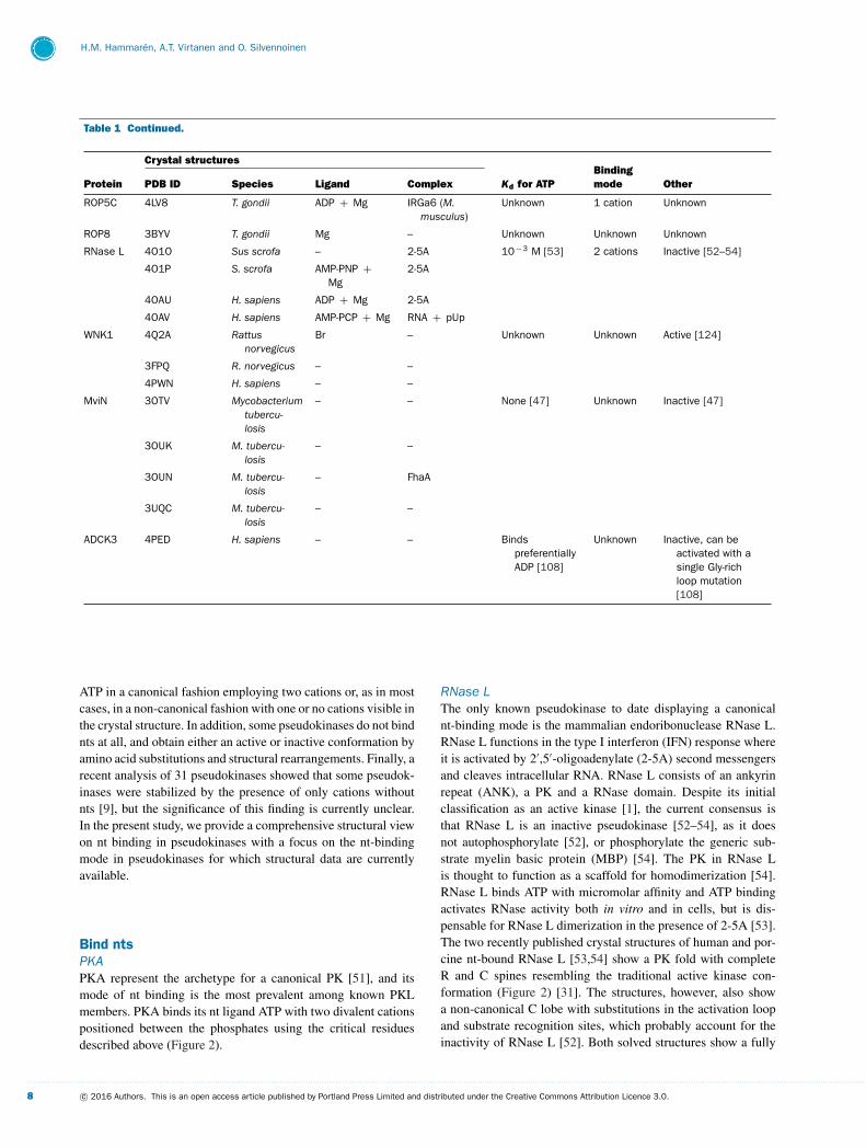
ATP in a canonical fashion employing two cations or, as in mostcases, in a non-canonical fashion with one or no cations visible inthe crystal structure. In addition, some pseudokinases do not bindnts at all, and obtain either an active or inactive conformation byamino acid substitutions and structural rearrangements. Finally, arecent analysis of 31 pseudokinases showed that some pseudok-inases were stabilized by the presence of only cations withoutnts [9], but the significance of this finding is currently unclear.In the present study, we provide a comprehensive structural viewon nt binding in pseudokinases with a focus on the nt-bindingmode in pseudokinases for which structural data are currentlyavailable.
Bind ntsPKAPKA represent the archetype for a canonical PK [51], and itsmode of nt binding is the most prevalent among known PKLmembers. PKA binds its nt ligand ATP with two divalent cationspositioned between the phosphates using the critical residuesdescribed above (Figure 2).
RNase LThe only known pseudokinase to date displaying a canonicalnt-binding mode is the mammalian endoribonuclease RNase L.RNase L functions in the type I interferon (IFN) response whereit is activated by 2′,5′-oligoadenylate (2-5A) second messengersand cleaves intracellular RNA. RNase L consists of an ankyrinrepeat (ANK), a PK and a RNase domain. Despite its initialclassification as an active kinase [1], the current consensus isthat RNase L is an inactive pseudokinase [52–54], as it doesnot autophosphorylate [52], or phosphorylate the generic sub-strate myelin basic protein (MBP) [54]. The PK in RNase Lis thought to function as a scaffold for homodimerization [54].RNase L binds ATP with micromolar affinity and ATP bindingactivates RNase activity both in vitro and in cells, but is dis-pensable for RNase L dimerization in the presence of 2-5A [53].The two recently published crystal structures of human and por-cine nt-bound RNase L [53,54] show a PK fold with completeR and C spines resembling the traditional active kinase con-formation (Figure 2) [31]. The structures, however, also showa non-canonical C lobe with substitutions in the activation loopand substrate recognition sites, which probably account for theinactivity of RNase L [52]. Both solved structures show a fully
. . . . . . . . . . . . . . . . . . . . . . . . . . . . . . . . . . . . . . . . . . . . . . . . . . . . . . . . . . . . . . . . . . . . . . . . . . . . . . . . . . . . . . . . . . . . . . . . . . . . . . . . . . . . . . . . . . . . . . . . . . . . . . . . . . . . . . . . . . . . . . . . . . . . . . . . . . . . . . . . . . . . . . . . . . . . . . . . . . . . . . . . . . . . . . . . . . . . . . . . . . . . . . . . . . . . . . . . . . . . . . . . . . . . . . . . . . . . . . . . . . . . . . . . . . . . . . . . . . . . . . . . . . . . . . . . . . . . . . . . . . . . . . . . . . . . . . . . . . . . . . . . . . . . . . . . . . . . . . . . . . . . . . . . . . . . . . . . . . . . . . . . . . . . . . . . . . . . . . . . . .
8 c© 2016 Authors. This is an open access article published by Portland Press Limited and distributed under the Creative Commons Attribution Licence 3.0.
Nucleotide binding in pseudokinases
ordered nt in the ATP-binding pocket, which resembles RNaseL’s closest active homologue inositol-requiring protein 1 (IRE1).Nt binding is coordinated by conserved PK residues, except oneuncommon substitution at Gly186 (Asp505, RNase L), which formsan additional interaction to one of the cations (Figure 2). Thebinding pocket is also flanked by the structured PK-ANK linker,which participates in 2-5A binding and interdomain interactions[53,54].
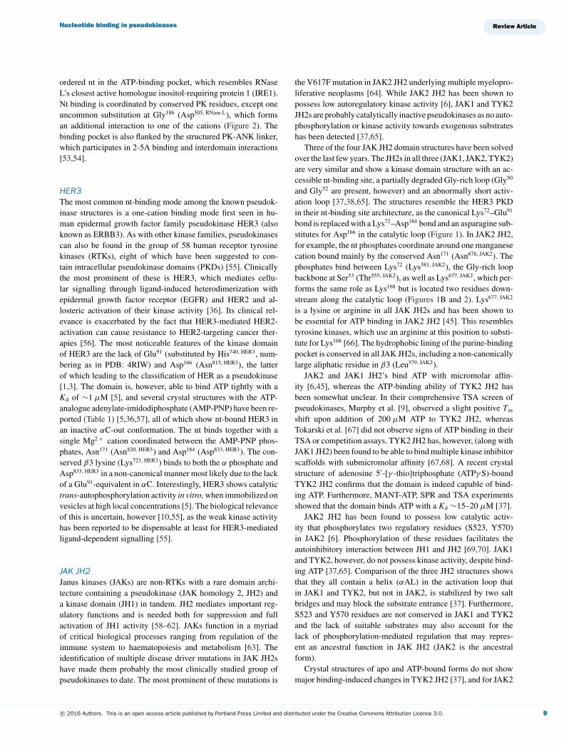
HER3The most common nt-binding mode among the known pseudok-inase structures is a one-cation binding mode first seen in hu-man epidermal growth factor family pseudokinase HER3 (alsoknown as ERBB3). As with other kinase families, pseudokinasescan also be found in the group of 58 human receptor tyrosinekinases (RTKs), eight of which have been suggested to con-tain intracellular pseudokinase domains (PKDs) [55]. Clinicallythe most prominent of these is HER3, which mediates cellu-lar signalling through ligand-induced heterodimerization withepidermal growth factor receptor (EGFR) and HER2 and al-losteric activation of their kinase activity [36]. Its clinical rel-evance is exacerbated by the fact that HER3-mediated HER2-activation can cause resistance to HER2-targeting cancer ther-apies [56]. The most noticeable features of the kinase domainof HER3 are the lack of Glu91 (substituted by His740, HER3, num-bering as in PDB: 4RIW) and Asp166 (Asn815, HER3), the latterof which leading to the classification of HER as a pseudokinase[1,3]. The domain is, however, able to bind ATP tightly with aKd of ∼1 μM [5], and several crystal structures with the ATP-analogue adenylate-imidodiphosphate (AMP-PNP) have been re-ported (Table 1) [5,36,57], all of which show nt-bound HER3 inan inactive αC-out conformation. The nt binds together with asingle Mg2 + cation coordinated between the AMP-PNP phos-phates, Asn171 (Asn820, HER3) and Asp184 (Asp833, HER3). The con-served β3 lysine (Lys723, HER3) binds to both the α phosphate andAsp833, HER3 in a non-canonical manner most likely due to the lackof a Glu91-equivalent in αC. Interestingly, HER3 shows catalytictrans-autophosphorylation activity in vitro, when immobilized onvesicles at high local concentrations [5]. The biological relevanceof this is uncertain, however [10,55], as the weak kinase activityhas been reported to be dispensable at least for HER3-mediatedligand-dependent signalling [55].
JAK JH2Janus kinases (JAKs) are non-RTKs with a rare domain archi-tecture containing a pseudokinase (JAK homology 2, JH2) anda kinase domain (JH1) in tandem. JH2 mediates important reg-ulatory functions and is needed both for suppression and fullactivation of JH1 activity [58–62]. JAKs function in a myriadof critical biological processes ranging from regulation of theimmune system to haematopoiesis and metabolism [63]. Theidentification of multiple disease driver mutations in JAK JH2shave made them probably the most clinically studied group ofpseudokinases to date. The most prominent of these mutations is
the V617F mutation in JAK2 JH2 underlying multiple myelopro-liferative neoplasms [64]. While JAK2 JH2 has been shown topossess low autoregulatory kinase activity [6], JAK1 and TYK2JH2s are probably catalytically inactive pseudokinases as no auto-phosphorylation or kinase activity towards exogenous substrateshas been detected [37,65].
Three of the four JAK JH2 domain structures have been solvedover the last few years. The JH2s in all three (JAK1, JAK2, TYK2)are very similar and show a kinase domain structure with an ac-cessible nt-binding site, a partially degraded Gly-rich loop (Gly50
and Gly52 are present, however) and an abnormally short activ-ation loop [37,38,65]. The structures resemble the HER3 PKDin their nt-binding site architecture, as the canonical Lys72–Glu91
bond is replaced with a Lys72–Asp184 bond and an asparagine sub-stitutes for Asp166 in the catalytic loop (Figure 1). In JAK2 JH2,for example, the nt phosphates coordinate around one manganesecation bound mainly by the conserved Asn171 (Asn678, JAK2). Thephosphates bind between Lys72 (Lys581, JAK2), the Gly-rich loopbackbone at Ser53 (Thr555, JAK2), as well as Lys677, JAK2, which per-forms the same role as Lys168 but is located two residues down-stream along the catalytic loop (Figures 1B and 2). Lys677, JAK2
is a lysine or arginine in all JAK JH2s and has been shown tobe essential for ATP binding in JAK2 JH2 [45]. This resemblestyrosine kinases, which use an arginine at this position to substi-tute for Lys168 [66]. The hydrophobic lining of the purine-bindingpocket is conserved in all JAK JH2s, including a non-canonicallylarge aliphatic residue in β3 (Leu579, JAK2).
JAK2 and JAK1 JH2’s bind ATP with micromolar affin-ity [6,45], whereas the ATP-binding ability of TYK2 JH2 hasbeen somewhat unclear. In their comprehensive TSA screen ofpseudokinases, Murphy et al. [9], observed a slight positive Tm
shift upon addition of 200 μM ATP to TYK2 JH2, whereasTokarski et al. [67] did not observe signs of ATP binding in theirTSA or competition assays. TYK2 JH2 has, however, (along withJAK1 JH2) been found to be able to bind multiple kinase inhibitorscaffolds with submicromolar affinity [67,68]. A recent crystalstructure of adenosine 5′-[γ -thio]triphosphate (ATPγ S)-boundTYK2 JH2 confirms that the domain is indeed capable of bind-ing ATP. Furthermore, MANT-ATP, SPR and TSA experimentsshowed that the domain binds ATP with a Kd ∼15–20 μM [37].
JAK2 JH2 has been found to possess low catalytic activ-ity that phosphorylates two regulatory residues (S523, Y570)in JAK2 [6]. Phosphorylation of these residues facilitates theautoinhibitory interaction between JH1 and JH2 [69,70]. JAK1and TYK2, however, do not possess kinase activity, despite bind-ing ATP [37,65]. Comparison of the three JH2 structures showsthat they all contain a helix (αAL) in the activation loop thatin JAK1 and TYK2, but not in JAK2, is stabilized by two saltbridges and may block the substrate entrance [37]. Furthermore,S523 and Y570 residues are not conserved in JAK1 and TYK2and the lack of suitable substrates may also account for thelack of phosphorylation-mediated regulation that may repres-ent an ancestral function in JAK JH2 (JAK2 is the ancestralform).
Crystal structures of apo and ATP-bound forms do not showmajor binding-induced changes in TYK2 JH2 [37], and for JAK2
. . . . . . . . . . . . . . . . . . . . . . . . . . . . . . . . . . . . . . . . . . . . . . . . . . . . . . . . . . . . . . . . . . . . . . . . . . . . . . . . . . . . . . . . . . . . . . . . . . . . . . . . . . . . . . . . . . . . . . . . . . . . . . . . . . . . . . . . . . . . . . . . . . . . . . . . . . . . . . . . . . . . . . . . . . . . . . . . . . . . . . . . . . . . . . . . . . . . . . . . . . . . . . . . . . . . . . . . . . . . . . . . . . . . . . . . . . . . . . . . . . . . . . . . . . . . . . . . . . . . . . . . . . . . . . . . . . . . . . . . . . . . . . . . . . . . . . . . . . . . . . . . . . . . . . . . . . . . . . . . . . . . . . . . . . . . . . . . . . . . . . . . . . . . . . . . . . . . . . . . . .
c© 2016 Authors. This is an open access article published by Portland Press Limited and distributed under the Creative Commons Attribution Licence 3.0. 9
H.M. Hammaren, A.T. Virtanen and O. Silvennoinen
JH2 only a stabilization of αC has been observed [38,45]. Inter-estingly, however, recent mutagenesis experiments indicate thatATP-binding to JAK2 JH2 is critical for the pathogenic activationof JAK, while being largely dispensable for wild-type functions[45]. This finding, along with the recent identification of a TYK2JH2 binding inhibitor able to modulate TYK2 activity [67] makeJAK JH2s tempting pharmacological targets.
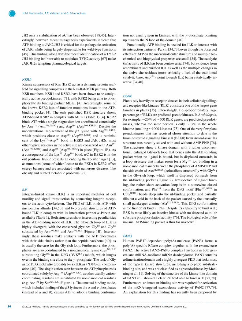
KSR2Kinase suppressors of Ras (KSR) act as a dynamic protein scaf-fold for signalling complexes in the Ras-Raf-MEK pathway. BothKSR members, KSR1 and KSR2, have been shown to be catalyt-ically active pseudokinases [71], with KSR2 being able to phos-phorylate its binding partner MEK1 [4]. Accordingly, some ofthe known KSR2 loss-of-function mutations locate to the ATP-binding pocket [4]. The only published KSR structure showsATP-bound KSR2 in complex with MEK1 (Table 1) [4]. KSR2binds ATP with a single magnesium ion coordinated canonicallyby Asn171 (Asn791, KSR2) and Asp184 (Asp803, KSR2). Despite theunconventional replacement of the β3 lysine with Arg692, KSR2,which positions close to Asp184 (Asp803, KSR2) and is reminis-cent of the Lys72–Asp184 bond in HER3 and JAK JH2s, mostother typical residues in the active site are conserved with Asn171
(Asn791, KSR2) and Asp166 (Asp786, KSR2) in place (Figure 1B). Asa consequence of the Lys72–Asp184 bond, αC in KSR2 is in theout position. KSR2 presents an enticing therapeutic target [13],as mutations (some of which locate to the PKD) in KSR2 affectenergy balance and are associated with numerous diseases, likeobesity and related metabolic problems [72].
ILKIntegrin-linked kinase (ILK) is an important mediator of cellmotility and signal transduction by connecting integrin recept-ors to the actin cytoskeleton. The PKD of ILK binds ATP withmicromolar affinity [34,50], and two crystal structures of ATP-bound ILK in complex with its interaction partner α-Parvin areavailable (Table 1). Both structures show interesting peculiaritiesin the ATP-binding mode of ILK. The Gly-rich loop of ILK ishighly divergent, with the conserved glycines Gly50 and Gly52
substituted by Asn200, ILK and Asn202, ILK (Figure 1B). Interest-ingly, these residues make contacts with the ATP phosphateswith their side chains rather than the peptide backbone [40], asis usually the case for the Gly-rich loop. Furthermore, the phos-phates are also coordinated by a noncanonical lysine (Lys341, ILK
substituting Gly186 in the DFG (DVKILK) motif), which lungesover in the binding site close to the γ -phosphate. The lack of Glyin the DFG motif also probably locks ILK in a ‘DFG-in’ conform-ation [40]. The single cation seen between the ATP phosphates iscoordinated solely by Asp184 (Asp339, ILK), as other usually cation-coordinating residues are substituted by non-canonical residues(e.g. Asn171 by Ser324, ILK, Figure 1). The unusual binding mode,which includes binding of the β3 lysine to the α and γ phosphates(instead of α and β), causes ATP to adopt a binding conforma-
tion not usually seen in kinases, with the γ -phosphate pointingup towards the N lobe of the domain [40].
Functionally, ATP binding is needed for ILK to interact withits interaction partner α-Parvin [34,73], even though the observedeffects of ATP on the macromolecular structure and multiple bio-chemical and biophysical properties are small [34]. The catalytic(in)activity of ILK has been controversial [74], but evidence fromrecombinant and purified ILK as well as the multiple changes inthe active site residues (most critically a lack of the traditionalcatalytic base, Asp166), point towards ILK being catalytically in-active [34,40].
BSK8Plants rely heavily on receptor kinases in their cellular signalling,and receptor-like kinases (RLK) constitute one of the largest genefamilies in plants [75]. Interestingly, a disproportionately largepercentage of RLKs are predicted pseudokinases. In Arabidopsis,for example, ∼20 % of ∼600 RLK genes, are predicted pseudok-inases, whereas the same portion is only ∼13 % in the wholekinome (totalling ∼1000 kinases) [75]. One of the very few plantpseudokinases that has received closer attention to date is thebrassinosteroid signalling kinase 8 (BSK8) from Arabidopsis. Itsstructure was recently solved with and without AMP-PNP [76].The structures show a kinase domain with a rather unconven-tional, enlarged Gly-rich loop that bends into the ATP-bindingpocket when no ligand is bound, but is displaced outwards ina loop structure that makes room for a Mg2 + ion binding in anon-canonical manner between the phosphates of AMP-PNP andthe side chain of Asn71, BSK8 (colocalizes structurally with Gly52)in the Gly-rich loop, which itself is displaced outwards fromthe nt-binding pocket (Figure 2). Irrespective of ligand bind-ing, the rather short activation loop is in a somewhat closedconformation, and Phe185 from the DFG motif (Phe200, BSK8 inCFGBSK8) bends deep into the nt-binding pocket and partiallyfills out a void in the back of the pocket caused by the unusuallysmall gatekeeper alanine (Ala132, BSK8). This DFG conformationis the first of its kind in PKs. Despite its capability to bind ATP,BSK is most likely an inactive kinase with no detected auto- orsubstrate phosphorylation activity [76]. The biological role of theunusual ATP-binding pocket is thus far unknown.
PAN3Human PAB1P-dependent poly(A)-nuclease (PAN3) forms apoly(A)-specific RNase complex together with the exonucleasePAN2. The active PAN2–PAN3 complex functions in both gen-eral and miRNA-mediated mRNA deadenylation. PAN3 containsa dimerization domain and a highly divergent PKD that lacks mostof the typical kinase structures, including a peptide substrate-binding site, and was not classified as a (pseudo)kinase by Man-ning et al., [1]. Solving of the structure of the kinase-like domainof PAN3 still showed a clear PK fold able to bind ATP [77,78].Furthermore, an intact nt-binding site was required for activationof the mRNA-targeted exonuclease activity of PAN2 [77,79].An explanation for this finding has recently been proposed by
. . . . . . . . . . . . . . . . . . . . . . . . . . . . . . . . . . . . . . . . . . . . . . . . . . . . . . . . . . . . . . . . . . . . . . . . . . . . . . . . . . . . . . . . . . . . . . . . . . . . . . . . . . . . . . . . . . . . . . . . . . . . . . . . . . . . . . . . . . . . . . . . . . . . . . . . . . . . . . . . . . . . . . . . . . . . . . . . . . . . . . . . . . . . . . . . . . . . . . . . . . . . . . . . . . . . . . . . . . . . . . . . . . . . . . . . . . . . . . . . . . . . . . . . . . . . . . . . . . . . . . . . . . . . . . . . . . . . . . . . . . . . . . . . . . . . . . . . . . . . . . . . . . . . . . . . . . . . . . . . . . . . . . . . . . . . . . . . . . . . . . . . . . . . . . . . . . . . . . . . . .
10 c© 2016 Authors. This is an open access article published by Portland Press Limited and distributed under the Creative Commons Attribution Licence 3.0.
Nucleotide binding in pseudokinases
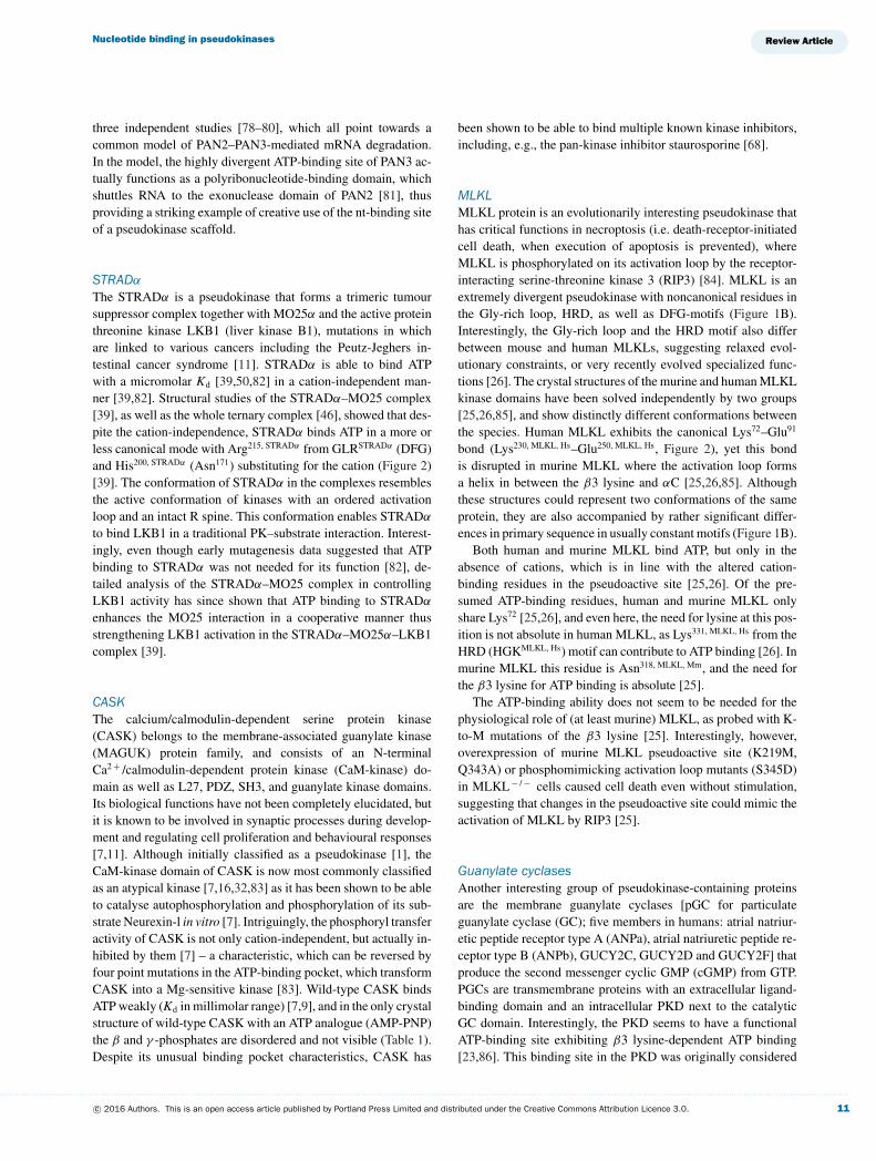
three independent studies [78–80], which all point towards acommon model of PAN2–PAN3-mediated mRNA degradation.In the model, the highly divergent ATP-binding site of PAN3 ac-tually functions as a polyribonucleotide-binding domain, whichshuttles RNA to the exonuclease domain of PAN2 [81], thusproviding a striking example of creative use of the nt-binding siteof a pseudokinase scaffold.
STRADα
The STRADα is a pseudokinase that forms a trimeric tumoursuppressor complex together with MO25α and the active proteinthreonine kinase LKB1 (liver kinase B1), mutations in whichare linked to various cancers including the Peutz-Jeghers in-testinal cancer syndrome [11]. STRADα is able to bind ATPwith a micromolar Kd [39,50,82] in a cation-independent man-ner [39,82]. Structural studies of the STRADα–MO25 complex[39], as well as the whole ternary complex [46], showed that des-pite the cation-independence, STRADα binds ATP in a more orless canonical mode with Arg215, STRADα from GLRSTRADα (DFG)and His200, STRADα (Asn171) substituting for the cation (Figure 2)[39]. The conformation of STRADα in the complexes resemblesthe active conformation of kinases with an ordered activationloop and an intact R spine. This conformation enables STRADα
to bind LKB1 in a traditional PK–substrate interaction. Interest-ingly, even though early mutagenesis data suggested that ATPbinding to STRADα was not needed for its function [82], de-tailed analysis of the STRADα–MO25 complex in controllingLKB1 activity has since shown that ATP binding to STRADα
enhances the MO25 interaction in a cooperative manner thusstrengthening LKB1 activation in the STRADα–MO25α–LKB1complex [39].
CASKThe calcium/calmodulin-dependent serine protein kinase(CASK) belongs to the membrane-associated guanylate kinase(MAGUK) protein family, and consists of an N-terminalCa2 + /calmodulin-dependent protein kinase (CaM-kinase) do-main as well as L27, PDZ, SH3, and guanylate kinase domains.Its biological functions have not been completely elucidated, butit is known to be involved in synaptic processes during develop-ment and regulating cell proliferation and behavioural responses[7,11]. Although initially classified as a pseudokinase [1], theCaM-kinase domain of CASK is now most commonly classifiedas an atypical kinase [7,16,32,83] as it has been shown to be ableto catalyse autophosphorylation and phosphorylation of its sub-strate Neurexin-l in vitro [7]. Intriguingly, the phosphoryl transferactivity of CASK is not only cation-independent, but actually in-hibited by them [7] – a characteristic, which can be reversed byfour point mutations in the ATP-binding pocket, which transformCASK into a Mg-sensitive kinase [83]. Wild-type CASK bindsATP weakly (Kd in millimolar range) [7,9], and in the only crystalstructure of wild-type CASK with an ATP analogue (AMP-PNP)the β and γ -phosphates are disordered and not visible (Table 1).Despite its unusual binding pocket characteristics, CASK has
been shown to be able to bind multiple known kinase inhibitors,including, e.g., the pan-kinase inhibitor staurosporine [68].
MLKLMLKL protein is an evolutionarily interesting pseudokinase thathas critical functions in necroptosis (i.e. death-receptor-initiatedcell death, when execution of apoptosis is prevented), whereMLKL is phosphorylated on its activation loop by the receptor-interacting serine-threonine kinase 3 (RIP3) [84]. MLKL is anextremely divergent pseudokinase with noncanonical residues inthe Gly-rich loop, HRD, as well as DFG-motifs (Figure 1B).Interestingly, the Gly-rich loop and the HRD motif also differbetween mouse and human MLKLs, suggesting relaxed evol-utionary constraints, or very recently evolved specialized func-tions [26]. The crystal structures of the murine and human MLKLkinase domains have been solved independently by two groups[25,26,85], and show distinctly different conformations betweenthe species. Human MLKL exhibits the canonical Lys72–Glu91
bond (Lys230, MLKL, Hs–Glu250, MLKL, Hs, Figure 2), yet this bondis disrupted in murine MLKL where the activation loop formsa helix in between the β3 lysine and αC [25,26,85]. Althoughthese structures could represent two conformations of the sameprotein, they are also accompanied by rather significant differ-ences in primary sequence in usually constant motifs (Figure 1B).
Both human and murine MLKL bind ATP, but only in theabsence of cations, which is in line with the altered cation-binding residues in the pseudoactive site [25,26]. Of the pre-sumed ATP-binding residues, human and murine MLKL onlyshare Lys72 [25,26], and even here, the need for lysine at this pos-ition is not absolute in human MLKL, as Lys331, MLKL, Hs from theHRD (HGKMLKL, Hs) motif can contribute to ATP binding [26]. Inmurine MLKL this residue is Asn318, MLKL, Mm, and the need forthe β3 lysine for ATP binding is absolute [25].
The ATP-binding ability does not seem to be needed for thephysiological role of (at least murine) MLKL, as probed with K-to-M mutations of the β3 lysine [25]. Interestingly, however,overexpression of murine MLKL pseudoactive site (K219M,Q343A) or phosphomimicking activation loop mutants (S345D)in MLKL− / − cells caused cell death even without stimulation,suggesting that changes in the pseudoactive site could mimic theactivation of MLKL by RIP3 [25].
Guanylate cyclasesAnother interesting group of pseudokinase-containing proteinsare the membrane guanylate cyclases [pGC for particulateguanylate cyclase (GC); five members in humans: atrial natriur-etic peptide receptor type A (ANPa), atrial natriuretic peptide re-ceptor type B (ANPb), GUCY2C, GUCY2D and GUCY2F] thatproduce the second messenger cyclic GMP (cGMP) from GTP.PGCs are transmembrane proteins with an extracellular ligand-binding domain and an intracellular PKD next to the catalyticGC domain. Interestingly, the PKD seems to have a functionalATP-binding site exhibiting β3 lysine-dependent ATP binding[23,86]. This binding site in the PKD was originally considered
. . . . . . . . . . . . . . . . . . . . . . . . . . . . . . . . . . . . . . . . . . . . . . . . . . . . . . . . . . . . . . . . . . . . . . . . . . . . . . . . . . . . . . . . . . . . . . . . . . . . . . . . . . . . . . . . . . . . . . . . . . . . . . . . . . . . . . . . . . . . . . . . . . . . . . . . . . . . . . . . . . . . . . . . . . . . . . . . . . . . . . . . . . . . . . . . . . . . . . . . . . . . . . . . . . . . . . . . . . . . . . . . . . . . . . . . . . . . . . . . . . . . . . . . . . . . . . . . . . . . . . . . . . . . . . . . . . . . . . . . . . . . . . . . . . . . . . . . . . . . . . . . . . . . . . . . . . . . . . . . . . . . . . . . . . . . . . . . . . . . . . . . . . . . . . . . . . . . . . . . . .
c© 2016 Authors. This is an open access article published by Portland Press Limited and distributed under the Creative Commons Attribution Licence 3.0. 11
H.M. Hammaren, A.T. Virtanen and O. Silvennoinen
to be the allosteric site causing the well-known ATP-sensitivity ofGC catalytic activity [87]. Recently, however, Robinson and Pot-ter [88] showed that the ATP-binding site exerting the allostericregulation site is, in fact, most likely in the catalytic domain itself.According to their model, two pGC catalytic domains form anasymmetric dimer upon activation, and the nt-binding site of onedomain acts as the allosteric ATP-binding site, whereas the otheracts as the catalytic site binding substrate GTP [88]. Intriguingly,this leaves the pGC PKDs with functional ATP-binding siteswithout a known physiological function.
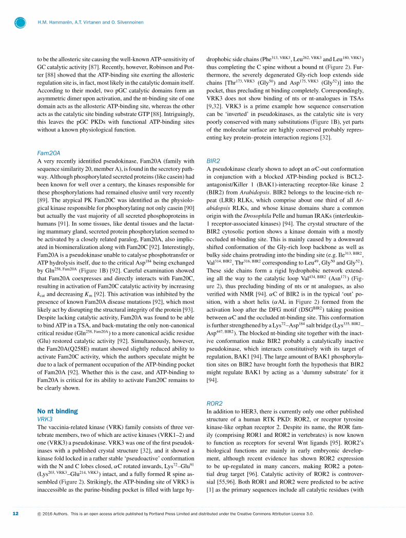
Fam20AA very recently identified pseudokinase, Fam20A (family withsequence similarity 20, member A), is found in the secretory path-way. Although phosphorylated secreted proteins (like casein) hadbeen known for well over a century, the kinases responsible forthese phosphorylations had remained elusive until very recently[89]. The atypical PK Fam20C was identified as the physiolo-gical kinase responsible for phosphorylating not only casein [90]but actually the vast majority of all secreted phosphoproteins inhumans [91]. In some tissues, like dental tissues and the lactat-ing mammary gland, secreted protein phosphorylation seemed tobe activated by a closely related paralog, Fam20A, also implic-ated in biomineralization along with Fam20C [92]. Interestingly,Fam20A is a pseudokinase unable to catalyse phosphotransfer orATP hydrolysis itself, due to the critical Asp184 being exchangedby Gln258, Fam20A (Figure 1B) [92]. Careful examination showedthat Fam20A coexpresses and directly interacts with Fam20C,resulting in activation of Fam20C catalytic activity by increasingkcat and decreasing Km [92]. This activation was inhibited by thepresence of known Fam20A disease mutations [92], which mostlikely act by disrupting the structural integrity of the protein [93].Despite lacking catalytic activity, Fam20A was found to be ableto bind ATP in a TSA, and back-mutating the only non-canonicalcritical residue (Gln258, Fam20A) to a more canonical acidic residue(Glu) restored catalytic activity [92]. Simultaneously, however,the Fam20A(Q258E) mutant showed slightly reduced ability toactivate Fam20C activity, which the authors speculate might bedue to a lack of permanent occupation of the ATP-binding pocketof Fam20A [92]. Whether this is the case, and ATP-binding toFam20A is critical for its ability to activate Fam20C remains tobe clearly shown.
No nt bindingVRK3The vaccinia-related kinase (VRK) family consists of three ver-tebrate members, two of which are active kinases (VRK1–2) andone (VRK3) a pseudokinase. VRK3 was one of the first pseudok-inases with a published crystal structure [32], and it showed akinase fold locked in a rather stable ‘pseudoactive’ conformationwith the N and C lobes closed, αC rotated inwards, Lys72–Glu91
(Lys203, VRK3–Glu214, VRK3) intact, and a fully formed R spine as-sembled (Figure 2). Strikingly, the ATP-binding site of VRK3 isinaccessible as the purine-binding pocket is filled with large hy-
drophobic side chains (Phe313, VRK3, Leu262, VRK3 and Leu180, VRK3)thus completing the C spine without a bound nt (Figure 2). Fur-thermore, the severely degenerated Gly-rich loop extends sidechains [Thr173, VRK3 (Gly50) and Asp175, VRK3 (Gly52)] into thepocket, thus precluding nt binding completely. Correspondingly,VRK3 does not show binding of nts or nt-analogues in TSAs[9,32]. VRK3 is a prime example how sequence conservationcan be ‘inverted’ in pseudokinases, as the catalytic site is verypoorly conserved with many substitutions (Figure 1B), yet partsof the molecular surface are highly conserved probably repres-enting key protein–protein interaction regions [32].
BIR2A pseudokinase clearly shown to adopt an αC-out conformationin conjunction with a blocked ATP-binding pocked is BCL2-antagonist/Killer 1 (BAK1)-interacting receptor-like kinase 2(BIR2) from Arabidopsis. BIR2 belongs to the leucine-rich re-peat (LRR) RLKs, which comprise about one third of all Ar-abidopsis RLKs, and whose kinase domains share a commonorigin with the Drosophila Pelle and human IRAKs (interleukin-1 receptor-associated kinases) [94]. The crystal structure of theBIR2 cytosolic portion shows a kinase domain with a mostlyoccluded nt-binding site. This is mainly caused by a downwardshifted conformation of the Gly-rich loop backbone as well asbulky side chains protruding into the binding site (e.g. Ile313, BIR2,Val314, BIR2, Thr316, BIR2 corresponding to Leu49, Gly50 and Gly52).These side chains form a rigid hydrophobic network extend-ing all the way to the catalytic loop Val434, BIR2 (Asn171) (Fig-ure 2), thus precluding binding of nts or nt analogues, as alsoverified with NMR [94]. αC of BIR2 is in the typical ‘out’ po-sition, with a short helix (αAL in Figure 2) formed from theactivation loop after the DFG motif (DSGBIR2) taking positionbetween αC and the occluded nt-binding site. This conformationis further strengthened by a Lys72–Asp184 salt bridge (Lys335, BIR2–Asp447, BIR2). The blocked nt-binding site together with the inact-ive conformation make BIR2 probably a catalytically inactivepseudokinase, which interacts constitutively with its target ofregulation, BAK1 [94]. The large amount of BAK1 phosphoryla-tion sites on BIR2 have brought forth the hypothesis that BIR2might regulate BAK1 by acting as a ‘dummy substrate’ for it[94].
ROR2In addition to HER3, there is currently only one other publishedstructure of a human RTK PKD: ROR2, or receptor tyrosinekinase-like orphan receptor 2. Despite its name, the ROR fam-ily (comprising ROR1 and ROR2 in vertebrates) is now knownto function as receptors for several Wnt ligands [95]. ROR2’sbiological functions are mainly in early embryonic develop-ment, although recent evidence has shown ROR2 expressionto be up-regulated in many cancers, making ROR2 a poten-tial drug target [96]. Catalytic activity of ROR2 is controver-sial [55,96]. Both ROR1 and ROR2 were predicted to be active[1] as the primary sequences include all catalytic residues (with
. . . . . . . . . . . . . . . . . . . . . . . . . . . . . . . . . . . . . . . . . . . . . . . . . . . . . . . . . . . . . . . . . . . . . . . . . . . . . . . . . . . . . . . . . . . . . . . . . . . . . . . . . . . . . . . . . . . . . . . . . . . . . . . . . . . . . . . . . . . . . . . . . . . . . . . . . . . . . . . . . . . . . . . . . . . . . . . . . . . . . . . . . . . . . . . . . . . . . . . . . . . . . . . . . . . . . . . . . . . . . . . . . . . . . . . . . . . . . . . . . . . . . . . . . . . . . . . . . . . . . . . . . . . . . . . . . . . . . . . . . . . . . . . . . . . . . . . . . . . . . . . . . . . . . . . . . . . . . . . . . . . . . . . . . . . . . . . . . . . . . . . . . . . . . . . . . . . . . . . . . .
12 c© 2016 Authors. This is an open access article published by Portland Press Limited and distributed under the Creative Commons Attribution Licence 3.0.
Nucleotide binding in pseudokinases
the exception of Gly52, Figure 1B), and even two Tyr residuesin the activation loop. Also, numerous groups have reported(auto)phosphorylation of ROR2 from immunoprecipitated ex-tracts [97–101]. However, whether the apparent catalytic activitystems from interacting kinases or ROR2 itself is in most cases un-clear [96]. Efforts using purified recombinant ROR1 and ROR2cytoplasmic portions, including the kinase domains, have thusfar been unable to detect catalytic activity [102] or nt binding[55].
The two published crystal structures of the ROR2 kinase do-main (Table 1) show the domain in an inactive, DFG-out, con-formation which resembles the inactive conformation of IRK (in-sulin receptor kinase) [103], where the activation loop bends intothe nt-binding pocket occupying the position of the nt phosphates(Figure 2) [104]. However, the inactive ROR2 conformation dif-fers from the canonical IRK-like conformation in three ways.Firstly, the adenine-binding pocket is occluded by Tyr555, ROR2
from the hinge region, which adopts an uncommon conform-ation (Figure 2), possibly making up for the unusually smallLeu634, ROR2 (Phe185) from the DLG (DFG) motif in blockingthe adenine pocket [55,104]. Secondly, Leu636, ROR2 blocks thephosphate-binding pocket causing Asp633, ROR2 (Asp184) to movecloser to αC and thus (thirdly) enabling a direct salt bridge linkto an Arg residue in αC (Arg528, ROR2), thus presumably furtherstabilizing the inactive conformation [104]. Although the DFG-αC link is not observed in all solved ROR2 structures, the firsttwo are most likely sufficient to block out nts and explain the lackof binding [55]. Interestingly, however, the prominent role of theunusual conformation of Tyr555, ROR2 in occluding the nt-bindingpocket has been suggested to potentially enable activation ofROR2 via phosphorylation of this residue [55,104]. This hypo-thesis remains yet to be tested, however.
Variable binding modes and abilities, or nt bindingundefinedTRIBsThere are four mammalian pseudokinases related to the Droso-phila Tribbles: TRIB1–3 and SgK495 [24]. All TRIBs are linkedto various forms of cancer [11], and they represent an interestinggroup of pseudokinases with a myriad of implicated biologicalfunctions [24,105,106], and possible therapeutic potential [13].TRIBs 1–3 consist of variable N-terminal extensions (NTEs),a highly conserved PKD and a C-terminal extension includinga conserved region for binding the E3 ligase constitutive pho-tomorphogenesis protein 1 (COP1) [105]. This COP1-bindingdomain seems critical for the physiological function of TRIBs,and the current model is that TRIBs act as scaffolds recruit-ing target proteins using their PKD, and linking them with COP1for ubiquitination [105,107]. TRIB pseudokinases have degradedacidic motifs in place of the Gly-rich loop [24], and highly non-canonical deviations in the DFG motif (e.g. SLETRIB1 and TRIB2,Figure 1B). The drastic structural consequences of these changeswere revealed recently by Murphy et al. [105], who managedto solve the crystal structure of human TRIB1 thus generatingthe first structure of a Tribbles protein. The structure shows a
kinase-like fold with a traditional C lobe, but a highly altered Nlobe. The degraded Gly-rich loop is short and contains a bend-inducing proline (Pro98, TRIB1) causing the whole loop (normallyforming the top of the nt-binding pocket) to be in a retracted posi-tion. TRIB1 also exhibits a shortened αC along with a practicallynon-existent β3-αC loop, which locks αC into a distorted, bentconformation (Figure 2) [105]. Additionally, the noncanonicalDFG motif (SLETRIB1, Figure 1B) is in a conformation resem-bling the ‘DFG-out’ state [28] (Figure 2). This unusual configura-tion seems to be stabilized by multiple salt bridges, including theβ3 lysine (Lys120, TRIB1) connecting to the Leu226, TRIB1 backbone(there is no Glu91 homologue in αC) and Lys210, TRIB1 (Asn171)connecting to Glu224, TRIB1, which precedes the SLE motif and isunique among human kinases [105]. Lys210, TRIB1 is itself stabil-ized by the catalytic aspartate Asp205, TRIB1 (Asp166). All of thesefactors combine to form a wide, yet obstructed, ATP-bindingpocket incompatible with nt binding – a conclusion backed up byTSA measurements showing no observable nt binding for TRIB1[105].
Despite sharing over >70 % amino acid sequence identitywith TRIB1, TRIB2 has, in contrast, been shown to exhibit sig-nificant Tm shifts in intrinsic fluorescence and TSA at 1 mMATP in the absence of cations [24], and slight Tm shifts at200 μM ATP [9], indicative of relatively weak ATP binding[24]. Furthermore, TRIB2 and TRIB3 have both been reported toshow low cation-independent autophosphorylation activity, butthe biological significance of this is unclear [24]. Murphy etal. [105] speculate that this difference could be due to a crit-ical alteration (Tyr134, TRIB1 is Cys104, TRIB2) in the TRIB2 C helixthat could enable the SLETRIB1 and TRIB2 motif to acquire a moreDFG-in-like state, thus enabling nt binding. Interestingly, muta-genesis experiments in cells have shown that an intact ATP-binding pocket is needed for the transforming ability of TRB2[107].
ADCK3Most of PK research has focused on eukaryotic protein kinases(ePKs), and approximately half of the 20 PKL families are yet tobe structurally explored [14]. One of those families, the evolu-tionarily ancient UbiB family, recently had its first protein struc-ture solved, with the crystal structure of human aarF domain-containing protein kinase (ADCK)3 [108], a mitochondrial pro-tein involved in isoprenoid lipid metabolism. The human ADCKfamily (comprising ADCK1–5) had been classified as an ‘atyp-ical kinase’ [1], but experimental validation of enzyme activityremained elusive [108]. The structure shows a conserved core PKfold with a C lobe insert and two additions to the N lobe fromconserved UbiB-specific features: an NTE before β1 (helicesGQα1–4), and an insert between β3 and αC (GQα5–6, Figure 2).UbiB proteins also share a striking alteration in the Gly-rich loop,which is replaced by an Ala-rich loop. The NTE and the N-lobeinsertion both form α helices that fold on to the Ala-rich loop andpeptide substrate-binding cleft. Specifically, a conserved KxGQmotif from the NTE and a conserved ExD motif (Glu405, ADCK3)
. . . . . . . . . . . . . . . . . . . . . . . . . . . . . . . . . . . . . . . . . . . . . . . . . . . . . . . . . . . . . . . . . . . . . . . . . . . . . . . . . . . . . . . . . . . . . . . . . . . . . . . . . . . . . . . . . . . . . . . . . . . . . . . . . . . . . . . . . . . . . . . . . . . . . . . . . . . . . . . . . . . . . . . . . . . . . . . . . . . . . . . . . . . . . . . . . . . . . . . . . . . . . . . . . . . . . . . . . . . . . . . . . . . . . . . . . . . . . . . . . . . . . . . . . . . . . . . . . . . . . . . . . . . . . . . . . . . . . . . . . . . . . . . . . . . . . . . . . . . . . . . . . . . . . . . . . . . . . . . . . . . . . . . . . . . . . . . . . . . . . . . . . . . . . . . . . . . . . . . . . .
c© 2016 Authors. This is an open access article published by Portland Press Limited and distributed under the Creative Commons Attribution Licence 3.0. 13
H.M. Hammaren, A.T. Virtanen and O. Silvennoinen
from the N lobe insertion fold into the active site of the pro-tein (Figure 2) leaving the substrate-binding pocket completelyoccluded. Also, catalytic activity seems to be further prohib-ited as the catalytic Asp166 (Asp488, ADCK3) points away from theactive site towards αF, where it makes a double salt bridge toArg611, ADCK3 (Figure 2) – a residue strictly conserved as eitherarginine or lysine in the UbiB family [108]. Despite these al-terations, however, all traditional catalytic residues are present(Figure 1B), the Lys72–Glu91 (Lys358, ADCK3–Glu411, ADCK3) saltbridge is visible, and the regulatory spine is intact. Furthermore,Stefely and colleagues could show that ADCK3 was stabilizedby nts in a TSA, but with a striking preference towards ADPover ATP [108]. Careful mutagenesis studies further showed thatthis preference could be reversed by a single mutation restor-ing Gly52 (Ala339, ADCK3) to the Ala-rich loop [108]. Additionallythe A339GADCK3 mutation also enabled Mg-ATP-mediated auto-phosphorylation, which was enhanced by removal of the KxGQmotif from the NTE, clearly demonstrating that the domain hascatalytic potential, but is inhibited by its preference for ADP overATP and the inhibitory insertion into the active site. Intriguingly,these regions inhibiting kinase activity were also critical for thephysiological function of the yeast ADCK3 homologue Coq8pin vivo [108]. Whether this means that ADCK3 is truly a pseudok-inase, or whether it is a PK that is activated through interactionswith other proteins is currently unclear. Stefely and colleaguesput forward a further hypothesis, proposing that ADCK3 wouldnot be a PK after all, but rather could be able to phosphorylatesmall molecules, like lipids, which might still be able to bind tothe substrate-binding pocket despite the NTE [108].
ROPKsAn interesting group of pseudokinases outside of vertebrates isthe rhoptry kinase (ROPK) family of apicomplexans – a phylumof parasitous protists including, e.g. Toxoplasma gondii, in whichroughly one third of all kinases are ROPKs. Many ROPKs arekey virulence factors and they are exported into the parasite’shost cell during infection, where they assist in transforming thehost cell environment to a more permissive state for the para-site [109]. Roughly half of the 42 known ROPK subfamilies arepseudokinases [109], and ROPK genes are often found in expan-ded loci that are likely sites of local tandem duplication with highsequence polymorphism [110]. Furthermore, members of ROPKsubfamilies probably acquired the inactivating changes individu-ally, thus repeatedly generating new and varying pseudokinases[109]. Thus far, four ROP pseudokinase structures have beenpublished (Table 1): ROP2 [43], ROP8 [111], ROP5B [110] andROP5C (PDB: 4LV8, Reese M.L., Boothroyd J.C., unpublishedwork). All ROPKs have a common NTE subdomain, which loopsaround the N lobe and down next to the hinge towards the C lobe.This extension has been hypothesized to be a regulatory regionphosphorylated by active ROPKs, such as the key virulence factorROP18 [111]. In the case of ROP2 and ROP8, the NTE runs overthe nt-binding site inserting a large side chain (Trp215, ROP2 [43],Arg228, ROP8 [111]) into the site, thus precluding ATP binding.Accordingly, most of the residues in and around the nt-binding
site are highly variable among a subset of ROP2-like kinases(including ROP8), suggesting lack of evolutionary pressure ascan be expected for a catalytically inactive pseudokinase [43].Interestingly, however, there are still some sequence motifs inthe pseudoactive sites of ROPKs, which many pseudokinaseshave converged to. Namely, a change of the HRD motif to HGX,where X is often a basic residue (e.g. HGHROP5B and C) [110]. Sim-ilar converging substitutions are also seen in the human kinome,where 19 out of ∼50 human pseudokinases have the R-to-G sub-stitution, whereas it is seen in only 8 out of the ∼500 activehuman kinases [110].
Despite changes in the nt-binding region, the peptide-bindingsite of ROP2 and ROP8 seem rather conserved, suggesting thatthese proteins might still be capable of kinase–substrate-likeinteractions [43]. As another example of the extensive hetero-geneity among ROPKs is ROP5, which, although able to bindATP, is probably inactive [110]. Structural analysis of ROP5B re-vealed a noncanonical mode of ATP binding with two magnesiumions unusually coordinated between the ATP phosphates. Despitethe conservation of Asn171 (Asn394, ROP5B) the residue next to it(Asp393, ROP5B) actually takes its place in coordinating the ions.Furthermore, Asp184 (Asp407, ROP5B) is shifted towards αC andonly coordinates one of the bound Mg ions compared with thetwo in the canonical mode of binding. Also, a basic side chainconnecting the γ -phosphate usually provided from the catalyticloop (Lys168) seems to be substituted for by Arg245, ROP5B from theGly-rich loop in ROP5B [110]. ROP5B also shows other featuresmore commonly seen in other pseudokinases, like the lack of acorrectly oriented Glu91 analogue, and thus a possible salt bridgebetween the equivalents of Lys72 and Asp184 (as seen in the JAKJH2s, HER3 and KSR2).
TitinAn example of a protein where the kinase/pseudokinase de-bate has taken a recent turn, is the giant myofilament proteintitin (Mr up to 4.2 MDa) found in sarcomeres. Titin containsover 200 immunoglobulin- or fibronectin type-III-like domains(actual numbers depend on the isoform in question) as well asa kinase domain (titin kinase, TK) near its C-terminus. TK con-sists of a mostly canonical kinase domain with a C-terminalinsertion folding into its active site thus effectively blocking po-tential activity [112]. Early results suggested that TK could beactivated by removal of the blocking C-terminal extension andsimultaneous tyrosine phosphorylation of the activation loop, andthat TK could phosphorylate telethonin/Tcap [112]. More recentsingle-molecule experiments indicated, that TK was able to bindATP (rather weakly, Kd ∼350 μM), but only when mechanic-ally stretched, possibly due to removal of the C-terminal exten-sion from the active site [113]. Titin’s position as a mechano-sensitive, active kinase has, however, been recently questioned,as the telethonin/Tcap-phosphorylating kinase activity in insectcell preparations was shown to be likely due to a contam-inating kinase [114], as bacterially produced TK lacks activ-ity [113,114], and the telethonin/Tcap-phosphorylating kinase
. . . . . . . . . . . . . . . . . . . . . . . . . . . . . . . . . . . . . . . . . . . . . . . . . . . . . . . . . . . . . . . . . . . . . . . . . . . . . . . . . . . . . . . . . . . . . . . . . . . . . . . . . . . . . . . . . . . . . . . . . . . . . . . . . . . . . . . . . . . . . . . . . . . . . . . . . . . . . . . . . . . . . . . . . . . . . . . . . . . . . . . . . . . . . . . . . . . . . . . . . . . . . . . . . . . . . . . . . . . . . . . . . . . . . . . . . . . . . . . . . . . . . . . . . . . . . . . . . . . . . . . . . . . . . . . . . . . . . . . . . . . . . . . . . . . . . . . . . . . . . . . . . . . . . . . . . . . . . . . . . . . . . . . . . . . . . . . . . . . . . . . . . . . . . . . . . . . . . . . . . .
14 c© 2016 Authors. This is an open access article published by Portland Press Limited and distributed under the Creative Commons Attribution Licence 3.0.
Nucleotide binding in pseudokinases
activity does not strictly copurify with recombinant TK [114].Furthermore, two noncanonical substitutions in TK’s VAIK andDFG motifs (Y/FMAK and EFG, respectively in vertebrate titin,Figure 1B) have been suggested to preclude kinase activity [114].This example highlights some of the technical challenges in atyp-ical kinase and pseudokinase research, as choice of experimentalsystem and purity of preparations are critical determinants for theconclusions about potential activity.
IRAKsIRAKs are a family of serine/threonine kinases with two activekinases (IRAK1 and 4) and two, likely inactive, pseudokinases(IRAK2 and 3). IRAK2 functions in the sustained activation of theToll-like receptor (TLR) pathway [115], and has been implicatedin colorectal cancer [116] and in the unfolded protein response[117]. IRAK2 shows cation-dependent nt binding in TSA [9], yetthe molecular details of this are still unclear due to a lack of crys-tal structures. IRAK2 was shown to have autophosphorylationactivity when immunoprecipitated from activated macrophages,raising the question of atypical kinase activity despite noncanon-ical kinase motifs (Gly52, Asp166 and Asp184 missing) [115]. Thekinase activity could, however, only be observed in macrophagesalso expressing IRAK4 [115], suggesting that the activity ob-served was derived from IRAK4, as IRAK2 and IRAK4 interactin vivo [118]. This would be in line with early results reportingnegligible autophosphorylation activity for recombinant IRAK2[119].
Whereas IRAKs 1, 2 and 4 are active components of the proin-flammatory TLR and IL-1R family signalling pathways, IRAK3(also called IRAK-M) functions as a key modulator of inflam-matory responses. The expression of IRAK3 is regulated by theother IRAK members in a negative feedback loop, where IRAK3associates with the same signalling molecules as IRAK1, 2 and 4and negatively regulates the pathways [120–122]. Furthermore,IRAK3 expression is up-regulated by glucocorticoid receptor ac-tivation, and has been shown to play a key role in the immunosup-pressive action of glucocorticoids [123]. Recent evidence has alsoidentified a role for IRAK3 in proinflammatory signalling [122].The kinase domain of IRAK3 does not show nt binding in TSA[9], but it evidently has an accessible binding pocket, as it bindsa number of known kinase inhibitors [9,68]. IRAK3 completelylacks an HRD motif (CGSIRAK3, Figure 1B) and is most likelyan inactive pseudokinase [119], which exerts its function throughnon-catalytic mechanisms [121,122].
Alternative catalytic mechanismWNK, PRPK and HaspinSome kinases, originally classified as pseudokinases, have laterbeen found to possess activity via atypical mechanisms. Althoughthis activity is often low and only detectable in in vitro settings,biologically significant kinase activity has been established for anumber of PKs initially thought to be inactive. The most prom-inent example of these is WNK, which is an active kinase whereLys72 is substituted with a second lysine (Lys233, WNK1) from β2
[27,124]. Interestingly, the unusual position of Lys233, WNK1 hasbeen proposed to enable WNK1 to bind a halide ion (e.g. Cl− ) atthe back of its nt-binding pocket (Figure 2) [125]. Biochemicaldata further suggest that when this ion is present, activation ofWNK1 by autophosphorylation is inhibited, essentially makingWNK1 a chloride sensor. This could explain the apparent regula-tion by chloride ions observed for WNK1’s downstream targets:sodium-coupled chloride cotransporters (NCCs) and potassium-coupled chloride cotransporters (KCCs) [125].
Similarly, p53-related protein kinase (PRPK), a human homo-logue of yeast Bud32, is catalytically active [126] despite lack-ing the typical β3 lysine and phosphorylates its only substratep53 when pre-phosphorylated by Akt [127]. Another noteworthyatypical kinase is Haspin, which belongs to its own PKL fam-ily outside of ePKs [14]. Despite having a modified DFG motif(DYTHaspin) Haspin is an active kinase [1], contains practically allcatalytically critical residues, and binds ATP in a mostly classicalmanner [128].
CONCLUSIONS AND PERSPECTIVES
Most pseudokinases have evolved from active kinases by obtain-ing regulatory functions where catalytic function is dispensable.However, a significant proportion of pseudokinases have retainedATP-binding ability. Presently, detailed and structural informa-tion exists for only a part of these proteins (Table 1), but mountingevidence suggests a structurally and functionally important func-tion for nt binding in several pseudokinases. Indeed, noncatalyticoccupation of the nt-binding pocket is known to regulate multipleactive kinases [129–131], and as pseudokinases share the sameprotein fold, similar functional significance for ATP binding canbe expected.
As a structural cofactor, nt binding seems to regulate or enableacquisition of certain conformations in multiple pseudokinases,e.g. allowing the domain to adopt a pseudoactive conformation(like STRADα) by completing the C spine and stabilizing thedomain. Other pseudokinases, like VRK3 or BIR2 (from Ara-bidopsis), have given up the need for bound nts altogether byfilling out the nt-binding pocket constitutively with bulky aminoacid substitutions (Figure 2). Completion of the spines (R andC), which is usually predictive of catalytic activity in canonicalkinases [16,30,31], thus cannot be used as a structural predictor ofcatalytic activity in pseudokinases, but is still useful in describ-ing a conformation adopted by a pseudokinase. Furthermore,most pseudokinases lack the main mode of regulation for activeePKs, namely a phosphorylated activation loop. Indeed, activ-ation loops in many pseudokinases are very short to the pointof being practically non-existent. In such an environment, struc-tural importance of nt-binding pocket occupation in regulationor modulation of protein–protein interactions could be furtherincreased.
The binding affinity for ATP is rather low in many pseudok-inases, which poses technical challenges. However, cellular ATP
. . . . . . . . . . . . . . . . . . . . . . . . . . . . . . . . . . . . . . . . . . . . . . . . . . . . . . . . . . . . . . . . . . . . . . . . . . . . . . . . . . . . . . . . . . . . . . . . . . . . . . . . . . . . . . . . . . . . . . . . . . . . . . . . . . . . . . . . . . . . . . . . . . . . . . . . . . . . . . . . . . . . . . . . . . . . . . . . . . . . . . . . . . . . . . . . . . . . . . . . . . . . . . . . . . . . . . . . . . . . . . . . . . . . . . . . . . . . . . . . . . . . . . . . . . . . . . . . . . . . . . . . . . . . . . . . . . . . . . . . . . . . . . . . . . . . . . . . . . . . . . . . . . . . . . . . . . . . . . . . . . . . . . . . . . . . . . . . . . . . . . . . . . . . . . . . . . . . . . . . . .
c© 2016 Authors. This is an open access article published by Portland Press Limited and distributed under the Creative Commons Attribution Licence 3.0. 15
H.M. Hammaren, A.T. Virtanen and O. Silvennoinen
concentrations are in the millimolar range [132], so nt bindingshould still occur in a physiological milieu even at relativelylow affinities. Some very low affinity ATP binders (e.g. TRIB2,CASK) have even been hypothesized to function as cellular ntconcentration sensors [24].
Functional ATP-binding pockets are required for the physiolo-gical function of multiple pseudokinases, even in the absence ofcatalytic activity. The detailed structural consequences of nt oc-cupation, however, are in many cases not obvious even whenusing multiple biophysical methods (e.g. ILK [34]) or whencrystallographic structural data with and without nt are avail-able (see, e.g. JAK2 JH2 [45]). In these cases methods able todetect subtle changes in protein dynamics (e.g. NMR or compu-tational methods) could provide new insights. Nevertheless, inmost cases structure-guided, targeted, carefully controlled muta-genesis studies, complemented where possible with a chemicalgenetics approach [133], are the key to answering questions ofimportance of nt-binding pocket occupation.
Pseudokinases play an important regulatory role in cellularsignalling, and abnormal function of several human pseudok-inases has been associated with human diseases [12]. The mostprominent examples of this are probably the JAK2 JH2 V617Fmutation underlying multiple myeloproliferative neoplasms [64],and HER3 overexpression in many cancers [134]. In total, over60 mutations in the human pseudokinome have been shown tocause, or be linked to, various malignancies [11–13]. With itsdirect link to pseudokinase regulatory functions, the nt-bindingsite of many pseudokinases poses an interesting option for phar-macological interventions. For example, the E351K mutation inMLKL increases affinity for ATP and is associated with hu-man lung carcinoma [26], and mutations in the ATP-bindingpocket of JAK JH2 revert the hyperactivation of pathogenic JAKJH2 mutants [45]. To date, only a few pseudokinases, namelyHER3 [135], MLKL [136] and TYK2 [67], have been attemp-ted to target pharmacologically, providing important proof-of-principle results that targeting pseudokinases could be a viabletherapeutic option. The results of pharmacological targeting ofMLKL and TYK2 JH2 are significant and encouraging, but itshould be kept in mind that the functions of pseudokinases arevery diverse and not as directly predictable as for active kinases.Further detailed analysis is required to understand the physiolo-gical functions of different pseudokinases, and insights thusgained will lay the basis for potential future therapeutic targetingattempts.
ACKNOWLEDGEMENT
We thank J.M. Murphy for sharing the TRIB1 structure.
FUNDING
This work was supported by the Medical Research Council ofAcademy of Finland, Sigrid Juselius Foundation, Finnish CancerFoundation, Competitive Research Funding of Tampere UniversityHospital and Tampere Tuberculosis Foundation.
REFERENCES
1 Manning, G., Whyte, D.B., Martinez, R., Hunter, T. andSudarsanam, S. (2002) The protein kinase complement of thehuman genome. Science 298, 1912–1934 CrossRef PubMed
2 Hanks, S.K., Quinn, A.M. and Hunter, T. (1998) The proteinkinase family: conserved features and deduced phylogeny of thecatalytic domains. Science 241, 42–52 CrossRef
3 Boudeau, J., Miranda-Saavedra, D., Barton, G.J. and Alessi, D.R.(2006) Emerging roles of pseudokinases. Trends Cell Biol 16,443–452 CrossRef PubMed
4 Brennan, D.F., Dar, A.C., Hertz, N.T., Chao, W.C., Burlingame,A.L., Shokat, K.M. and Barford, D. (2011) A Raf-induced allosterictransition of KSR stimulates phosphorylation of MEK. Nature472, 366–369 CrossRef PubMed
5 Shi, F., Telesco, S.E., Liu, Y., Radhakrishnan, R. and Lemmon,M.A. (2010) ErbB3/HER3 intracellular domain is competent tobind ATP and catalyze autophosphorylation. Proc. Natl. Acad. Sci.U.S.A. 107, 7692–7697 CrossRef PubMed
6 Ungureanu, D., Wu, J., Pekkala, T., Niranjan, Y., Young, C.,Jensen, O.N., Xu, C.F., Neubert, T.A., Skoda, R.C., Hubbard, S.R.and Silvennoinen, O. (2011) The pseudokinase domain of JAK2 isa dual-specificity protein kinase that negatively regulates cytokinesignaling. Nat. Struct. Mol. Biol. 18, 971–976 CrossRef PubMed
7 Mukherjee, K., Sharma, M., Urlaub, H., Bourenkov, G.P., Jahn, R.,Sudhof, T.C. and Wahl, M.C. (2008) CASK Functions as aMg2 + -independent neurexin kinase. Cell 133, 328–339CrossRef PubMed
8 Eyers, P.A. and Murphy, J.M. (2013) Dawn of the dead: proteinpseudokinases signal new adventures in cell biology. Biochem.Soc. Trans. 41, 969–974 CrossRef PubMed
9 Murphy, J.M., Zhang, Q., Young, S.N., Reese, M.L., Bailey, F.P.,Eyers, P.A., Ungureanu, D., Hammaren, H., Silvennoinen, O.,Varghese, L.N. et al. (2014) A robust methodology to subclassifypseudokinases based on their nucleotide-binding properties.Biochem. J. 457, 323–334 CrossRef PubMed
10 Zeqiraj, E. and van Aalten, D.M. (2010) Pseudokinases-remnantsof evolution or key allosteric regulators? Curr. Opin. Struct. Biol.20, 772–781 CrossRef PubMed
11 Zhang, H., Photiou, A., Grothey, A., Stebbing, J. and Giamas, G.(2012) The role of pseudokinases in cancer. Cell Signal 24,1173–1184 CrossRef PubMed
12 Reiterer, V., Eyers, P.A. and Farhan, H. (2014) Day of the dead:pseudokinases and pseudophosphatases in physiology anddisease. Trends Cell Biol 24, 489–505 CrossRef PubMed
13 Bailey, F.P., Byrne, D.P., McSkimming, D., Kannan, N. and Eyers,P.A. (2015) Going for broke: targeting the human cancerpseudokinome1. Biochem. J. 465, 195–211 CrossRef PubMed
14 Kannan, N., Taylor, S.S., Zhai, Y., Venter, J.C. and Manning, G.(2007) Structural and functional diversity of the microbialkinome. PLoS Biol 5, e17 CrossRef PubMed
15 Cheek, S., Ginalski, K., Zhang, H. and Grishin, N.V. (2005) Acomprehensive update of the sequence and structureclassification of kinases. BMC Struct. Biol. 5, 6 CrossRef PubMed
16 Taylor, S.S. and Kornev, A.P. (2011) Protein kinases: evolution ofdynamic regulatory proteins. Trends Biochem. Sci. 36, 65–77CrossRef PubMed
17 Grant, B.D., Hemmer, W., Tsigelny, I., Adams, J.A. and Taylor, S.S.(1998) Kinetic analyses of mutations in the glycine-rich loop ofcAMP-dependent protein kinase. Biochemistry 37, 7708–7715CrossRef PubMed
18 Taylor, S., Yang, J., Wu, J., Haste, N., Radzio-Andzelm, E. andAnand, G. (2004) PKA: a portrait of protein kinase dynamics.Biochim. Biophys. Acta 1697, 259–269 CrossRef PubMed
. . . . . . . . . . . . . . . . . . . . . . . . . . . . . . . . . . . . . . . . . . . . . . . . . . . . . . . . . . . . . . . . . . . . . . . . . . . . . . . . . . . . . . . . . . . . . . . . . . . . . . . . . . . . . . . . . . . . . . . . . . . . . . . . . . . . . . . . . . . . . . . . . . . . . . . . . . . . . . . . . . . . . . . . . . . . . . . . . . . . . . . . . . . . . . . . . . . . . . . . . . . . . . . . . . . . . . . . . . . . . . . . . . . . . . . . . . . . . . . . . . . . . . . . . . . . . . . . . . . . . . . . . . . . . . . . . . . . . . . . . . . . . . . . . . . . . . . . . . . . . . . . . . . . . . . . . . . . . . . . . . . . . . . . . . . . . . . . . . . . . . . . . . . . . . . . . . . . . . . . . .
16 c© 2016 Authors. This is an open access article published by Portland Press Limited and distributed under the Creative Commons Attribution Licence 3.0.
Nucleotide binding in pseudokinases
19 Carrera, A.C., Alexandrov, K. and Roberts, T.M. (1993) Theconserved lysine of the catalytic domain of protein kinases isactively involved in the phosphotransfer reaction and not requiredfor anchoring ATP. Proc. Natl. Acad. Sci. U.S.A. 90, 442–446CrossRef PubMed
20 Robinson, M.J., Harkins, P.C., Zhang, J., Baer, R., Haycock, J.W.,Cobb, M.H. and Goldsmith, E.J. (1996) Mutation of position 52 inERK2 creates a nonproductive binding mode for adenosine5′ -triphosphate. Biochemistry 35, 5641–5646 CrossRef PubMed
21 Cheng, K. and Koland, J. (1998) Nucleotide-binding properties ofkinase-deficient epidermal-growth-factor-receptor mutants.Biochem. J. 330, 353–359 CrossRef PubMed
22 Iyer, G.H., Garrod, S., Woods, V.L. and Taylor, S.S. (2005)Catalytic independent functions of a protein kinase as revealedby a kinase-dead mutant: study of the Lys72His mutant ofcAMP-dependent kinase. J. Mol. Biol. 351, 1110–1122CrossRef PubMed
23 Jaleel, M., Saha, S., Shenoy, A.R. and Visweswariah, S.S. (2006)The kinase homology domain of receptor guanylyl cyclase C: ATPbinding and identification of an adenine nucleotide sensitive site.Biochemistry 45, 1888–1898 CrossRef PubMed
24 Bailey, F.P., Byrne, D.P., Oruganty, K., Eyers, C.E., Novotny, C.,Shokat, K.M., Kannan, N. and Eyers, P.A. (2015) The Tribbles 2(TRB2) pseudokinase binds to ATP and autophosphorylates in ametal-independent manner. Biochem. J. 467, 47–62CrossRef PubMed
25 Murphy, J.M., Czabotar, P.E., Hildebrand, J.M., Lucet, I.S., Zhang,J., Alvarez-Diaz, S., Lewis, R., Lalaoui, N., Metcalf, D., Webb, A.I.et al. (2013) The pseudokinase MLKL mediates necroptosis via amolecular switch mechanism. Immunity 39, 443–453CrossRef PubMed
26 Murphy, J.M., Lucet, I.S., Hildebrand, J.M., Tanzer, M.C., Young,S.N., Sharma, P., Lessene, G., Alexander, W.S., Babon, J.J., Silke,J. et al. (2014) Insights into the evolution of divergentnucleotide-binding mechanisms among pseudokinases revealedby crystal structures of human and mouse MLKL. Biochem. J.457, 369–377 CrossRef PubMed
27 Min, X., Lee, B., Cobb, M.H. and Goldsmith, E.J. (2004) Crystalstructure of the kinase domain of WNK1, a kinase that causes ahereditary form of hypertension. Structure 12, 1303–1311CrossRef PubMed
28 Mobitz, H. (2015) The ABC of protein kinase conformations.Biochim. Biophys. Acta 1854, 1555–1566 CrossRef PubMed
29 Valiev, M., Kawai, R., Adams, J.A. and Weare, J.H. (2003) Therole of the putative catalytic base in the phosphoryl transferreaction in a protein kinase: first-principles calculations. J. Am.Chem. Soc. 125, 9926–9927 CrossRef PubMed
30 Kornev, A.P., Haste, N.M., Taylor, S.S. and Ten Eyck, L.F. (2006)Surface comparison of active and inactive protein kinasesidentifies a conserved activation mechanism. Proc. Natl. Acad.Sci. U.S.A. 103, 17783–17788 CrossRef PubMed
31 Kornev, A.P., Taylor, S.S. and Ten Eyck, L.F. (2008) A helix scaffoldfor the assembly of active protein kinases. Proc. Natl. Acad. Sci.U.S.A. 105, 14377–14382 CrossRef PubMed
32 Scheeff, E.D., Eswaran, J., Bunkoczi, G., Knapp, S. and Manning,G. (2009) Structure of the pseudokinase VRK3 reveals adegraded catalytic site, a highly conserved kinase fold, and aputative regulatory binding site. Structure 17, 128–138CrossRef PubMed
33 Lucet, I.S., Babon, J.J. and Murphy, J.M. (2013) Techniques toexamine nucleotide binding by pseudokinases. Biochem. Soc.Trans. 41, 975–980 CrossRef PubMed
34 Fukuda, K., Knight, J.D., Piszczek, G., Kothary, R. and Qin, J.(2011) Biochemical, proteomic, structural, and thermodynamiccharacterizations of integrin-linked kinase (ILK): cross-validationof the pseudokinase. J. Biol. Chem. 286, 21886–21895CrossRef PubMed
35 Suijkerbuijk, S.J., van Dam, T.J., Karagoz, G.E., von Castelmur, E.,Hubner, N.C., Duarte, A., Vleugel, M., Perrakis, A., Rudiger,S.G.D., Snel, B. et al. (2012) The vertebrate mitotic checkpointprotein BUBR1 is an unusual pseudokinase. Dev. Cell 22,1321–1329 CrossRef PubMed
36 Jura, N., Shan, Y., Cao, X., Shaw, D.E. and Kuriyan, J. (2009)Structural analysis of the catalytically inactive kinase domain ofthe human EGF receptor 3. Proc. Natl. Acad. Sci. U.S.A. 106,21608–21613 CrossRef PubMed
37 Min, X., Ungureanu, D., Maxwell, S., Hammaren, H., Thibault, S.,Hillert, E.K., Ayres, M., Greenfield, B., Eksterowicz, J., Gabel, C.et al. (2015) Structural and functional characterization of the JH2pseudokinase domain of JAK family tyrosine kinase 2 (TYK2). J.Biol. Chem. 290, 27261–27270 CrossRef PubMed
38 Bandaranayake, R.M., Ungureanu, D., Shan, Y., Shaw, D.E.,Silvennoinen, O. and Hubbard, S.R. (2012) Crystal structures ofthe JAK2 pseudokinase domain and the pathogenic mutantV617F. Nat. Struct. Mol. Biol. 19, 754–759CrossRef PubMed
39 Zeqiraj, E., Filippi, B.M., Goldie, S., Navratilova, I., Boudeau, J.,Deak, M., Alessi, D.R. and van Aalten, D.M.F. (2009) ATP andMO25α regulate the conformational state of the STRADα
pseudokinase and activation of the LKB1 tumour suppressor.PLoS Biol 7, e1000126 CrossRef PubMed
40 Fukuda, K., Gupta, S., Chen, K., Wu, C. and Qin, J. (2009) Thepseudoactive site of ILK is essential for its binding toalpha-Parvin and localization to focal adhesions. Mol. Cell 36,819–830 CrossRef PubMed
41 Arrowsmith, C.H. and Yu-Sung, W. (1998) NMR of large (s>25 kDa) proteins and protein complexes. Prog. Nucl. Magn.Reson. Spectrosc. 32, 277–286 CrossRef
42 Snyder, D.A., Chen, Y., Denissova, N.G., Acton, T., Aramini, J.M.,Ciano, M., Karlin, R., Liu, J., Manor, P., Rajan, P.A. et al. (2005)Comparisons of NMR spectral quality and success incrystallization demonstrate that NMR and X-ray crystallographyare complementary methods for small protein structuredetermination. J. Am. Chem. Soc. 127, 16505–16511CrossRef PubMed
43 Labesse, G., Gelin, M., Bessin, Y., Lebrun, M., Papoin, J.,Cerdan, R., Arold, S.T. and Dubremetz, J.-F. (2009) ROP2 fromToxoplasma gondii: a virulence factor with a protein-kinase foldand no enzymatic activity. Structure 17, 139–146CrossRef PubMed
44 de Mol, N.J. (2012) Surface plasmon resonance for proteomics.Chemical Genomics and Proteomics (Zanders, E.D., ed.),Methods in Molecular Biology, 800, 33–53, Humana Press, NewYork City CrossRef
45 Hammaren, H.M., Ungureanu, D., Grisouard, J., Skoda, R.C.,Hubbard, S.R. and Silvennoinen, O. (2015) ATP binding to thepseudokinase domain of JAK2 is critical for pathogenicactivation. Proc. Natl. Acad. Sci. U.S.A. 112, 4642–4647CrossRef PubMed
46 Zeqiraj, E., Filippi, B.M., Deak, M., Alessi, D.R. and van Aalten,D.M. (2009) Structure of the LKB1–STRAD–MO25 complexreveals an allosteric mechanism of kinase activation. Sci. Signal.326, 1707
47 Gee, C.L., Papavinasasundaram, K.G., Blair, S.R., Baer, C.E.,Falick, A.M., King, D.S., Griffin, J.E., Venghatakrishnan, H.,Zukauskas, A., Wei, J.-R. et al. (2012) A phosphorylatedpseudokinase complex controls cell wall synthesis inmycobacteria. Sci. Signal. 5, ra7 CrossRef PubMed
48 Matulis, D., Kranz, J.K., Salemme, F.R. and Todd, M.J. (2005)Thermodynamic stability of carbonic anhydrase: measurementsof binding affinity and stoichiometry using ThermoFluor.Biochemistry 44, 5258–5266 CrossRef PubMed
49 Garbett, N.C. and Chaires, J.B. (2012) Thermodynamic studiesfor drug design and screening. Expert Opin. Drug. Discov. 7,299–314 CrossRef PubMed
. . . . . . . . . . . . . . . . . . . . . . . . . . . . . . . . . . . . . . . . . . . . . . . . . . . . . . . . . . . . . . . . . . . . . . . . . . . . . . . . . . . . . . . . . . . . . . . . . . . . . . . . . . . . . . . . . . . . . . . . . . . . . . . . . . . . . . . . . . . . . . . . . . . . . . . . . . . . . . . . . . . . . . . . . . . . . . . . . . . . . . . . . . . . . . . . . . . . . . . . . . . . . . . . . . . . . . . . . . . . . . . . . . . . . . . . . . . . . . . . . . . . . . . . . . . . . . . . . . . . . . . . . . . . . . . . . . . . . . . . . . . . . . . . . . . . . . . . . . . . . . . . . . . . . . . . . . . . . . . . . . . . . . . . . . . . . . . . . . . . . . . . . . . . . . . . . . . . . . . . . .
c© 2016 Authors. This is an open access article published by Portland Press Limited and distributed under the Creative Commons Attribution Licence 3.0. 17
H.M. Hammaren, A.T. Virtanen and O. Silvennoinen
50 Becher, I., Savitski, M.M., Savitski, M.F., Hopf, C., Bantscheff, M.and Drewes, G. (2013) Affinity profiling of the cellular kinome forthe nucleotide cofactors ATP, ADP, and GTP. ACS Chem. Biol. 8,599–607 CrossRef PubMed
51 Taylor, S.S., Zhang, P., Steichen, J.M., Keshwani, M.M. andKornev, A.P. (2013) PKA: lessons learned after twenty years.Biochim. Biophys. Acta 1834, 1271–1278 CrossRef PubMed
52 Dong, B. and Silverman, R.H. (1999) Alternative function of aprotein kinase homology domain in 2′,5′ -oligoadenylatedependent RNase L. Nucleic Acids Res 27, 439–445CrossRef PubMed
53 Huang, H., Zeqiraj, E., Dong, B., Jha, B.K., Duffy, N.M., Orlicky,S., Thevakumaran, N., Talukdar, M., Pillon, M.C., Ceccarelli, D.F.et al. (2014) Dimeric structure of pseudokinase RNase L boundto 2-5A reveals a basis for interferon-induced antiviral activity.Mol. Cell 53, 221–234 CrossRef PubMed
54 Han, Y., Donovan, J., Rath, S., Whitney, G., Chitrakar, A. andKorennykh, A. (2014) Structure of human RNase L reveals thebasis for regulated RNA decay in the IFN response. Science 343,1244–1248 CrossRef PubMed
55 Mendrola, J.M., Shi, F., Park, J.H. and Lemmon, M.A. (2013)Receptor tyrosine kinases with intracellular pseudokinasedomains. Biochem. Soc. Trans. 41, 1029–1036CrossRef PubMed
56 Mujoo, K., Choi, B.K., Huang, Z., Zhang, N. and An, Z. (2014)Regulation of ERBB3/HER3 signaling in cancer. Oncotarget 5,10222–10236 CrossRef PubMed
57 Littlefield, P., Moasser, M.M. and Jura, N. (2014) AnATP-competitive inhibitor modulates the allosteric function of theHER3 pseudokinase. Chem. Biol. 21, 453–458CrossRef PubMed
58 Chen, M., Cheng, A., Candotti, F., Zhou, Y.J., Hymel, A., Fasth, A.,Notarangelo, L.D. and O’Shea, J.J. (2000) Complex effects ofnaturally occurring mutations in the JAK3 pseudokinase domain:evidence for interactions between the kinase and pseudokinasedomains. Mol. Cell Biol. 20, 947–956 CrossRef PubMed
59 Saharinen, P., Takaluoma, K. and Silvennoinen, O. (2000)Regulation of the Jak2 tyrosine kinase by its pseudokinasedomain. Mol. Cell Biol. 20, 3387–3395 CrossRef PubMed
60 Yeh, T.C., Dondi, E., Uze, G. and Pellegrini, S. (2000) A dual rolefor the kinase-like domain of the tyrosine kinase Tyk2 ininterferon-α signaling. Proc. Natl. Acad. Sci. U.S.A. 97,8991–8996 CrossRef PubMed
61 Saharinen, P. and Silvennoinen, O. (2002) The pseudokinasedomain is required for suppression of basal activity of Jak2 andJak3 tyrosine kinases and for cytokine-inducible activation ofsignal transduction. J. Biol. Chem. 277, 47954–47963CrossRef PubMed
62 Saharinen, P., Vihinen, M. and Silvennoinen, O. (2003)Autoinhibition of Jak2 tyrosine kinase is dependent on specificregions in its pseudokinase domain. Mol. Biol. Cell 14,1448–1459 CrossRef PubMed
63 Yamaoka, K., Saharinen, P., Pesu, M., Holt, 3rd V., Silvennoinen,O. and O’Shea, J.J. (2004) The Janus kinases (Jaks). Genome.Biol. 5, 253 CrossRef PubMed
64 Vainchenker, W. and Constantinescu, S. (2013) JAK/STATsignaling in hematological malignancies. Oncogene 32,2601–2613 CrossRef PubMed
65 Toms, A.V., Deshpande, A., McNally, R., Jeong, Y., Rogers, J.M.,Kim, C.U., Gruner, S.M., Ficarro, S.B., Marto, J.A., Sattler, M.et al. (2013) Structure of a pseudokinase-domain switch thatcontrols oncogenic activation of Jak kinases. Nat. Struct. Mol.Biol. 20, 1221–1223 CrossRef PubMed
66 Knight, J.D., Qian, B., Baker, D. and Kothary, R. (2007)Conservation, variability and the modeling of active proteinkinases. PLoS One 2, e982 CrossRef PubMed
67 Tokarski, J.S., Zupa-Fernandez, A., Tredup, J.A., Pike, K., Chang,C., Xie, D., Cheng, L., Pedicord, D., Muckelbauer, J., Johnson,S.R. et al. (2015) Tyrosine kinase 2-mediated signal transductionin T lymphocytes is blocked by pharmacological stabilization of itspseudokinase domain. J. Biol. Chem. 290, 11061–11074CrossRef PubMed
68 Davis, M.I., Hunt, J.P., Herrgard, S., Ciceri, P., Wodicka, L.M.,Pallares, G., Hocker, M., Treiber, D.K. and Zarrinkar, P. (2011)Comprehensive analysis of kinase inhibitor selectivity. Nat.Biotechnol. 29, 1046–1051 CrossRef PubMed
69 Lupardus, P.J., Ultsch, M., Wallweber, H., Bir Kohli, P., Johnson,A.R. and Eigenbrot, C. (2014) Structure of thepseudokinase–kinase domains from protein kinase TYK2 revealsa mechanism for Janus kinase (JAK) autoinhibition. Proc. Natl.Acad. Sci. U.S.A. 111, 8025–8030 CrossRef PubMed
70 Shan, Y., Gnanasambandan, K., Ungureanu, D., Kim, E.T.,Hammaren, H., Yamashita, K., Silvennoinen, O., Shaw, D.E. andHubbard, S.R. (2014) Molecular basis forpseudokinase-dependent autoinhibition of JAK2 tyrosine kinase.Nat. Struct. Mol. Biol. 21, 579–584 CrossRef PubMed
71 Zhang, H., Koo, C.Y., Stebbing, J. and Giamas, G. (2013) Thedual function of KSR1: a pseudokinase and beyond. Biochem.Soc. Trans. 41, 1078–1082 CrossRef PubMed
72 Pearce, L.R., Atanassova, N., Banton, M.C., Bottomley, B., vander Klaauw, A.A., Revelli, J.-P., Hendricks, A., Keogh, J.M.,Henning, E., Doree, D. et al. (2013) KSR2 mutations areassociated with obesity, insulin resistance, and impaired cellularfuel oxidation. Cell 155, 765–777 CrossRef PubMed
73 Lange, A., Wickstrom, S.A., Jakobson, M., Zent, R., Sainio, K.and Fassler, R. (2009) Integrin-linked kinase is an adaptor withessential functions during mouse development. Nature 461,1002–1006 CrossRef PubMed
74 Hannigan, G., McDonald, P., Walsh, M. and Dedhar, S. (2011)Integrin-linked kinase: not so ’pseudo’ after all. Oncogene 30,4375–4385 CrossRef PubMed
75 Castells, E. and Casacuberta, J.M. (2007) Signalling throughkinase-defective domains: the prevalence of atypical receptor-likekinases in plants. J. Exp. Bot. 58, 3503–3511CrossRef PubMed
76 Grutter, C., Sreeramulu, S., Sessa, G. and Rauh, D. (2013)Structural characterization of the RLCK family member BSK8: apseudokinase with an unprecedented architecture. J. Mol. Biol.425, 4455–4467 CrossRef PubMed
77 Christie, M., Boland, A., Huntzinger, E., Weichenrieder, O. andIzaurralde, E. (2013) Structure of the PAN3 pseudokinase revealsthe basis for interactions with the PAN2 deadenylase and theGW182 proteins. Mol. Cell 51, 360–373CrossRef PubMed
78 Wolf, J., Valkov, E., Allen, M.D., Meineke, B., Gordiyenko, Y.,McLaughlin, S.H., Olsen, T.M., Robinson, C.V., Bycroft, M.,Stewart, M. et al. (2014) Structural basis for Pan3 binding toPan2 and its function in mRNA recruitment and deadenylation.EMBO J 33, 1514–1526 CrossRef PubMed
79 Schafer, I.B., Rode, M., Bonneau, F., Schussler, S. and Conti, E.(2014) The structure of the Pan2–Pan3 core complex revealscross-talk between deadenylase and pseudokinase. Nat. Struct.Mol. Biol. 21, 591–598 CrossRef PubMed
80 Jonas, S., Christie, M., Peter, D., Bhandari, D., Loh, B.,Huntzinger, E., Weichenrieder, O. and Izaurralde, E. (2014) Anasymmetric PAN3 dimer recruits a single PAN2 exonuclease tomediate mRNA deadenylation and decay. Nat. Struct. Mol. Biol.21, 599–608 CrossRef PubMed
81 Martin, S. and Coller, J. (2014) PAN-orama: three convergentviews of a eukaryotic deadenylase. Nat. Struct. Mol. Biol. 21,577–578 CrossRef PubMed
. . . . . . . . . . . . . . . . . . . . . . . . . . . . . . . . . . . . . . . . . . . . . . . . . . . . . . . . . . . . . . . . . . . . . . . . . . . . . . . . . . . . . . . . . . . . . . . . . . . . . . . . . . . . . . . . . . . . . . . . . . . . . . . . . . . . . . . . . . . . . . . . . . . . . . . . . . . . . . . . . . . . . . . . . . . . . . . . . . . . . . . . . . . . . . . . . . . . . . . . . . . . . . . . . . . . . . . . . . . . . . . . . . . . . . . . . . . . . . . . . . . . . . . . . . . . . . . . . . . . . . . . . . . . . . . . . . . . . . . . . . . . . . . . . . . . . . . . . . . . . . . . . . . . . . . . . . . . . . . . . . . . . . . . . . . . . . . . . . . . . . . . . . . . . . . . . . . . . . . . . .
18 c© 2016 Authors. This is an open access article published by Portland Press Limited and distributed under the Creative Commons Attribution Licence 3.0.
Nucleotide binding in pseudokinases
82 Boudeau, J., Scott, J.W., Resta, N., Deak, M., Kieloch, A.,Komander, D., Hardie, D.G., Prescott, A.R., van Aalten, D.M.F. andAlessi, D.R. (2004) Analysis of the LKB1–STRAD–MO25 complex.J. Cell Sci. 117, 6365–6375 CrossRef PubMed
83 Mukherjee, K., Sharma, M., Jahn, R., Wahl, M.C. and Sudhof, T.C.(2010) Evolution of CASK into a Mg2 + -sensitive kinase. Sci.Signal. 3, ra33 CrossRef PubMed
84 Czabotar, P.E. and Murphy, J.M. (2015) A tale of two domains – astructural perspective of the pseudokinase, MLKL. FEBS J. 282,4268–4278
85 Xie, T., Peng, W., Yan, C., Wu, J., Gong, X. and Shi, Y. (2013)Structural insights into RIP3-mediated necroptotic signaling. CellRep 5, 70–78 CrossRef PubMed
86 Joubert, S., Jossart, C., McNicoll, N. and De Lean, A. (2005)Atrial natriuretic peptide-dependent photolabeling of a regulatoryATP-binding site on the natriuretic peptide receptor-A. FEBS J272, 5572–5583 CrossRef PubMed
87 Kurose, H., Inagami, T. and Ui, M. (1987) Participation ofadenosine 5′ -triphosphate in the activation of membrane-boundguanylate cyclase by the atrial natriuretic factor. FEBS Lett 219,375–379 CrossRef PubMed
88 Robinson, J.W. and Potter, L.R. (2012) Guanylyl cyclases A and Bare asymmetric dimers that are allosterically activated by ATPbinding to the catalytic domain. Sci. Signal. 5, ra65CrossRef PubMed
89 Sreelatha, A., Kinch, L.N. and Tagliabracci, V.S. (2015) Thesecretory pathway kinases. Biochim. Biophys. Acta 1854,1687–1693 CrossRef PubMed
90 Tagliabracci, V.S., Engel, J.L., Wen, J., Wiley, S.E., Worby, C.A.,Kinch, L.N., Xiao, J., Grishin, N.V. and Dixon, J.E. (2012) Secretedkinase phosphorylates extracellular proteins that regulatebiomineralization. Science 336, 1150–1153CrossRef PubMed
91 Tagliabracci, V.S., Wiley, S.E., Guo, X., Kinch, L.N., Durrant, E.,Wen, J., Xiao, J., Cui, J., Nguyen, K.B., Engel, J.L. et al. (2015) Asingle kinase generates the majority of the secretedphosphoproteome. Cell 161, 1619–1632CrossRef PubMed
92 Cui, J., Xiao, J., Tagliabracci, V.S., Wen, J., Rahdar, M. and Dixon,J.E. (2015) A secretory kinase complex regulates extracellularprotein phosphorylation. Elife 4, e06120CrossRef PubMed
93 Xiao, J., Tagliabracci, V.S., Wen, J., Kim, S.A. and Dixon, J.E.(2013) Crystal structure of the Golgi casein kinase. Proc. Natl.Acad. Sci. U.S.A. 110, 10574–10579CrossRef PubMed
94 Blaum, B.S., Mazzotta, S., Noldeke, E.R., Halter, T., Madlung, J.,Kemmerling, B. and Stehle, T. (2014) Structure of thepseudokinase domain of BIR2, a regulator of BAK1-mediatedimmune signaling in Arabidopsis. J. Struct. Biol. 186, 112–121CrossRef PubMed
95 Green, J.L., Kuntz, S.G. and Sternberg, P.W. (2008) Ror receptortyrosine kinases: orphans no more. Trends Cell Biol 18, 536–544CrossRef PubMed
96 Debebe, Z. and Rathmell, W.K. (2015) Ror2 as a therapeutictarget in cancer. Pharmacol. Ther. 150, 143–148CrossRef PubMed
97 Masiakowski, P. and Carroll, R.D. (1992) A novel family of cellsurface receptors with tyrosine kinase-like domain. J. Biol. Chem.267, 26181–26190 PubMed
98 Oishi, I., Takeuchi, S., Hashimoto, R., Nagabukuro, A., Ueda, T.,Liu, Z., Hatta, T., Akira, S., Matsuda, Y., Yamamura, H. et al.(1999) Spatio-temporally regulated expression of receptortyrosine kinases, mRor1, mRor2, during mouse development:implications in development and function of the nervous system.Genes Cells 4, 41–56 CrossRef PubMed
99 Liu, Y., Ross, J.F., Bodine, P.V. and Billiard, J. (2007)Homodimerization of Ror2 tyrosine kinase receptor induces14-3-3β phosphorylation and promotes osteoblast differentiationand bone formation. Mol. Endocrinol. 21, 3050–3061CrossRef PubMed
100 Akbarzadeh, S., Wheldon, L.M., Sweet, S.M., Talma, S.,Mardakheh, F.K. and Heath, J.K. (2008) The deleted inbrachydactyly B domain of ROR2 is required for receptoractivation by recruitment of Src. PLoS One 3, e1873CrossRef PubMed
101 Mikels, A., Minami, Y. and Nusse, R. (2009) Ror2 receptorrequires tyrosine kinase activity to mediate Wnt5A signaling. J.Biol. Chem. 284, 30167–30176 CrossRef PubMed
102 Bainbridge, T.W., DeAlmeida, V.I., Izrael-Tomasevic, A., Chalouni,C., Pan, B., Goldsmith, J., Schoen, A.P., Quinones, G.A., Kelly, R.,Lill, J.R. et al. (2014) Evolutionary divergence in the catalyticactivity of the CAM-1, ROR1 and ROR2 kinase domains. PLoSOne 9, e102695 CrossRef PubMed
103 Hubbard, S.R., Wei, L., Ellis, L. and Hendrickson, W.A. (1994)Crystal structure of the tyrosine kinase domain of the humaninsulin receptor. Nature 372, 746–754 CrossRef PubMed
104 Artim, S.C., Mendrola, J.M. and Lemmon, M.A. (2012) Assessingthe range of kinase autoinhibition mechanisms in the insulinreceptor family. Biochem. J. 448, 213–220 CrossRef PubMed
105 Murphy, J.M., Nakatani, Y., Jamieson, S.A., Dai, W., Lucet, I.S.and Mace, P.D. (2015) Molecular mechanism of CCAAT-enhancerbinding protein recruitment by the TRIB1 pseudokinase. Structure23, 2111–2121 CrossRef PubMed
106 Kiss-Toth, E. (2011) Tribbles: ’puzzling’ regulators of cellsignalling. Biochem. Soc. Trans. 39, 684–687 CrossRef PubMed
107 Keeshan, K., Bailis, W., Dedhia, P.H., Vega, M.E., Shestova, O.,Xu, L., Toscano, K., Uljon, S.N., Blacklow, S.C. and Pear, W.S.(2010) Transformation by Tribbles homolog 2 (Trib2) requires boththe Trib2 kinase domain and COP1 binding. Blood 116,4948–4957 CrossRef PubMed
108 Stefely, J.A., Reidenbach, A.G., Ulbrich, A., Oruganty, K., Floyd,B.J., Jochem, A., Saunders, J.M., Johnson, I.E., Minogue, C.E.,Wrobel, R.L. et al. (2015) Mitochondrial ADCK3 employs anatypical protein kinase-like fold to enable coenzyme Qbiosynthesis. Mol. Cell 57, 83–94 CrossRef PubMed
109 Talevich, E. and Kannan, N. (2013) Structural and evolutionaryadaptation of rhoptry kinases and pseudokinases, a family ofcoccidian virulence factors. BMC Evol. Biol. 13, 117CrossRef PubMed
110 Reese, M.L. and Boothroyd, J.C. (2011) A conservednon-canonical motif in the pseudoactive site of the ROP5pseudokinase domain mediates its effect on toxoplasmavirulence. J. Biol. Chem. 286, 29366–29375CrossRef PubMed
111 Qiu, W., Wernimont, A., Tang, K., Taylor, S., Lunin, V., Schapira,M., Fentress, S., Hui, R. and Sibley, L.D. (2009) Novel structuraland regulatory features of rhoptry secretory kinases inToxoplasma gondii. EMBO J. 28, 969–979CrossRef PubMed
112 Mayans, O., van der Ven, P.F.M., Wilm, M., Mues, A., Young, P.,Furst, D.O., Wilmanns, M. and Gautel, M. (1998) Structural basisfor activation of the titin kinase domain during myofibrillogenesis.Nature 395, 863–869 CrossRef PubMed
113 Puchner, E.M., Alexandrovich, A., Kho, A.L., Hensen, U., Schafer,L.V., Brandmeier, B., Grater, F., Grubmuller, H., Gaub, H.E. andGautel, M. (2008) Mechanoenzymatics of titin kinase. Proc. Natl.Acad. Sci. U.S.A. 105, 13385–13390 CrossRef PubMed
114 Bogomolovas, J., Gasch, A., Simkovic, F., Rigden, D.J., Labeit, S.and Mayans, O. (2014) Titin kinase is an inactive pseudokinasescaffold that supports MuRF1 recruitment to the sarcomericM-line. Open. Biol. 4, 140041 CrossRef PubMed
. . . . . . . . . . . . . . . . . . . . . . . . . . . . . . . . . . . . . . . . . . . . . . . . . . . . . . . . . . . . . . . . . . . . . . . . . . . . . . . . . . . . . . . . . . . . . . . . . . . . . . . . . . . . . . . . . . . . . . . . . . . . . . . . . . . . . . . . . . . . . . . . . . . . . . . . . . . . . . . . . . . . . . . . . . . . . . . . . . . . . . . . . . . . . . . . . . . . . . . . . . . . . . . . . . . . . . . . . . . . . . . . . . . . . . . . . . . . . . . . . . . . . . . . . . . . . . . . . . . . . . . . . . . . . . . . . . . . . . . . . . . . . . . . . . . . . . . . . . . . . . . . . . . . . . . . . . . . . . . . . . . . . . . . . . . . . . . . . . . . . . . . . . . . . . . . . . . . . . . . . .
c© 2016 Authors. This is an open access article published by Portland Press Limited and distributed under the Creative Commons Attribution Licence 3.0. 19
H.M. Hammaren, A.T. Virtanen and O. Silvennoinen
115 Kawagoe, T., Sato, S., Matsushita, K., Kato, H., Matsui, K.,Kumagai, Y., Saitoh, T., Kawai, T., Takeuchi, O. and Akira, S.(2008) Sequential control of Toll-like receptor-dependentresponses by IRAK1 and IRAK2. Nat. Immunol. 9, 684–691CrossRef PubMed
116 Wang, H., Flannery, S.M., Dickhofer, S., Huhn, S., George, J.,Kubarenko, A.V., Lascorz, J., Bevier, M., Willemsen, J., Pichulik, T.et al. (2014) A coding IRAK2 protein variant compromises Toll-likereceptor (TLR) signaling and is associated with colorectal cancersurvival. J. Biol. Chem. 289, 23123–23131CrossRef PubMed
117 Benosman, S., Ravanan, P., Correa, R.G., Hou, Y., Yu, M., Gulen,M.F., Li, X., Thomas, J., Cuddy, M., Matsuzawa, Y. et al. (2013)Interleukin-1 receptor-associated kinase-2 (IRAK2) is a criticalmediator of endoplasmic reticulum (ER) stress signaling. PLoSOne 8, e64256 CrossRef PubMed
118 Pauls, E., Nanda, S.K., Smith, H., Toth, R., Arthur, J.S. andCohen, P. (2013) Two phases of inflammatory mediatorproduction defined by the study of IRAK2 and IRAK1 knock-inmice. J. Immunol. 191, 2717–2730 CrossRef PubMed
119 Wesche, H., Gao, X., Li, X., Kirschning, C.J., Stark, G.R. and Cao,Z. (1999) IRAK-M is a novel member of the Pelle/interleukin-1receptor-associated kinase (IRAK) family. J. Biol. Chem. 274,19403–19410 CrossRef PubMed
120 Kim, Y., Park, J., Kwon, K.H., Hong, C.Y. and Yi, A. (2012)Interleukin-1 receptor-associated kinase 2-and protein kinaseD1-dependent regulation of IRAK-monocyte expression by CpGDNA. PLoS One 7, e43970 CrossRef PubMed
121 Kobayashi, K., Hernandez, L.D., Galan, J.E., Janeway, C.A.,Medzhitov, R. and Flavell, R.A. (2002) IRAK-M is a negativeregulator of Toll-like receptor signaling. Cell 110, 191–202CrossRef PubMed
122 Zhou, H., Yu, M., Fukuda, K., Im, J., Yao, P., Cui, W., Bulek, K.,Zepp, J., Wan, Y., Kim, T.W. et al. (2013) IRAK-M mediatesToll-like receptor/IL-1R-induced NFkappaB activation and cytokineproduction. EMBO J 32, 583–596 CrossRef PubMed
123 Miyata, M., Lee, J., Susuki-Miyata, S., Wang, W.Y., Xu, H., Kai, H.,Kobayashi, K.S., Flavell, R.A. and Li, J.-D. (2015) Glucocorticoidssuppress inflammation via the upregulation of negative regulatorIRAK-M. Nat. Commun. 6, 6062 CrossRef PubMed
124 Xu, B., English, J.M., Wilsbacher, J.L., Stippec, S., Goldsmith, E.J.and Cobb, M.H. (2000) WNK1, a novel mammalianserine/threonine protein kinase lacking the catalytic lysine insubdomain II. J. Biol. Chem. 275, 16795–16801CrossRef PubMed
125 Piala, A.T., Moon, T.M., Akella, R., He, H., Cobb, M.H. andGoldsmith, E.J. (2014) Chloride sensing by WNK1 involvesinhibition of autophosphorylation. Sci. Signal. 7, ra41CrossRef PubMed
126 Abe, Y., Matsumoto, S., Wei, S., Nezu, K., Miyoshi, A., Kito, K.,Ueda, N., Shigemoto, K., Hitsumoto, Y., Nikawa, J. et al. (2001)Cloning and characterization of a p53-related protein kinaseexpressed in interleukin-2-activated cytotoxic T-cells, epithelialtumor cell lines, and the testes. J. Biol. Chem. 276,44003–44011 CrossRef PubMed
127 Facchin, S., Ruzzene, M., Peggion, C., Sartori, G., Carignani, G.,Marin, O., Brustolon, F., Lopreiato, R. and Pinna, L.A. (2007)Phosphorylation and activation of the atypical kinase p53-relatedprotein kinase (PRPK) by Akt/PKB. Cell Mol. Life Sci. 64,2680–2689 CrossRef PubMed
128 Eswaran, J., Patnaik, D., Filippakopoulos, P., Wang, F., Stein, R.L.,Murray, J.W., JMG, Higgins and Knapp, S. (2009) Structure andfunctional characterization of the atypical human kinase Haspin.Proc. Natl. Acad. Sci. U.S.A. 106, 20198–20203CrossRef PubMed
129 Dar, A.C. and Shokat, K.M. (2011) The evolution of proteinkinase inhibitors from antagonists to agonists of cellularsignaling. Annu. Rev. Biochem. 80, 769–795CrossRef PubMed
130 Cameron, A.M. (2011) Occupational hazards: allosteric regulationof protein kinases through the nucleotide-binding pocket.Biochem. Soc. Trans. 39, 472–476 CrossRef PubMed
131 Taylor, S.S., Shaw, A., Hu, J., Meharena, H.S. and Kornev, A.(2013) Pseudokinases from a structural perspective. Biochem.Soc. Trans. 41, 981–986 CrossRef PubMed
132 Traut, T.W. (1994) Physiological concentrations of purines andpyrimidines. Mol. Cell Biochem. 140, 1–22 CrossRef PubMed
133 Lopez, M.S., Kliegman, J.I. and Shokat, K.M. (2014) The logicand design of analog-sensitive kinases and their small moleculeinhibitors. Methods Enzymol 548, 189–213 CrossRef PubMed
134 Baselga, J. and Swain, S.M. (2009) Novel anticancer targets:revisiting ERBB2 and discovering ERBB3. Nat. Rev. Cancer 9,463–475 CrossRef PubMed
135 Xie, T., Lim, S.M., Westover, K.D., Dodge, M.E., Ercan, D., Ficarro,S.B., Udayakumar, D., Gurbani, D., Tae, H.S., Riddle, S.M. et al.(2014) Pharmacological targeting of the pseudokinase Her3. Nat.Chem. Biol. 10, 1006–1012 CrossRef PubMed
136 Hildebrand, J.M., Tanzer, M.C., Lucet, I.S., Young, S.N., Spall,S.K., Sharma, P., Pierotti, C., Garnier, J.-M., Dobson, R.C.J.,Webb, A.I. et al. (2014) Activation of the pseudokinase MLKLunleashes the four-helix bundle domain to induce membranelocalization and necroptotic cell death. Proc. Natl. Acad. Sci.U.S.A. 111, 15072–15077 CrossRef PubMed
137 Larkin, M.A., Blackshields, G., Brown, N.P., Chenna, R.,McGettigan, P.A., McWilliam, H., Valentin, F., Wallace, I.M., Wilm,A., Lopez, R. et al. (2007) Clustal W and Clustal X version 2.0.Bioinformatics 23, 2947–2948 CrossRef PubMed
138 Goujon, M., McWilliam, H., Li, W., Valentin, F., Squizzato, S.,Paern, J. and Lopez, R. (2010) A new bioinformatics analysistools framework at EMBL-EBI. Nucleic Acids Res 38,W695–W699 CrossRef PubMed
139 Biswas, K.H., Shenoy, A.R., Dutta, A. and Visweswariah, S.S.(2009) The evolution of guanylyl cyclases as multidomainproteins: conserved features of kinase-cyclase domain fusions. J.Mol. Evol. 68, 587–602 CrossRef PubMed
140 Baas, A.F., Boudeau, J., Sapkota, G.P., Smit, L., Medema, R.,Morrice, N.A., Alessi, D.R. and Clevers, H.C. (2003) Activation ofthe tumour suppressor kinase LKB1 by the STE20-likepseudokinase STRAD. EMBO J 22, 3062–3072CrossRef PubMed
Received 1 September 2015/11 November 2015; accepted 20 November 2015
Accepted Manuscript online 20 November 2015, doi 10.1042/BSR20150226
. . . . . . . . . . . . . . . . . . . . . . . . . . . . . . . . . . . . . . . . . . . . . . . . . . . . . . . . . . . . . . . . . . . . . . . . . . . . . . . . . . . . . . . . . . . . . . . . . . . . . . . . . . . . . . . . . . . . . . . . . . . . . . . . . . . . . . . . . . . . . . . . . . . . . . . . . . . . . . . . . . . . . . . . . . . . . . . . . . . . . . . . . . . . . . . . . . . . . . . . . . . . . . . . . . . . . . . . . . . . . . . . . . . . . . . . . . . . . . . . . . . . . . . . . . . . . . . . . . . . . . . . . . . . . . . . . . . . . . . . . . . . . . . . . . . . . . . . . . . . . . . . . . . . . . . . . . . . . . . . . . . . . . . . . . . . . . . . . . . . . . . . . . . . . . . . . . . . . . . . . .
20 c© 2016 Authors. This is an open access article published by Portland Press Limited and distributed under the Creative Commons Attribution Licence 3.0.