Embed Size (px)
Citation preview
www.e-mja.finki.ukim.mk
Number 8. December 2019
Macedonian Journal of AnaesthesiaA Journal on Anaesthesiology, Resuscitation, Analgesia and Critical Care
Editor-in-ChiefMirjana Shosholcheva
Deputy Editors:Andrijan Kartalov
Biljana Kuzmanovska
Associate EditorMarija Jovanovski-Srceva
Editorial Board:
Marija Sholjakova (Macedonia)Nikola Jankulovski (Macedonia) Karin Khuenl-Brady (Austria)Quirino Piacevoli (Italy)Zorka Nikolova-Todorova (Macedonia) Radmilo Jankovic (Serbia) Olegs Sabelnikovs (Latvia)Jannicke Mellin-Olsen (Norway) Meral Kanbak (Turkey)Nebojsa Ladjevich (Serbia)Zoran Karadjov (Macedonia)Hristo Bozov (Bulgaria)Zuhal Aykaç (Turkey)Katarina Sakic (Hrvatska)
Jasmina Jakupovich-Smajich (BIH)Jasminka Nancheva (Macedonia)Vojislava Neskovic (Serbia)Daniela Miladinova (Macedonia)Jordan Nojkov (Macedonia)Paul Zilberman (Israel) Antigona Hasani (Kosovo)Biljana Shirgovska (Macedonia) Atanas Sivevski (Macedonia)Hülya Bilgin (Turkey)Sinisha Stojanoski (Macedonia)Berrin Günaydin - (Turkey)Rudin Domi (Albanija)
Production Editor Vanja Gievski
Publisher:Department of Anaesthesia and Reanimation, Faculty of Medicine, “Ss. Cyril and Methodius”
University, Skopje, Macedonia
Proofreading Daniela Nacevska Stojcevska
Printed by ASKOLOR
EDITORIALIS THE BIOIMPEDANCE, AFTER ULTRASOUND A NEW TOOL FOR ANESTHESIOLOGISTS? . . . . . . . . . . . . . . . . . . . . . . . . 7Sholjakova M
ORIGINAL RESEARCHPATIENT SATISFACTION - PATIENT CONTROLLED INTRAVENOUS REMIFENTANIL VERSUS INTERMITTENT EPIDURAL FOR LABOR ANALGESIA . . . . . . . . . . . . . . . . . . . . . . . . . . . . . 12Karadjova D, Shosholcheva M, Sivevski А, Ivanov Е, Kjaev I, Aleksiovska-Papestiev I
ORIGINAL RESEARCHDIABETES MELLITUS AND SMOKING AS MAIN RISK FACTORS FOR PROLONGED POSTOPERATIVE ILEUS FOLLOWING COLORECTAL SURGERY . . . . . . . . . . . . . . . . . . . . . . . . . . . . . . . . . . . . . . . 23Osmani B
ORIGINAL RESEARCHAIRTRAQ® IS THE PREFERRED DEVICE FOR DIFFICULT INTUBATION BY RESIDENTS? . . . . . . . . . . . . . . . . . . . . . . . . . . . . . . . . . . . 28Gjorchevska E, Gavrilovska-Brzanov A, Ilieva E, Mojsova-Mijovska M, Petrusheva A
CASE REPORTLIFE-THREATENING LARINGEAL KAPOSI SARCOMA IN PATIENT WITH LATE HIV DIAGNOSIS . . . . . . . . . . . . . . . . . . . . . . . . 33Stevanovic M, Duganova M, Grozdanovski K, Demiri M, Marinkovic S, Petreska B
CONTENT
Number 8. December 2019 | 7 |
EDITORIAL
IS THE BIOIMPEDANCE, AFTER ULTRASOUND A NEW TOOL FOR ANESTHESIOLOGISTS?
Sholjakova M
With aim to apostolate the unavoidable changes in our practice from my days until today, I have the pleasure to prepare this Editorial. Looking back, I think that the new era brings too many satisfactions to the anesthesiologists.
The advances in medical technology made a prompt change in everyday anesthesiologists practice. The anesthetic machines became robust, complicated, with a lot of added parts. Looking at the list of the tools used in anesthesiology you will apperceive that it is too long. The classical anesthesia machines provide continuous supply of gases and anesthetics and are equipped with many additional components as: vaporizer, respiratory maintenance circuit, flow meter, bag, valves, ventilator, monitors for respiration, vital signs and anesthetics, aspirator (mucus sucker), scavenge system, peripheral nerve stimulator TOF monitor and different devices for intubation.
The contemporary professional responsibility of anesthesiologist is in an increased trend. The bound per-operative care of the homeostasis of the patients ordered practicing new ways in anesthesiology (1). With the expectance of calm, painless and free of complications postopera-tive period, the anesthesiologists started to combine general anesthesia with different regional anesthetic techniques. They become common procedures and the need for understanding more sophisticated structure of the body, ordered to the engineers to create portable and more per-fect equipment for quick diagnoses and imaging. The difficulties in identification of peripheral nerves for their block, the high percentage of failure or partial success, dictated the use of new imaging techniques. So, in anesthesia practice there were introduced the effects of sound waves and biological electrical currents which are flowing through the soft tissue (2).
The anesthesia machine which was already overloaded seems that will accept some additional parts as ultrasound machine and bio-impedance.
The place of ultrasound in anesthesiology
The use of ultrasound in medical practice started in the earliest 70-ties in the last century. Today it is used for diagnostic procedures or therapeutic needs. The medical reports from the last 10 years assign that the ultrasound is an indispensable necessity for anesthesiologists. The most of them highlight the importance of using ultrasound for verification the anatomical structures of central vessels, peripheral nerves, epidural space, the shape and functioning of the heart or other organs (3).
ORIGINAL RESEARCH 42TREATMENT OF OSTEOARTHRITIS OF THE FIRST CARPOMETACARPAL JOINT WITH EXCISION OF THE TRAPEZIUM. . . . . . . . . . . . . . . . . . . . . . . . . . . . . . . . . . . . . . . . . . . . 42Foteva M
CASE REPORTTRACHEAL DIVERTICULUM – A POSSIBILITY OF INTRAOPERATIVE AND POSTOPERATIVE COMPLICATIONS. . . . . . 48Tusheva I, Adjami B, Nikolova S, Buklioska D, Poposki B, Malinovska-Nikolovska L
ORIGINAL RESEARCH 56BEDSIDE ECHOCARDIOGRAPHY IN A HEMODYNAMICALLY UNSTABLE CHILD . . . . . . . . . . . . . . . . . . . . . . . . . . . . . . . . . . . . . . . . . . . . . . 56Naumovski F, Toleska M, Kuzmanovska B, Kartalov A, Trposka A, Donev Lj
CASE REPORT 62MANAGEMENT OF DIABETIC KETOACIDOSIS IN INTENSIVE CARE UNIT . . . . . . . . . . . . . . . . . . . . . . . . . . . . . . . . . . . . . . 62Nedelkova I, Gjorchevska E, Asprovska K, Donev Lj, Micunovic Lj, Leshi A
| 8 |
Macedonian Journal of Anaesthesia
Number 8. December 2019 | 9 |
Otherwise, from the safety point of the patient, many of the procedures previously made by the topographic marks, today are easier performed and in a safer manner done under ultrasound guided conditions. That is why ultrasound entered on “great door” in everyday anesthetic prac-tice. The executive summary from the ASRA meeting in 2010 was that the use of ultrasound for guided regional anesthesia and pain treatment was evident and it becomes a part of evidence-based medicine (4).
Ultrasound imaging is based on the use of sound waves. For diagnostic procedures the fre-quencies used for ultrasound typically vary from 2 to 15 MHz (up to 22 MHz in modern probe) and are higher than those in the audible range. In the body, the transmission of the sound waves is made possible by fluids and soft tissues. These waves have power to penetrate different tis-sues of the body at different speed and to reflect back from the tissue interface. The amplitude of reflection and transmission are forming the shapes of internal architecture; those signals are controlled by the computer software which generates the ultrasound images (5).
The benefits of the use of ultrasound are great. In comparison to the others imaging tech-niques, ultrasound provides images in real-time. It is portable, it can be brought to the bedside, it is low-cost and provides images in a non-ionizing technique. The use of ultrasound is universal. It is used in all medical specialties.
Perfect imaging 3D/4D
Quality medical supplies have the mode to help to provide excellent patient’s care. The development of be – planar ultrasound provided more efficient localization and detection of the searched region. The probe was with two planes (2D) that were perpendicular to each other. The use of 2D ultrasound imaging was limited by the clinicians because of uncertain diagnostic accuracy. The interpretation of the images mostly was depending on the experience and knowledge of the specialists. In order to solve the problem, the need for advanced imaging technique appeared and that was necessary.
At 1997, Morrimot AK and his team in “Studies in Health Technology and Informatics” announced that they are working on the development of a novel scanning system integrated in a commercial ultrasound machine with high definition and improved resolution. The result of this was a new generation of equipment with volumetric 3D ultrasound data set that can be visualized using the standard techniques, as panoramic view of the region of interest. In 3D ultrasound many 2D planes are added to create a three dimension images; the image quality is improved from standard 2D data, providing a full understandable sophisticated imaging of the spatial anatomic relationships, in trunk, abdominal organs, extremities, head and the neck (6).
The possibilities of the 3D ultrasound are enormous and many different types of images can be formed. In one reference, the ultrasound imaging system was used, to reconstruct high-reso-lution (<50μm) three-dimensional (3D) surface images of periodontal defects in human jawbone
IS THE BIOIMPEDANCE, AFTER ULTRASOUND A NEW TOOL FOR ANESTHESIOLOGISTS?
[Mahmoud AM]. The system was able to reconstruct 3D images for the mandible’s outer surface with superior spatial resolution down to 24 μm, and to perform the whole scanning in <30s (7).
Other types of 3D ultrasound with Doppler, are able to display the blood flow, motion of tissue over time, the location of blood, the presence of specific molecules, the softness of tissue, or the anatomy of a region. It is very practical for regional anesthesia because the anatomical structures are very well recognized. The placement of needles for peripheral blocks, near to peripheral nerves, is guided by ultrasound, where the optimal dose of local anesthetic solution is injected. This approach decreases the number of unsuccessful blocks, the anesthesiologists become more self-confident and less amount of local anesthetics was used. Almost each puncture for central venous cannulation is guided by ultrasound (8). It is also used for obtaining informa-tion about flow-velocity in the regional circulation. Blood velocity can be measured in various blood vessels using ultrasound Doppler probes. It is very valuable for assessing the lungs and for evaluation of many abnormalities in the thorax. The ultrasound is also very sensitive for the detection of water retention or the development of pulmonary edema.
From the previous, it is clear that the benefits from ultrasound are enormous, but for its use skills and practice are necessary, which is obvious. That is why the training of the ultrasound skills should be incorporated within the training programs of anesthesiologists.
Bioelectrical impedance analysis (BIA)Bio-electro-magnetism – bio impedance
In everyday practice of an anesthesiologist, the management of fluid balance presents a challenge. The postoperative weight gain, with or without edema forming, is frequently found after major surgery. The question of how much fluids are sufficient during surgery is still present. The incidence of postoperative fluid overload and fluid retention is common, even that is not clinically evident. There are reports of affecting up near 40% of patients after major surgery. It was shown that “target directed fluid therapy” after colon surgery provided better outcome (1). The standard methods for discovering the total body water, the calculation of daily water balance, the preoperative needs and postoperative measurement of the weight are too complicated for everyday practice. In recent years technological improvements of the equipment for measurement of bio impedance and its more precise results encourage the clinicians to use them for more accurate researches in clinical practice, particularly for determination of the preoperative and postoperative fluid status (9).
Bioelectrical impedance analysis (BIA) is a commonly used method in human nutrition and clinical research for estimating body composition, body fat, water and muscle mass. Bioelectrical impedance analysis is described in the relation between the electrical properties of tissues and tissue structure. Bio impedance deals with the measurement of electrical conductivity of biological electrical currents. It is a safe, non-invasive, rapid, reproducible cheap method, used in various
| 10 |
Macedonian Journal of Anaesthesia
Number 8. December 2019 | 11 |
clinical settings to estimate total body water (TBW). In last time it becomes very popular due to its easy usage and portability of the equipment (10).
The method is based on several different physical mechanisms when introducing a small undetectable, alternating electrical current through the body. The current flows through the phys-iologic fluids by the movement of the ions which are depended to several effects that produced a resistance. From the other side the applied current is capable to charge the cell membranes and the others interfaces, which worked as capacitors. The electrical impedance is measuring the potential difference that results. The tissue structures have different conductive and resis-tive properties for this small current. The main component of the BIA are measures of tissue resistance (R) and reactance (Xc), which are inversely related to fluid volume and are directly related to the square of the conducting length. The use of special equations allows measurement of preoperative fluid depletion and postoperative fluid overload (11).
BIA measurements could be performed at the bedside, after a minimum of 30 minutes supine rest. For a standard analyzes are used four electrodes (tetra polar) in whole-body (hand—foot) techniques, with a single-frequency (50 kHz) analyzer (12).
Today used instruments in clinical, nutritional and medical practice are more accurate than those previously developed in early 80es. It is a convenient method for measuring total body water (TBW) and resting energy expenditure (REE). By BIA measurements it could be noticed an increase in the body’s electrical resistance recognized as dehydration or a decrease in resistance, as over fluid administration. The most precise results occurred when 8 electrodes measurement is obtained. It has been found 94% exactness in measurement of body fat percentage and 99% when measuring Lean Mass (13).
The process of the improvement of the BIA is still in progress. More recently, segmental BIA and Vector BIA (or BIVA) have been developed. The measurements of impedance of whole body or segmental allow direct assessment of soft tissue hydration and mass, and estimation of compartment volumes in patients with fluid overloading or with dehydration.
As a conclusion, I will say that the new technologies provide new approaches in bedside imaging and routine monitoring of the body fluid variation in the patient, enabling safer anesthetic practice.
The duty of biomedical engineering is not finished and we are looking forward for developing of novel medical devices as well as the study of biological rhythms.
Financial support - not declared.Conflict of interest - none to declare.
Marija V. SholjakovaRetired professor of AnesthesiologyActive professor of Doctoral studies at Medical Faculty ofThe University “Ss Cyril and Methodius”
References:1. Angjusev D. Comparative analysis of standard protocol of fluid administration versus goal
directed protocol with restrictive fluid administration according to the outcome in colorectal surgery, UKIM, Med fac. 2018, Master thesis.
2. Martinsen OG, Grimnes S. Bio-impedance and bioelectricity bases, 2011, books. Google. com.
3. Mesbah A, Yeung J, GaoF. Pain after thoracotomy. BJA 2016; 16(1):1-7.4. Neal JM, Brull R, Chan VW at al. The ASRA evidence-based assessment of ultrasound-guid-
ed regional anesthesia and pain medicine: Executive summary. Reg Anesth Pain Med 2010;35:S1-9.
5. Huang K, Zeng Z. A Review on Real-Time 3D Ultrasound Imaging Technology. BioMed Res Int. Volume 2017 |Article ID 6027029 | 20 pages | https://doi.org/10.1155/2017/6027029.
6. Morimoto AK, Krumm JC, Kozlowski DM at al. High definition 3D ultrasound imaging. Stud Health Technol Inform. 1997;39:90-8.
7. Mahmoud AM, Ngan P, Crout R et al. High-resolution 3D ultrasound jawbone surface im-aging for diagnosis of periodontal bony defects: an in vitro study. Ann Biomed Eng. 2010 Nov;38(11):3409-22. doi: 10.1007/s10439-010-0089-0. Epub 2010 Jun 8.
8. Sazdov D. The influence of combined use of positive End-expiratory pressure and Trendelenburg position on success of ultrasound guided trans-pectoral catheterization of Axilar vain in patients on mechanical ventilation, UKIM, Med fac. 2019, Doctor thesis.
9. Piccoli et al: A new method for monitoring body fluid variation by bioimpedance analysis: The RXc graph. Kidney International, Vol. 46 (1994), pp. 534-539.
10. Foster KR, Lukaski HC. Whole-body impedance--what does it measure? Am J Clin Nutr. 1996 Sep;64 (3 Suppl):388S-396S. doi: 10.1093/ajcn/64.3.388S.
11. Bosy-Westphal A, Later W, Hitze B et al (2008). “Accuracy of bioelectrical impedance consumer devices for measurement of body composition in comparison to whole body magnetic resonance imaging and dual X-ray absorptiometry”. Obesity Facts. 2008; 1 (6): 319–24. doi:10.1159/000176061.
12. CodognottoM, Piccoli A, Piazza M et al. Sensitivity of hole body bioelectrical impedance to edema in one leg. In. Hermann Scharfetter, Robert Merva (Eds.): ICEBI 2007, IFMBE Proceedings 17, pp. 791–794, 2007 Springer-Verlag Berlin Heidelberg 2007.
13. Cagini L at al. Fluid and electrolyte balance after major thoracic surgery by bioimpedance and endocrine evaluation. European Journal of Cardio-thoracic Surgery 40 (2011) e71—e76.
IS THE BIOIMPEDANCE, AFTER ULTRASOUND A NEW TOOL FOR ANESTHESIOLOGISTS?
| 12 |
Macedonian Journal of Anaesthesia
Number 8. December 2019 | 13 |
ORIGINAL RESEARCH UDK: 618.4-089.5-032:611.819.59
PATIENT SATISFACTION - PATIENT CONTROLLED INTRAVENOUS REMIFENTANIL VERSUS INTERMITTENT EPIDURAL FOR LABOR ANALGESIA
Karadjova D1, Shosholcheva M2, Sivevski А1, Ivanov Е1, Kjaev I1, Aleksiovska-Papestiev I1
1University Clinic for Gynecology and Obstetrics, Skopje;2University Clinic for Surgical Diseases “St. Naum Ohridski”, Skopje
ABSTRACTIntroduction: The neuraxial techniques are the most effective methods for labor analgesia,
while epidural analgesia using ultra diluted anesthetics is considered the gold standard in obstetric anesthesia promoting excellent analgesia with minimal side effects. Remifentanil is becoming more and more popular for labor analgesia as an alternative for neuroaxial anesthesia in moments when it is contraindicated, unwanted by the patient or simply unavailable.
Materials and methods: 155 pregnant women were included in the study and randomized into 2 groups: a remifentanil group (RG), and an epidural group (EG). Patients in the RG (80 patients) received intravenous PCA with remifentanil, starting with 0.2 μg/kgTT, gradually increasing the dose by 0.1 μg/kg TT to maximal dose of 1 μg/kgTT. Patients in the ЕG (75 patients) received epidural analgesia with programmed intermittent bolus dosing. Our primary outcome was patient satisfaction and efficacy. During labor we analyzed patient pain scores and satisfaction scores through 2 VAS scales in different time points. Patient and neonatal safety was monitored through complete hemodynamic monitoring (SaO2, respiratory rate, non-invasive blood pressure, heart rate, sedation, continuous cardiotocograph recording).
Results: VAS pain scores were significantly higher in the remifentanil group at all time points, the average VAS pain score in the RG was 46.44 ± 8.5, and in EG 28.33 ± 11.8 (p <0.0001). On the other hand, VAS satisfaction scores were all the time almost the same in both groups, the average VAS satisfaction score during the entire monitoring period was 93.41 ± 9.1 in the RG, and 94.01 ± 9.5 in the EG, without statistically significant difference between the two groups (p = 0.688). During the entire follow-up period, there was a significantly lower SaO2 value, lower respiratory rate per minute and more frequent sedation in the RG at all time points after the onset of analgesia.
Conclusion: PCA with remifentanil is less effective for pain relief in patients during labor compared to epidural analgesia, but the satisfaction of patients is equal in both groups. Continuous respiratory monitoring and oxygen supply are mandatory.
IntroductionIn modern obstetrics the possibility to obtain pain relief during labor is one of the most important goals in women’s satisfaction related to medical care. The neuraxial techniques are the most effective methods for labor analgesia1, while epidural analgesia using ultra diluted anesthetics is considered the gold standard in obstetric anesthesia, promoting excellent analgesia with minimal side effects2. However, these techniques are not always applicable for every patient. In some cases, they are impossible because of the existence of maternal contraindications, discomfort towards and even refusal of such invasive procedures or unavailability of skilled professionals. In the search for alternative methods for pain relief during labor, obstetric anesthesiologists are more and more directed towards systemic opioids and finding an ideal opioid for intravenous analgesia.
Remifentanil with its pharmacokinetic capacities can be ideal medicine for labor analgesia. It is an ultra-short acting, μ-1 opioid receptor agonist, metabolized to an inactive metabolite by plasma and tissue esterases. It is characterized with fast onset of analgesia (30-60 seconds), with a maximum effect in 2.5 minutes, analgesic half-life of 3.5-6 minutes and without accumula-tion effect after long term use. Plasma concentrations of remifentanil in pregnant women are approximately half of those found in women not pregnant due to the greater volume of distribu-tion and higher clearance. It crosses the placenta very quickly, but it is rapidly metabolized and redistributed in the fetus2. All these characteristics make remifentanil ideal for labor analgesia3.
The goal of our study is to analyze patient satisfaction with PCA remifentanil compared with epidural for labor analgesia.
Materials and methodsThis is a prospective, randomized clinical study performed at the University Clinic for Gynecology and Obstetrics in the period from January 2016 to January 2018. The study was approved by the Ethic Committee for Human Research of the Ss Cyril and Methodius University - Medical Faculty, Skopje, on 09.12.2015. Inclusion criteria for the study were: primiparous, patients older than 18 years of age, healthy or with a mild systemic disease (ASA 1 or 2), singleton pregnancy, vertex presentation and at term for birth (gestational age > 36 weeks). All patients signed an in-formed consent before entering the study, and it was explained to them that their participation is voluntary and that they can change their mind at any time and cross over to another type of labor analgesia. The randomization took place after the patients were admitted in the hospital; there was not blindness and patients knew exactly in which group they were randomized. Patients were randomized into two groups: remifentanil intravenous analgesia group, and epidural analgesia group. Parturients in both groups, before the start of the analgesia, were prepared by placing a peripheral venous line and basic monitoring (non-invasive blood pressure, pulse oximetry, respiratory rate). We began with pain relief at 4-5 cm cervical dilatation, always in agreement with the attending obstetrician.
| 14 |
Macedonian Journal of Anaesthesia
Number 8. December 2019 | 15 |
Patient in the remifentanil group (RG) received intravenous remifentanil in bolus doses on a pump for patient controlled analgesia (PCA) with 2 minutes locked interval. We started the remifentanil analgesia with smaller doses and increased them gradually. We started with 0,2 μg/kg remifentanil (solution 20 μg/ml), gradually increased for 0.1 μg/kg up to the maximum bolus dose of 1 μg/kg. All patients were explained how to operate the pump and when to give the bolus dose. We advise all patients to apply the bolus when they feel that there is pain coming. Few labor pains were enough and the patients knew when to give the bolus. Analgesia was stopped 10 minutes before the expected expulsion of the newborn. All the time during the analgesia with remifentanil anesthesiologist or experienced nurse remained in the delivery box with the laboring parturient.
Patients distributed in the epidural group (EG) received epidural analgesia with programmed intermittent bolus dosing. After placement of the epidural catheter on level L2 – L3 or level L3 – L4 and negative test dose (3 ml 0.25% Bupivacain), all patients received a bolus dose of 10 ml 0.1% Bupivacain with Fentanil 0.05 mg. Further on, all patients received epidural bolus of 10 ml 0.0625% Bupivacain with Fentanil 2 μg/ml on every 60 minutes, starting 60 minutes after the initial dose. If needed, extra boluses of 5 ml 0.1% Bupivacain were given for the treatment of breakthrough pain. Last bolus dose patients received at least 30 minutes before the expected completion of the birth.
At all times during labor analgesia parturients were monitored: oxygen saturation (SaO2), heart rate and fetal heart rate continuously, respiratory rate and noninvasive systolic and dia-stolic blood pressure (SBP, DBP) every 15 minutes, the level of sedation was evaluated every 30 minutes by the Ramsey sedation score - RSS.
During labor the both groups of patients were asked to answer two separate questions. First they were asked to determine the level of pain on a specially designed scale for pain (visual analogue scale - VAS) from 0 (no pain) to 100 (highest possible pain) of “how strong pain is during contraction” in every 30 minutes during childbirth starting with the first ques-tion before starting analgesia. The second question was designed to determine the patient satisfaction with analgesia. Parturients were asked every hour after the start of analgesia to determine their satisfaction with labor analgesia on different VAS scale from 0 (extremely dissatisfied) to 100 (very satisfied) with the answer to the question “are you satisfied with the analgesia”. 8 – 16 hours after delivery patients were asked to score overall satisfaction with labor analgesia on VAS scale from 0 (dissatisfied) to 10 (very satisfied) as a measure of whole birth experience.
Results155 patients were randomized to receive either PCA with remifentanil (80 patients) or intermittent epidural analgesia (75 patients) for painless delivery. Patients characteristics are given in table 1.
PCA remifentanil group (N=80) Epidural group (N=75) p valueAge (years) 29.85±5.2 31.3±3.8 0.0497*Weight (kg) 80.34±8.6 81.55±8.6 0.38 nsLevel of educationHighMediumBasic
37 (46.25%)34 (42.5%)9 (11.25%)
51 (68%)22 (29.33%)2 (2.67%) 0.01*
ASA classificationASA 1ASA 2
61 (76.25%)19 (23.75%)
61 (81.33%)14 (18.67%) 0.44 ns
Duration of analgesia (minutes) 165.22±63.1 194.75±60.1 0.0078**Delivery ended with cesarean section 13 (16.25%) 14 (18.67%)
Table 1. Patient characteristicsgroup 1 – PCA with remifentanil group; group 2 – epidural analgesia group *sig<0.05, **sig<0.01(Chi-square test); (Student t-test)
VAS pain scores immediately after the initiation of analgesia were significantly reduced in both groups, but still remained higher in RG. As the labor progressed pain scores were elevated in both groups, more in RG, but still far from initial scores (table 2, figure 1). Mean values of the VAS pain scores after onset of analgesia in the remifentanil group were 46.44±8.5, while in the epidural group they were 28.33±11.8. The difference of 18.11 was statistically significant for p<0.0001. Analyzing the VAS pain scores in period of 4 hours, we concluded that remifentanil is significantly less effective than epidural analgesia.
Remifentanil group Epidural groupp value
N mean ± SD min - max N mean ± SD min – maxBefore start of analgesia 80 88.1±11.6 50-100 75 90.13±10.6 10-100 0.26 ns30 min 80 42.97±14.8 10-60 75 32.77±16.7 10-70 0.00009**60 min 79 32.76±8.9 10-50 70 6.23±8.9 0-55 <0.0001**120 min 50 37.08±9.5 10-60 61 10.68±8.5 0-50 <0.0001**240 min 9 40.62±10.5 15-60 12 17.68±12.4 5-65 <0.0001**
Тable 2. VAS pain scores – PCA remifentanil group and epidural group**sig<0.01; p (Student t-test)
Figure 1. Comparison of VAS pain scores between the two groups of patients
PATIENT SATISFACTION - PATIENT CONTROLLED INTRAVENOUS REMIFENTANIL VERSUS INTERMITTENT EPIDURAL FOR LABOR ANALGESIA
| 16 |
Macedonian Journal of Anaesthesia
Number 8. December 2019 | 17 |
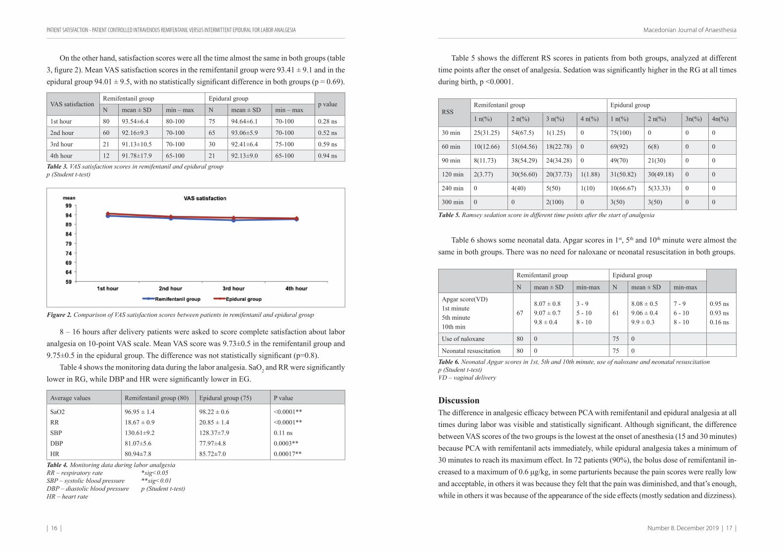
On the other hand, satisfaction scores were all the time almost the same in both groups (table 3, figure 2). Mean VAS satisfaction scores in the remifentanil group were 93.41 ± 9.1 and in the epidural group 94.01 ± 9.5, with no statistically significant difference in both groups (p = 0.69).
VAS satisfactionRemifentanil group Epidural group
p valueN mean ± SD min – max N mean ± SD min – max
1st hour 80 93.54±6.4 80-100 75 94.64±6.1 70-100 0.28 ns
2nd hour 60 92.16±9.3 70-100 65 93.06±5.9 70-100 0.52 ns
3rd hour 21 91.13±10.5 70-100 30 92.41±6.4 75-100 0.59 ns
4th hour 12 91.78±17.9 65-100 21 92.13±9.0 65-100 0.94 nsTable 3. VAS satisfaction scores in remifentanil and epidural groupp (Student t-test)
Figure 2. Comparison of VAS satisfaction scores between patients in remifentanil and epidural group
8 – 16 hours after delivery patients were asked to score complete satisfaction about labor analgesia on 10-point VAS scale. Mean VAS score was 9.73±0.5 in the remifentanil group and 9.75±0.5 in the epidural group. The difference was not statistically significant (p=0.8).
Table 4 shows the monitoring data during the labor analgesia. SaO2 and RR were significantly lower in RG, while DBP and HR were significantly lower in EG.
Average values Remifentanil group (80) Epidural group (75) P value
SaO2RRSBPDBPHR
96.95 ± 1.418.67 ± 0.9130.61±9.281.07±5.680.94±7.8
98.22 ± 0.620.85 ± 1.4128.37±7.977.97±4.885.72±7.0
<0.0001**<0.0001**0.11 ns0.0003**0.00017**
Table 4. Monitoring data during labor analgesiaRR – respiratory rate *sig<0.05SBP – systolic blood pressure **sig<0.01DBP – diastolic blood pressure p (Student t-test)HR – heart rate
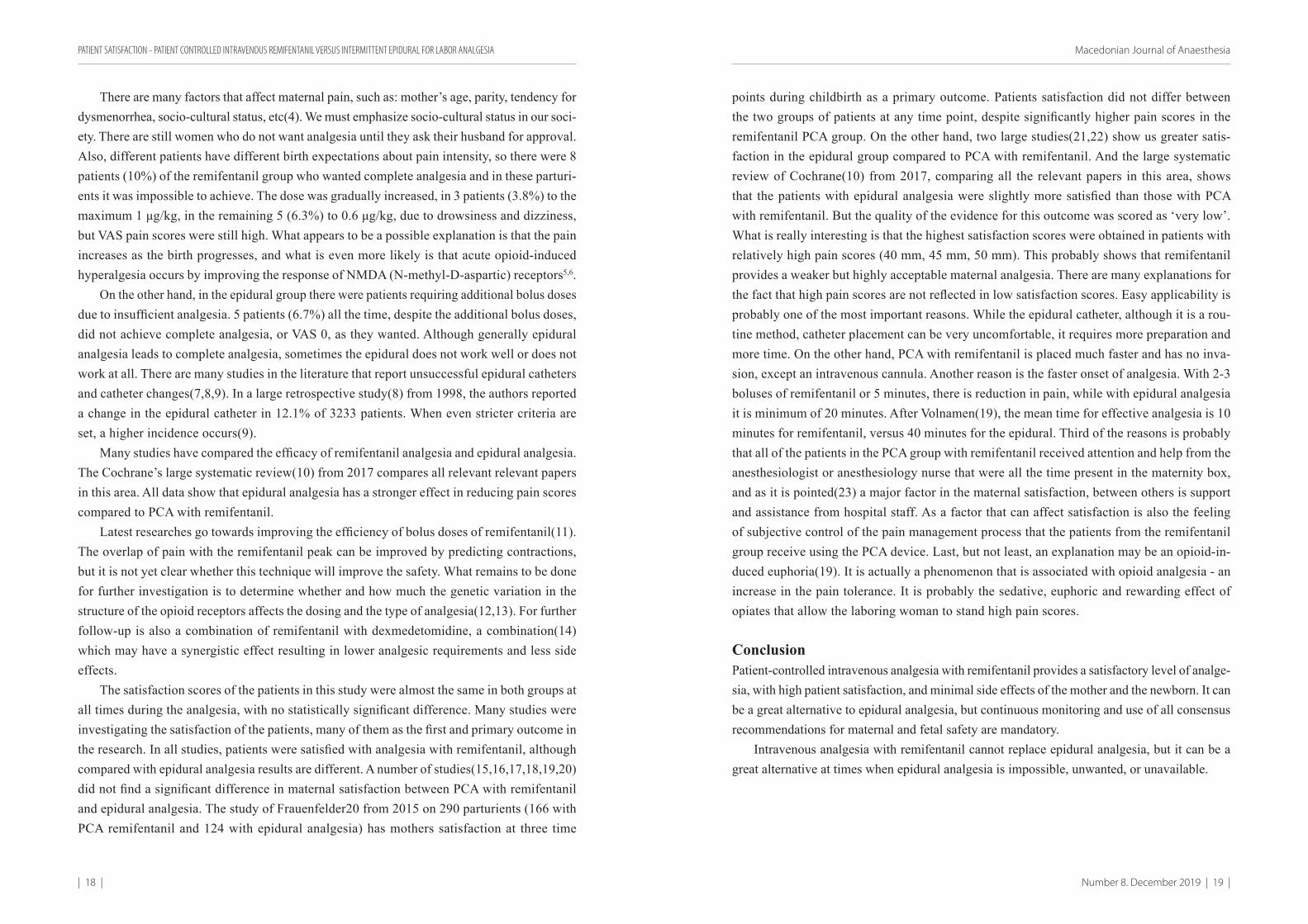
Table 5 shows the different RS scores in patients from both groups, analyzed at different time points after the onset of analgesia. Sedation was significantly higher in the RG at all times during birth, p <0.0001.
RSSRemifentanil group Epidural group
1 n(%) 2 n(%) 3 n(%) 4 n(%) 1 n(%) 2 n(%) 3n(%) 4n(%)
30 min 25(31.25) 54(67.5) 1(1.25) 0 75(100) 0 0 0
60 min 10(12.66) 51(64.56) 18(22.78) 0 69(92) 6(8) 0 0
90 min 8(11.73) 38(54.29) 24(34.28) 0 49(70) 21(30) 0 0
120 min 2(3.77) 30(56.60) 20(37.73) 1(1.88) 31(50.82) 30(49.18) 0 0
240 min 0 4(40) 5(50) 1(10) 10(66.67) 5(33.33) 0 0
300 min 0 0 2(100) 0 3(50) 3(50) 0 0
Тable 5. Ramsey sedation score in different time points after the start of analgesia
Table 6 shows some neonatal data. Apgar scores in 1st, 5th and 10th minute were almost the same in both groups. There was no need for naloxane or neonatal resuscitation in both groups.
Remifentanil group Epidural group
N mean ± SD min-max N mean ± SD min-max
Apgar score(VD)1st minute5th minute10th min
678.07 ± 0.89.07 ± 0.79.8 ± 0.4
3 - 95 - 108 - 10
618.08 ± 0.59.06 ± 0.49.9 ± 0.3
7 - 96 - 108 - 10
0.95 ns0.93 ns0.16 ns
Use of naloxane 80 0 75 0
Neonatal resuscitation 80 0 75 0
Table 6. Neonatal Apgar scores in 1st, 5th and 10th minute, use of naloxane and neonatal resuscitationp (Student t-test)VD – vaginal delivery
DiscussionThe difference in analgesic efficacy between PCA with remifentanil and epidural analgesia at all times during labor was visible and statistically significant. Although significant, the difference between VAS scores of the two groups is the lowest at the onset of anesthesia (15 and 30 minutes) because PCA with remifentanil acts immediately, while epidural analgesia takes a minimum of 30 minutes to reach its maximum effect. In 72 patients (90%), the bolus dose of remifentanil in-creased to a maximum of 0.6 μg/kg, in some parturients because the pain scores were really low and acceptable, in others it was because they felt that the pain was diminished, and that’s enough, while in others it was because of the appearance of the side effects (mostly sedation and dizziness).
PATIENT SATISFACTION - PATIENT CONTROLLED INTRAVENOUS REMIFENTANIL VERSUS INTERMITTENT EPIDURAL FOR LABOR ANALGESIA
| 18 |
Macedonian Journal of Anaesthesia
Number 8. December 2019 | 19 |
There are many factors that affect maternal pain, such as: mother’s age, parity, tendency for dysmenorrhea, socio-cultural status, etc(4). We must emphasize socio-cultural status in our soci-ety. There are still women who do not want analgesia until they ask their husband for approval. Also, different patients have different birth expectations about pain intensity, so there were 8 patients (10%) of the remifentanil group who wanted complete analgesia and in these parturi-ents it was impossible to achieve. The dose was gradually increased, in 3 patients (3.8%) to the maximum 1 μg/kg, in the remaining 5 (6.3%) to 0.6 μg/kg, due to drowsiness and dizziness, but VAS pain scores were still high. What appears to be a possible explanation is that the pain increases as the birth progresses, and what is even more likely is that acute opioid-induced hyperalgesia occurs by improving the response of NMDA (N-methyl-D-aspartic) receptors5,6.
On the other hand, in the epidural group there were patients requiring additional bolus doses due to insufficient analgesia. 5 patients (6.7%) all the time, despite the additional bolus doses, did not achieve complete analgesia, or VAS 0, as they wanted. Although generally epidural analgesia leads to complete analgesia, sometimes the epidural does not work well or does not work at all. There are many studies in the literature that report unsuccessful epidural catheters and catheter changes(7,8,9). In a large retrospective study(8) from 1998, the authors reported a change in the epidural catheter in 12.1% of 3233 patients. When even stricter criteria are set, a higher incidence occurs(9).
Many studies have compared the efficacy of remifentanil analgesia and epidural analgesia. The Cochrane’s large systematic review(10) from 2017 compares all relevant relevant papers in this area. All data show that epidural analgesia has a stronger effect in reducing pain scores compared to PCA with remifentanil.
Latest researches go towards improving the efficiency of bolus doses of remifentanil(11). The overlap of pain with the remifentanil peak can be improved by predicting contractions, but it is not yet clear whether this technique will improve the safety. What remains to be done for further investigation is to determine whether and how much the genetic variation in the structure of the opioid receptors affects the dosing and the type of analgesia(12,13). For further follow-up is also a combination of remifentanil with dexmedetomidine, a combination(14) which may have a synergistic effect resulting in lower analgesic requirements and less side effects.
The satisfaction scores of the patients in this study were almost the same in both groups at all times during the analgesia, with no statistically significant difference. Many studies were investigating the satisfaction of the patients, many of them as the first and primary outcome in the research. In all studies, patients were satisfied with analgesia with remifentanil, although compared with epidural analgesia results are different. A number of studies(15,16,17,18,19,20) did not find a significant difference in maternal satisfaction between PCA with remifentanil and epidural analgesia. The study of Frauenfelder20 from 2015 on 290 parturients (166 with PCA remifentanil and 124 with epidural analgesia) has mothers satisfaction at three time
points during childbirth as a primary outcome. Patients satisfaction did not differ between the two groups of patients at any time point, despite significantly higher pain scores in the remifentanil PCA group. On the other hand, two large studies(21,22) show us greater satis-faction in the epidural group compared to PCA with remifentanil. And the large systematic review of Cochrane(10) from 2017, comparing all the relevant papers in this area, shows that the patients with epidural analgesia were slightly more satisfied than those with PCA with remifentanil. But the quality of the evidence for this outcome was scored as ‘very low’. What is really interesting is that the highest satisfaction scores were obtained in patients with relatively high pain scores (40 mm, 45 mm, 50 mm). This probably shows that remifentanil provides a weaker but highly acceptable maternal analgesia. There are many explanations for the fact that high pain scores are not reflected in low satisfaction scores. Easy applicability is probably one of the most important reasons. While the epidural catheter, although it is a rou-tine method, catheter placement can be very uncomfortable, it requires more preparation and more time. On the other hand, PCA with remifentanil is placed much faster and has no inva-sion, except an intravenous cannula. Another reason is the faster onset of analgesia. With 2-3 boluses of remifentanil or 5 minutes, there is reduction in pain, while with epidural analgesia it is minimum of 20 minutes. After Volnamen(19), the mean time for effective analgesia is 10 minutes for remifentanil, versus 40 minutes for the epidural. Third of the reasons is probably that all of the patients in the PCA group with remifentanil received attention and help from the anesthesiologist or anesthesiology nurse that were all the time present in the maternity box, and as it is pointed(23) a major factor in the maternal satisfaction, between others is support and assistance from hospital staff. As a factor that can affect satisfaction is also the feeling of subjective control of the pain management process that the patients from the remifentanil group receive using the PCA device. Last, but not least, an explanation may be an opioid-in-duced euphoria(19). It is actually a phenomenon that is associated with opioid analgesia - an increase in the pain tolerance. It is probably the sedative, euphoric and rewarding effect of opiates that allow the laboring woman to stand high pain scores.
ConclusionPatient-controlled intravenous analgesia with remifentanil provides a satisfactory level of analge-sia, with high patient satisfaction, and minimal side effects of the mother and the newborn. It can be a great alternative to epidural analgesia, but continuous monitoring and use of all consensus recommendations for maternal and fetal safety are mandatory.
Intravenous analgesia with remifentanil cannot replace epidural analgesia, but it can be a great alternative at times when epidural analgesia is impossible, unwanted, or unavailable.
PATIENT SATISFACTION - PATIENT CONTROLLED INTRAVENOUS REMIFENTANIL VERSUS INTERMITTENT EPIDURAL FOR LABOR ANALGESIA

| 20 |
Macedonian Journal of Anaesthesia
Number 8. December 2019 | 21 |
Literature:1. Wong CA - Epidural and Spinal Analgesia/Anesthesia for Labor and Vaginal Delivery, em:
Chestnut DH et al. - Obstetric Anesthesia: Principles and Practice, 4ª Ed, Philadelphia, Mosby Elsevier, 2009; 429-492.
2. Kan RE, Hughes SC, Rosen MA et al. - Intravenous remifentanil: placental transfer, maternal and neonatal effects. Anesthesiology, 1998; 88:1467-1474.
3. Egan TD. Pharmacokinetics and pharmacodynamics of remifentanil: an update in the year 2000. Curr Opin Anaesthesiol 2000;13:449–55.
4. Lowe NK. The nature of labor pain. Am J Obstet Gynecol 2002; 186: S16–24.5. Koppert W, Sittl R, Scheuber K, Alsheimer M, Schmeltz M, Schuttler J. Differential mod-
ulation of remifentanil-induced analgesia and postinfusion hyperalgesia by S-ketamine and clonidine in humans. Anesthesiology 2003; 99: 152–159.
6. Angst MS, Chu LF, Tingle MS, Shafer SL, Clark JD, Drover DR. No evidence for devel-opment of acute tolerance to analgesic, respiratory depressant and sedative opioid effects in humans. Pain 2009; 142: 17–26.
7. Norris MC, Fogel ST, Conway-Long C. Combined spinal-epidural versus epidural labor analgesia. Anesthesiology 2001; 95: 913–920.
8. Eappen S, Blinn A, Segal S. Incidence of epidural catheter replecement in parturients: a retrospective chat review. Int J Obstet Anesth 1998 7: 220–225.
9. Beilin Y, Arnold I, Telfeyan C, Bernstein H, Hossain S. Quality of analgesia when air versus saline is used for identification of the epidural space in the parturient. Reg Anesth Pain Med 2000; 25: 596–599.
10. Weibel S, Jelting Y, Afshari A, Pace NL, Eberhart LHJ, Jokinen J, Artmann T, Kranke P. Patient-controlled analgesia with remifentanil versus alternative parenteral methods for pain management in labour. Cochrane Database of Systematic Reviews 2017, Issue 4. Art. No.: CD011989.
11. Rehberg B, Wickboldt N, Juillet C, Savoldelli G. Can remifentanil use in obstetrics be improved by optimal patient-controlled analgesia bolus timing? Br J Anaesth. 2015 Feb;114(2):281-9.
12. Landau R, Kern C, Columb MO, Smiley RM, Blouin JL. Genetic variability of the mu-opi-oid receptor influences intrathecal fentanyl analgesia requirements in laboring women. Pain 2008; 139: 5–14.
13. Landau R. Ortner C. The impact on genetics and other factors on intra and post-partum pain. Current Anesthesiology Reports 2013 Dec: 3(4).
14. Abdalla W, Ammar MA, Tharwat AI. Combination of dexmedetomidine and remifentanil for labor analgesia: A double blinded, randomized, contolled study. Saudi J Anaesth. 2015 Oct-Dec;9(4):433-8.
15. Stocki D, Matot I, Elnav S; A randomized controlled trial of the efficacy and respiratory effects of patient-controlled intravenous remifentanil analgesia and patient-controlled epi-dural analgesia in laboring women. Anesth Analg 2014; 118;589-97.
16. Douma MR, Middeldorp JM, Verwey RA, Dahan A, Stienstra R. A randomised comparison of intravenous remifentanil patient-controlled analgesia with epidural ropivacaine/sufentanil during labour. Int J Obstet Anesth. 2011;20:118–23.
17. Douma MR, Stienstra R, Middeldorp JM, Arbous MS, Dahan A. Differences in maternal temperatureduring labour with remifentanil patient-controlled analgesia or epidural analgesia: a randomised controlled trial. Int J Obstet Anesth. 2015; 24(4):313-22.
18. Stourac P, Suchomelova H, Stodulkova M et al. Comparasion of parturient-controlled
remifentanil with epidural bupivacaine and sufentanil for labor analgesia, randomized con-trolled trial. Biomed Pap Med Fac Univ Palacky Olom Czech Rep 2014; 158(2):227-32.
19. Volmanen P, Sarvela J, Akural EI, Raudaskoski T, Korttila K, Alahuhta S. Intravenous remifentanil vs. epidural levobupivacaine with fentanyl for pain relief in early labour: a randomised, controlled, double-blinded study. Acta Anaesthesiologica Scandinavica 2008;52(2):249–55.
20. Frauenfelder S, van Rijan R, Radder CM, de Vries MC, Dijksman LM, Godfried MB; Patient satisfaction between remifentanil patient-controlled analgesia and epidural analgesia for labor pain; Acta Obstet Gynecol Scand 2015 94: 1014-21.
21. Freeman LM, Bloemenkamp KW, Fransen MT et al. Patient controlled analgesia with remifentanil versus epidural analgesia in labour: randomised multicentre equivalence trial BMJ 2015;350:h846.
22. SLM Logtenberg, K Oude Rengerink, CJ Verhoeven, LM Freeman, ESA van den Akker, MB Godfried et al. Labour pain with remifentanil patient-controlled analgesia versus epidural analgesia: a randomised equivalence trial. BJOG An International Journal of Obstetrics and Gynecology, 2017; 124,4:652–660.
23. Hodnett ED. Pain and women’s satisfaction with the experience of childbirth: a systematic review. Am J Obstet Gynecol. 2002 May;186(5 Suppl Nature):S160-72.
PATIENT SATISFACTION - PATIENT CONTROLLED INTRAVENOUS REMIFENTANIL VERSUS INTERMITTENT EPIDURAL FOR LABOR ANALGESIA
| 22 |
Macedonian Journal of Anaesthesia
Number 8. December 2019 | 23 |
АПСТРАКТВовед: Невроаксијалните техники се најефикасните методи за аналгезија во тек на
породување, додека епидуралната аналгезија со употреба на силно разредени анестетици се смета за златен стандард во акушерската анестезија промовирајќи одлична аналгезија со минимални несакани ефекти. Ремифентанил станува сè попопуларен за аналгезија во тек на породувањето, како алтернатива за невроаксијална анестезија во моментите кога таа е контраиндицирана, несакана од пациентот или едноставно недостапна.
Материјали и методи: 155 бремени жени беа вклучени во студијата и рандомизирани во 2 групи: ремифентанил група (РГ) и епидурална група (ЕГ). Пациентките во РГ (80 пациенти) добија интравенска пациент/контролирана аналгезија (ПКА) со ремифентанил, започнувајќи со 0,2 μg/kgTT, постепено зголемувајќи ја дозата за 0,1 μg/kg ТТ до максимална доза од 1 μg/kgTT. Пациентите во ЕГ (75 пациенти) добија епидурална аналгезија со програмирано наизменично болусно дозирање. Нашата главна цел беше задоволството на пациентките и ефикасноста на обезболувањето. Во текот на породувањето, анализиравме резултати за болка и резултати за задоволство преку 2 VAS скали во различни временски точки. Безбедноста на пациентите и новороденчињата се набљудуваше преку целосен хемодинамски мониторинг (SaO2, респираторна фреквенција, неинвазивен крвен притисок, пулс, седација, континуиран кардиотокографски запис).
Резултати: VAS скоровите за болка беа значително повисоки во РГ во сите временски точки, просечниот VAS скор за болка во РГ беше 46.44 ± 8.5, а во ЕГ 28.33 ± 11.8 (p <0.0001). Од друга страна, VAS скоровите за задоволство цело време беа скоро исти во двете групи, просечниот VAS скор за задоволство во текот на целиот период на набљудување беше 93.41 ± 9.1 во РГ и 94.01 ± 9.5 во ЕГ, без статистички значајна разлика помеѓу двете групи (p = 0,688). За време на целиот период на аналгезија во текот на породување, имаше значително пониска вредност на SaO2, пониска респираторна фреквенција во минута и почеста седација во РГ во сите временски точки по почетокот на аналгезија.
Заклучок: ПКА со ремифентанил е помалку ефикасна за обезболување кај пациенти во тек на породување во споредба со епидурална аналгезија, но задоволството на пациентите е еднакво и во двете групи. Континуиран респираторен мониторинг и достапност на кислород е задолжително.
PATIENT SATISFACTION - PATIENT CONTROLLED INTRAVENOUS REMIFENTANIL VERSUS INTERMITTENT EPIDURAL FOR LABOR ANALGESIA
DIABETES MELLITUS AND SMOKING AS MAIN RISK FACTORS FOR PROLONGED POSTOPERATIVE ILEUS FOLLOWING COLORECTAL SURGERY
Osmani B1
1 Clinic for Digestive Surgery, Clinical Center Mother Teresa, University Ss Cyril and Methodius, Skopje, N Macedonia
ABSTRACTProlonged postoperative ileus (POI) is common complication after colorectal surgery. It is
defined as two or more of nausea/vomiting, inability to tolerate oral diet over 24 h, and absence of flatus over 24 h.
The aim of this study is to identify the risk factors for development of prolonged postoper-ative ileus after colorectal surgery.
Material and methods: This study is retrospective analysis of data obtained from medical records of the patients subjected to colorectal surgery for malignancy over 12 months period in tertiary digestive surgical clinic “Mother Teresa”, Skopje, N Macedonia. Exclusion criteria were history of previous laparotomy and patients who were subjected to surgical revision due to surgical complications, or hospital readmission due to surgical complications. Demographic data, smoking history, obesity (BMI > 25), cardiac and pulmonary comorbidity and occurrence of prolonged postoperative ileus were analyzed.
Data were analyzed using descriptive statistic reported as counts and percentage for demo-graphics, and chi – square test was used to evaluate the association between various risk factors and prolonged POI, with a significance level of P < 0.05.
Results: A total number of 280 patients, aged 40 – 76 years, subjected to colorectal surgery for malignancy over the period of 12 months were included in the study.
Prolonged postoperative ileus developed in 62 patients (22%).Demographic data: Total number of male patients in the study was 156, total number of
female patients was 124. In the group that developed prolonged POI, male patients were 36 (58%), and female 26 (42%).
There is no statistical significance between sex and occurrence of prolonged POI (p > 0.05).The analysis of the possible associations of the clinical data with development of prolonged
postoperative ileus showed no statistical significance between cardiac morbidity and prolonged POI (p > 0.05), respiratory morbidity and prolonged POI (p > 0.05) and obesity and prolonged POI (p > 0.05).
ORIGINAL RESEARCH UDK: 616.345/.35-089.168:616.34-007.272]-02

| 24 |
Macedonian Journal of Anaesthesia
Number 8. December 2019 | 25 |
There is statistical significance between smoking and occurrence of prolonged POI (p < 0.05) and diabetes mellitus and occurrence of prolonged POI (p < 0.05).
Conclusion: The risk of prolonged POI is increased in patients with diabetes mellitus and smokers. Taking these factors in consideration, identifying the patients at risk could be beneficial in prevention and treatment of POI.
Key words: diabetes mellitus, postoperative ileus, smokingCorresponding author: Bujar Osmani, Clinic for Digestive Surgery, Clinical Center “Mother
Teresa”, University “Ss Cyril and Methodius”, Skopje, N. Macedonia
IntroductionPostoperative ileus (POI) is defined as: “interval from surgery until passage of flatus/stool AND tolerance of an oral diet”; while prolonged postoperative ileus (POI) as: “two or more of nausea/vomiting, inability to tolerate oral diet over 24 h, absence of flatus over 24 h, distension, radio-logic confirmation occurring on or after day 4 postoperatively without prior resolution of POI” (1)Prolonged postoperative ileus is common complication after colorectal surgery, reportedly affecting 10 - 15% of the patients (2). Although in most cases it can spontaneously resolve within 2 to 4 days, and is not considered a life threatening complication, for affected patients it causes discomfort due to nausea, vomiting, inability to take food orally and prolonged hospital stay. Prolonged postoperative ileus causes financial burden on healthcare system due to prolonged hospital stay (3). The pathogenesis of POI involves neurogenic mechanisms; prevalence of sympathetic over parasympathetic system leading to decreased motility and ileus, inflammatory mechanisms and pharmacologic mechanisms (4). Clinical management of POI mainly relies on intravenous fluids, correction of electrolyte imbalance, prokinetic agents, such as metocloprim-ide and insertion of nasogastric tube. Some authors dispute the efficiency of nasogastric tube, suggesting that it can actually prolong the postoperative ileus, and instead, propose starting an early clear liquid diet (5, 6). Because of the limited therapeutic options, the best approach are prevention strategies, i.e. identifying the patients at risk and preventing the development of POI. Male sex, age, smoking, cardiac and pulmonary comorbidity, as well as previous laparotomy are most common risk factors for prolonged postoperative Ileus in colorectal surgery (7).
The Aim of this study is to identify the risk factors for development of prolonged postoper-ative ileus after colorectal surgery.
Material and methodsThis study is retrospective analysis of data obtained from medical records of the patients subject-ed to colorectal surgery for malignancy over 12monthsperiodin tertiary digestive surgical clinic “Mother Teresa”, Skopje, N Macedonia. Exclusion criteria were history of previous laparotomy and patients who were subjected to surgical revision due to surgical complications, or hospital readmission due to surgical complications. Demographic data, smoking history, obesity (BMI >
25), cardiac and pulmonary comorbidity and occurrence of prolonged postoperative ileus were analyzed.
Data were analyzed using descriptive statistic reported as counts and percentage for demo-graphics, and chi – square test was used to evaluate the association between various risk factors and prolonged POI, with a significance level of P < 0.05.
ResultsA total number of 280 patients, aged 40 – 76 years, subjected to colorectal surgery for malignancy over the period of 12 months were included in the study.
Prolonged postoperative ileus developed in 62 patients (22%).Demographic data: Total number of male patients in the study was 156, total number of
female patients was 124. In the group that developed prolonged POI, male patients were 36 (58%), and female 26 (42%).
There is no statistical significance between sex and occurrence of prolonged POI (p > 0.05).The analysis of the possible associations of the clinical data with development of prolonged
postoperative ileus showed no statistical significance between cardiac morbidity and prolonged POI (p > 0.05), respiratory morbidity and prolonged POI (p > 0.05) and obesity and prolonged POI (p > 0.05).
There is statistical significance between smoking and occurrence of prolonged POI (p < 0.05) and diabetes mellitus and occurrence of prolonged POI (p < 0.05).
Patients without prolonged POI Patients with prolonged POI P - Value
Sex Male 120Female 98
Male 36Female 26
p=.6728p > 0.05
Cardiac comorbidity 75 27 p =.1867p> 0.05
Respiratory comorbidity 30 12 p=.2764p > 0.05
Obesity (BMI > 25) 70 24 p =.3315p > 0.05
Diabetes mellitus 50 29 p=.0002p < 0.05
Smoking 69 38 p =.00002p <0.05
Table 1. Association between patients’ demographics and clinical data with the occurrence of prolonged postop-erative ileus.Bold text indicates statistically significant findings (association).
DiscussionThis conduction of this study was aimed to find possible correlation between demographic char-acteristics and certain medical conditions with development of prolonged postoperative ileus in patients subjected to colorectal surgery. The incidence of prolonged POI in this study was 22%
DIABETES MELLITUS AND SMOKING AS MAIN RISK FACTORS FOR PROLONGED POSTOPERATIVE ILEUS FOLLOWING COLORECTAL SURGERY
| 26 |
Macedonian Journal of Anaesthesia
Number 8. December 2019 | 27 |
which is higher than the reported incidence by Quiroga-Centeno et al, and Millan et al., (2, 7), but lower than the reported incidence by Howe Mao et al., (3). This study showed no correlation between sex and development of prolonged POI, while the meta – analysis from Lee et al, showed that male sex was associated with prolonged POI (8). According to Quiroga-Centeno et al, and Millan et al., cardiac and respiratory comorbidities were associated with prolonged POI (2, 7), but in this study we found no such correlation. This study showed strong association between smoking and development of prolonged POI, which correlates with the findings of Sugawara et al. (9) Afore mentioned authors didn’t include diabetes mellitus in risk factors for prolonged POI, but this study showed significant correlation between them.
Postoperative ileus can develop after any kind of surgery, but surgeries of the gastrointestinal tract are in particular associated with prolonged postoperative ileus. The duration of POI may vary from two days to several weeks. It is associated with prolonged hospital stay, in average of 5 days (6).
The treatment options are limited and with uncertain results, and there is no consensus on benefits of traditional treatments with nasogastric tube and prokinetic drugs. Some pharmacologic management options are still under investigation for effectiveness. Neostigmine, endrophonium chloride, cisaprid and metocloprimide are used to minimize the sympathetic inhibition of gas-trointestinal motility, but their effect in treatment of POI is still uncertain. (6)
Therefore, the best approach to management of this postoperative complication is prevention. Identifying the risk factors and patients at risk of developing POI is a first step of prevention. Quitting smoking several weeks prior to surgery, maintaining good glycemic status, fluid and electrolyte balance, as well as early clear fluid diet and ambulation are recommended in order to minimize the risk of POI.
Conclusion: The risk of prolonged POI is increased in patients with diabetes mellitus and smokers. Taking these factors in consideration, identifying the patients at risk could be beneficial in prevention and treatment of POI.
References:1. Vather, R., Trivedi, S. & Bissett, I. Defining Postoperative Ileus: Results of a Systematic
Review and Global Survey. J Gastrointest Surg 17,2013 962–9722. Quiroga-Centeno, A.C., Jerez-Torra, K.A. et al. Risk Factors for Prolonged Postoperative
Ileus in Colorectal Surgery: A Systematic Review and Meta-analysis. World J Surg (2020)3. Howe Mao, TonyMilne, GregoryO’Grady et al. Prolonged Postoperative Ileus Significantly
Increases the Cost of Inpatient Stay for Patients Undergoing Elective Colorectal Surgery: Results of a Multivariate Analysis of Prospective Data at a Single Institution. Diseases of the Colon & Rectum. 62(5)2019.631–637
4. Mattej P, Rombeaus JL. Review of the pathophysiology and management of postoperative ileus. World J Surg.30(8)2006.1382-91
5. Delaney C, Kehlet H, Senagore A et al. Postoperative ileus: profiles, risk factors, and definitions—a framework for optimizing surgical outcomes in patients undergoing major abdominal colorectal surgery. In: Bosker G, editor. Clinical consensus update in general
surgery. Roswell (GA): Pharmatecture, LLC); 2006. Available: www.clinicalwebcasts.com/pdfs/GenSurg_WEB.pdf
6. Zeinalli F, Stulberg J, Delaney C. Pharmacological management of postoperative ileus. Can J Surg 52(2) 2009.153–157
7. Millan, M., Biondo, S., Fraccalvieri, D. et al. Risk Factors for Prolonged Postoperative Ileus After Colorectal Cancer Surgery. World J Surg 2012.36 : 179
8. Lee MJ, Vaughan- Shaw P, Vimalachandran D et al. A systematic review and meta- analysis of baseline risk factors for the development of postoperative ileus in patients undergoing gastrointestinal surgery. Ann R Coll Surg Engl. 2019 Dec 20:1-10.
9. Sugawara K, Kawaguchi Y, Nomura Y et al. Perioperative factors predicting prolonged postoperative ileus after abdominal surgery. J Gastrointestinal Surg. 2018 22 (3): 508-515
АПСТРАКТПостоперативен илеус е честа компликација кај пациенти подложени на колоректална
хирургија. Пролонгиран постоперативен илеус е дефиниран како состојба на две или повеќе повраќања, неможност да се толерира орална исхрана и отсуство на гасови во период од 24 часа.
Целта на оваа студија е да се идентификуваат ризик факторите за развој на пролонгиран постоеративен илеус.
Материјали и методи: Ретроспективна анализа на медицинските истории на пациенти подложени на колоректална хирургија во период од 12 месеци во терциерна хируршка болница. Беа анализирани демографски карактеристики, историја на пушење, згоеност, срцеви и белодробни коморбидитети, дијабетес мелитус и појава на пролонгиран постоперативен илеус. Податоците беа анализирани со дескриптивни методи за демографските карактеристики, а компарација на квалитативните податоци со x2 тест.
Резултати: 280 пациенти беа вклучени во студијата, пролонгиран постоперативен илеус се јавил кај 62 пациенти (22%). Нема статистички значителна корелација помеѓу полот, згоеноста, срцевите и белодробни коморбидитети кај пациентите и појава на пролонгиран постоперативен илеус. Статистички значителна корелација (р < 0.05) се докажа помеѓу појавата на пролонгиран постоперативен илеус и пациентите кои се пушачи, како и пациентите кои имаат дијабетес мелитус.
Заклучок: Ризикот за појава на пролонгиран постоперативен илеус е зголемен кај пациенти кои имаат дијабетес мелитус и кај пациенти пушачи. Имајќи ги предвид овие фактори, идентификување на пациентите со зголемен ризик, може да е од бенефит за превенција и третман на постоперативен илеус.
Клучни зборови: дијабетес мелитус, постоперативен илеус, пушење.
DIABETES MELLITUS AND SMOKING AS MAIN RISK FACTORS FOR PROLONGED POSTOPERATIVE ILEUS FOLLOWING COLORECTAL SURGERY
| 28 |
Macedonian Journal of Anaesthesia
Number 8. December 2019 | 29 |
AIRTRAQ® IS THE PREFERRED DEVICE FOR DIFFICULT INTUBATION BY RESIDENTS?
Gjorchevska E1, Gavrilovska-Brzanov A1, Ilieva E1, Mojsova-Mijovska M1, Petrusheva A1
1 University Clinic for Anaesthesia Reanimation and Intensive Care Faculty of Medicine, Ss. Cyril and Methodius University of Skopje
ABSTRACTBackground: The Airtraq® optical laryngoscope is an intubation device designed to provide
a view of the glottis without alignment of the oro-pharyngeal and laryngeal axes. Recent liter-ature shows that, given its two significant features: time effectiveness and short learning curve, Airtraq® is the most favorable option when it comes to difficult intubation.
Objectives: The goal was to analyze Airtraq® effectiveness when used by inexperienced physicians in anticipated difficult intubation in adult patients.
Materials and methods: We conducted a prospective evaluation in ten medical residents using the Airtraq® device for the first time. All of them were experienced in using Macintosh. Each resident conducted laryngoscopy and intubation with the Airtraq® device after short didactic guidance. Eighteen patients were included, over a period of seven months. The patients showed four difficult intubation predictors: history of difficult intubation, thyromental distance less than 60 mm, mouth opening less than 35 mm and Mallampati class 3 or 4. All of them were clinically examined for difficult airway by an ENT specialist.
Results: Before induction of anaesthesia all residents received a short demonstration on the use of the Airtraq®. Every participant was supervised by an Airtraq® handling specialist for each intubation maneuver. In sixteen patients, Airtraq® insertion, glottis visualization and subsequent intubation were easy and rapid, without arterial oxygen desaturation. In two patients the trachea was intubated from the second and third attempt. There were two tracheal intubation failures, associated with extended tracheal intubation and an Airtraq® specialist had to continue with intu-bation. The Airtraq® reduced the duration of intubation attempts in all cases, reduced the number of optimization maneuvers required, and reduced the potential for dental trauma. However, the two intubation failures emphasize the fact that Airtraq® laryngoscopy requires a clinical training process, especially in the event of anticipated difficult airway management situations.
Conclusion: The residents participating the study, found the Airtraq® easier to use in all scenarios compared to the Macintosh laryngoscope. The Airtraq® may be the preferred device, required by inexperienced physicians in cases of difficult airway.
Corresponding Author: Gjorchevska Elena, University Clinic for Anesthesia Reanimation and Intensive Care, Faculty of Medicine, University of Ss. Cyril and Methodius - Skopje
*I declare that the abstract for this article has been published in the proceedings book of abstracts at the VI Macedonian Congress of Anaesthesiology, Reanimation and Intensive Care with International participation 24-27.10.2019.
ORIGINAL RESEARCH UDK: 616.5-006.3:[616.98:578.828.7
IntroductionResidents with limited clinical experience are frequently required to perform direct laryngoscopy in the clinical setting. In this context, difficult or failed intubation is an important cause of mor-bidity and mortality, due to direct airway trauma or systemic hypoxia complications (1). Novel intubation devices can reduce the morbidity and mortality risk in patients, when less experienced physicians are faced with difficult intubation scenario.
The Airtraq® optical laryngoscope is an intubation device designed to provide a view of the glottis without alignment of the oro-pharyngeal and laryngeal axes. Recent literature shows that, given its two significant features: time effectiveness and short learning curve, Airtraq® is the most favorable option when it comes to difficult intubation (2).
The Airtraq blade is anatomically shaped and made of two side-by-side channels. In one of the channels an endotracheal tube (ETT) in all sizes can be positioned and inserted. The other ends in a distal lens allowing visualization of the glottis, the surrounding structures and the tip of the ETT. There is a battery-operated light in the blade’s end. A proximal viewfinder uses lens and prism combinations, instead of fiber optics for transmission of the image. After the blade is inserted in the midline of the mouth over the base of the tongue, the viewfinder optimizes the view of the glottis and the tip of the ETT. The ETT does not obstruct the clear view of the vocal cords and in this way the Airtraq requires less manual skills to use (2).
ObjectivesThe goal was to analyze Airtraq® effectiveness when used by residents with limited difficult tracheal intubation skills in adult patients presenting for elective surgery. We hypothesize that Airtraq would be superior in comparison to Macintosh laryngoscope.
Materials and methodsWe conducted a prospective single center evaluation in the University Clinic for Anaesthesia, Reanimation and Intensive Care in Skopje. We performed indirect laryngoscopy and subsequent en-dotracheal intubation using the Airtraq® in patients presented with difficult intubation predictors for elective urology surgery under general endotracheal anesthesia. The patients were intubated by ten medical residents using the device for the first time. All of them were experienced in using Macintosh laryngoscope. Each resident conducted laryngoscopy and intubation with the Airtraq® device after short didactic and video guidance. As primary goal, we evaluated the time duration of tracheal intu-bation defined as the time from inserting the Airtraq in the mouth between the teeth to the moment of the visualization of the EET passing the vocal cords. Additionally we evaluated the number of intubation attempts and the rate of successful placement of the ETT in the trachea. All of the residents were closely monitored and guided by a senior anesthesiologist experienced in difficult intubation and Airtraq management. The position of the ETT tip was verified after each intubation attempt. When failed intubation attempt occurred, an Airtraq handling specialist proceeded with intubation.
| 30 |
Macedonian Journal of Anaesthesia
Number 8. December 2019 | 31 |
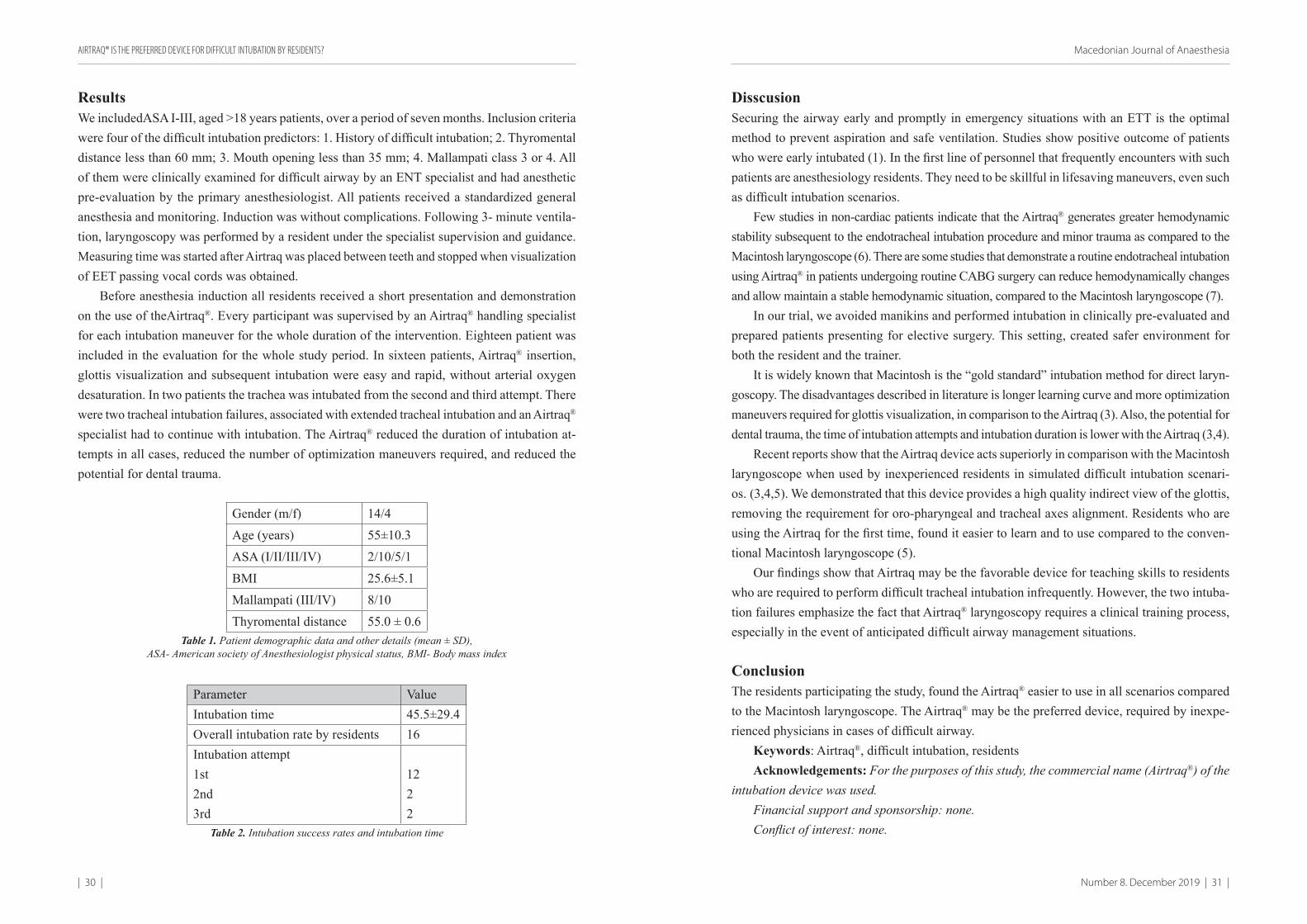
ResultsWe includedASA I-III, aged >18 years patients, over a period of seven months. Inclusion criteria were four of the difficult intubation predictors: 1. History of difficult intubation; 2. Thyromental distance less than 60 mm; 3. Mouth opening less than 35 mm; 4. Mallampati class 3 or 4. All of them were clinically examined for difficult airway by an ENT specialist and had anesthetic pre-evaluation by the primary anesthesiologist. All patients received a standardized general anesthesia and monitoring. Induction was without complications. Following 3- minute ventila-tion, laryngoscopy was performed by a resident under the specialist supervision and guidance. Measuring time was started after Airtraq was placed between teeth and stopped when visualization of EET passing vocal cords was obtained.
Before anesthesia induction all residents received a short presentation and demonstration on the use of theAirtraq®. Every participant was supervised by an Airtraq® handling specialist for each intubation maneuver for the whole duration of the intervention. Eighteen patient was included in the evaluation for the whole study period. In sixteen patients, Airtraq® insertion, glottis visualization and subsequent intubation were easy and rapid, without arterial oxygen desaturation. In two patients the trachea was intubated from the second and third attempt. There were two tracheal intubation failures, associated with extended tracheal intubation and an Airtraq® specialist had to continue with intubation. The Airtraq® reduced the duration of intubation at-tempts in all cases, reduced the number of optimization maneuvers required, and reduced the potential for dental trauma.
Gender (m/f) 14/4Age (years) 55±10.3ASA (I/II/III/IV) 2/10/5/1BMI 25.6±5.1Mallampati (III/IV) 8/10Thyromental distance 55.0 ± 0.6
Table 1. Patient demographic data and other details (mean ± SD),ASA- American society of Anesthesiologist physical status, BMI- Body mass index
Parameter ValueIntubation time 45.5±29.4Overall intubation rate by residents 16Intubation attempt1st2nd3rd
1222
Table 2. Intubation success rates and intubation time
DisscusionSecuring the airway early and promptly in emergency situations with an ETT is the optimal method to prevent aspiration and safe ventilation. Studies show positive outcome of patients who were early intubated (1). In the first line of personnel that frequently encounters with such patients are anesthesiology residents. They need to be skillful in lifesaving maneuvers, even such as difficult intubation scenarios.
Few studies in non-cardiac patients indicate that the Airtraq® generates greater hemodynamic stability subsequent to the endotracheal intubation procedure and minor trauma as compared to the Macintosh laryngoscope (6). There are some studies that demonstrate a routine endotracheal intubation using Airtraq® in patients undergoing routine CABG surgery can reduce hemodynamically changes and allow maintain a stable hemodynamic situation, compared to the Macintosh laryngoscope (7).
In our trial, we avoided manikins and performed intubation in clinically pre-evaluated and prepared patients presenting for elective surgery. This setting, created safer environment for both the resident and the trainer.
It is widely known that Macintosh is the “gold standard” intubation method for direct laryn-goscopy. The disadvantages described in literature is longer learning curve and more optimization maneuvers required for glottis visualization, in comparison to the Airtraq (3). Also, the potential for dental trauma, the time of intubation attempts and intubation duration is lower with the Airtraq (3,4).
Recent reports show that the Airtraq device acts superiorly in comparison with the Macintosh laryngoscope when used by inexperienced residents in simulated difficult intubation scenari-os. (3,4,5). We demonstrated that this device provides a high quality indirect view of the glottis, removing the requirement for oro-pharyngeal and tracheal axes alignment. Residents who are using the Airtraq for the first time, found it easier to learn and to use compared to the conven-tional Macintosh laryngoscope (5).
Our findings show that Airtraq may be the favorable device for teaching skills to residents who are required to perform difficult tracheal intubation infrequently. However, the two intuba-tion failures emphasize the fact that Airtraq® laryngoscopy requires a clinical training process, especially in the event of anticipated difficult airway management situations.
ConclusionThe residents participating the study, found the Airtraq® easier to use in all scenarios compared to the Macintosh laryngoscope. The Airtraq® may be the preferred device, required by inexpe-rienced physicians in cases of difficult airway.
Keywords: Airtraq®, difficult intubation, residentsAcknowledgements: For the purposes of this study, the commercial name (Airtraq®) of the
intubation device was used.Financial support and sponsorship: none.Conflict of interest: none.
AIRTRAQ® IS THE PREFERRED DEVICE FOR DIFFICULT INTUBATION BY RESIDENTS?
| 32 |
Macedonian Journal of Anaesthesia
Number 8. December 2019 | 33 |
References1. Mort TC. Esophageal intubation with indirect clinical tests during emergency tracheal in-
tubation: a report on patient morbidity. J ClinAnesth 2005;17:255-622. Di Marco P, Scattoni L, Spinoglio A et al. Learning and performance of tracheal intubation
by novice personnel: a comparison of the Airtraq and Macintosh laryngoscope. AnesthAnalg. 2011 Jan;112(1):122-5
3. Maharaj CH, Costello J, Higgins BD et al. Retention of tracheal intubation skills by novice personnel: a comparison of the Airtraq and Macintosh laryngoscopes. Anaesthesia. 2007 Mar;62(3):272-8.
4. Giquello JA, Humbert S, Duc F et al [Use of the Airtraq by inexperienced phy-sicians supervised during a series of tracheal intubation in adult patient with an-ticipated difficult airway]Ann Fr AnesthReanim. 2011 Nov; 30(11):804-8. doi: 10.1016/j.annfar.2011.05.006.
5. Maharaj CH, Costello JF, Harte BH et al. Anaesthesia. Evaluation of the Airtraq and Macintosh laryngoscopes in patients at increased risk for difficult tracheal intubation. 2008 Feb;63(2):182-8. doi: 10.1111/j.1365-2044.2007.05316.x.
6. Chalkeidis O, Kotsovolis G, Kalakonas A et al. A comparison between the Airtraq and Macintosh laryngoscopes for routine airway management by experienced anesthesiologists: a randomized clinical trial. Acta Anaesthesiol Taiwan. 2010;48:15–20.
7. Aleksandra Gavrilovska-Brzanov, Mohhamed Al Jarallah, Andrea Cogliati et al. Evaluation of the hemodynamic response to endotracheal intubation, comparing the Airtraq with Macintosh laryngoscopes in cardiac surgical patients] Acta inform med. 2015 oct23(5): 280-284 doi:10.5455/aim.2015.23.280-284
CASE REPORT UDK: 616.717.7/.8-002-089.853AIRTRAQ® IS THE PREFERRED DEVICE FOR DIFFICULT INTUBATION BY RESIDENTS?
LIFE-THREATENING LARINGEAL KAPOSI SARCOMA IN PATIENT WITH LATE HIV DIAGNOSIS
Stevanovic M1, Duganova M1, Grozdanovski K1, Demiri M1, Marinkovic S1, Petreska B1
1 University Clinic for Infectious diseases and febrile conditions, Skopje, Macedonia
ABSTRACTA 38-year-old woman presented with dark lesions on her upper body, weakness, and weight
loss, painful and difficult swallowing. Despite being seen at several different clinics for 6 months, no diagnosis was made. Patient was referred at the Clinic for infectious diseases (CID) because of a high fever. At her first visit at CID she was diagnosed with HIV infection. A lymph node extraction eventually identified Kaposi sarcoma (KS). A laryngeal tumor was also established. Since her condition was clinically defined as a late HIV infection, treatment with antibiotic and antiretroviral therapy was started. Following development of difficulties in breathing and swal-lowing, a tracheotomy was performed and chemotherapy with Doxorubicin 70 mg was initiated. The patient’s general condition improved after 4 courses of chemotherapy. Her condition suddenly deteriorated 4 months later, after chemotherapy was switched to cyclophosphamide 1000 mg. X-ray analysis showed recurrence of the KS with pleural involvement with seriously bad general conditions. Further treatment was initiated with paclitaxel 210 mg and her condition improved again. In countries with low HIV and KS prevalence a lack of experience among doctors of these conditions, results in delays in the diagnosis and initiation of therapy.
Keywords: HIV infection, Kaposi’s sarcoma, tracheotomyCoresponding author: Stevanovic Milena, University Clinic for infectious diseases and
febrile conditions, Skopje, Macedonia
IntroductionKaposi’s sarcoma (KS) is a well-known complication ofHIV infection and the most common-malignancy observed in patients with AIDS (1). KS is a vascular lesion of low-grade malignant potential caused by human herpes virus-8 (HHV8) infection. KS develops as a multifocal tumor that manifests most frequently in mucocutaneous sites, typically the skin of the lower extremities, face, trunk, genitalia and the oropharyngeal mucosa (2-4). KS also commonly involves lymph nodes and visceral organs, most notably the respiratory and gastrointestinal tracts. Peculiar pre-sentations of KS reported in relation to the gastrointestinal tract involvement include primary KS of the appendix, isolated rectal KS, and KS with mesenteric localization (5,6). Scores of authors have reported on the occurrence of KS in numerous unusual sites (i.e. anatomic locations other than the aforementioned sites) (2,3). KS is described most frequently among individuals with
| 34 |
Macedonian Journal of Anaesthesia
Number 8. December 2019 | 35 |
HIV exhibiting advanced immune suppression CD4 T lymphocyte (CD4) cell counts <200 cells/mm3, although they may occur at any CD4 cell count. Recent reports of KS occurring at higher CD4 cell countssuggest that clinicians caring for patients with HIV should be vigilant for the clinical manifestations of KS in patients at risk of HHV-8 infection, regardless of CD4 cell count may arise at any CD4 cell count (6). We report a rare case of KS presenting in a woman with advanced HIV infection as a life-threatening cause of airway obstruction and favorable outcome.
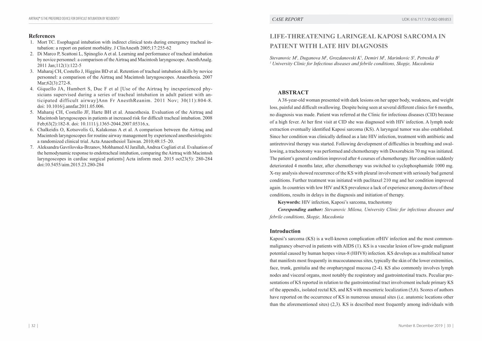
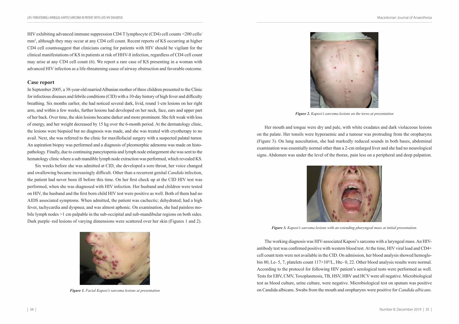
Case reportIn September 2005, a 38-year-old married Albanian mother of three children presented to the Clinic for infectious diseases and febrile conditions (CID) with a 10-day history of high fever and difficulty breathing. Six months earlier, she had noticed several dark, livid, round 1-cm lesions on her right arm, and within a few weeks, further lesions had developed on her neck, face, ears and upper part of her back. Over time, the skin lesions became darker and more prominent. She felt weak with loss of energy, and her weight decreased by 15 kg over the 6-month period. At the dermatology clinic, the lesions were biopsied but no diagnosis was made, and she was treated with cryotherapy to no avail. Next, she was referred to the clinic for maxillofacial surgery with a suspected palatal tumor. An aspiration biopsy was performed and a diagnosis of pleomorphic adenoma was made on histo-pathology. Finally, due to continuing pancytopenia and lymph node enlargement she was sent to the hematology clinic where a sub mandible lymph node extraction was performed, which revealed KS.
Six weeks before she was admitted at CID, she developed a sore throat, her voice changed and swallowing became increasingly difficult. Other than a recurrent genital Candida infection, the patient had never been ill before this time. On her first check up at the CID HIV test was performed, when she was diagnosed with HIV infection. Her husband and children were tested on HIV, the husband and the first born child HIV test were positive as well. Both of them had no AIDS associated symptoms. When admitted, the patient was cachectic; dehydrated; had a high fever, tachycardia and dyspnea; and was almost aphonic. On examination, she had painless mo-bile lymph nodes >1 cm palpable in the sub-occipital and sub-mandibular regions on both sides. Dark purple–red lesions of varying dimensions were scattered over her skin (Figures 1 and 2).
Figure 1. Facial Kaposi’s sarcoma lesions at presentation
Figure 2. Kaposi’s sarcoma lesions on the torso at presentation
Her mouth and tongue were dry and pale, with white exudates and dark violaceous lesions on the palate. Her tonsils were hyperaemic and a tumour was protruding from the oropharynx (Figure 3). On lung auscultation, she had markedly reduced sounds in both bases, abdominal examination was essentially normal other than a 2-cm enlarged liver and she had no neurological signs. Abdomen was under the level of the thorax, pain less on a peripheral and deep palpation.
Figure 3. Kaposi’s sarcoma lesions with an extending pharyngeal mass at initial presentation.
The working diagnosis was HIV-associated Kaposi’s sarcoma with a laryngeal mass. An HIV-antibody test was confirmed positive with western blood test. At the time, HIV viral load and CD4+ cell count tests were not available in the CID. On admission, her blood analysis showed hemoglo-bin 80, Le- 5, 7, platelets count 117×109/L, Htc- 0, 22. Other blood analysis results were normal. According to the protocol for following HIV patient’s serological tests were performed as well. Tests for EBV, CMV, Toxoplasmosis, TB, HSV, HBV and HCV were all negative. Microbiological test as blood culture, urine culture, were negative. Microbiological test on sputum was positive on Candida albicans. Swabs from the mouth and oropharynx were positive for Candida albicans.
LIFE-THREATENING LARINGEAL KAPOSI SARCOMA IN PATIENT WITH LATE HIV DIAGNOSIS

| 36 |
Macedonian Journal of Anaesthesia
Number 8. December 2019 | 37 |
After admission, the results of a sub-mandibular node biopsy taken a few weeks before at the department of haematology, was received, which showed Kaposi’s sarcoma.
An X-ray of the lungs revealed bilateral bronchopneumonia but no obvious tumour masses (Figure 4) and a computer tomography (CT) scan of her head and neck showed tumor formation in the pharyngeal area, posterior to the oropharynx with extension into the larynx, having a valve mechanism (Figure 5).
Figure 4. Lung X-ray at initial presentation showed bilateral bronchopneumonia
Figure 5. Computer tomography scan images of the head and neck on admission showed a tumor in the pharyn-geal area, posterior to the oropharynx and extending into the larynx (tumor mass denoted by arrow).
Since the patient was in very complex and difficult general condition she was treated inten-sively, rehydrated, received a blood transfusion, antibiotics (Ceftriakson 2 g per day iv during 14 days), antifungal therapy (sol. Diflason 200 mg x2 i.v during 14 days), tbl. trimethoprim-sulfa-methoxazole 480 mg once daily as prophylaxis therapy were commenced and oral combination of antiretroviral treatment (cARVT) was started immediately (tbl. Lamivudine 150 mg, tbl.
Stavudine 30 mg and Nevirapine 200 mg was administered twice daily, Nevirapine was titrated according to the Nevirapine summary of product characteristics).
Ten days after cARVT was commenced, it had to be interrupted due to the patient having compromised swallowing and breathing because of the growing tumor witch obstructed the pha-rynges region. She was transferred to the intensive care unit and a tracheotomy was performed and oxygen therapy was introduced. She was stabilized and intravenous Doxorubicin 80 mg was commenced every 2 weeks in consultation with colleagues from the oncology clinic; She developed severe anaemia while receiving the Doxorubicin and received daily one-unit blood transfusions for a week, and daily granulocyte colony-stimulating factor for the same period. Following the first three courses of chemotherapy, the tumour mass decreased enough for the patient to breathe and swallow normally. Antiretroviral treatment was recommenced, identical as previously used, since it was only available in R. Macedonia at the time. During her first admission in the hospital (12 weeks) her condition was improved.
Following the initial four courses of 80 mg doxorubicin, the dose was dropped to 70 mg for a further seven courses. The patient’s general condition improved and the skin lesions diminished in size after 4 course of Doxorubicin in 8 weeks. By that time, she had received the maximal dose of doxorubicin; treatment was switched to cyclophosphamide 1000 mg every 3 weeks. After the third course of Cyclophosphamide, her condition deteriorated. She was readmitted to the hospital with increasing breathlessness, enlarged and new skin lesions, facial and peripheral oedema, marked bruising of the skin, high fever and expectorating dark red material (Figures 6 and 7). Treatment with Cyclophosphamide was interapted.
Her blood analyses on readmission showed persistent anaemia, leucopoenia (Le-2, 3) and thrombocytopenia (Tr-64), severe hypoalbuminemia (Alb. 18), raised lactate dehydrogenize (LDH 2000) and C-reactive protein levels (CRP- 250).
Figure 6. Increased size and lividity of facial Kaposi’s sarcoma with marked
oedema.
Figure 7. Extensive facial Kaposi’s sarcoma.
X-ray of the lungs showed right-sided confluent lesions with pleural involvement with ef-fusion suggesting Kaposi’s sarcoma recurrence. Neither CT nor bronchoscopy was performed at this time (Figure 8).
LIFE-THREATENING LARINGEAL KAPOSI SARCOMA IN PATIENT WITH LATE HIV DIAGNOSIS
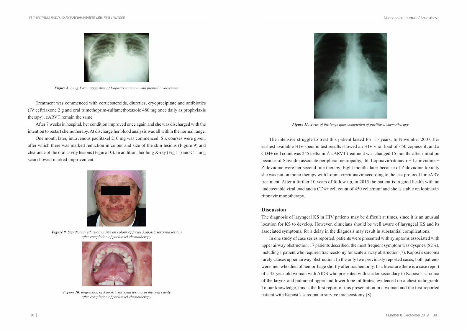
| 38 |
Macedonian Journal of Anaesthesia
Number 8. December 2019 | 39 |
Figure 8. Lung X-ray suggestive of Kaposi’s sarcoma with pleural involvement.
Treatment was commenced with corticosteroids, diuretics, cryoprecipitate and antibiotics (IV ceftriaxone 2 g and oral trimethoprim-sulfamethoxazole 480 mg once daily as prophylaxis therapy), cARVT remain the same.
After 7 weeks in hospital, her condition improved once again and she was discharged with the intention to restart chemotherapy. At discharge her blood analysis was all within the normal range.
One month later, intravenous paclitaxel 210 mg was commenced. Six courses were given, after which there was marked reduction in colour and size of the skin lesions (Figure 9) and clearance of the oral cavity lesions (Figure 10). In addition, her lung X-ray (Fig 11) and CT lung scan showed marked improvement.
Figure 9. Significant reduction in size an colour of facial Kaposi’s sarcoma lesions after completion of paclitaxel chemotherapy.
Figure 10. Regression of Kaposi’s sarcoma lesions in the oral cavity after completion of paclitaxel chemotherapy.
Figure 11. X-ray of the lungs after completion of paclitaxel chemotherapy
The intensive struggle to treat this patient lasted for 1.5 years. In November 2007, her earliest available HIV-specific test results showed an HIV viral load of <50 copies/mL and a CD4+ cell count was 245 cells/mm3. cARVT treatment was changed 15 months after initiation because of Stavudin associate peripheral neuropathy, tbl. Lopinavir/ritonavir + Lamivudine + Zidovudine were her second line therapy. Eight months later because of Zidovudine toxicity she was put on mono therapy with Lopinavir/ritonavir according to the last protocol for cARV treatment. After a further 10 years of follow up, in 2015 the patient is in good health with an undetectable viral load and a CD4+ cell count of 450 cells/mm3 and she is stable on lopinavir/ritonavir monotherapy.
DiscussionThe diagnosis of laryngeal KS in HIV patients may be difficult at times, since it is an unusual location for KS to develop. However, clinicians should be well aware of laryngeal KS and its associated symptoms, for a delay in the diagnosis may result in substantial complications.
In one study of case series reported, patients were presented with symptoms associated with upper airway obstruction, 17 patients described, the most frequent symptom was dyspnea (82%), including 1 patient who required tracheostomy for acute airway obstruction (7). Kaposi’s sarcoma rarely causes upper airway obstruction. In the only two previously reported cases, both patients were men who died of hemorrhage shortly after tracheotomy. In a literature there is a case report of a 45-year-old woman with AIDS who presented with stridor secondary to Kaposi’s sarcoma of the larynx and pulmonal upper and lower lobe infiltrates, evidenced on a chest radiograph. To our knowledge, this is the first report of this presentation in a woman and the first reported patient with Kaposi’s sarcoma to survive tracheostomy (8).
LIFE-THREATENING LARINGEAL KAPOSI SARCOMA IN PATIENT WITH LATE HIV DIAGNOSIS
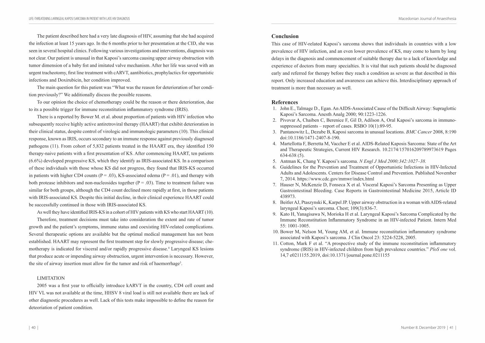
| 40 |
Macedonian Journal of Anaesthesia
Number 8. December 2019 | 41 |
The patient described here had a very late diagnosis of HIV, assuming that she had acquired the infection at least 15 years ago. In the 6 months prior to her presentation at the CID, she was seen in several hospital clinics. Following various investigations and interventions, diagnosis was not clear. Our patient is unusual in that Kaposi’s sarcoma causing upper airway obstruction with tumor dimension of a baby fist and imitated valve mechanism. After her life was saved with an urgent tracheotomy, first line treatment with cARVT, aantibiotics, prophylactics for opportunistic infections and Doxirubicin, her condition improved.
The main question for this patient was “What was the reason for deterioration of her condi-tion previously?” We additionally discuss the possible reasons.
To our opinion the choice of chemotherapy could be the reason or there deterioration, due to its a possible trigger for immune reconstitution inflammatory syndrome (IRIS).
There is a reported by Bower M. et al. about proportion of patients with HIV infection who subsequently receive highly active antiretroviral therapy (HAART) that exhibit deterioration in their clinical status, despite control of virologic and immunologic parameters (10). This clinical response, known as IRIS, occurs secondary to an immune response against previously diagnosed pathogens (11). From cohort of 5,832 patients treated in the HAART era, they identified 150 therapy-naive patients with a first presentation of KS. After commencing HAART, ten patients (6.6%) developed progressive KS, which they identify as IRIS-associated KS. In a comparison of these individuals with those whose KS did not progress, they found that IRIS-KS occurred in patients with higher CD4 counts (P = .03), KS-associated edema (P = .01), and therapy with both protease inhibitors and non-nucleosides together (P = .03). Time to treatment failure was similar for both groups, although the CD4 count declined more rapidly at first, in those patients with IRIS-associated KS. Despite this initial decline, in their clinical experience HAART could be successfully continued in those with IRIS-associated KS.
As well they have identified IRIS-KS in a cohort of HIV patients with KS who start HAART (10).Therefore, treatment decisions must take into consideration the extent and rate of tumor
growth and the patient’s symptoms, immune status and coexisting HIV-related complications. Several therapeutic options are available but the optimal medical management has not been established. HAART may represent the first treatment step for slowly progressive disease; che-motherapy is indicated for visceral and/or rapidly progressive disease.4 Laryngeal KS lesions that produce acute or impending airway obstruction, urgent intervention is necessary. However, the site of airway insertion must allow for the tumor and risk of haemorrhage3.
LIMITATION2005 was a first year to officially introduce kARVT in the country, CD4 cell count and
HIV VL was not available at the time, HHSV 8 viral load is still not available there are lack of other diagnostic procedures as well. Lack of this tests make impossible to define the reason for deteoriation of patient condition.
ConclusionThis case of HIV-related Kaposi’s sarcoma shows that individuals in countries with a low prevalence of HIV infection, and an even lower prevalence of KS, may come to harm by long delays in the diagnosis and commencement of suitable therapy due to a lack of knowledge and experience of doctors from many specialties. It is vital that such patients should be diagnosed early and referred for therapy before they reach a condition as severe as that described in this report. Only increased education and awareness can achieve this. Interdisciplinary approach of treatment is more than necessary as well.
References1. John E., Talmage D., Egan. An AIDS-Associated Cause of the Difficult Airway: Supraglottic
Kaposi’s Sarcoma. Anesth Analg 2000; 90:1223-1226.2. Pivovar A, Chaiben C, Berenice F, Gil D, Adilson A, Oral Kaposi’s sarcoma in immuno-
suppressed patients – report of cases. RSBO 10(1):89-95.3. Pantanowitz L, Dezube B, Kaposi sarcoma in unusual locations. BMC Cancer 2008, 8:190
doi:10.1186/1471-2407-8-190.4. Martellotta F, Berretta M, Vaccher E et al. AIDS-Related Kaposis Sarcoma: State of the Art
and Therapeutic Strategies, Current HIV Research. 10.2174/157016209789973619 Pages 634-638 (5).
5. Antman K, Chang Y. Kaposi’s sarcoma. N Engl J Med 2000;342:1027–38.6. Guidelines for the Prevention and Treatment of Opportunistic Infections in HIV-Infected
Adults and Adolescents. Centers for Disease Control and Prevention. Published November 7, 2014. https://www.cdc.gov/mmwr/index.html
7. Hauser N, McKenzie D, Fonseca X et al. Visceral Kaposi’s Sarcoma Presenting as Upper Gastrointestinal Bleeding. Case Reports in Gastrointestinal Medicine 2015, Article ID 438973.
8. Beitler AJ, Ptaszynski K, Karpel JP. Upper airway obstruction in a woman with AIDS-related laryngeal Kaposi’s sarcoma. Chest; 109(3):836-7.
9. Kato H, Yanagisawa N, Morioka H et al. Laryngeal Kaposi’s Sarcoma Complicated by the Immune Reconstitution Inflammatory Syndrome in an HIV-infected Patient. Intern Med 55: 1001-1005.
10. Bower M, Nelson M, Young AM, et al. Immune reconstitution inflammatory syndrome associated with Kaposi’s sarcoma. J Clin Oncol 23: 5224-5228, 2005.
11. Cotton, Mark F et al. “A prospective study of the immune reconstitution inflammatory syndrome (IRIS) in HIV-infected children from high prevalence countries.” PloS one vol. 14,7 e0211155.2019, doi:10.1371/journal.pone.0211155
LIFE-THREATENING LARINGEAL KAPOSI SARCOMA IN PATIENT WITH LATE HIV DIAGNOSIS
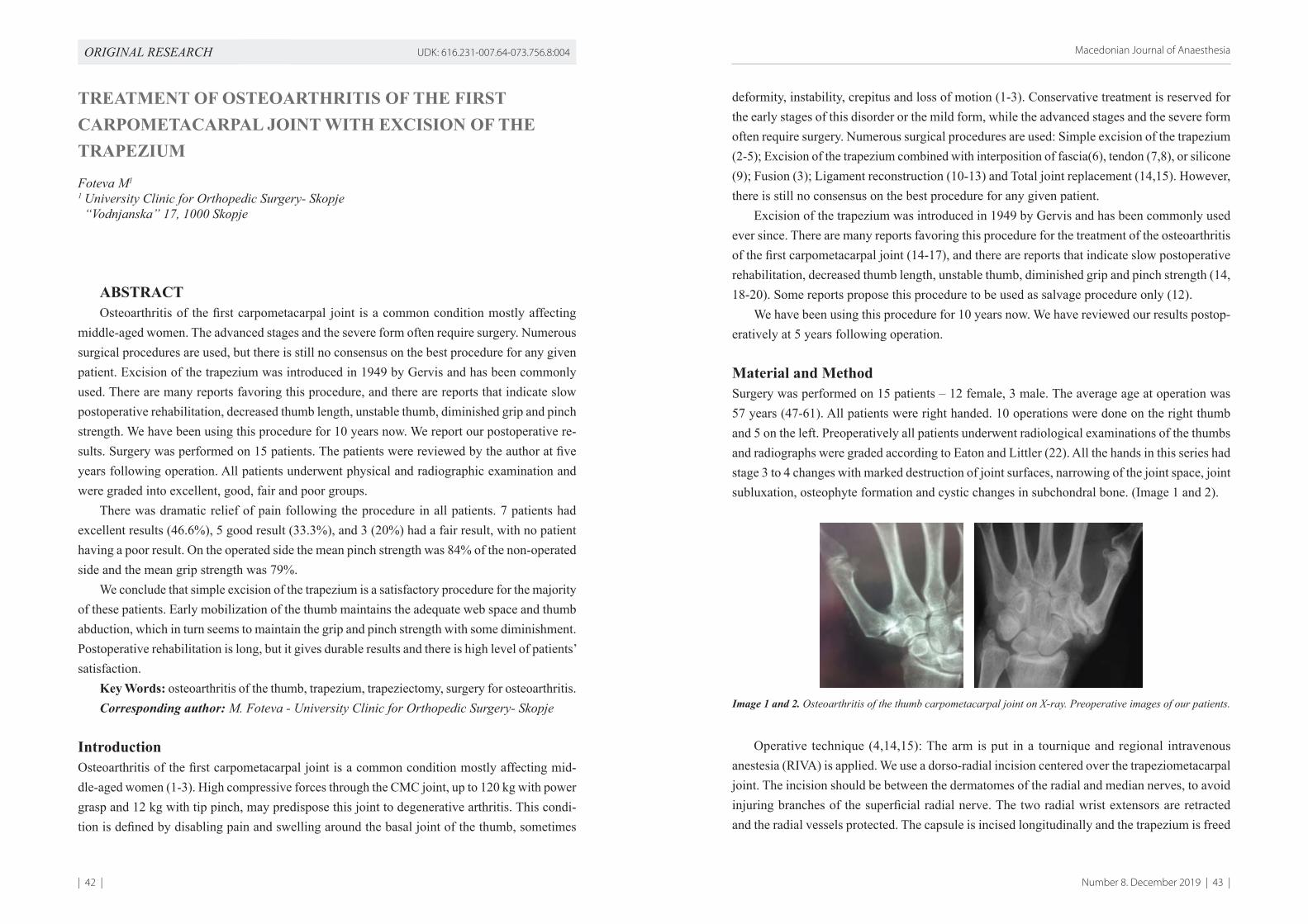
| 42 |
Macedonian Journal of Anaesthesia
Number 8. December 2019 | 43 |
ORIGINAL RESEARCH UDK: 616.231-007.64-073.756.8:004
TREATMENT OF OSTEOARTHRITIS OF THE FIRST CARPOMETACARPAL JOINT WITH EXCISION OF THE TRAPEZIUM
Foteva M1
1 University Clinic for Orthopedic Surgery- Skopje “Vodnjanska” 17, 1000 Skopje
ABSTRACTOsteoarthritis of the first carpometacarpal joint is a common condition mostly affecting
middle-aged women. The advanced stages and the severe form often require surgery. Numerous surgical procedures are used, but there is still no consensus on the best procedure for any given patient. Excision of the trapezium was introduced in 1949 by Gervis and has been commonly used. There are many reports favoring this procedure, and there are reports that indicate slow postoperative rehabilitation, decreased thumb length, unstable thumb, diminished grip and pinch strength. We have been using this procedure for 10 years now. We report our postoperative re-sults. Surgery was performed on 15 patients. The patients were reviewed by the author at five years following operation. All patients underwent physical and radiographic examination and were graded into excellent, good, fair and poor groups.
There was dramatic relief of pain following the procedure in all patients. 7 patients had excellent results (46.6%), 5 good result (33.3%), and 3 (20%) had a fair result, with no patient having a poor result. On the operated side the mean pinch strength was 84% of the non-operated side and the mean grip strength was 79%.
We conclude that simple excision of the trapezium is a satisfactory procedure for the majority of these patients. Early mobilization of the thumb maintains the adequate web space and thumb abduction, which in turn seems to maintain the grip and pinch strength with some diminishment. Postoperative rehabilitation is long, but it gives durable results and there is high level of patients’ satisfaction.
Key Words: osteoarthritis of the thumb, trapezium, trapeziectomy, surgery for osteoarthritis.Corresponding author: M. Foteva - University Clinic for Orthopedic Surgery- Skopje
IntroductionOsteoarthritis of the first carpometacarpal joint is a common condition mostly affecting mid-dle-aged women (1-3). High compressive forces through the CMC joint, up to 120 kg with power grasp and 12 kg with tip pinch, may predispose this joint to degenerative arthritis. This condi-tion is defined by disabling pain and swelling around the basal joint of the thumb, sometimes
deformity, instability, crepitus and loss of motion (1-3). Conservative treatment is reserved for the early stages of this disorder or the mild form, while the advanced stages and the severe form often require surgery. Numerous surgical procedures are used: Simple excision of the trapezium (2-5); Excision of the trapezium combined with interposition of fascia(6), tendon (7,8), or silicone (9); Fusion (3); Ligament reconstruction (10-13) and Total joint replacement (14,15). However, there is still no consensus on the best procedure for any given patient.
Excision of the trapezium was introduced in 1949 by Gervis and has been commonly used ever since. There are many reports favoring this procedure for the treatment of the osteoarthritis of the first carpometacarpal joint (14-17), and there are reports that indicate slow postoperative rehabilitation, decreased thumb length, unstable thumb, diminished grip and pinch strength (14, 18-20). Some reports propose this procedure to be used as salvage procedure only (12).
We have been using this procedure for 10 years now. We have reviewed our results postop-eratively at 5 years following operation.
Material and MethodSurgery was performed on 15 patients – 12 female, 3 male. The average age at operation was 57 years (47-61). All patients were right handed. 10 operations were done on the right thumb and 5 on the left. Preoperatively all patients underwent radiological examinations of the thumbs and radiographs were graded according to Eaton and Littler (22). All the hands in this series had stage 3 to 4 changes with marked destruction of joint surfaces, narrowing of the joint space, joint subluxation, osteophyte formation and cystic changes in subchondral bone. (Image 1 and 2).
Image 1 and 2. Osteoarthritis of the thumb carpometacarpal joint on X-ray. Preoperative images of our patients.
Operative technique (4,14,15): The arm is put in a tournique and regional intravenous anestesia (RIVA) is applied. We use a dorso-radial incision centered over the trapeziometacarpal joint. The incision should be between the dermatomes of the radial and median nerves, to avoid injuring branches of the superficial radial nerve. The two radial wrist extensors are retracted and the radial vessels protected. The capsule is incised longitudinally and the trapezium is freed
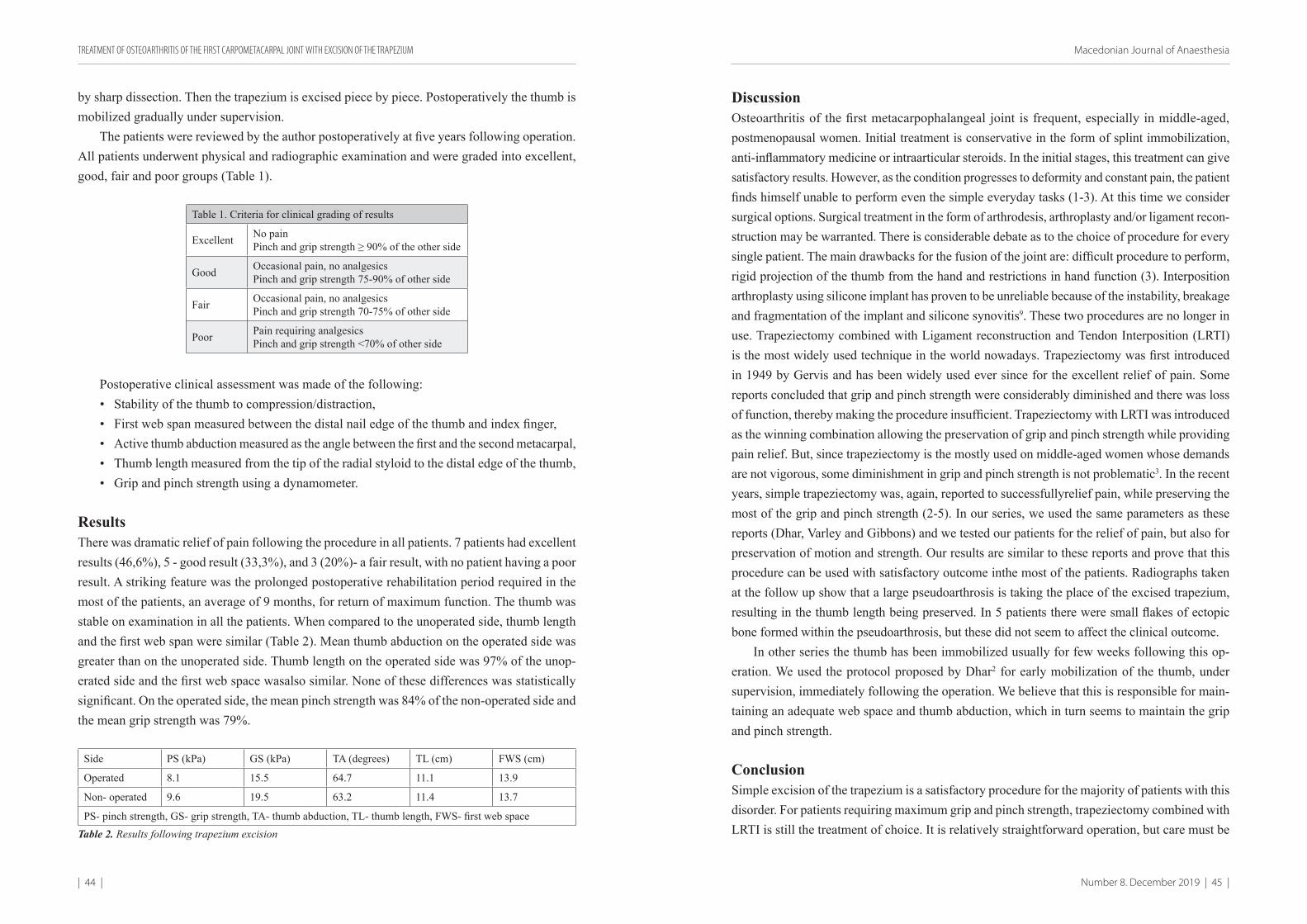
| 44 |
Macedonian Journal of Anaesthesia
Number 8. December 2019 | 45 |
by sharp dissection. Then the trapezium is excised piece by piece. Postoperatively the thumb is mobilized gradually under supervision.
The patients were reviewed by the author postoperatively at five years following operation. All patients underwent physical and radiographic examination and were graded into excellent, good, fair and poor groups (Table 1).
Table 1. Criteria for clinical grading of results
Excellent No painPinch and grip strength ≥ 90% of the other side
Good Occasional pain, no analgesicsPinch and grip strength 75-90% of other side
Fair Occasional pain, no analgesicsPinch and grip strength 70-75% of other side
Poor Pain requiring analgesicsPinch and grip strength <70% of other side
Postoperative clinical assessment was made of the following:• Stability of the thumb to compression/distraction,• First web span measured between the distal nail edge of the thumb and index finger,• Active thumb abduction measured as the angle between the first and the second metacarpal,• Thumb length measured from the tip of the radial styloid to the distal edge of the thumb,• Grip and pinch strength using a dynamometer.
ResultsThere was dramatic relief of pain following the procedure in all patients. 7 patients had excellent results (46,6%), 5 - good result (33,3%), and 3 (20%)- a fair result, with no patient having a poor result. A striking feature was the prolonged postoperative rehabilitation period required in the most of the patients, an average of 9 months, for return of maximum function. The thumb was stable on examination in all the patients. When compared to the unoperated side, thumb length and the first web span were similar (Table 2). Mean thumb abduction on the operated side was greater than on the unoperated side. Thumb length on the operated side was 97% of the unop-erated side and the first web space wasalso similar. None of these differences was statistically significant. On the operated side, the mean pinch strength was 84% of the non-operated side and the mean grip strength was 79%.
Side PS (kPa) GS (kPa) TA (degrees) TL (cm) FWS (cm)
Operated 8.1 15.5 64.7 11.1 13.9
Non- operated 9.6 19.5 63.2 11.4 13.7
PS- pinch strength, GS- grip strength, TA- thumb abduction, TL- thumb length, FWS- first web spaceTable 2. Results following trapezium excision
DiscussionOsteoarthritis of the first metacarpophalangeal joint is frequent, especially in middle-aged, postmenopausal women. Initial treatment is conservative in the form of splint immobilization, anti-inflammatory medicine or intraarticular steroids. In the initial stages, this treatment can give satisfactory results. However, as the condition progresses to deformity and constant pain, the patient finds himself unable to perform even the simple everyday tasks (1-3). At this time we consider surgical options. Surgical treatment in the form of arthrodesis, arthroplasty and/or ligament recon-struction may be warranted. There is considerable debate as to the choice of procedure for every single patient. The main drawbacks for the fusion of the joint are: difficult procedure to perform, rigid projection of the thumb from the hand and restrictions in hand function (3). Interposition arthroplasty using silicone implant has proven to be unreliable because of the instability, breakage and fragmentation of the implant and silicone synovitis9. These two procedures are no longer in use. Trapeziectomy combined with Ligament reconstruction and Tendon Interposition (LRTI) is the most widely used technique in the world nowadays. Trapeziectomy was first introduced in 1949 by Gervis and has been widely used ever since for the excellent relief of pain. Some reports concluded that grip and pinch strength were considerably diminished and there was loss of function, thereby making the procedure insufficient. Trapeziectomy with LRTI was introduced as the winning combination allowing the preservation of grip and pinch strength while providing pain relief. But, since trapeziectomy is the mostly used on middle-aged women whose demands are not vigorous, some diminishment in grip and pinch strength is not problematic3. In the recent years, simple trapeziectomy was, again, reported to successfullyrelief pain, while preserving the most of the grip and pinch strength (2-5). In our series, we used the same parameters as these reports (Dhar, Varley and Gibbons) and we tested our patients for the relief of pain, but also for preservation of motion and strength. Our results are similar to these reports and prove that this procedure can be used with satisfactory outcome inthe most of the patients. Radiographs taken at the follow up show that a large pseudoarthrosis is taking the place of the excised trapezium, resulting in the thumb length being preserved. In 5 patients there were small flakes of ectopic bone formed within the pseudoarthrosis, but these did not seem to affect the clinical outcome.
In other series the thumb has been immobilized usually for few weeks following this op-eration. We used the protocol proposed by Dhar2 for early mobilization of the thumb, under supervision, immediately following the operation. We believe that this is responsible for main-taining an adequate web space and thumb abduction, which in turn seems to maintain the grip and pinch strength.
ConclusionSimple excision of the trapezium is a satisfactory procedure for the majority of patients with this disorder. For patients requiring maximum grip and pinch strength, trapeziectomy combined with LRTI is still the treatment of choice. It is relatively straightforward operation, but care must be
TREATMENT OF OSTEOARTHRITIS OF THE FIRST CARPOMETACARPAL JOINT WITH EXCISION OF THE TRAPEZIUM
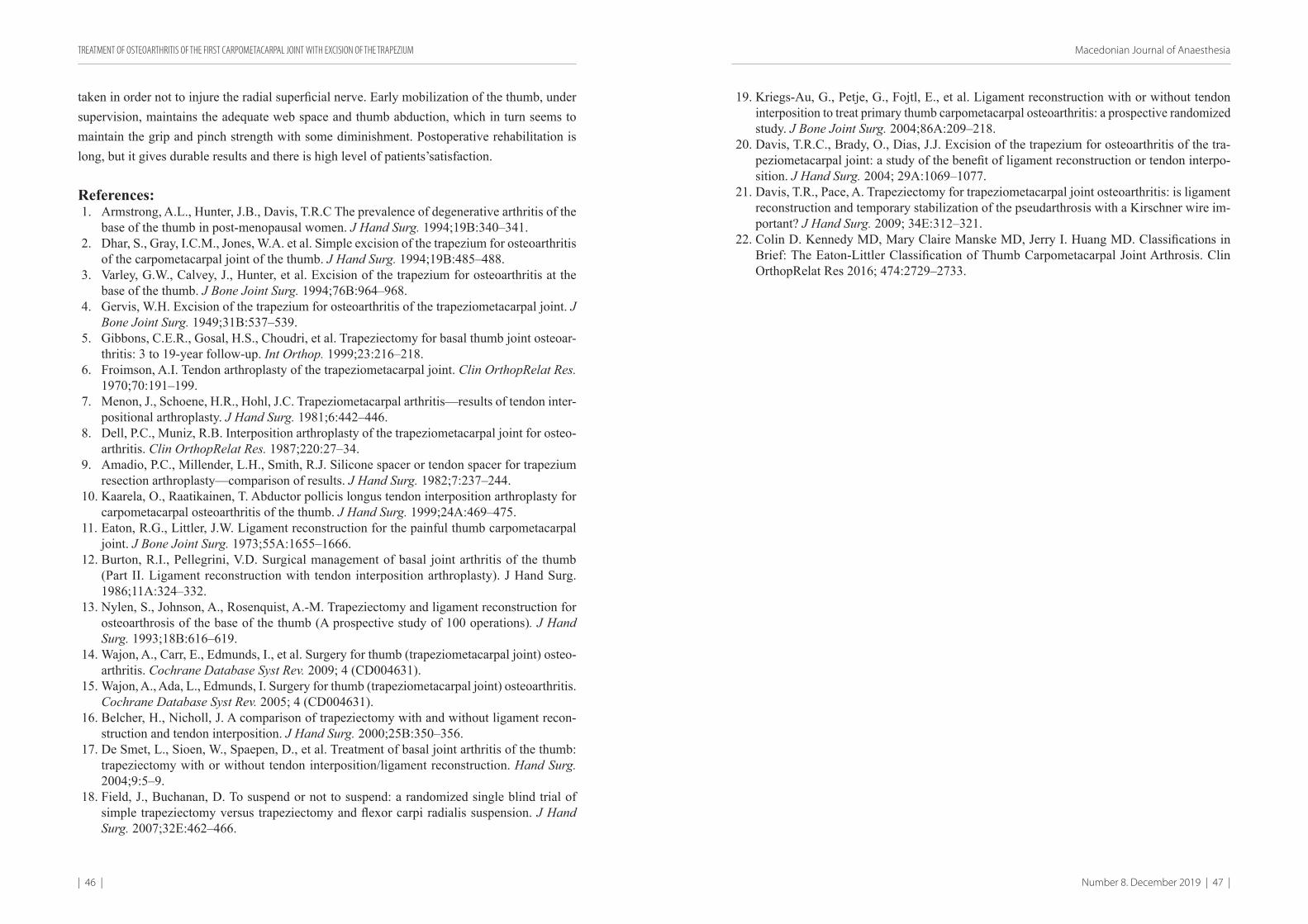
| 46 |
Macedonian Journal of Anaesthesia
Number 8. December 2019 | 47 |
taken in order not to injure the radial superficial nerve. Early mobilization of the thumb, under supervision, maintains the adequate web space and thumb abduction, which in turn seems to maintain the grip and pinch strength with some diminishment. Postoperative rehabilitation is long, but it gives durable results and there is high level of patients’satisfaction.
References:1. Armstrong, A.L., Hunter, J.B., Davis, T.R.C The prevalence of degenerative arthritis of the
base of the thumb in post-menopausal women. J Hand Surg. 1994;19B:340–341.2. Dhar, S., Gray, I.C.M., Jones, W.A. et al. Simple excision of the trapezium for osteoarthritis
of the carpometacarpal joint of the thumb. J Hand Surg. 1994;19B:485–488.3. Varley, G.W., Calvey, J., Hunter, et al. Excision of the trapezium for osteoarthritis at the
base of the thumb. J Bone Joint Surg. 1994;76B:964–968.4. Gervis, W.H. Excision of the trapezium for osteoarthritis of the trapeziometacarpal joint. J
Bone Joint Surg. 1949;31B:537–539.5. Gibbons, C.E.R., Gosal, H.S., Choudri, et al. Trapeziectomy for basal thumb joint osteoar-
thritis: 3 to 19-year follow-up. Int Orthop. 1999;23:216–218.6. Froimson, A.I. Tendon arthroplasty of the trapeziometacarpal joint. Clin OrthopRelat Res.
1970;70:191–199.7. Menon, J., Schoene, H.R., Hohl, J.C. Trapeziometacarpal arthritis—results of tendon inter-
positional arthroplasty. J Hand Surg. 1981;6:442–446.8. Dell, P.C., Muniz, R.B. Interposition arthroplasty of the trapeziometacarpal joint for osteo-
arthritis. Clin OrthopRelat Res. 1987;220:27–34.9. Amadio, P.C., Millender, L.H., Smith, R.J. Silicone spacer or tendon spacer for trapezium
resection arthroplasty—comparison of results. J Hand Surg. 1982;7:237–244.10. Kaarela, O., Raatikainen, T. Abductor pollicis longus tendon interposition arthroplasty for
carpometacarpal osteoarthritis of the thumb. J Hand Surg. 1999;24A:469–475.11. Eaton, R.G., Littler, J.W. Ligament reconstruction for the painful thumb carpometacarpal
joint. J Bone Joint Surg. 1973;55A:1655–1666.12. Burton, R.I., Pellegrini, V.D. Surgical management of basal joint arthritis of the thumb
(Part II. Ligament reconstruction with tendon interposition arthroplasty). J Hand Surg. 1986;11A:324–332.
13. Nylen, S., Johnson, A., Rosenquist, A.-M. Trapeziectomy and ligament reconstruction for osteoarthrosis of the base of the thumb (A prospective study of 100 operations). J Hand Surg. 1993;18B:616–619.
14. Wajon, A., Carr, E., Edmunds, I., et al. Surgery for thumb (trapeziometacarpal joint) osteo-arthritis. Cochrane Database Syst Rev. 2009; 4 (CD004631).
15. Wajon, A., Ada, L., Edmunds, I. Surgery for thumb (trapeziometacarpal joint) osteoarthritis. Cochrane Database Syst Rev. 2005; 4 (CD004631).
16. Belcher, H., Nicholl, J. A comparison of trapeziectomy with and without ligament recon-struction and tendon interposition. J Hand Surg. 2000;25B:350–356.
17. De Smet, L., Sioen, W., Spaepen, D., et al. Treatment of basal joint arthritis of the thumb: trapeziectomy with or without tendon interposition/ligament reconstruction. Hand Surg. 2004;9:5–9.
18. Field, J., Buchanan, D. To suspend or not to suspend: a randomized single blind trial of simple trapeziectomy versus trapeziectomy and flexor carpi radialis suspension. J Hand Surg. 2007;32E:462–466.
19. Kriegs-Au, G., Petje, G., Fojtl, E., et al. Ligament reconstruction with or without tendon interposition to treat primary thumb carpometacarpal osteoarthritis: a prospective randomized study. J Bone Joint Surg. 2004;86A:209–218.
20. Davis, T.R.C., Brady, O., Dias, J.J. Excision of the trapezium for osteoarthritis of the tra-peziometacarpal joint: a study of the benefit of ligament reconstruction or tendon interpo-sition. J Hand Surg. 2004; 29A:1069–1077.
21. Davis, T.R., Pace, A. Trapeziectomy for trapeziometacarpal joint osteoarthritis: is ligament reconstruction and temporary stabilization of the pseudarthrosis with a Kirschner wire im-portant? J Hand Surg. 2009; 34E:312–321.
22. Colin D. Kennedy MD, Mary Claire Manske MD, Jerry I. Huang MD. Classifications in Brief: The Eaton-Littler Classification of Thumb Carpometacarpal Joint Arthrosis. Clin OrthopRelat Res 2016; 474:2729–2733.
TREATMENT OF OSTEOARTHRITIS OF THE FIRST CARPOMETACARPAL JOINT WITH EXCISION OF THE TRAPEZIUM
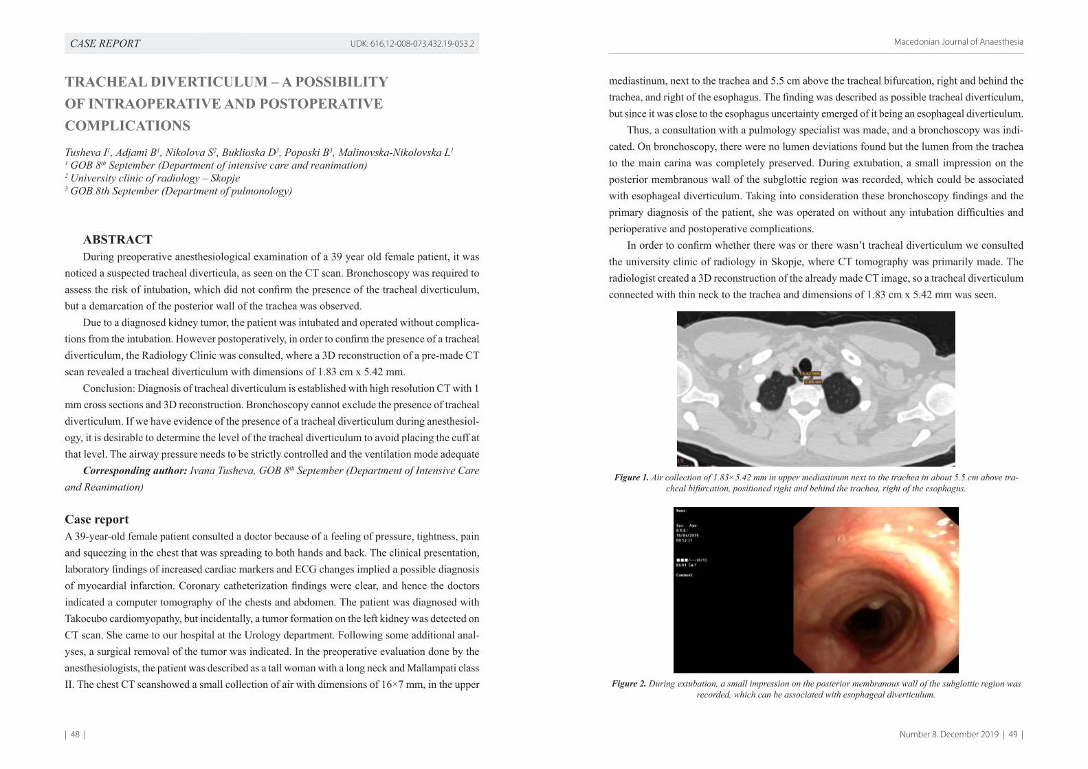
| 48 |
Macedonian Journal of Anaesthesia
Number 8. December 2019 | 49 |
CASE REPORT UDK: 616.12-008-073.432.19-053.2
TRACHEAL DIVERTICULUM – A POSSIBILITY OF INTRAOPERATIVE AND POSTOPERATIVE COMPLICATIONS
Tusheva I1, Adjami B1, Nikolova S2, Buklioska D3, Poposki B3, Malinovska-Nikolovska L1
1 GOB 8th September (Department of intensive care and reanimation)2 University clinic of radiology – Skopje3 GOB 8th September (Department of pulmonology)
ABSTRACTDuring preoperative anesthesiological examination of a 39 year old female patient, it was
noticed a suspected tracheal diverticula, as seen on the CT scan. Bronchoscopy was required to assess the risk of intubation, which did not confirm the presence of the tracheal diverticulum, but a demarcation of the posterior wall of the trachea was observed.
Due to a diagnosed kidney tumor, the patient was intubated and operated without complica-tions from the intubation. However postoperatively, in order to confirm the presence of a tracheal diverticulum, the Radiology Clinic was consulted, where a 3D reconstruction of a pre-made CT scan revealed a tracheal diverticulum with dimensions of 1.83 cm x 5.42 mm.
Conclusion: Diagnosis of tracheal diverticulum is established with high resolution CT with 1 mm cross sections and 3D reconstruction. Bronchoscopy cannot exclude the presence of tracheal diverticulum. If we have evidence of the presence of a tracheal diverticulum during anesthesiol-ogy, it is desirable to determine the level of the tracheal diverticulum to avoid placing the cuff at that level. The airway pressure needs to be strictly controlled and the ventilation mode adequate
Corresponding author: Ivana Tusheva, GOB 8th September (Department of Intensive Care and Reanimation)
Case reportA 39-year-old female patient consulted a doctor because of a feeling of pressure, tightness, pain and squeezing in the chest that was spreading to both hands and back. The clinical presentation, laboratory findings of increased cardiac markers and ECG changes implied a possible diagnosis of myocardial infarction. Coronary catheterization findings were clear, and hence the doctors indicated a computer tomography of the chests and abdomen. The patient was diagnosed with Takocubo cardiomyopathy, but incidentally, a tumor formation on the left kidney was detected on CT scan. She came to our hospital at the Urology department. Following some additional anal-yses, a surgical removal of the tumor was indicated. In the preoperative evaluation done by the anesthesiologists, the patient was described as a tall woman with a long neck and Mallampati class II. The chest CT scanshowed a small collection of air with dimensions of 16×7 mm, in the upper
mediastinum, next to the trachea and 5.5 cm above the tracheal bifurcation, right and behind the trachea, and right of the esophagus. The finding was described as possible tracheal diverticulum, but since it was close to the esophagus uncertainty emerged of it being an esophageal diverticulum.
Thus, a consultation with a pulmology specialist was made, and a bronchoscopy was indi-cated. On bronchoscopy, there were no lumen deviations found but the lumen from the trachea to the main carina was completely preserved. During extubation, a small impression on the posterior membranous wall of the subglottic region was recorded, which could be associated with esophageal diverticulum. Taking into consideration these bronchoscopy findings and the primary diagnosis of the patient, she was operated on without any intubation difficulties and perioperative and postoperative complications.
In order to confirm whether there was or there wasn’t tracheal diverticulum we consulted the university clinic of radiology in Skopje, where CT tomography was primarily made. The radiologist created a 3D reconstruction of the already made CT image, so a tracheal diverticulum connected with thin neck to the trachea and dimensions of 1.83 cm x 5.42 mm was seen.
Figure 1. Air collection of 1.83×5.42 mm in upper mediastinum next to the trachea in about 5.5.cm above tra-cheal bifurcation, positioned right and behind the trachea, right of the esophagus.
Figure 2. During extubation, a small impression on the posterior membranous wall of the subglottic region was recorded, which can be associated with esophageal diverticulum.
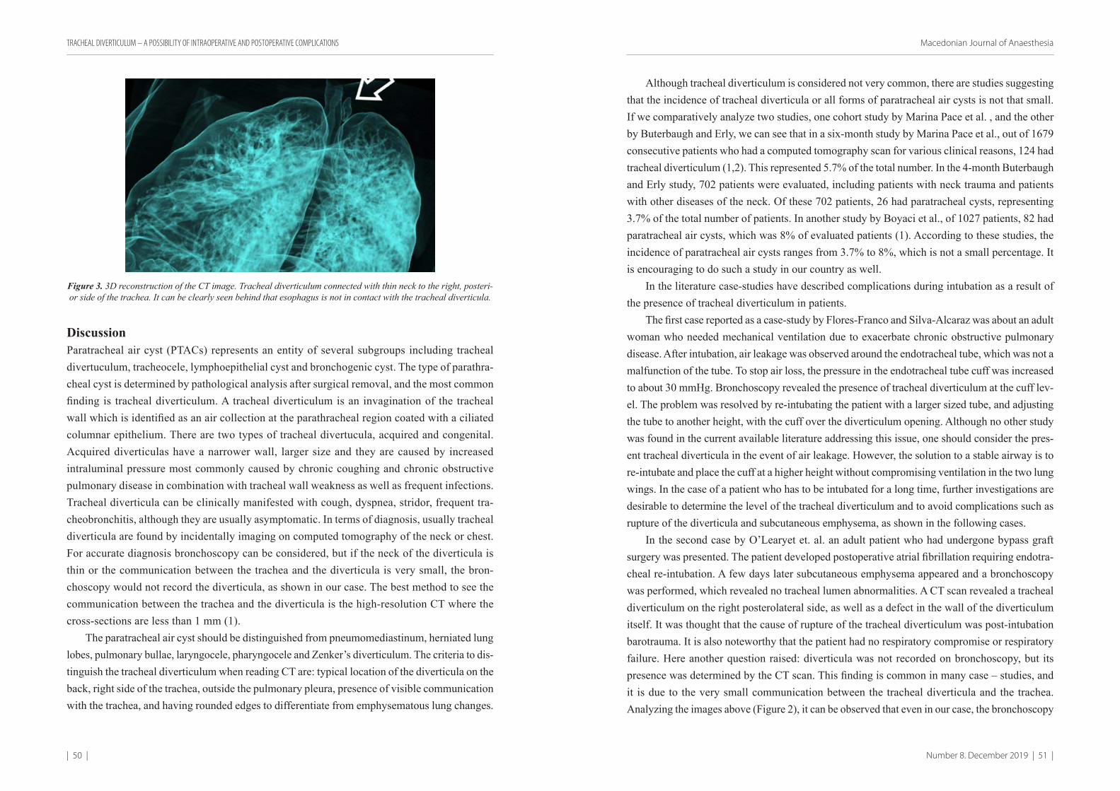
| 50 |
Macedonian Journal of Anaesthesia
Number 8. December 2019 | 51 |
Figure 3. 3D reconstruction of the CT image. Tracheal diverticulum connected with thin neck to the right, posteri-or side of the trachea. It can be clearly seen behind that esophagus is not in contact with the tracheal diverticula.
DiscussionParatracheal air cyst (PTACs) represents an entity of several subgroups including tracheal divertuculum, tracheocele, lymphoepithelial cyst and bronchogenic cyst. The type of parathra-cheal cyst is determined by pathological analysis after surgical removal, and the most common finding is tracheal diverticulum. A tracheal diverticulum is an invagination of the tracheal wall which is identified as an air collection at the parathracheal region coated with a ciliated columnar epithelium. There are two types of tracheal divertucula, acquired and congenital. Acquired diverticulas have a narrower wall, larger size and they are caused by increased intraluminal pressure most commonly caused by chronic coughing and chronic obstructive pulmonary disease in combination with tracheal wall weakness as well as frequent infections. Tracheal diverticula can be clinically manifested with cough, dyspnea, stridor, frequent tra-cheobronchitis, although they are usually asymptomatic. In terms of diagnosis, usually tracheal diverticula are found by incidentally imaging on computed tomography of the neck or chest. For accurate diagnosis bronchoscopy can be considered, but if the neck of the diverticula is thin or the communication between the trachea and the diverticula is very small, the bron-choscopy would not record the diverticula, as shown in our case. The best method to see the communication between the trachea and the diverticula is the high-resolution CT where the cross-sections are less than 1 mm (1).
The paratracheal air cyst should be distinguished from pneumomediastinum, herniated lung lobes, pulmonary bullae, laryngocele, pharyngocele and Zenker’s diverticulum. The criteria to dis-tinguish the tracheal diverticulum when reading CT are: typical location of the diverticula on the back, right side of the trachea, outside the pulmonary pleura, presence of visible communication with the trachea, and having rounded edges to differentiate from emphysematous lung changes.
Although tracheal diverticulum is considered not very common, there are studies suggesting that the incidence of tracheal diverticula or all forms of paratracheal air cysts is not that small. If we comparatively analyze two studies, one cohort study by Marina Pace et al. , and the other by Buterbaugh and Erly, we can see that in a six-month study by Marina Pace et al., out of 1679 consecutive patients who had a computed tomography scan for various clinical reasons, 124 had tracheal diverticulum (1,2). This represented 5.7% of the total number. In the 4-month Buterbaugh and Erly study, 702 patients were evaluated, including patients with neck trauma and patients with other diseases of the neck. Of these 702 patients, 26 had paratracheal cysts, representing 3.7% of the total number of patients. In another study by Boyaci et al., of 1027 patients, 82 had paratracheal air cysts, which was 8% of evaluated patients (1). According to these studies, the incidence of paratracheal air cysts ranges from 3.7% to 8%, which is not a small percentage. It is encouraging to do such a study in our country as well.
In the literature case-studies have described complications during intubation as a result of the presence of tracheal diverticulum in patients.
The first case reported as a case-study by Flores-Franco and Silva-Alcaraz was about an adult woman who needed mechanical ventilation due to exacerbate chronic obstructive pulmonary disease. After intubation, air leakage was observed around the endotracheal tube, which was not a malfunction of the tube. To stop air loss, the pressure in the endotracheal tube cuff was increased to about 30 mmHg. Bronchoscopy revealed the presence of tracheal diverticulum at the cuff lev-el. The problem was resolved by re-intubating the patient with a larger sized tube, and adjusting the tube to another height, with the cuff over the diverticulum opening. Although no other study was found in the current available literature addressing this issue, one should consider the pres-ent tracheal diverticula in the event of air leakage. However, the solution to a stable airway is to re-intubate and place the cuff at a higher height without compromising ventilation in the two lung wings. In the case of a patient who has to be intubated for a long time, further investigations are desirable to determine the level of the tracheal diverticulum and to avoid complications such as rupture of the diverticula and subcutaneous emphysema, as shown in the following cases.
In the second case by O’Learyet et. al. an adult patient who had undergone bypass graft surgery was presented. The patient developed postoperative atrial fibrillation requiring endotra-cheal re-intubation. A few days later subcutaneous emphysema appeared and a bronchoscopy was performed, which revealed no tracheal lumen abnormalities. A CT scan revealed a tracheal diverticulum on the right posterolateral side, as well as a defect in the wall of the diverticulum itself. It was thought that the cause of rupture of the tracheal diverticulum was post-intubation barotrauma. It is also noteworthy that the patient had no respiratory compromise or respiratory failure. Here another question raised: diverticula was not recorded on bronchoscopy, but its presence was determined by the CT scan. This finding is common in many case – studies, and it is due to the very small communication between the tracheal diverticula and the trachea. Analyzing the images above (Figure 2), it can be observed that even in our case, the bronchoscopy
TRACHEAL DIVERTICULUM – A POSSIBILITY OF INTRAOPERATIVE AND POSTOPERATIVE COMPLICATIONS
| 52 |
Macedonian Journal of Anaesthesia
Number 8. December 2019 | 53 |
revealed no communication with the trachea and its diverticulum. Тhe impression clearly visible on the back wall of the trachea, is compared to Figure 3, is exactly at the level of the tracheal diverticulum. It can be concluded that bronchoscopy does not always determine the presence of tracheal diverticulum, and the best way to diagnose tracheal diverticulum is CT scan where the cross sections are less than 1 mm, as mentioned above.
In the third case presents a patient at the age of 55 who comes to the doctor for arthroscopic shoulder surgery. Because of Cormack-Lehane classification 3, the patient was intubated using a boogie, and the tube number 8. After the intubation, no ETCO2 curve initially appeared, while saturation remained 99%. With the help of a laryngoscope, the location of the endotracheal tube was checked. After it was confirmed that it was in the right place the ETCO2 curve showed up. After 15 minutes, while interscalene block was applied, deviations in the parameters were observed, the peak increased to 42 cm H2O, ETCO2 60 mmHg and SpO2 dropped to 88%. On auscultation crackles and rhonchi were heard all over the chest. Six hours postoperatively, the patient developed subcutaneous emphysema, and chest x-ray revealed pneumomediastinum and right pneumothorax. Bronchoscopy revealed no tracheal damage, but CT scan revealed a tracheal cyst on the tracheal posterior wall 10-20 mm in size along with a 20 mm tracheal wall defect located 2 cm above the carina. With 3D computed tomography reconstruction, very little communication between the trachea and the cyst was seen. The cause of all these complications was a rupture of the tracheal cyst. This is the first study in the literature to show deviation of the parameters during a tracheal diverticulum (cyst) rupture, probably due to the location of the diverticulum.
In the fourth case reported by Chauhan et al. a 77-year-old patient was presented, in whom a cardiopulmonary resuscitation was necessary because of a cardiopulmonary arrest. Difficulties occurred during intubation and discreet hemorrhage from the endotracheal tube was observed after hospitalization. Bronchoscopy revealed a rupture in the wall on the right lateral side of the trachea, covered with thrombotic masses. On computed tomography, there were discrepancies in the morphology of the trachea, but no trachea rupture was detected. Bronchoscopy was again performed and determined the presence of a large acquired tracheal diverticulum without apparent rupture. The patient was extubated without difficulty and consequences. In this case, the compli-cation was detected at the act of intubation itself. Such bleeding may be associated with rupture of the trachea, but the overall condition of the patient should be taken into account, as well as the correctness of the parameters, such as ETCO2 curve, respiratory volume and, a good SpO2.
The fifth case reported as a case study by Davies R. is also a 77-year-old patient who came to the doctor for a thigh bone fracture. Anesthesiological evaluation established Mallampati grade one with good neck mobility although it had thoracic kyphosis. The patient had pre-viously reported some difficulties in breathing, but this was not clinically examined because doctors thought it was a consequence of kyphosis. Manual ventilation was easy, but there was difficulty in intubation, so a tube of size 8 could not pass more than 2 cm from the vocal cords. Anesthesiologists had the same problem with tubes size 7 as well as size 6. After these attempts,
a laryngeal mask was inserted. Postoperatively a bronchoscopy attempt was made, which was also not successful. On computed tomography of the neck, a tracheal diverticulum about 2 cm below the vocal cords was detected as well as a 90-degree bend in the subglottic trachea due to pronounced cervical lordosis resulting from kyphosis. Although the reason for the impossibility of intubation in this case was not only the tracheal diverticulum, the tracheal diverticulum is still the reason that the tube does not go deeper than 2 cm. Most likely, the tube entered the diverticula because of the angle at which it entered. However, there is another paper published by Kim HJ. et al. showing difficulties in intubation in a patient with severe kyphosis. It is interesting that in this patient, beside kyphosis, CT scan revealed a narrowing of the tracheal lumen as a result of previous pulmonary diseases. This is significant because it suggests that not only the tracheal diverticulum may be the cause of difficult intubation but also other changes of the trachea such as stenosis. In any case, if there is a patient with severe kyphosis, it is desirable to pre-operatively evaluate the airways in order to determine the risk of aggravated intubation. Patients with long neck, frequent respiratory infections and cough, pronounced kyphosis and deformity of the spinal cord should be associated with the possibility of present tracheal diverticulum.
The reported cases showed complications from the presence of a tracheal diverticulum. However, in our case there were no complications although it was not concluded that the tracheal diverticulum did not exist. If we analyze the images, especially Figure 3. where the demarcation made by the diverticula is very high on the tracheal wall and Figure 4 which determines the height of the tracheal diverticulum, it can be concluded that the complications did not occur because the endotracheal tube cuff was placed below the neck of the tracheal diverticulum, ventilation was performed with adequate pressure and the throat of the tracheal diverticulum was tight.
ConclusionsAlthough neglected as a finding, the tracheal diverticulum and its rupture can lead to significant consequences such as insufficient ventilation, subcutaneous emphysema, difficult or impossible intubation. The diagnosis of tracheal diverticulum is made with high-resolution CT scan and 1 mm cross-sections, and it is desirable to perform a 3D reconstruction. Bronchoscopy can be used in emergencies, especially if tracheal rupture is suspected, but it cannot rule out the presence of tracheal diverticulum as well as its defect. These patients require good anesthesiological pre-op-erative evaluation, a prepared anesthesia plan, careful anesthesia management and prolonged postoperative supervision.
References1. Tanrivermis Sayit, A., Elmali, M., Saglam, et al. The diseases of airway-tracheal divertic-
ulum: a review of the literature. Journal of thoracic disease, (2016), 8(10), E1163–E1167.2. Marina Pace MD, ∗ Annarita Dapoto MD, Alessandra Surace MD, et al. Tracheal divertic-
ula - A retrospective analysis of patients referred for thoracic CTMDMedicine (Baltimore). 2018 Sep; 97(39): e12544.
TRACHEAL DIVERTICULUM – A POSSIBILITY OF INTRAOPERATIVE AND POSTOPERATIVE COMPLICATIONS
| 54 |
Macedonian Journal of Anaesthesia
Number 8. December 2019 | 55 |
3. J.E. Buterbaugh, W.K. Erly. Paratracheal Air Cysts: A Common Finding on Routine CT Examinations of the Cervical Spine and Neck That May Mimic Pneumomediastinum in Patients With Traumatic Injuries. American Journal of Neuroradiology Jun 2008, 29 (6) 1218-1221;
4. Boyaci, N., Sen Dokumaci, D., Karakas, et al. Paratracheal air cysts: prevalence and relevance to pulmonary emphysema and bronchiectasis using thoracic multidetector CT. Diagnostic and interventional radiology (Ankara, Turkey)2015. 21(1), 42–46.
5. Flores-Franco, R.A., & Silva-Alcaraz, J. Acquired Tracheal Diverticulum as an Unexpected Cause of Endotracheal Tube Cuff Leak. Journal of critical care medicine (Universitatea de Medicina si Farmacie din Targu-Mures) 2015. 1(4), 171–173.
6. O’Leary, C.N., Ryan, J.W., Corbett, et al. Barotrauma induced tracheal diverticulum rupture: imaging findings. BMJ case reports, 2016, bcr2016217518.
7. Allaert, S., Lamont, J., Kalmar, A.F. et al. Tracheal diverticulum as a cause of subcutaneous emphysema following positive-pressure ventilation. Can J Anesth/J Can Anesth. 2016. 63: 1098.
8. Carmona Soto P, et al. Divertículo traquealadquirido como causa de intubación orotraqueal complicada. Arch Bronconeumol. 2011. 48:64–5.
9. Davies R. Difficult tracheal intubation secondary to a tracheal diverticulum and a 90 degree deviation in the trachea. Anaesthesia 2000; 55: 923–925.
10. Kim, H.J., Choi, Y.S., Park, et al. Difficult endotracheal intubation secondary to tracheal deviation and stenosis in a patient with severe kyphoscoliosis: a case report. Korean journal of anesthesiology. 2016. 69(4), 386–389.
11. Sharma M, Bulathsinghala CP, Khan A, et al. An Unusual Case of Iatrogenic Tracheal Diverticulum Found in a Mechanically Ventilated Patient: To Treat or Not to Treat. Cureus. 2019;11(10):e5911.
12. Kim HY, Lee KH, Kim YJ, et al. Incidental paratracheal air cyst on thoracic CT and their association with chronic inflammatory lung disease. Biomed Res Int 2017;2017:8740635.
13. Cheng HM, Chang PY, Chiang KH, et al. Prevalence and characteristics of paratracheal air cysts and their association with emphysema in a general population. Eur J Radiol. 2012;81:2673–2677.
14. Silvie A., Jan L., Alain F.K., et al. Tracheal diverticulum as a cause of subcutaneous emphy-sema following positive-pressure ventilation. Can J Anesth. 2016 63:1098–1099.
15. Kang Y, Lee YK, Lee HS, et al. Tracheal injury as a perforation of a newly formed tracheal diverticulum after tracheal intubation: A case report. Anesth Pain Med. 2015 Jan;10(1):32-35
16. Tanrivermis Sayit A, Elmali M, Saglam D, et al. The diseases of airway-tracheal diverticu-lum: a review of the literature. J Thorac Dis. 2016;8(10):E1163–E1167.
17. B. Mazul‐Sunko D. Zdenčar I. Kožul J. Špiček‐Macan D. Stančić‐Rokotov. Pneumomediastinum related to distal tracheal diverticulum. Anaesthesia. 2013. 68, 3; 432-433.
АПСТРАКТПри анестезиолошки преглед на 39 годишна пациентка, беше детектиран наод за
суспектен трахеален дивертикулум, виден на компјутерска томографија. За процена на ризикот за интубација беше побарана бронхоскопија, која не го потврди присуството на трахеалниот дивертикум, но беше забележана демаркација на задниот ѕид на трахеата. Поради поставената дијагноза, тумор на бубрег, пациентка беше интубирана и оперирана без компликации од интубацијата. Сепак постоперативно, со цел да се потврди присуството на трахеалниот дивертикум беше консултирана Клиника за Радиологија, каде на 3D реконструкција од претходно направената компјутерска томографија, се детектираше трахеален дивертикулум со димензии од 1.83 cm × 5.42 mm.
Заклучок: Дијагнозата за трахеален дивертикулум се поставува со CT со висока резолуција и пресекци од 1 mm и 3D реконструкција на истата. Бронхоскопија не може да исклучи присуство на трахеален дивертикулум. Доколку добиеме податок за присуство на трахеален дивертикулум при анестезиолошкиот преглед, пожелно е да се утврди нивото на трахеалниот дивертикулум за да се избегне пласирање на кафот на тоа ниво. Притисокот во дишните патишта потребно е да биде строго контролиран и модусот на вентилација соодветен.
TRACHEAL DIVERTICULUM – A POSSIBILITY OF INTRAOPERATIVE AND POSTOPERATIVE COMPLICATIONS
| 56 |
Macedonian Journal of Anaesthesia
Number 8. December 2019 | 57 |
ORIGINAL RESEARCH UDK: 616.379-008.64-06:616.153.284
BEDSIDE ECHOCARDIOGRAPHY IN A HEMODYNAMICALLY UNSTABLE CHILD
Naumovski F1, Toleska M1, Kuzmanovska B1, Kartalov A1, Trposka A1, Donev Lj1
1 PHI – University Clinic “Mother Theresa” – Skopje Department of Anesthesia, Reanimation and Intensive Care
Introduction:Echocardiography may be considered as an important tool due to hemodynamic assessment of a hemodynamically unstable child in a Pediatric Intensive Care Units (PICU), which could provide identification of the etiology and pathophysiology of hemodynamic insta-bility but also to help due to guiding therapy.
Objectives: The aim of our cohort study is to determine the impact of usage of fast focused echocardiography examination in the Surgical PICU done by Anesthesiologist/Intensivist.
Material and Methods: We’ve made a fast focused bedside transthoracic echocardiographic examination of 11 hemodynamically unstable children using the regular parasternal long and short axis view and four chamber subcostal view of the heart, LVOT, aorta, truncus pulmonalis and Inferior vena cava (IVC) for qualitative assessment. Dimensions and collapsibility of IVC were measured. The youngest patient was premature neonate born in 28th gestation week and the oldest was 12 years old child.
Results: In 9 out of 11 children we’ve identified abnormality by the qualitative assessment of the heart due to performing focused echocardiography. In 4 children we found global myocardial hypokinesia, one child with insignificant pericardial effusion (2 mm) and 4 children with Inferior vena cava collapsibility greater than 50%. In the children with hypokinesia inotropic support was established while the children with IVC collapsibility greater than 50% were treated with fluid boluses. Persistence of foramen ovale as a random finding was seen in 5 neonates, insignificant for the hemodynamic instability.
Conclusion: Several studies have shown the positive effect of the echocardiography usage in the management of critically ill children, changing their treatment in 30%–60% of cases after the test is performed (2). From our study we can conclude that performing focused echocardi-ography is feasible and provides valuable data which could lead to improved care of severely ill children and thus should be encouraged in daily ICU practice.
Key words: bedside echocardiography, hemodynamic instability, transthoracic echocardiography
Corresponding author: Filip Naumovski, University Clinic “Mother Theresa” – SkopjeDepartment of Anesthesia, Reanimation and Intensive Care
IntroductionEchocardiography may be considered as an important tool due to hemodynamic assessment of a hemodynamically unstable child in a Pediatric Intensive Care Units (PICU). As a diagnostic modality provides valuable information about the type of shock, selecting a suitable therapeutic intervention and evaluation of the patient’s response to therapy (1). Evaluation of the cardiac function, contractility, cardiac output, fluid responsiveness and cardiac anatomy can be helpful for the Pediatric Intensivist in understanding the etiology and pathophysiology of the critical illness but also could help in guiding therapy. Using the transthoracic echocardiography (TTE) as a non-invansive, risk free and easily repeatable method of bedside monitoring of the hemo-dynamic status in the critically ill children has shown a promising positive effect changing the therapy in 30-60% of the patients after the exam is performed (2).
ObjectivesThe aim of our cohort study is to determine the role of bedside echocardiography in monitoring of the hemodynamics and the impact of usage of fast focused transthoracic echocardiography examination on the process of guiding therapy in hemodynamically unstable children admitted in the Surgical PICU, done by Anesthesiologist/Intensivist.
Material and MethodsWe’ve made a fast focused bedside transthoracic echocardiographic examination of 11 hemody-namically unstable children using the regular parasternal long and short axis view, four chamber and subcostal view of the heart, LVOT, aorta, truncus pulmonalis and Inferior vena cava (IVC) for qualitative assessment. Dimensions and collapsibility of IVC near to inlet in right ventricule were measured in M-mode. Color Doppler for identification of regurgitation existence, turbulent blood flow or persistence of pathological interatrial or interventricular communication was used. For this purpose we’ve used echocardiography machine SIEMENS Aucson S1000 and cardiac probe due to performing the exam. The youngest patient included in the study was premature neonate born in 28th gestation week and the oldest was 12 years old child while the value for the mean age among children included in the study was 3.4 years. The basic inclusion criterion was the presence of hemodynamic instability in the children admitted in our surgical PICU. Under the term “hemodynamic instability” we consider all the children with pathological values for the vital signs as heart rate (HR) and/or systolic blood pressure (SBP) according to the values provided by the American Heart Association (AHA). Respectively, all the neonates with HR <85bpm or >205bpm and/or systolic BP <60mmHg, infants with HR <100bpm or >190bpm and/or systolic BP <70mmHg, children from 1-10 years old with HR <60bpm or >140bpm and/or systolic BP <70+(age in years x2) and children >10 years old with HR <60bpm and/or >100bpm or systol-ic BP <90mmHg were included in the study. The examination of the children was made by an Anesthesiologist/Intensivist. Due to examination the children were in the supine position and
| 58 |
Macedonian Journal of Anaesthesia
Number 8. December 2019 | 59 |
all of them were demanding mechanical ventilatory support. During the qualitative examination we’ve analyzed the cardiac contractility, existence of pericardial effusion, possible existence of tricuspidal, mitral or aortic regurgitation registrated with color Dopller or existence of any other anatomical abnormalities. Collapsibility of IVC was analyzed also. The cardiac contractility was qualitatively assessed and described as hypercontractility, normal contractility or hypocontractility (hypokinesia, global or segmental). Existence of pericardial effusion was described as significant (>4 mm) or non significant (<4mm). Diameter of IVC was measured but also collapsibility of IVC was analyzed and described as greater or less than 50%. The highest and the lowest IVC diam-eter according to the respiratory cycle were measured in M-mode and collapsibility of IVC was calculated by IVCd(min)/IVCd(max)x100. All patients with collapsibility greater than 50% were considered as a fluid depleted or fluid responders. We’ve performed a bedside echocardiographyc examination in a critically ill children with signs of haemodynamical instability tending to reveal the presence of any pathological signs as a possible cause for the hemodynamical instability.
ResultsIn 9 out of 11 children (81%) we’ve identified abnormality only by the qualitative assessment of the heart. According to the findings of the echocardiographic examination in 5 out of 11 (45%) children we’ve made a change in the therapy strategy. In 4 children we’ve identified impairment of the myocardial contractility described as global myocardial hypokinesia. In two of the children the global myocardial hypokinesia was result of existence of a cardiac trauma due to polythrauma and one of those children had a massive bilateral pleural effusion verified ultrasonografically as a random finding. In the other two children with verified myocardial hypokinesia the possible cause of the impaired contractility was the existence of severe septic shock. Only in one child with polyserositis due to Ketoacidosis we’ve found existence of pericardial effusion which was described as insignificant according to the effusion measurements (2 mm). In another 4 chil-dren we found an Inferior vena cava collapsibility greater than 50%. All those children were tachycardic with normal myocardial contractility or slightly hypercontractile movements of the cardiac walls. According to the findings they were considered as volume depleted or volume responders. In the children with verified hypokinesia, an inotropic support with Dopamine or Dobutamine was established immediately. While in the children with IVC collapsibility greater than 50% we considered them as volume depleted and volume responders, so they were treat-ed with fluid boluses. We found existence of foramen ovale apertum as a random finding in 5 neonates. Three out of 5 neonates were born as a preterm babies. Foramen ovale apertum was diagnosed while examining the children using two dimensional four chamber view of the heart and the persistence of foramen ovale was confirmed with using color Doppler technique. We consider the existence of foramen ovale as a insignificant for the hemodynamic instability in all of the children because of its small size measured with mean value of 2.9 mm and because of normal right and left cardiac dimensions (3).
DiscussionEchocardiography is a widely used diagnostic modality. Assessing the myocardial structure and function in pediatric intensive care settings could be done by using bedside echocardiography (4). The usage of bedside echocardiography for diagnostic purposes still remains important, but it’s use as a real time hemodynamic monitoring tool has not been well established yet (4). According to Spencer KT the goal of using focused echocardiography as a real time monitor of the hemodynamics is to recognize a narrow list of abnormalities which are detectable by phy-sicians with limited ultrasound training and could have high clinical value (5). In contrast of using the central venous pressure (CVP) and arterial pressure (AP) measurements as an invansive modalities of monitoring of the hemodynamics the bedside echocardiography is a non invansive, easy, repeatable and non risky mode of assessment of the actual hemodynamic state providing significant information not only for diagnosing shock and hemodynamic instability but rather more deep information about the etiology and pathophysiology of the shock. We’ve identified presence of abnormal finding and impaired cardiac function in 81% of the examined children due to performed bedside focused echocardiography exam and the information gained from the examination of the children guided us to make changes in the therapy strategy in 45% of the examined children. Also, using bedside echocardiography provided information that could lead to changing the mode of therapy in 37% of the patients according to Manasia AR et al.(6) and 30-60% of the patients according to Helosia AG (2). Assessment of the myocardial contractility is more than essential in a hemodynamically unstable children, especially the contractility of the left ventricule which could be assessed qualitatively and quantitatively. Actually, the qual-itative analysis of the left ventricular (LV) systolic function consists of the visual analysis of the myocardial contractile function and is the method of choice for the assessment of the LV function by nonechocardiographers (7). We performed qualitative assessment of the myocardial contractility only because we considered that is more rapid and time saving procedure tat could provide sufficient information about the patophysiology of the hemodynamic instability in the children. According to Ünlüer E et al. the qualitative assessment of the myocardial contractility is readily and more quickly performed instead of the formal quantitative methods of estimation, which are in fact more time consuming (7). Using qualitative assessment of the myocardial contractility only, we found global myocardial hypokinesia in 4 children. Bedside echocardiog-raphy offers possibility for rapid diagnosis of pericardial effusion (PE) in critically ill children where PE is identified as an echolucent space adjacent to the cardiac structures (2,8). We found a small pericardial effusion (2 mm) only in one child and it was considered as a insignificant for the hemodynamic instability in the examined child. According to Raux O et al. and Michard F et al. the assessment of the preload and fluid responsiveness during the bedside echocardio-graphic examination is the key in the management of critically ill patients. Several pediatric studies demonstrated that 40–69% of examined critically ill children responded to intravascu-lar volume expansion after diagnosing them as a volume depleated using static and dynamic
BEDSIDE ECHOCARDIOGRAPHY IN A HEMODYNAMICALLY UNSTABLE CHILD
| 60 |
Macedonian Journal of Anaesthesia
Number 8. December 2019 | 61 |
echocardiographic parameters (9,10). Measuring not only the diameters of IVC but rather its collapsibility provides more accurate data about the circulating volume and could differentiate the patients into fluid responders and non responders. Because static measurements of IVC diameter poorly correlate with the patient’s individual response to fluid resuscitation and because that is especially seen in children in whom the IVC diameter is related to their weight and height we measured the IVC collapsibility during the changes in the respiratory cycle(11). Basically, the respiratory changes in IVC diameter during inspiration and expiration are the most frequently used echocardiographic method for the assessment of the fluid responsiveness in children and adults. According to our examination 36% of the children were diagnosed with IVC collapsibil-ity greater than 50% and were considered as fluid depleted or fluid responders and were treated with fluid boluses and high rate infusions per kg, per hour. Bedside focused echocardiography could provide valuable data for the cardiac anatomy and structures and sometimes could reveal existence of different cardiac anatomy as atrial septal defect (ASD), ventricular sepal defect (VSD), valve abnormalities or great vessels pathology. ASD, VSD and turbulent blood flow due to valve abnormalities could be verified using Color Doppler technique. In our study we used Color Doppler for detecting existence of any interatrial and interventricular pathologic communi-cations or existence of any valve pathology. We found existence of ASD in 5 neonates. According to Torres AJ. transthoracic echocardiography is the primary diagnostic method to assess ASD including location and size while echocardiography is the single method for estimation of the hemodynamics in children with ASD (12). Using bedside echocardiography as a point of care diagnostic modality in a daily practice in the surgical PICU have helped us to differentiate does the children are volume depleted and boluses of fluids must be given or maybe there is a state of a myocardial hypocontractillity demanding inotropic support. We believe that the bedside echocardiographic evaluation helped us in detecting the possible pathophysiological mechanism of a haemodynamic instability and guided us in changing the therapy regimens.
ConclusionSeveral studies have shown the positive effect of the echocardiography usage in the manage-ment of the critically ill children, changing their treatment in 30%–60% of cases after the test is performed (2). From the study that we’ve performed we can conclude that performing focused bedside echocardiography in daily practice is feasible, non risky, repeatable and provides valuable and essential data for the patient’s hemodynamic state which could lead to improvement in care of the severely ill children. The role of the bedside TTE as a real time monitor is not only into diagnosing the type of shock but rather understanding the undergoing mechanism of hemody-namic instability and guiding therapy regimens. Thus Anesthesiologists/Intensivists working in a PICU should be encouraged to use bedside echocardiography in daily ICU practice.
Conflict of interests: None.
Reference1. Maurizio Cecconi et al. Consensus on circulatory shock and hemodynamic monitoring.
Task force of the European Society of Intensive Care Medicine, Intensive Care Med (2014) 40:1795–1815, DOI 10.1007/s00134-014-3525-z
2. Heloisa Amaral Gaspar1 and Samira SaadyMorhy2, The Role of Focused Echocardiography in Pediatric Intensive Care: A Critical Appraisal. Hindawi Publishing Corporation BioMed Research International Volume 2015, Article ID 596451
3. Alejandro Javier Torres Hemodynamic assessment of atrial septal defects. J Thorac Dis. 2018 Sep;10 (Suppl 24): S2882–S2889. doi: 10.21037/jtd.2018.02.17
4. Klugman D., Berger J.T. Echocardiography as a hemodynamic monitor in critically ill children. Pediatric Critical Care Medicine. 2011;12(4):S50–S54. doi: 10.1097/pcc. 0b013e3182211c17
5. Spencer K.T. Focused cardiac ultrasound: where do we stand? Current Cardiology Reports. 2015;17, article 14 doi: 10.1007/s11886-015-0567-y.
6. Manasia A.R., Nagaraj H.M., Kodali R.B., et al. Feasibility and potential clinical utility of goal-directed transthoracic echocardiography performed by noncardiologist intensivists using a small hand-carried device (SonoHeart) in critically ill patients. Journal of Cardiothoracic and Vascular Anesthesia. 2005;19(2):155–159. doi: 10.1053/j.jvca.2005.01.023.
7. Ünlüer E.E., Karagöz A., Akoğlu H., Bayata S. Visual estimation of bedside echocardio-graphic ejection fraction by emergency physicians. Western Journal of Emergency Medicine. 2014;15(2):221–226. doi: 10.5811/westjem.2013.9.16185.
8. Goodman A., Perera P., Mailhot T., Mandavia D. The role of bedside ultrasound in the diagnosis of pericardial effusion and cardiac tamponade. Journal of Emergencies, Trauma and Shock. 2012;5(1):72–75. doi: 10.4103/0974-2700.93118.
9. Raux O., Spencer A., Fesseau R., et al. Intraoperative use of transoesophageal doppler to predict response to volume expansion in infants and neonates. British Journal of Anaesthesia. 2012;108(1):100–107. doi: 10.1093/bja/aer336.
10. Michard F., Teboul J.-L. Predicting fluid responsiveness in ICU patients: a critical analysis of the evidence. Chest. 2002;121(6):2000–2008. doi: 10.1378/chest.121.6.2000.
11. Haines E.J., Chiricolo G.C., Aralica K., et al. Derivation of a pediatric growth curve for inferior vena caval diameter in healthy pediatric patients: brief report of initial curve devel-opment. Critical Ultrasound Journal. 2012;4, article 12 doi: 10.1186/2036-7902-4-
12. Torres AJ. Hemodynamic assessment of atrial septal defects. J Thorac Dis 2018;10 (Suppl 24):S2882- S2889. doi: 10.21037/jtd.2018.02.17
BEDSIDE ECHOCARDIOGRAPHY IN A HEMODYNAMICALLY UNSTABLE CHILD
| 62 |
Macedonian Journal of Anaesthesia
Number 8. December 2019 | 63 |
CASE REPORT UDK: 616.22-072.1:616.231-089.819.3
MANAGEMENT OF DIABETIC KETOACIDOSIS IN INTENSIVE CARE UNIT
Nedelkova I1, Gjorchevska E1, Asprovska K1, Donev Lj1, Micunovic Lj1, Leshi A1
1 University Clinic of Anesthesia, Reanimation and Intensive Care Unit, Skopje, Republic of North Macedonia
ABSTRACTDiabetic ketoacidosis (DKA) is a complex metabolic disorder following hyperglycemia
(>11mmol/L, >13,9 mmol/L), acidosis (venous or arterial pH <7.30), and ketonaemia (≥ 3 mmol/L or positive) (1). The treatment of DKA includes optimization of volume status, hyperglycemia, ketoacidosis and electrolyte abnormalities (5). Adequate fluid replacement followed by insulin administration is the most important initial treatment in DKA (1, 5). We present 62-yr-old male patient with DKA and GCS 9, severe metabolic acidosis of 7.08, ketouria and hyperglycemia of 64.42 mmol/l admitted to our ICU. We have monitored the patient with standard monitoring and have evaluated the APACHE II score of 19 with mortality rate of 32.2%. IV fluid therapy was started with 0.9% NaCl, 15–20 mL/kg/h for the first hour and after that we have decreased the rate of 4–14 mL/kg/h. After rehydration therapy we gave bolus dose of IV insulin of 0,1 U/kg and then continuous infusion of 0,1 U/kg/hr. During the stay we monitored the patient close-ly with frequent ABG tests, blood glucose tests and electrolytes. We had one respiratory crisis which leaded to mechanical ventilation but we managed to extubate the patient the next day. The patient was 9 days in the ICU and then he was transferred to the endocrinology department where subcutaneous doses of insulin were given. In our case report we were able to manage the patient with DKA in the ICU according to protocols and we have had positive outcome. We concluded that APACHE II score had significant association with the outcome of the patient.
Keywords: DKA treatment in ICU, DKA protocols, APACHE II score for DKACorresponding author: Nedelkova I University Clinic of Anesthesia, Reanimation and
Intensive Care Unit, Skopje, Republic of North Macedonia; email: [email protected]
IntroductionDiabetic ketoacidosis (DKA) is a complex metabolic disorder following hyperglycemia (>11mmol/L, >13,9 mmol/L), acidosis (venous or arterial pH <7.30), and ketonaemia (≥ 3 mmol/L or positive) (1). Insulin deficiency, elevated counter-regulatory hormones of insulin (cortisol, glucagon, growth hormone and catecholamine) and peripheral insulin resistance contribute to DKA’s pathophysiology: hyperglycemia, dehydration, ketosis, and electrolyte imbalance (2). The treatment of DKA includes optimization of volume status, hyperglycemia,
ketoacidosis and electrolyte abnormalities (5). Adequate fluid replacement followed by insulin administration is the most important initial treatment in DKA (1, 5). It is not advised to infuse insulin without resuscitating the initial volume as it can only intensify dehydration. Serum potassium should be closely monitored during treatment with DKA as the resulting hypoka-lemia can result in arrhythmias and cardiac arrest. Bicarbonate therapy in mild and moderate forms of DKA is not indicated and it is controversial to use bicarbonate in severe DKA (4). DKA has been treated routinely in ICU environments, including guidelines from the American Diabetes Association for DKA treatment, especially when there is severe metabolic acidosis, even when patients are grouped as mild or moderate in severity (9). Frequent monitoring of blood glucose, the need for intravenous insulin infusion and the need for frequent measure-ments of vital signs is essential for this level of ICU care (10). In accordance with established guidelines, DKA’s treatment, particularly if it is in its severe form, requires proper monitoring and care at the ICU.
Case reportHereby we present 62 yr old male patient (70 kg weight, 170 cm height) who was admitted in our intensive care unit (ICU) with diabetic ketoacidosis (DKA), transferred from the endo-crinology department. According to heterogenic anamnesis he has had insulin depended DM type II almost 30 years but he missed the s.c. doses for several days. The patient was admitted in bad general condition with GCS 9. Immediately after admission we have drawn blood for lab tests and ABG test and urine for urine ketones. According ABG test we concluded severe metabolic acidosis (PH 7, 08, pO2=10, 45 kPa, pCO2=4, 03 kPa, BE (B)=19,7 mmol/L, HCO3=9,8mmol/L). Glucose level was immeasurable in the ABG test and on stick glucose meter. The first measurable glucose level we got was 64, 42 mmol/l. We monitored the patient with standard noninvasive (NIBP, SpO2, ECG) and invasive monitoring (urine output, CVP, ABG tests, LAB tests). Rebreathing oxygen mask was put on and the set for intubation was on standby. We evaluated Acute Physiology and Chronic Health Evaluation II (APACHE II) score of 19 with 32, 2% predicted death rate. According to the DKA protocols we gave 1 L NaCl 0,9% bolus IV in the first hour and after fluid resuscitation therapy we gave 0,1 U/kg insulin bolus dose IV. Then we gave continuous infusion insulin with dose of 0,1 U/kg/hr. Potassium levels were corrected with KCL 7,4%. In the first 24hrs we managed to control the hypergly-cemia from 64, 42 mmol/L to 9,4 mmol/L. After passing the initial crisis we put insulin in 5% Glucose 500 ml (20, 25, 40 IU according glucose levels) on continuous infusion on 22 ml/h. On the 3rd day of stay we made chest and abdomen contrast enhanced computed tomography (CTCM). The patient condition had deteriorated on the 4th day and he had to be intubated and put on mechanical ventilation. The next day the patient was extubated and O2 mask was put on. Transthoracic ultrasound (US) and thoracocentesis were performed on the 5th day of stay. Pleural effusion was found and 450 ml serous fluid from the right pleural cavity was evacuated.
| 64 |
Macedonian Journal of Anaesthesia
Number 8. December 2019 | 65 |
Enteral nutrition with water and clear liquids was started on the 3rd day of stay. The patient was 9 days in the ICU. During the stay we managed to control the glucose levels with intravenous insulin and after stabilization of the condition we transferred the patient on the endocrinology department, where he continued with subcutaneous insulin doses.
DiscussionAccording to UK DKA guidelines, diagnosis of DKA is based on ketonemia ≥ 3 mmol/l or ke-tonuria ≥ 3+, blood glucose > 11 mmol/l (or established diabetes mellitus), venous plasma bicar-bonates (HCO3−) < 15 mmol/l and/or venous pH < 7.30. The USA guidelines recommend that a glucose level of >13.9 mmol/L, presence of positive serum and urine ketones with an anion gap, and arterial pH <7.3 should be used to diagnose DKA (1, 3,4). Based on the initial diagnostic criteria, DKA has been classified as mild, moderate and severe. Mild DKA can be categorized by a pH level of 7.25-7.3 and a serum bicarbonate level of 15-18 mEq / L, moderate DKA can be graded by a pH of 7.0-7.24 and a serum bicarbonate level of 10-15 mEq / L and extreme DKA has a pH of less than 7.0 and bicarbonate of less than 10 mEq / L. (2). We diagnosed the patient with positive ketonuria, blood glucose level of 64,42 mmol/L, plasma bicarbonates of 9,8 mmol/L and arterial pH of 7,08. According to the severity of the disease our patient was in between mild and severe DKA. Bicarbonate levels were less than 10 mmol/L which placed it to the severe group but arterial pH was 7, 08 which placed it to the mild form. Acute disease severity can be measured by quantifying the Acute Physiology and Chronic Health Evaluation II (APACHE II) score (8). APACHE II score among ICU patients showed that the higher the mortality rate, the higher the score (7). Avinash A. at all (2016) found that the The calculated APACHE II score at presentation was significantly associated with DKA’s final outcome. Patients with a 25.0±9.74 APACHE II score had significantly higher mortality compared to those with a lower 15.0±5.95 score.(6) We have had APACHE II score of 19 with predicted mortality rate of 32,2%. According to the APACHE II score and the positive outcome of our patient we can conclude significant association between the score and the outcome. When it comes to treatment, most protocols recommend an initial isotonic crystalloid solution bolus (0.9% saline) for the first hour at a starting rate of 15–20 mL/kg/h (1–1.5 L/h) and then at a decreased rate of 4–14 mL/kg / h (4). Isotonic saline (0.9% NaCl) is the initial fluid of choice. A fixed rate intravenous insulin infusion calculated on 0.1 units/kg is recommended (1). Extreme DKA and mental obtundation patients should be treated with continuous intravenous insulin or, if less extreme, with hourly subcutaneous insulin injection until ketoacidosis is resolved to maintain insulin levels at ~100 μU / ml. During treatment, serum glucose values must be monitored every 1-2 h. Depending on the patient’s clinical response, serum electrolytes, phosphate and venous pH must be evaluated every 2-6 h (11). Serum potassium should be closely monitored during DKA treatment. Insulin administration and correction of acidemia and hyperosmolality drive potassi-um intracellularly, leading to hypokalemia that may result with arrhythmias and cardiac arrest.
During DKA therapy if serum potassium drops to < 3.3 mEq / L, insulin should be stopped and potassium should be intravenously administered. When serum potassium is between 3.3 and 5.3 mmol/L, small amounts of potassium (20–30 mEq/L) are routinely added to intravenous fluids. Potassium levels > 5.3 mmol/L do not require replacement. The transition to subcutaneous insulin must be initiated when the patient is able to tolerate oral intake and DKA is resolved (5). We have measured glucose levels every 2 hours in the first 24hrs. ABG test were made every 6hrs and electrolyte tests were made every 8hrs in the first 24hrs. Insulin was given as continuous infusion intravenously because of the severity of the disease. Ketoacidosis resolution criteria include blood glucose < 200 mg/dl (11 mmol/L), serum bicarbonate level 18 mEq/l, venous pH >7.3, and anion gap of 12 mEq/l calculated. Subcutaneous regular insulin therapy starts every 4 hours after DKA has been resolved. A multiple daily injection schedule that uses a combination of regular (short-acting) and intermediate or long-acting insulin as needed to control plasma glucose should be established when the patient is able to eat (11). We managed to resolve the ketoacidosis with glucose level below 11 mmol/l, normal pH and normal bicarbonates levels.
Clinical RelevanceThe overall mortality rate for DKA is 0.2-2 percent, with people living in developing countries at the upper end of the range. Signs of poor prognosis are the development of deep coma during diagnosis, hypothermia and oliguria. When DKA is properly treated, it rarely produces residual effects. When insulin was discovered in 1922, the mortality rate was 100%. DKA mortality rates have declined markedly in developed countries over the past 3 decades, from 7.96% to 0.67% (12). The best results are found in patients treated in intensive care units during the first 1-2 days of hospitalization, although some hospitals are successful in the emergency room treatment of mild cases of DKA. Cerebral edema continues to be the most common cause of death, especial-ly among young children and adolescents. Rapid intracellular fluid shifts frequently result in cerebral edema. Other mortality causes include severe hypokalemia, adult respiratory distress syndrome and comorbid conditions (e.g., pneumonia, acute myocardial infarction) (13). In this case report, our careful approach to the DKA according to guidelines and ICU treatment resulted with positive outcome which are essential for the prognosis.
References:1. Savage MW, Dhatariya KK, Kilvert A et al for the Joint British Diabetes Societies Joint
British Diabetes Societies guideline for the management of diabetic ketoacidosis. Second Edition Update: September 2013;
2. Kitabchi AE, Umpierrez GE, Murphy MB et al. Hyperglycemic crises in adult patients with diabetes: a consensus statement from the American Diabetes Association. Diabetes Care. 2006;29(12):2739–2748.
3. Dhatariya KK, Vellanki P. Treatment of Diabetic Ketoacidosis (DKA)/Hyperglycemic Hyperosmolar State (HHS): Novel Advances in the Management of Hyperglycemic Crises (UK Versus USA). Curr Diab Rep. 2017;17(5):33. doi:10.1007/s11892-017-0857-4
MANAGEMENT OF DIABETIC KETOACIDOSIS IN INTENSIVE CARE UNIT
| 66 |
Macedonian Journal of Anaesthesia
Number 8. December 2019 | 67 |
4. Kitabchi AE, Umpierrez GE, Miles JM et al. Hyperglycemic crises in adult patients with diabetes. Diabetes Care. 2009;32:1335–1343. doi: 10.2337/dc09-9032.
5. Gosmanov AR, Gosmanova EO, Dillard-Cannon E. Management of adult diabetic ketoac-idosis. Diabetes Metab Syndr Obes. 2014;7:255–264. Published 2014 Jun 30. doi:10.2147/DMSO.S50516
6. Agarwal A, Yadav A, Gutch M, et al. Prognostic Factors in Patients Hospitalized with Diabetic Ketoacidosis. Endocrinol Metab (Seoul). 2016;31(3):424–432. doi:10.3803/EnM.2016.31.3.424
7. Freire AX, Umpierrez GE, Afessa B et al. Predictors of intensive care unit and hospital length of stay in diabetic ketoacidosis. J Crit Care. 2002;17(4):207–11. doi: 10.1053/jcrc.2002.36755.
8. Knaus WA, Draper EA, Wagner DP, Zimmerman JE. APACHE II: a severity of disease classification system. Crit Care Med. 1985;13(10):818–29.
9. Smith G, Nielsen M. ABC of intensive care. Criteria for admission. BMJ. 1999;318:1544–1547.10. Hurlock-Chorostecki C. Managing diabetic ketoacidosis: the role of the ICU nurse in an
endocrine emergency. Dynamics. 2004;15:18–22.11. Kitabchi AE, Umpierrez GE, Murphy MB et al. Management of Hyperglycemic Crises in
Patients With Diabetes 2001, 24 (1) 131-153; doi: 10.2337/diacare.24.1.13112. Lin SF; Lin JD; Huang YY Diabetic ketoacidosis: comparisons of patient characteristics,
clinical presentations and outcomes today and 20 years ago. Chang Gung Med J. 2005; 28(1):24-30
13. Glaser NS; Marcin JP; Wootton-Gorges SL et al. Correlation of clinical and biochemical findings with diabetic ketoacidosis-related cerebral edema in children using magnetic res-onance diffusion-weighted imaging. J Pediatr. 2008; 153(4):541-6
Guidelines for Authors
Macedonian Journal of Anaesthesia (MJA) is a scientific journal of the Macedonian Society of Anaesthesia (MSA) and Macedonian Society of Critical Care Medicine (MSCCM). The aim of this specialized medical journal is to speed and promote scientific achievements, novelties’, clinical experience’s, reviews, controversial topics in anesthesia, reanimation and intensive care.
The Journal is published twice a year (April and November), but additional supplements might be published when needed. MJA publishes original (professional and scientific) articles, review articles, case reports, therapeutic and technological innovation, discussions, critics, impressions from meetings, information for international conferences and reviews of new books or variate.
Manuscripts that are published should have not been published previously. Manuscripts that have been previously published only in form of abstracts are eligible for publishing in the journal but should be followed by additional letter send to the Editor, where the abstract details are noted (abstract number, which book of proceeding or doi, date and place).
The authors are responsible for respecting the ethical guidelines for medical researches, as well as for all that is explained, attitudes, analyses and shown results.
The redaction sends the manuscripts to anonymous reviewing process from international or domestic reviewers and the Editors determine the definitive categorization of the manuscript. Once reviewed manuscript together with the reviewers’ remarks is send back to the corresponding author, after what the author is omitted to deliver the final form of the manuscript to the Editorial board within one week.
Editorial board has the right to publish the manuscripts according to the reviewer’s priority. The order in which the manuscripts are published does not reflect the scientific value of the arti-cle. The Editorial board keeps the rights to make changes to the manuscript to fulfil the criteria.
MANUSCRIPT PREPARATION
Manuscript should be sent together with the accompanying letter from the corresponding authors where declaration that the text has not been published previously is signed. Additional conflict of interests and confirmation by all the authors should be declared in this letter (example: Annex no.1).
The guidelines for authors adhere to the uniform Requirements for Manuscripts submitted to Biomedical Journals:www.nlm.nih.gov.
Language and style of the manuscripts should be clear, simple to according the language, anesthesiological and medical taxonomy.
The manuscript has to be written in English, followed by an abstract in Macedonia (after the references section).
Manuscripts should be written in Microsoft Word (*.doc format) with Times New Roman font and size 12. Margins on left, up and bottom should be 3cm and right margin should be 2,5cm.
MANAGEMENT OF DIABETIC KETOACIDOSIS IN INTENSIVE CARE UNIT
| 68 |
Guidelines for Authors Macedonian Journal of Anaesthesia
Number 8. December 2019 | 69 |
the inline space should be 2. Do not use Bold or Italic letters for the whole text (only for parts that have to be emphasized). Manuscript should not exceed 10 pages (without the references).
Abbreviations and correct medical terms should be used according to the International Committee of Editors of Medical Journals (http://www.icmje.org). Use only standard abbrevia-tions; use of nonstandard abbreviations can be confusing to readers. Avoid abbreviations in the title of the manuscript. The spelled-out abbreviation followed by the abbreviation in parenthesis should be used on first mention unless the abbreviation is a standard unit of measurement.
MANUSCRIPT SECTIONS
• Manuscript should be organized in:• Title page• Text organized in IMRaD • Acknowledgments• Authors Contribution• References• Abstract in Macedonian only (For Macedonian Natives)Review articles, case reports, therapeutic and technological innovation, discussions, critics,
impressions from meetings, information for international conferences and reviews of new books or variate may be written in different sequences and manners.
TITLE PAGE
The title of the manuscript written in CAPITAL LETTERS. Authors Surname and Name initial ( Jovanov J, maximum 6 authors), without academic
or other titles.Name and address of the institution where the authors come from whit the subscribed digits Abstract in English. Abstract should include up to 250 words and should contain goals of
the paper, important elements from the methodology, concisely displayed results and conclusion. Each abstract at the end must have Key words: in alphabetical order.
TEXT
• Introduction, • Matherial and Method,• Results • Discussion• Conclusion Review articles, case reports, therapeutic and technological innovation, discussions, critics,
impressions from meetings, information for international conferences and reviews of new books or variate may be written in different sequences and manners.
Introduction section should include a literature overview in relevance to the elaborated problem. In this sections 3-5 key references are citied and this section should not be longer than 2 pages.
Material and method sections includes detailed description of the performances in the research as well as the statistical analyses used. This section should include: time during what the research was conducted, type of the study, place of where the research was undertaken, randomization or stratification used (clear description of the examined groups), exclusion and inclusion criteria, method, analysis types, apparatus and instruments used and referent values of the examined features (in SI-International System units).
Results are displayed in simple manner with text, images, tables and charts that are submit-ted in the text where author wants to stand, titled and numbered appropriately. Additionally, on separate document all carts images and tables are send together with the manuscript.
Title and the number of the charts and tables are placed above them while the explanations, abbreviations and comments are placed below. Images title and number is placed below and the image should include proper explanation.
Discussion section emphasize the key finding of the actual research and compares these result to other relevant literature data.
Conclusion section should not include more than 150 words and shoul be drown from the relevant elaborated results.
Acknowledgment and Author contributions sections are displayed after the conclusion and before the reference section.
REFFERENCES
This sections include only the citied references. The references are listed in order of ap-pearance in the paper and the citation is standard numbers enclosed in small brackets in the same line with the text ( ).
For each reference if more than three authors appear provide the names of the first three authors and followed by et al.
Examples:Journal references:
Nirmala BC, Kumari G. Foot drop after spinal anaesthesia: a rare complication. Indian J Anaesth. 2011; 55: 78–79.
Lynch EP, Lazor MA, Gellius JE, et al. The Impact of Posoperative Pain on the Development of Postoperative Delirium. Anesth Analg 1998; 86:781-785.
2. Journal supplements:AzmanJ, Frkovic V, Bilic L, et al. Korelacija I regresija. Acta Med Croat 2006;60 (suppl I):81-89.

| 70 |
Guidelines for Authors
3. BooksBrown, D.L. Spinal, epidural, and caudal anesthesia. In R.D. Miller Miller’s Anesthesia, 6th
edition. Philadelphia: Elsevier Churchill Livingstone; 2005.p 98-198
4. Doctoral or master thesisJelisavac Cosic S.Urokinazni I tkivni aktivator plazminogena i njihov inhibitor u raku dojke
(Master thesis).Zagreb: Farmaceutsko-biohemijski fakultet 2004, p.50
5. Electronic referenceDag Stat. Mackinnon A. Available from :http//www.mhri.cdu.au/biostats.Accessed May 5th 2006.Webster NR. The anaesthetist as peri-operative physician.Anaesthesia. http://dx.doi.
org/10.1046/j.1365- 2044.2000.01722.x
References used from abstracts are marked as (abstr)., and from letters with (letter)
АПСТРАКТ НА МАКЕДОНСКИ
Macedonian abstract should include title (in capital letters) and all the needed features as the English abstract only written in Macedonian with Times New Roman, font size 12 with Macedonian support in Microsoft Word.
Prepared manuscript should be submitted electronically to [email protected].
All manuscripts that don’t fulfil the above criteria will not be accepted for reviewing
Accompany letterANNEX no.1
I ___________________. Here by declare that the article ____________________ (NAME OF
THE ARTICLE) has not been previously published (fully or partialy) previously.
We autors____________________________________________________________________
_____________________________(FULL NAME AND SURNAMES OF THE AUTORS AND
SIGNATURES) are responsible for the etic, profesional and scientific content of the study.
I ________________________( THE FIRST AUTHOR FULL NAME) declare Conflict of
interest or declare non Conflict of interest.
______________________________(FIRST AND SURNAME OF THE CORESPONDING
AUTHOR, …….. (ADRESS), ………,….. (TELEPHONE NUMBER) ……., E-MAIL

www.e-mja.finki.ukim.mk
Number 8. December 2019


![“Aide mémoire” for MJA/MJV of OMCL...AIDE MEMOIRE - ISO/IEC 17025:2017 Page 3/28 FORM/158 - Rev. 04 [25/02/2019] MJA/MJV Number: 0 REFERENCES OMCL STATUS ISO 17025:2017 Chapter](https://img.pdfslide.net/doc/110x75/60d27f8def1a5f774713e27e/aoeaide-mmoirea-for-mjamjv-of-omcl-aide-memoire-isoiec-170252017-page.jpg)