Embed Size (px)
Citation preview
Instructions for use
Title NUTRITIONAL SECONDARY HYPERPARATHYROIDISM OCCURRING IN A STRAIN OF GERMANSHEPHERD PUPPIES
Author(s) KAWAGUCHI, Kayo; BRAGA, Ignacia Syjuco III; TAKAHASHI, Akemi; OCHIAI, Kenji; ITAKURA, Chitoshi
Citation Japanese Journal of Veterinary Research, 41(2-4), 89-96
Issue Date 1993-11-30
DOI 10.14943/jjvr.41.2-4.89
Doc URL http://hdl.handle.net/2115/2454
Type bulletin (article)
File Information KJ00002377673.pdf
Hokkaido University Collection of Scholarly and Academic Papers : HUSCAP
lPn. l· Vet. Res. 41 (2-4): 89-96 (1993)
NUTRITIONAL SECONDARY HYPERPARATHYROIDISM OCCURRING IN A STRAIN OF GERMAN SHEPHERD PUPPIES
Kayo KAWAGUCHI, Ignacia Syjuco BRAGA m, Akemi TAKAHASHI, Kenji OCHIAI and Chitoshi ITAKURA
(Accepted for pUblication: Sept. 9, 1993.)
ABSTRACT
Five German shepherd puppies, aged between 66 and 174 days (4 of which had the same parents) had ananastasia with deformation of limbs. Histopathologically, all the puppies showed moderate to marked fibrous osteodystrophy with parathyroid gland activation. These puppies were fed a diet consisting of 80 %
steamed rice and 20 % raw meat. It was implied that though the diet contained the required level of calcium, it contained more than the required level of phosphorus. From the results, the present cases were diagnosed as nutrtional secondary hyperparathyroidism caused by a diet containing too much phosphorus. Based on the clinical aspects concerning the development of this condition a
familial factor was suggested.
Key words: Hyperparathyroidism, bone disease, fibrous osteodystrophy, dog, nutrition
INTRODUCTION
Nutritional secondary hyperparathyroidism (NSHP) is a common disease in
dogsll-
13), and puppies were found to be especially susceptible to it due to the high
calcium requirement10). Previous studies on NSHP reported lesions such as osteo
clastic bone resorption with occasional fractures, osteoblastic activation, and fibrous tissue replacement of bone4,5,9,13).
We encountered 5 German shepherd puppies which were histopathologically di
agnosed as having fibrous osteodystrophy following NSHP. Of these cases, 4 had the
same parents and were fed an unbalanced diet. The pathology is described and the
possible etiologies are discussed in this study.
Department of Comparative Pathology, Faculty of Veterinary Medicine,Hokkaido University, Sapporo 060, Japan
90
MATERIALS AND METHODS
Five German shepherd puppies, with ages between 66 and 174 days (Nos. 1-4 and
7), which had been kept by a commercial breeder, became ananastatic or wobbly
(Table 1). They were submitted to our department for pathological examination. Two German shepherd puppies aged 64 days (No.6) and 9 months (No.5) were supplied by the breeder as controls.. Of the 7 puppies, 5, including 1 control, had the same parents (litters A and B, Table 1). According to the breeder, the other puppies had grown up normally except No.4.
Case No.
1
2
3
4
5 6
7
Litter a Sex
A F
A M
A M
M M
B M
B M
Table 1. Case history
Termination
Diet
Killed
Killed
Killed Killed Killed
Killed
Necropsy
date
'90/6/7
6111
7114
11124 11129 12/22
'91/4110
Age (days)
81
85 116
66 9M 64
174
Main symptom
Ananastasia
Ananastasia Ananastasia
Wobbling
Ananastasia
Remarks
Fed commercial dog food for 1 month prior to nee· ropsy
Control Control, fed commercial dog food for 25 days prior to necropsy
a Litters A and B had the same parents. Litter A : born on 18th March, 1990 and litter B : born on 19th October, 1990.
They were fed a diet consisting of 80 % steamed rice and 20 % raw beef and horse meat. The puppies in litter B were given calcium supplements.
Blood biochemistry was done on Nos. 2-4, 6 and 7. Inorganic phosphorus (lP)
was 4.9 mg / dl in No.2, and 7.7 to 10.7 mg I dl in Nos. 3, 4 and 7 (10.5 mg Idl in control dog No.6). Calcium (Ca) levels were 7.6 to 13.2 mg I dl in Nos. 2-4 and 7 (10.8 mg / dl in control dog No.6).
The puppies were necropsied within 24 hr after death or euthanasia. Tissues were fixed in 10 % buffered formalin. Some formalin-fixed bone samples were immersed in 0.5 % cyanuric chloride in anhydrous methanol containing 1 % N-methyl
morpholine to stain for osteoid14). The bones were decalcified in 5 % formic acid.
Tissues including bones immersed in cyanuric chloride were routinely processed and embedded in paraffin. Sections were stained with hematoxylin and eosin (H & E). Selected bone tissue sections were stained with Masson trichrome.
91
RESULTS
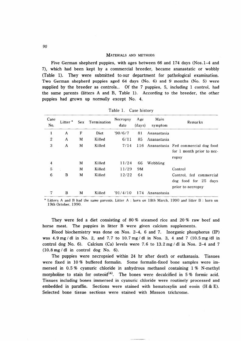
Before necropsy, all the puppies manifested similar physical features. Marked bilateral dorsal flexion and swelling of carpal joints resulted in horizontal orientation of the metacarpal bones. The hindlimbs were wobbly and bandy-legged with muscular atrophy. The posterior trunk was underdeveloped, making the head appear large (Fig. 1). There was mild curvature of the spine.
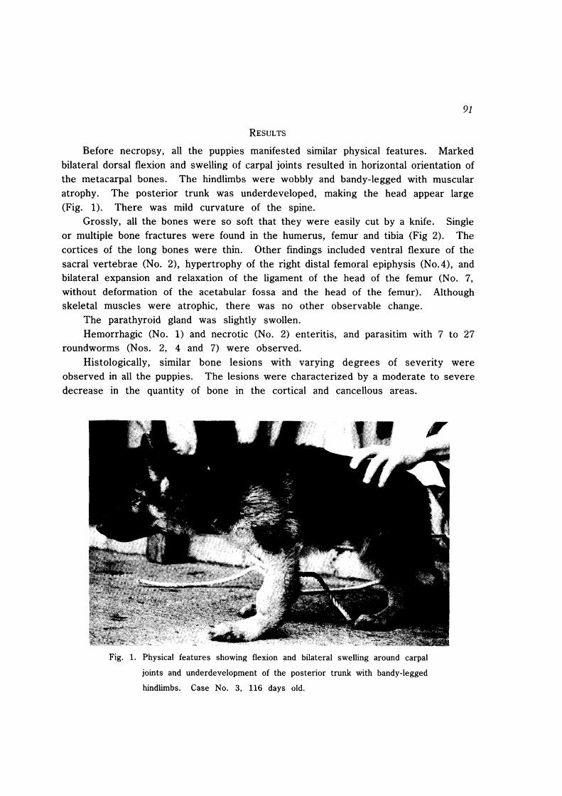
Grossly, all the bones were so soft that they were easily cut by a knife. Single or mUltiple bone fractures were found in the humerus, femur and tibia (Fig 2). The cortices of the long bones were thin. Other findings included ventral flexure of the sacral vertebrae (No.2), hypertrophy of the right distal femoral epiphysis (No.4), and bilateral expansion and relaxation of the ligament of the head of the femur (No.7, without deformation of the acetabular fossa and the head of the femur). Although skeletal muscles were atrophic, there was no other observable change.
The parathyroid gland was slightly swollen. Hemorrhagic (No.1) and necrotic (No.2) enteritis, and parasitim with 7 to 27
roundworms (Nos. 2, 4 and 7) were observed. Histologically, similar bone lesions with varying degrees of severity were
observed in all the puppies. The lesions were characterized by a moderate to severe decrease in the quantity of bone in the cortical and cancellous areas.
Fig. 1. Physical features showing flexion and bilateral swelling around carpal
joints and underdevelopment of the posterior trunk with bandy-legged
hindlimbs. Case No.3, 116 days old.
92
Fig. 2. Longitudinal cut of the humerus. Note severe thinning of the cortical
bone with a recovering fracture (arrow). Case No.2, 85 days old.
Formation of irregular cavities in the cortices of long bones was seen, and in severe cases, their compact bone resembled trabecular bone (porotic). The lesions were variable in degree even among bones of the same animal. In early lesions, small cavities were formed by active osteoclasts without osteoblastic activation resulting in resorption of cortical bone. In the latter stages, large osteoclasts with clear nuclei and abundant
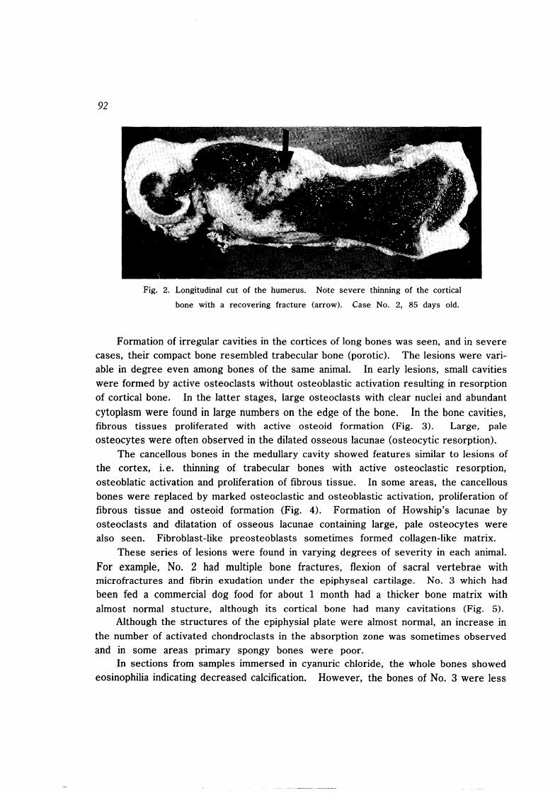
cytoplasm were found in large numbers on the edge of the bone. In the bone cavities, fibrous tissues proliferated with active osteoid formation (Fig. 3). Large, pale osteocytes were often observed in the dilated osseous lacunae (osteocytic resorption).
The cancellous bones in the medullary cavity showed features similar to lesions of the cortex, i. e. thinning of trabecular bones with active osteoclastic resorption, osteoblatic activation and proliferation of fibrous tissue. In some areas, the cancellous bones were replaced by marked osteoclastic and osteoblastic activation, proliferation of fibrous tissue and osteoid formation (Fig. 4). Formation of Howship's lacunae by osteoclasts and dilatation of osseous lacunae containing large, pale osteocytes were also seen. Fibroblast-like preosteoblasts sometimes formed collagen-like matrix.
These series of lesions were found in varying degrees of severity in each animal.
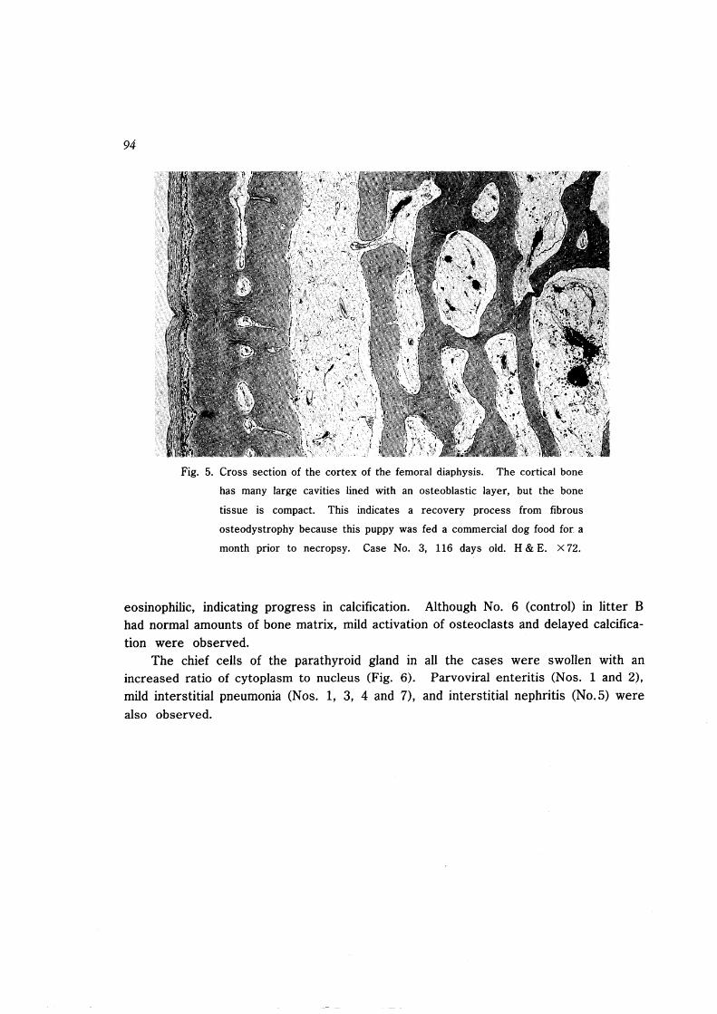
For example, No. 2 had multiple bone fractures, flexion of sacral vertebrae with microfractures and fibrin exudation under the epiphyseal cartilage. No. 3 which had been fed a commercial dog food for about 1 month had a thicker bone matrix with almost normal stucture, although its cortical bone had many cavitations (Fig. 5).
Although the structures of the epiphysial plate were almost normal, an increase in the number of activated chondroclasts in the absorption zone was sometimes observed and in some areas primary spongy bones were poor.
In sections from samples immersed in cyanuric chloride, the whole bones showed eosinophilia indicating decreased calcification. However, the bones of No.3 were less
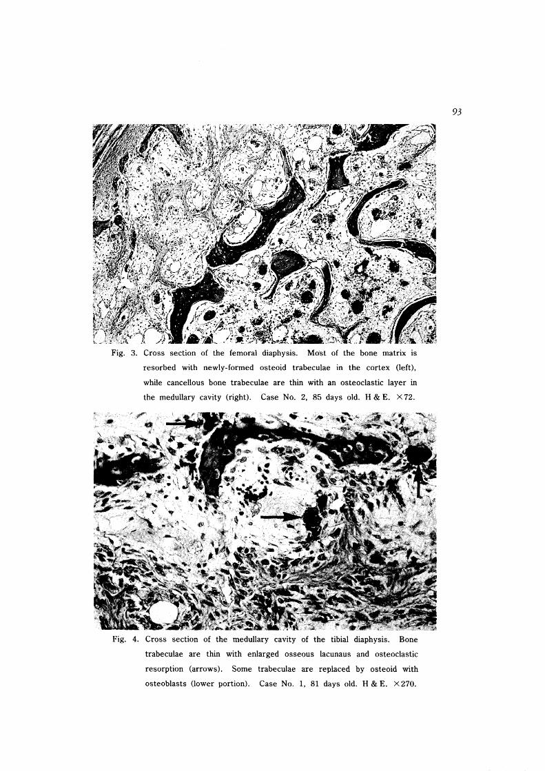
Fig. 3. Cross section of the femoral diaphysis. Mo'st of the bone matrix is
resorbed with newly-formed osteoid trabeculae in the cortex (left),
while cancellous bone trabeculae are thin with an osteoclastic layer in
the medullary cavity (right). Case No.2, 85 days old. H & E. X 72.
Fig. 4. Cross section of the medullary cavity of the tibial diaphysis. Bone
trabeculae are thin with enlarged osseous lacunaus and osteoclastic
resorption (arrows). Some trabeculae are replaced by osteoid with
osteoblasts (lower portion). Case No.1, 81 days old. H & E. X 270.
93
94
Fig. 5. Cross section of the cortex of the femoral diaphysis. The cortical bone
has many large cavities lined with an osteoblastic layer, but the bone
tissue is compact. This indicates a recovery process from fibrous
osteodystrophy because this puppy was fed a commercial dog food for a
month prior to necropsy. Case No.3, 116 days old. H & E. X 72.
eosinophilic, indicating progress in calcification. Although No. 6 (control) in litter B had normal amounts of bone matrix, mild activation of osteoclasts and delayed calcification were observed.
The chief cells of the parathyroid gland in all the cases were swollen with an increased ratio of cytoplasm to nucleus (Fig. 6). Parvoviral enteritis (Nos. 1 and 2),
mild interstitial pneumonia (Nos. 1, 3, 4 and 7), and interstitial nephritis (No.5) were also observed.
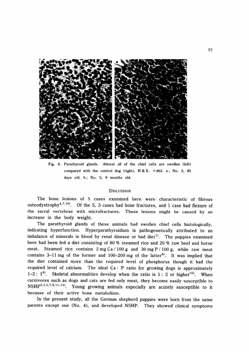
Fig. 6. Parathyroid glands. Almost all of the chief cells are swollen (left)
compared with the control dog (right). H & E. X 462. a; No.2, 85
days old. b; No.5, 9 months old.
DISCUSSION
95
The bone lesions of 5 cases examined here were characteristic of fibrous osteodystrophy4,7, 10). Of the 5, 3 cases had bone fractures, and 1 case had flexure of the sacral vertebrae with microfractures. These lesions might be caused by an increase in the body weight.
The parathyroid glands of these animals had swollen chief cells histologically, indicating hyperfunction. Hyperparathyroidism is pathogenetically attributed to an imbalance of minerals in blood by renal disease or bad diet!}. The puppies examined here had been fed a diet consisting of 80 % steamed rice and 20 % raw beef and horse meat. Steamed rice contains 2 mg Ca / 100 g and 30 mg P / 100 g, while raw meat contains 3-11 mg of the former and 100-200 mg of the latter6
}. It was implied that the diet contained more than the required level of phosphorus though it had the required level of calcium. The ideal Ca: P ratio for growing dogs is approximately 1-2: IS}. Skeletal abnormalities develop when the ratio is 1: 2 or higher lO
). When carnivores such as dogs and cats are fed only meat, they become easily susceptible to NSHp2,3,5,7,8,1l-13). Young growing animals especially are acutely susceptible to it
because of their active bone metabolism. In the present study, all the German shepherd puppies were born from the same
parents except one (No.4), and developed NSHP. They showed clinical symptoms
96
related to severe bone lesions observed histopathologically 10 to 15 days after weaning. Age and nutrition factors contributing to the development of the lesions were discussed above. In the same breeding kennel, other puppies born from differ~nt parents were normal. One young dog (No.5, aged 9 months), submitted as a control, had no bone lesions histopathologically. A genetic factor was therefore considered as a contributing factor to the development of NSHP. Heritable parathyroid sensitivity has been suggested in dogs, cats and horses lO
). NSHP is also reported to be more common in German shepherd dogs33
•
REFERENCES
1) BENJAMIN, M. M.: Outline of Veterinary Clinical Pathology, 3rd ed., Iowa State
Univ. Press, Ames, 1978. 2) BENNETT, D.: Nutrition and bone disease in the dog and cat. Vet. Rec. 98:
313-320, 1976.
3) CAMPBELL, J. R.: Bone dystrophy in puppies. Vet. Rec. 74: 1340-1348, 1962. 4) DAVIES, G. O. : Osteodystrophia fibrosa (osteofibrosis) in domesticated animals. Vet.
Rec.48: 1399-1404, 1936.
5) GODDARD, K. M., WILLIAMS, G. D., NEWBERNE, P. M. & WILSON, R. B.: A comparison of all-meat, semimoist, and dry-type dog food as diets for growing
beagles. J. Am. Vet. Med. Assoc. 157: 1233-1236, 1970.
6) KAGAKUGUUTSUCHO-SHIGENCHOSAKAI: Tables of Japan Food Components. Hitotsubas hi, Tokyo, 1984 (in Japanese).
7) KROOK, L., BARRETT, R. B., VSUI, K & WOLKE, R. E.: Nutritional secondary
hyperparathyroidism in the cat. Cornell Vet. 53: 224-240, 1963. 8) LEWIS, L. D., MORRIS, M. L. Jr. & HAND, M. S. : Small Animal Clinical Nutrition
DI, 3rd ed., Mark Morris Associates, Topeka, 1987.
9) MORRIS, M. L. Jr., TEETER, S. M. & COLLINS, D. R.: The effects of exclusive feeding of an all-meat dog food. J. Am. Vet. Assoc. 158: 477-488, 1971.
10) PALMER, N. : Metabolic diseases of bones. In: Jubb, K. V. F., Kennedy, P. C. &
Palmer, N. eds., Pathology of Domestic Animals, 4th ed., Vol. 1, pp. 55-93. Academic Press, California, 1993.
11) PEDERSEN, N. C. : Nutritional secondary hyperparathyroidism in a cattery associated
with the feeding of a bad diet containing horse meat. Feline Pract. 13: 19-26, 1983.
12) PRICI, D. A. : Dogs need more than meat. J. Am. Vet. Med. Assoc. 156: 681-685, 1970.
13) SAVILLE, P. D., KROOK, L., GUSTAFSSON, P., MARSHALL, J. L. & FIGARO LA, F.: Nutritional secondary hyperparathyroidism in a dog. Morphologic and radioisotope
studies with treatment. Cornell Vet. 59: 155-167, 1969.
14) YOSHIKI, S., VENO, T., AKITA, T. & YAMANOUCHI, M.: Improved procedure for
histological identification of osteoid matrix in decalcified bone. Stain Technol. 58: 85-89, 1983.