Embed Size (px)
Citation preview
Vox Sanguinis (2014) 106, 31–37
ORIGINAL PAPER © 2013 International Society of Blood TransfusionDOI: 10.1111/vox.12073
Occult hepatitis B infections among blood donorsin Lao PDRP. Jutavijittum,1 I. E. Andernach,2 A. Yousukh,1 B. Samountry,3 K. Samountry,3 T. Thammavong,4 J. Keokhamphue,4
K. Toriyama5 & C. P. Muller2,61Department of Pathology, Faculty of Medicine, Chiang Mai University, Chiang Mai, Thailand2Institute of Immunology, Centre de Recherche Public de la Sant�e/Laboratoire National de Sant�e, Luxembourg, Grand-Duchy of Luxembourg3Department of Pathology, Faculty of Medicine, University of Health Sciences, Vientiane, Lao PDR4National Blood Transfusion Center, Lao Red Cross, Vientiane, Lao PDR5Department of Pathology, Japanese Red-Cross Nagasaki Atomic Bomb Hospital, Nagasaki, Japan6Institut Pasteur du Laos, Vientiane, Lao PDR
Received: 4 February 2013,revised 1 July 2013,accepted 2 July 2013,published online 12 August 2013
Background and Objectives In Lao People’s Democratic Republic, hepatitis Bvirus is highly endemic. However, blood donations are only screened for HBsAg,leaving a risk of transmission by HBsAg-negative occult infected donors. Here,we characterized first-time blood donors to assess prevalence of hepatitis B virusinfections and occult infected donors.
Materials and Methods Sera were screened for HBsAg, HBeAg and anti-HBs,anti-HBc and anti-HBe antibodies. Occult HBV infections (OBIs) were assessed inHBsAg-negative sera by PCR, and sera of HBsAg positive and occult infecteddonors were phylogenetically characterized.
Results 9�6% of the donors were HBsAg positive, and 45.5% were positive for atleast one of the hepatitis B virus serum markers. More than 40% HBsAg carrierswere HBeAg positive, with HBeAg seroconversion occurring around 30 years ofage. Furthermore, 10�9% of HBsAg-negative, anti-HBc and/or anti-HBs-positivedonors were occult infected with hepatitis B virus. Thus, at least 3�9% of blooddonations would potentially be unsafe, but hepatitis B virus DNA copy numbersgreatly varied between donors.
Conclusion In Lao People’s Democratic Republic, a sizable proportion of HBsAg-negative and anti-HBc antibody-positive blood donations are potentially DNApositive and infective for hepatitis B.
Key words: chronic hepatitis B infection, hepatitis B virus, occult hepatitis Binfection, seroclearance, serological markers.
Introduction
More than one-third of the world’s population has been
infected with hepatitis B virus (HBV) at some point during
their life, and 75% of the estimated 360 million chronic
carriers worldwide live in the Asia-Pacific region alone
[1, 2]. Chronically infected individuals with detectable
HBV surface antigen (HBsAg) represent a significant risk
of virus transmission by blood donation. The Asian geno-
types B and C are often found at higher viral loads in the
blood (>107–108 copies/ml) [3], whereas other genotypes,
such as genotype E in Africa, are associated with HBeAg
seroconversion before the age of 15 or 16 and lower viral
loads [3, 4]. An additional risk for transfusion-transmitted
infections (TTI) results from occult HBV infections (OBI),
defined as the presence of HBV DNA in the blood or liver
without detectable HBsAg. The patho-physiological basis
of OBI is still poorly understood, but is probably due to
an incomplete immune control of the infection. Most
individuals with OBI have antibodies against HBV core
antigen (anti-HBc) possibly reflecting an ongoing chronic
Correspondence: Claude P. Muller: Institute of Immunology, Centre deRecherche Public de la Sant�e/Laboratoire National de Sant�e, 20A rueAuguste Lumi�ere, L-1950 Luxembourg, Grand-Duchy of LuxembourgE-mail: [email protected]
31
infection with HBsAg below detection limits. This is par-
ticularly common in endemic countries where individuals
are infected during early childhood. OBI with antibodies
against HBV surface antigen (anti-HBs), with or without
anti-HBc antibodies, sometimes occur during the recovery
phase of the infection. Rare sero-negative cases of OBI
occur in individuals with no markers of HBV infection
other than HBV DNA. Individuals with chronic hepatitis
who have mutated HBsAg that is not detected by diag-
nostic assays also contribute to OBI. While OBI infections
are generally characterized by very low levels of HBV
DNA in the blood (usually <1000 copies/ml or <200 IU/
ml [5]), they nevertheless are a concern for recipients of
blood donations [6–8].
In Western countries with a low prevalence of HBV
infections, anti-HBc antibody testing is included in the
screening of blood donations for TTI to exclude individu-
als with past exposure to HBV (including the vast major-
ity of OBI). However, in highly endemic countries, this
approach would exclude a large proportion of healthy
donors that have cleared HBV infections, severely under-
mining the blood supply [9, 10]. In these settings, screen-
ing for TTI commonly includes HBsAg testing [6, 10].
However, this strategy does not detect OBI or acute infec-
tions during the initial infection period. Thus, nucleic acid
testing (NAT) has been proposed and tested as a highly
sensitive technique for HBV DNA detection in OBI blood
donors. However, in low-income countries, NAT may not
be possible due to limited resources [6, 9–11].
In Lao PDR, HBsAg is highly endemic, with a preva-
lence of 8.7% in Lao blood donors [12]. As in its neigh-
bouring countries Cambodia and Vietnam [10], blood
donations in Lao PDR are screened for HBsAg only, with
the risk of TTI by HBsAg-negative blood donors with OBI.
In this study, we determined the serological profile of Lao
blood donors and the prevalence of OBI. We discuss pos-
sible implications for the safety of Lao blood donations.
Materials and methods
Study group
A total of 906 first-time blood donors, including 296
females, were randomly recruited between March and
June 2006. In Lao PDR, all blood donors are routinely
asked for sexual and medical risk factors related to TTI
including clinical hepatitis in an interview and by ques-
tionnaire. Donors with previous hepatitis B, C, HIV and
syphilis were excluded. Donors are not routinely asked
whether they are vaccinated against HBV, and sera are
not routinely tested for ALT. The mean age of the donors
was 23.8 – 7.4 years (range 16–53 years), with about
80% of donors being <30 years old. Serum samples were
obtained from Vientiane municipality, Vientiane Province
and Bolikhamsay Province, either at the National Blood
Transfusion Centre of the Lao Red Cross in Vientiane or
by mobile collecting units and were stored at -20°C until
further testing.
Serological analyses
All assays were performed on an automated AxSYM�
system (Abbott, Wiesbaden, Germany). HBsAg, HBeAg
and anti-HBs, anti-HBc, anti-HBe antibodies were
detected by AxSYM� HBsAg V2, AxSYM� HBe 2.0,
AxSYM� AUSAB, AxSYM� CORE and AxSYM� Anti-HBe
2.0 assays, respectively.
Molecular and phylogenetic analyses
DNA was extracted using the QIAGEN DNA Blood Mini
Kit (QIAGEN, Venlo, the Netherlands), according to the
manufacturer’s instructions. The HBV genome was
amplified as four overlapping fragments (preS, S, X and
C) as described previously [13]. The sensitivity of the
PCR reactions for preS, S, X and C was 1424, 1009,
2240 and 950 copies per reaction (or 285, 202, 448 and
190 IU/ml), respectively. Phylogenetic analyses were per-
formed using MEGA v.5 (Tempe, AZ, USA) [14] and the
neighbour-joining method of the Kimura 2-parameter
model with 1000 bootstrap replicates. Genotyping was
performed by analysing at least one of the four frag-
ments. For detection of OBI, DNA was extracted from
200 ll of serum from HBsAg-negative, anti-HBc and/or
anti-HBs-positive donors. The DNA was eluted in 60 ll,and 5 ll was used in quantitative real-time PCR. The
PCR was performed using forward primer 5′-ACTCA-
CCAACCTCTTGTCCT-3′ (nt 333–352 in relation to the
EcoR1 site), reverse primer 5′-GACAAACGGG CAACA-
TACCT-3′ (nt 476–457) and TaqMan Probe (5′-FAM
and 3′-TAMRA) 5′-TATCGCTGGATGTGTCTGCGGCGT-3′
(nt 368–391). The reaction was performed at 50°C for
2 min, 95°C for 10 min and 50 cycles of 95°C for 15 s
and 60°C for 60 s [15] and compared with a HBV
plasmid dilution series ranging from 18.5 to
1.85 9 1010 copies in the PCR corresponding to 1110 to
111 9 1010 copies/ml or 222 to 22 9 1010 IU/ml serum.
Although viral DNA can be detected below the linear
range, they were not quantifiable. Therefore, values
below this linear range (CT values above 34) are
expressed as ‘<1110 copies/ml’. The estimated limit of
detection using our in-house plasmid was about
110 copies/ml or approximately 20 IU/ml. Samples with
CT values above 37 were considered negative. Negative
controls were included in all extractions and PCR
experiments.
� 2013 International Society of Blood TransfusionVox Sanguinis (2014) 106, 31–37
32 P. Jutavijittum et al.
Statistical analyses
A computerized data sheet was used for record keeping;
all data were evaluated with SPSS 17.0 for Windows statis-
tic software package (SPSS Inc., Chicago, IL, USA) or
SigmaPlot 12.0 (Systat Software Inc., San Jose, CA, USA).
Parameters were compared by chi-square test or Fisher’s
exact test where appropriate.
Ethical statement
This study was approved by the competent Research
Ethics Committee of the Faculty of Medicine of Chiang
Mai University, Thailand, and the Ethics Committee of the
Faculty of Medical Sciences, National University of Laos,
Lao PDR.
Results
HBsAg, anti-HBs and anti-HBc antibodies
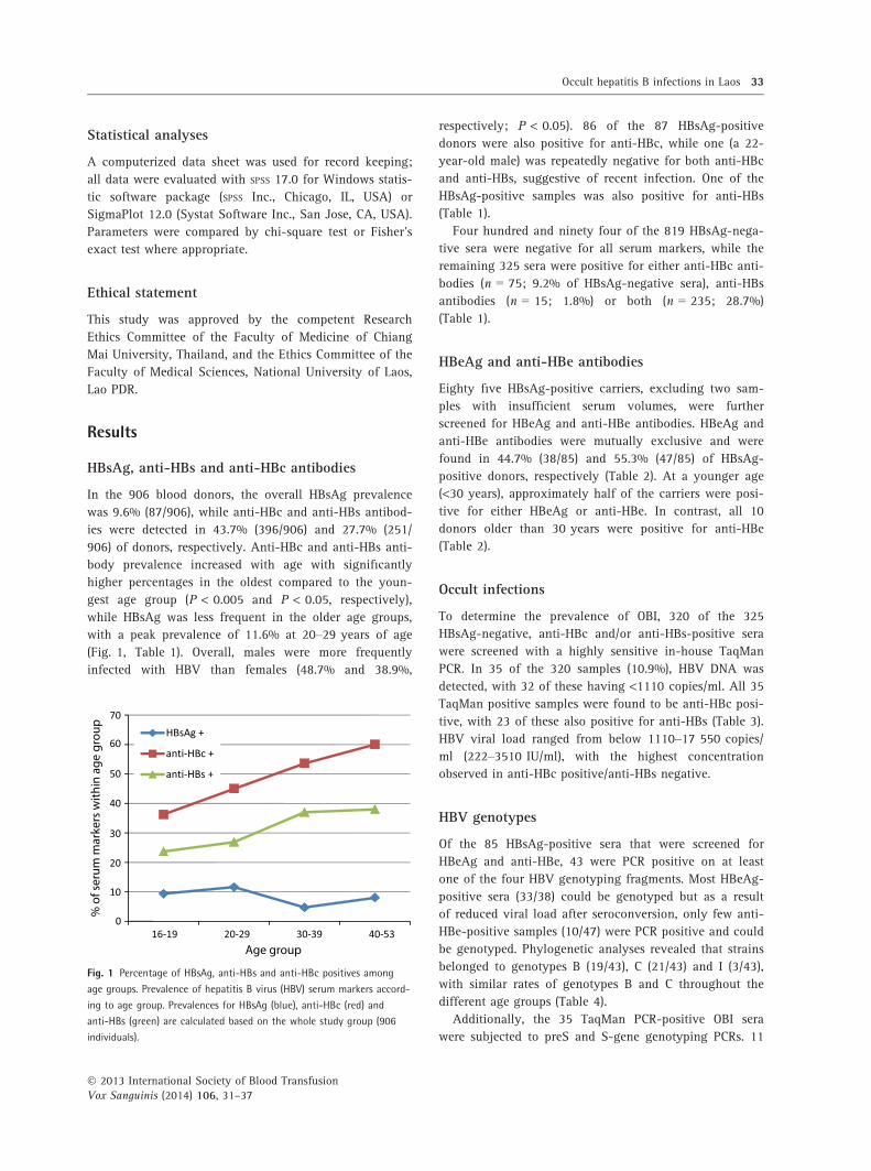
In the 906 blood donors, the overall HBsAg prevalence
was 9.6% (87/906), while anti-HBc and anti-HBs antibod-
ies were detected in 43.7% (396/906) and 27.7% (251/
906) of donors, respectively. Anti-HBc and anti-HBs anti-
body prevalence increased with age with significantly
higher percentages in the oldest compared to the youn-
gest age group (P < 0.005 and P < 0.05, respectively),
while HBsAg was less frequent in the older age groups,
with a peak prevalence of 11.6% at 20–29 years of age
(Fig. 1, Table 1). Overall, males were more frequently
infected with HBV than females (48.7% and 38.9%,
respectively; P < 0.05). 86 of the 87 HBsAg-positive
donors were also positive for anti-HBc, while one (a 22-
year-old male) was repeatedly negative for both anti-HBc
and anti-HBs, suggestive of recent infection. One of the
HBsAg-positive samples was also positive for anti-HBs
(Table 1).
Four hundred and ninety four of the 819 HBsAg-nega-
tive sera were negative for all serum markers, while the
remaining 325 sera were positive for either anti-HBc anti-
bodies (n = 75; 9.2% of HBsAg-negative sera), anti-HBs
antibodies (n = 15; 1.8%) or both (n = 235; 28.7%)
(Table 1).
HBeAg and anti-HBe antibodies
Eighty five HBsAg-positive carriers, excluding two sam-
ples with insufficient serum volumes, were further
screened for HBeAg and anti-HBe antibodies. HBeAg and
anti-HBe antibodies were mutually exclusive and were
found in 44.7% (38/85) and 55.3% (47/85) of HBsAg-
positive donors, respectively (Table 2). At a younger age
(<30 years), approximately half of the carriers were posi-
tive for either HBeAg or anti-HBe. In contrast, all 10
donors older than 30 years were positive for anti-HBe
(Table 2).
Occult infections
To determine the prevalence of OBI, 320 of the 325
HBsAg-negative, anti-HBc and/or anti-HBs-positive sera
were screened with a highly sensitive in-house TaqMan
PCR. In 35 of the 320 samples (10.9%), HBV DNA was
detected, with 32 of these having <1110 copies/ml. All 35
TaqMan positive samples were found to be anti-HBc posi-
tive, with 23 of these also positive for anti-HBs (Table 3).
HBV viral load ranged from below 1110–17 550 copies/
ml (222–3510 IU/ml), with the highest concentration
observed in anti-HBc positive/anti-HBs negative.
HBV genotypes
Of the 85 HBsAg-positive sera that were screened for
HBeAg and anti-HBe, 43 were PCR positive on at least
one of the four HBV genotyping fragments. Most HBeAg-
positive sera (33/38) could be genotyped but as a result
of reduced viral load after seroconversion, only few anti-
HBe-positive samples (10/47) were PCR positive and could
be genotyped. Phylogenetic analyses revealed that strains
belonged to genotypes B (19/43), C (21/43) and I (3/43),
with similar rates of genotypes B and C throughout the
different age groups (Table 4).
Additionally, the 35 TaqMan PCR-positive OBI sera
were subjected to preS and S-gene genotyping PCRs. 11
HBsAg +
anti-HBc +
anti-HBs +
0
10
20
30
40
50
60
70
16-19 20-29 30-39 40-53
% o
f ser
um
mar
kers
wit
hin
ag
e g
rou
p
Age group
Fig. 1 Percentage of HBsAg, anti-HBs and anti-HBc positives among
age groups. Prevalence of hepatitis B virus (HBV) serum markers accord-
ing to age group. Prevalences for HBsAg (blue), anti-HBc (red) and
anti-HBs (green) are calculated based on the whole study group (906
individuals).
� 2013 International Society of Blood TransfusionVox Sanguinis (2014) 106, 31–37
Occult hepatitis B infections in Laos 33
of these, with viral copies ranging from less than 1110
to 17 550 per ml (222–3510 IU/ml), were found to be
positive in at least one of the PCRs. Phylogenetic anal-
yses revealed a predominance of HBV genotype C (8/
11), while one strain each was attributed to genotypes
B and I. Another strain was suspected to be mixed
infected with multiple genotypes and was not further
analysed. While the proportion of genotype C infections
in the occult group was significantly higher than in the
HBsAg-positive group (P < 0.05), this is likely to be a
selection bias, since HBV/C infections tend to have
higher viral loads than HBV/B infections. Thus, OBI
with HBV/B variants is more likely to remain unde-
tected.
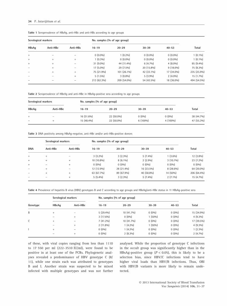
Table 2 Seroprevalence of HBeAg and anti-HBe in HBsAg-positive sera according to age groups
Serological markers No. samples (% of age group)
TotalHBeAg Anti-HBe 16–19 20–29 30–39 40–53
+ - 16 (51.6%) 22 (50.0%) 0 (0%) 0 (0%) 38 (44.7%)
- + 15 (48.4%) 22 (50.0%) 6 (100%) 4 (100%) 47 (55.3%)
Table 3 DNA positivity among HBsAg-negative, anti-HBc and/or anti-HBs-positive donors
DNA
Serological markers No. samples (% of age group)
TotalAnti-HBc Anti-HBs 16–19 20–29 30–39 40–53
+ + - 3 (3.2%) 3 (2.3%) 5 (7.4%) 1 (3.6%) 12 (3.8%)
+ + + 10 (10.8%) 8 (6.1%) 2 (2.9%) 3 (10.7%) 23 (7.2%)
+ - + 0 (0%) 0 (0%) 0 (0%) 0 (0%) 0 (0%)
- + - 12 (12.9%) 28 (21.4%) 16 (23.5%) 8 (28.6%) 64 (20.0%)
- + + 63 (67.7%) 89 (67.9%) 40 (58.8%) 14 (50%) 206 (64.4%)
- - + 5 (5.4%) 3 (2.3%) 5 (7.4%) 2 (7.1%) 15 (4.7%)
Table 1 Seroprevalence of HBsAg, anti-HBc and anti-HBs according to age groups
Serological markers No. samples (% of age group)
TotalHBsAg Anti-HBc Anti-HBs 16–19 20–29 30–39 40–53
+ - - 0 (0.0%) 1 (0.2%) 0 (0.0%) 0 (0.0%) 1 (0.1%)
+ + + 1 (0.3%) 0 (0.0%) 0 (0.0%) 0 (0.0%) 1 (0.1%)
+ + - 31 (9.0%) 44 (11.4%) 6 (4.7%) 4 (8.0%) 85 (9.4%)
- + - 17 (5.0%) 29 (7.5%) 20 (15.8%) 9 (18.0%) 75 (8.3%)
- + + 75 (21.9%) 101 (26.1%) 42 (33.1%) 17 (34.0%) 235 (25.9%)
- - + 5 (1.5%) 3 (0.8%) 5 (3.9%) 2 (4.0%) 15 (1.7%)
- - - 213 (62.3%) 209 (54.0%) 54 (42.5%) 18 (36.0%) 494 (54.5%)
Table 4 Prevalence of hepatitis B virus (HBV) genotypes B and C according to age groups and HBeAg/anti-HBe status in 11 HBsAg-positive sera
Genotype
Serological markers No. samples (% of age group)
TotalHBeAg Anti-HBe 16–19 20–29 30–39 40–53
B + - 5 (29.4%) 10 (41.7%) 0 (0%) 0 (0%) 15 (34.9%)
- + 3 (17.6%) 0 (0%) 1 (50%) 0 (0%) 4 (9.3%)
C + - 7 (41.2%) 10 (41.7%) 0 (0%) 0 (0%) 17 (39.5%)
- + 2 (11.8%) 1 (4.2%) 1 (50%) 0 (0%) 4 (9.3%)
I + - 0 (0%) 1 (4.2%) 0 (0%) 0 (0%) 1 (2.3%)
- + 0 (0%) 2 (8.3%) 0 (0%) 0 (0%) 2 (4.7%)
� 2013 International Society of Blood TransfusionVox Sanguinis (2014) 106, 31–37
34 P. Jutavijittum et al.
Discussion
In healthy first-time blood donors, we found a high
prevalence of HBV serum markers. We determined that
45.5% of the blood donors were positive for any of the
HBV markers, suggesting that almost half of the Lao
population has been in contact with HBV at one point
during their lives. 9.6% of the donors were HBsAg
positive. This is consistent with the 8.7% HBsAg
prevalence reported before from Lao PDR [12] as well as
from neighbouring countries such as Cambodia (7.7%
HBsAg and 58.6% anti-HBc prevalence) and Vietnam
(11.4% HBsAg and 51.7% anti-HBc) [10].
The presence of anti-HBc and also anti-HBs antibodies
in the investigated cohort increased with age and was
significantly higher in the oldest age group, while HBsAg
positivity had a tendency to decrease in the higher age
groups. This is likely to represent spontaneous HBsAg
seroclearance during late stages of HBV infections and is
consistent with clearance rates of up to 45% observed in
endemic regions [16]. In Lao PDR, a country with a high
prevalence of chronic HBV carriers, most infections
are acquired during early life, perpetuating chronic
infections. Anti-HBs antibodies were found in 27.7% of
all donors, and only 6.1% of these were anti-HBc
negative and therefore potentially vaccinated. This is in
accordance with the low HBV vaccine coverage in adults
in Lao PDR where HBV vaccine was only introduced in
2001 for infants. Furthermore, 8.3% of donors were
anti-HBc positive, but negative for HBsAg and anti-HBs,
indicating high numbers of later stage chronic HBV
infections with loss or reduced levels of HBsAg, or
resolved infections (HBV DNA negative) with anti-HBs
antibodies below the detection limit (Fig. 1 and Table 1).
Interestingly, one sample was repeatedly positive for
HBsAg as well as for anti-HBc and anti-HBs antibodies.
While this may be a case of exposure to heterologous
HBsAg serotypes, we have no further basis for such
speculations [17]. This serological profile may also be
indicative of acute HBV infection or the case of a chronic
carrier with severe liver disease, although the latter is
unlikely as blood donors are normally healthy volunteers.
More than 40% (38/85) of HBsAg carriers in this study
were HBeAg positive. Interestingly, while about half of
the carriers below 30 years of age were positive for either
HBeAg or anti-HBe, all 10 donors older than 30 years
were positive for anti-HBe but not HBeAg (Table 2). This
suggests that seroconversion from HBeAg to anti-HBe, an
indicator of sustained remission, occurs at around
30 years of age in Lao PDR. Although previous studies
indicated earlier HBeAg seroconversion in patients
infected with HBV genotype B than for those infected
with HBV genotype C [18, 19], we could not observe a
difference in the prevalence of HBV B and C genotypes in
the different age groups (Table 4).
In our study, 10.9% (35/320) of HBsAg-negative, anti-
HBV antibody-positive donors were HBV DNA positive in
a highly sensitive real-time PCR. Thus, at least 3.9% of
all 906 investigated donors were occult infected with
HBV (OBI). When a less sensitive commercial assay was
used, only four samples, including the three with the
highest DNA levels, were DNA positive (data not shown).
This would correspond to an OBI prevalence of 1.3% of
HBsAg-negative, anti-HBV antibody-positive donors and
0.4% of all donors. It is possible that the individuals
detected with the commercial assay represent the greatest
risk of onwards HBV transmission. This apparent differ-
ence in prevalence highlights the difficulty of comparing
OBI prevalence data from studies using different detection
techniques. Previous studies in highly endemic regions
also showed high prevalences of OBI. In Indonesia, where
9.4% of the population is chronically infected, a study
using nested PCR found 8.1% of blood donors to be
occult infected [20]. Similarly, in a highly endemic popu-
lation in India, an in-house PCR detected OBI in 30% of
anti-HBc antibody-positive donors [21]. This is in contrast
to data from regions where HBV is not endemic, such as
the USA, where OBI can be as low as 3.7% of anti-HBc
antibody-positive donors [22]. While the prevalence of
OBI is dependent on epidemiological aspects such as the
route of infection, the clinical evolution of the disease
and interventions, the infectivity of OBI blood as well as
the determinants of this infectivity still remain unclear.
The majority of OBIs that could be genotyped in the
current study were attributed to genotype C (8/11).
Significantly, fewer HBV genotype B infections were
observed in the OBI group (1/11) than in HBsAg carriers
(19/43), where equal proportions of HBV genotype C and B
were observed. One study of OBI in individuals from
south-east China, where genotype B is dominant, also
showed that the prevalence of genotype C was signifi-
cantly higher in OBI than in HBsAg-positive individuals
[23], while others have not seen this difference [24]. Stud-
ies on larger cohorts are needed to further determine the
significance of a genotype bias in OBI.
All OBI donors were anti-HBc positive while the
prevalence of OBI was somewhat lower in anti-HBs/HBc
antibody double positive (23/229; 10%) than in anti-HBc
single positive samples (12/76; 15.8%; not statistically
significant). In other studies, OBI prevalences were also
higher in individuals with anti-HBc antibodies only and
lower in double positive or anti-HBs antibody only
donors [25]. In general, high levels of anti-HBs antibodies
in donated blood forestall HBV infections of recipients
(reviewed in [9]). Thus, recipient infection may not be
prevented in the presence of low levels of anti-HBs
� 2013 International Society of Blood TransfusionVox Sanguinis (2014) 106, 31–37
Occult hepatitis B infections in Laos 35
antibodies (<100 IU/ml), in particular, when levels of
HBV DNA are high or when the recipients are immuno-
compromised [9]. In our cohort, OBI sera had a range of
anti-HBs levels from 0 to 768 IU/ml. Importantly, the
three donors with highest DNA levels of 1790, 12 800
and 17 550 copies per ml (358, 2560 and 3510 IU/ml)
had undetectable anti-HBs antibodies, re-emphasizing the
higher risk of TTI from these donors. Rare sero-negative
cases of OBI occur in individuals with no markers of HBV
infection other than HBV DNA. For example, in Thai
blood donors, about one in 3000 had HBV DNA and was
antibody and HBsAg negative. Approximately 20% of
these were window period cases [26, 27]. Since we only
investigated HBV DNA in antibody-positive cases, our
study would have missed DNA-positive (antibody-nega-
tive) window cases.
The significance of OBI for the donor and the recipient
is poorly understood. In particular, there are very few
studies investigating the infectivity of transfusion with
OBI blood. The few available studies seem to agree that
only subsets of recipients of OBI blood become infected.
For example, one study in Taiwan showed that two of 11
recipients of OBI blood became subsequently infected [28].
In another study from the Japanese Red Cross, OBI blood
was infectious in 19% of recipients [29]. Recently, it was
suggested that the 50% infectious dose of OBI blood prod-
ucts would be about 1000 HBV DNA copies [30]. However,
the relationship between viral DNA concentration or sero-
logy of the donor and infection of the recipient was not
determined in these studies. Thus, certainly a large propor-
tion of the OBIs observed in the current study, and espe-
cially the ‘anti-HBc only’ donors with higher serum DNA
levels, represent a considerable risk of TTI.
To eliminate the risk of HBV transmission by blood
transfusion, additional analyses besides HBsAg are
required in Lao PDR. While anti-HBc antibody screening
would largely exclude the HBsAg-negative/DNA-positive
sera, it would also unnecessarily exclude anti-HBc anti-
body-positive blood with undetectable HBV DNA, that is,
approximately 30% of all donors. In Thailand, a country
with an emerging economy, blood banks systematically
screen for HBV, hepatitis C virus and HIV-1/2 by nucleic
acid testing (NAT) since 2006. Together with a broad hep-
atitis B vaccination strategy, this has led to a reduction in
the incidence of new HBV infections in that country [26].
While this triple NAT screening may be cost-effective in
Thailand due to its high prevalence of HIV-1 infections,
its cost-benefit in resource poor Lao PDR requires careful
evaluation.
Acknowledgements
The authors would like to thank Miss Praijitr Tanan,
(Blood Bank of Maharaj Nakorn Chiang Mai Hospital and
Faculty of Medicine, Chiang Mai University) for her excel-
lent technical assistance and Antony Black (Institute of
Immunology, CRP-Sant�e, Luxembourg and Institut Pasteur
du Laos, Vientiane, Lao PDR) for his help in performing
some of the experiments and help with the manuscript.
This work was partially supported by the Faculty of
Medicine Research Fund, Chiang Mai University, Thailand
and by a grant from the Ministry of Cooperation and
Humanitarian Aid, Luxembourg. I.E.A. was supported by a
fellowship from the National Research Fund, Luxembourg
(TR-PHD-BFR07-129).
References1 Lavanchy D: Hepatitis B virus epide-
miology, disease burden, treatment,
and current and emerging prevention
and control measures. J Viral Hepat
2004; 11:97–107
2 WHO: Hepatitis B fact sheet. 2000
3 Liaw YF, Leung N, Kao JH, et al.:
Chronic Hepatitis BGWPotA-PAftSotL:
Asian-Pacific consensus statement on
the management of chronic hepatitis
B: a 2008 update. Hep Intl 2008;
2:263–283
4 Candotti D, Danso K, Allain JP:
Maternofetal transmission of hepatitis
B virus genotype E in Ghana, west
Africa. J Gen Virol 2007; 88:
2686–2695
5 Raimondo G, Allain JP, Brunetto MR,
et al.: Statements from the Taormina
expert meeting on occult hepatitis B
virus infection. J Hepatol 2008;
49:652–657
6 Liu CJ, Chen DS, Chen PJ: Epidemiol-
ogy of HBV infection in Asian blood
donors: emphasis on occult HBV
infection and the role of NAT. J Clin
Virol 2006; 36(Suppl. 1):S33–S44
7 Ocana S, Casas ML, Buhigas I, et al.:
Diagnostic strategy for occult hepatitis
B virus infection. World J Gastroenter-
ol 2011; 17:1553–1557
8 Alavian SM, Miri SM, Hollinger FB,
et al.: Occult Hepatitis B (OHB) in Clini-
cal Settings. Hepat Mon 2012; 12:e6126.
9 Candotti D, Allain JP: Transfusion-
transmitted hepatitis B virus infection.
J Hepatol 2009; 51:798–809
10 Ol HS, Bjoerkvoll B, Sothy S, et al.:
Prevalence of hepatitis B and hepatitis
C virus infections in potential blood
donors in rural Cambodia. Southeast
Asian J Trop Med Public Health 2009;
40:963–971
11 Phikulsod S, Oota S, Tirawatnapong T,
et al.: Working Group for NATSiTBD:
One-year experience of nucleic acid
technology testing for human immu-
nodeficiency virus Type 1, hepatitis C
virus, and hepatitis B virus in Thai
blood donations. Transfusion 2009;
49:1126–1135
� 2013 International Society of Blood TransfusionVox Sanguinis (2014) 106, 31–37
36 P. Jutavijittum et al.
12 Jutavijittum P, Yousukh A, Samoun-
try B, et al.: Seroprevalence of hepa-
titis B and C virus infections among
Lao blood donors. Southeast Asian J
Trop Med Public Health 2007;
38:674–679
13 Olinger CM, Venard V, Njayou M,
et al.: Phylogenetic analysis of the
precore/core gene of hepatitis B virus
genotypes E and A in West Africa:
new subtypes, mixed infections and
recombinations. J Gen Virol 2006;
87:1163–1173
14 Tamura K, Peterson D, Peterson N,
et al.: MEGA5: molecular evolutionary
genetics analysis using maximum like-
lihood, evolutionary distance, and
maximum parsimony methods. Mol
Biol Evol 2011; 28:2731–2739
15 Liu Y, Hussain M, Wong S, et al.: A
genotype-independent real-time PCR
assay for quantification of hepatitis B
virus DNA. J Clin Microbiol 2007;
45:553–558
16 Chu CM, Liaw YF: Chronic hepatitis B
virus infection acquired in childhood:
special emphasis on prognostic and
therapeutic implication of delayed
HBeAg seroconversion. J Viral Hepati-
tis 2007; 14:147–152
17 Andernach IE, Jutavijittum P,
Samountry B, et al.: A high variability
of mixed infections and recent recom-
binations of hepatitis B virus in Laos.
PLoS ONE 2012; 7:e30245
18 Kao JH, Chen PJ, Lai MY, et al.:
Hepatitis B virus genotypes and
spontaneous hepatitis B e antigen sero-
conversion in Taiwanese hepatitis B
carriers. J Med Virol 2004; 72:363–369
19 Yuen MF, Sablon E, Tanaka Y, et al.:
Epidemiological study of hepatitis B
virus genotypes, core promoter and
precore mutations of chronic hepatitis
B infection in Hong Kong. J Hepatol
2004; 41:119–125
20 Thedja MD, Roni M, Harahap AR,
et al.: Occult hepatitis B in blood
donors in Indonesia: altered antigenic-
ity of the hepatitis B virus surface pro-
tein. Hepatol Int 2010; 4:608–614
21 Panigrahi R, Biswas A, Datta S, et al.:
Anti-hepatitis B core antigen testing
with detection and characterization of
occult hepatitis B virus by an in-house
nucleic acid testing among blood
donors in Behrampur, Ganjam, Orissa
in southeastern India: implications for
transfusion. Virol J 2010; 7:204
22 Kleinman SH, Kuhns MC, Todd DS,
et al.: Retrovirus Epidemiology Donor
S: Frequency of HBV DNA detection
in US blood donors testing positive for
the presence of anti-HBc: implications
for transfusion transmission and donor
screening. Transfusion 2003; 43:
696–704
23 Yuan Q, Ou SH, Chen CR, et al.:
Molecular characteristics of occult
hepatitis B virus from blood donors in
southeast China. J Clin Microbiol
2010; 48:357–362
24 Candotti D, Lin CK, Belkhiri D, et al.:
Occult hepatitis B infection in blood
donors from South East Asia:
molecular characterisation and poten-
tial mechanisms of occurrence. Gut
2012; 61:1744–1753
25 Torbenson M, Thomas DL: Occult
hepatitis B. Lancet Infect Dis 2002;
2:479–486
26 Chimparlee N, Oota S, Phikulsod S, et al.:
Hepatitis B and hepatitis C virus in Thai
blood donors. Southeast Asian J Trop
Med Public Health, 2011; 42:609–615
27 Louisirirotchanakul S, Oota S,
Khuponsarb K, et al.: Working Group
for NATSiTBD: Occult hepatitis B virus
infection in Thai blood donors. Trans-
fusion 2011; 51:1532–1540
28 Wang JT, Lee CZ, Chen PJ, et al.: Trans-
fusion-transmitted HBV infection in an
endemic area: the necessity of more sen-
sitive screening for HBV carriers. Trans-
fusion 2002; 42:1592–1597.
29 Satake M, Taira R, Yugi H, et al.:
Infectivity of blood components with
low hepatitis B virus DNA levels iden-
tified in a lookback program. Trans-
fusion 2007; 47:1197–1205
30 Allain JP, Mihaljevic I, Gonzalez-Fraile
MI, et al.: Infectivity of blood products
from donors with occult hepatitis B
virus infection. Transfusion 2013;
7:1405–1415
� 2013 International Society of Blood TransfusionVox Sanguinis (2014) 106, 31–37
Occult hepatitis B infections in Laos 37