Embed Size (px)
Citation preview

Saudi Journal of Ophthalmology (2016) 30, 39–43
Original Article
Ocular manifestation of Ichthyosis
Peer review under responsibilityof Saudi Ophthalmological Society,King Saud University Production and hosting by Elsevier
Access this article onlinwww.saudiophthaljournwww.sciencedirect.com
Received 12 January 2015; received in revised form 26 November 2015; accepted 14 December 2015; available online 18 December 2015.
Anterior Segment Division, King Khaled Eye Specialist Hospital, Riyadh, Saudi Arabia
⇑ Address: Anterior Segment/Emergency Room, King Khaled Eye Specialist Hospital, PO Box 7191, Riyadh 11462, Saudi Arabia. Tel.:4821234x3861.e-mail address: [email protected]
Mohammad A. Al-Amry ⇑
Abstract
Purpose: Ichthyosis is a rare dermato-ocular disease. This study evaluates the presenting ocular signs, symptoms, complicationsand prognosis of ichthyosis in a case series from Saudi Arabia.Methods: A retrospective chart review was performed for 11 patients with ichthyosis who presented to King Khaled Eye SpecialistHospital, Riyadh, Saudi Arabia, over the last 20 years.Results: The most common presenting ocular diagnosis was ectropion of both the lids. Two patients developed corneal perfora-tion with poor prognosis. Most of the patients underwent skin grafting to repair eyelid ectropion. The visual prognosis was excel-lent because timely surgical interventions were performed. Hence the rate of corneal complications such as perforation was low.Conclusion: The most ocular presentation of ichthyosis is ectropion of both the upper and lower lids. Despite good visual prog-nosis, there were some devastating corneal complications such as perforation with unpredictable outcomes.
Keywords: Ichthyosis, Ectropion, Congenital, Skin graft, Collodion
� 2015 The Author. Production and hosting by Elsevier B.V. on behalf of Saudi Ophthalmological Society, King Saud University.This is an open access article under the CC BY-NC-ND license (http://creativecommons.org/licenses/by-nc-nd/4.0/).
http://dx.doi.org/10.1016/j.sjopt.2015.12.004
Introduction
Ichthyosis is an inherited group of skin disorders character-ized by the skin thickening and scale formation. Some ofthese disorders include, ichthyosis vulgaris and epidermolytichyperkeratosis (EHK) which are autosomal dominant andlamellar type which is autosomal recessive and X-linkedichthyosis.1 Harlequin ichthyosis is an inherited autosomalrecessive disorder with mutations in the abca12 gene. It isfatal and is characterized by deeply fissured skin and deformi-ties of the hands and feet. Harlequin ichthyosis may beaccompanied with lipid dysfunctions of the epidermal layerof the skin, starting prenatally, and has many names such asalligator baby and malignant keratosis.2 KID syndrome standsfor keratitis, ichthyosis and sensorineural deafness. It mani-fests itself as alopecia, dental disorders, susceptibility to bac-terial and mycotic infections and squamous cell carcinoma.3,4
Ichthyosis with confetti is a very rare type of ichthyosis char-acterized by dermatological features of collodion baby mixedwith patches of confetti-like healthy skin.5
The most common ocular manifestation of ichthyosis iscicatricial ectropion. We present the ocular signs, symptoms,complications and prognosis of ichthyosis in a case seriesfrom Saudi Arabia.
Patients and methods
The medical records were reviewed for all patients diag-nosed with ichthyosis who presented to King Khaled Eye Spe-cialist Hospital, Riyadh, Saudi Arabia. This hospital is referralbased eye care hospital that treats patients nationwide andfrom the Gulf region. A retrospective chart review was per-formed for cases of ichthyosis from 1994–2014. Data werecollected on patient demographics, age at presentation, type
e:al.com
+966 1
40 M.A. Al-Amry
of ichthyosis, the type of referral to the hospital and if thepatient was treated by a local dermatologist. Other datawere collected on skin and ocular presenting signs includinglid ectropion and corneal complications, the medical and sur-gical treatment as well as the type of the surgical procedureand the incidence of repeat lid correcting procedures such asskin grafting.
Patients were excluded if they underwent surgical repairof the lids and/or cornea that was not due to ichthyosis orif the presenting or follow-up data were insufficient.
Patients (pediatric or adults) were included if they haddocumented or suspected ichthyosis and documented orsuspected cases of congenital lid ectropion, corneal stemcell deficiency and/or skin manifestations of ichthyosis andany cases of ichthyosis with documented histopathologystudies.
The study was registered with the institutional reviewboard and approval was obtained from the ethics committee.
Results
Eleven out of 16 patients met the inclusion criteria. Fourpatients were excluded because there were no physical med-ical records located to retrieve the data, and 1 patient had adiagnosis of orbital inflammatory syndrome not ichthyosis.
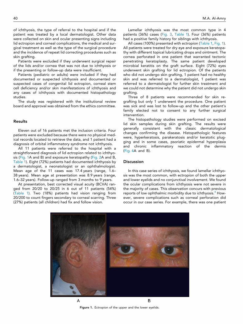
All 11 patients were referred to the hospital with astraightforward diagnosis of lid ectropion related to ichthyo-sis (Fig. 1A and B) and exposure keratopathy (Fig. 2A and B,Table 1). Eight (72%) patients had documented ichthyosis bya dermatologist, a neonatologist or an ophthalmologist.Mean age of the 11 cases was 17.4 years (range, 1.6–38 years). Mean age at presentation was 8.9 years (range,1.6–32 years). Follow-up ranged from 3 months to 9 years.
At presentation, best corrected visual acuity (BCVA) ran-ged from 20/20 to 20/25 in 6 out of 11 patients (54%)(Table 1). Two (18%) patients had vision ranging from20/200 to count fingers secondary to corneal scarring. Three(27%) patients (all children) had fix and follow vision.
Figure 1. Ectropion of the up
Lamellar ichthyosis was the most common type in 4patients (36%) cases (Fig. 3, Table 1). Four (36%) patientshad a positive family history for siblings with ichthyosis.
All cases (100%) presented with ectropion (Table 1, Fig. 1).All patients were treated for dry eye and exposure keratopa-thy with different topical lubricating drops and ointment. Thecornea perforated in one patient that warranted tectonicpenetrating keratoplasty. The same patient developedmicrobial keratitis on the graft surface. Eight (72%) eyesunderwent skin grafting for lid ectropion. Of the patientswho did not undergo skin grafting, 1 patient had no healthyskin and was referred to a dermatologist, 1 patient wasreferred to a dermatologist for further skin treatment andwe could not determine why the patient did not undergo skingrafting.
Three of 8 patients were recommended for skin re-grafting but only 1 underwent the procedure. One patientwas sick and was lost to follow-up and the other patient’sfamily elected not to consent to any further surgicalintervention.
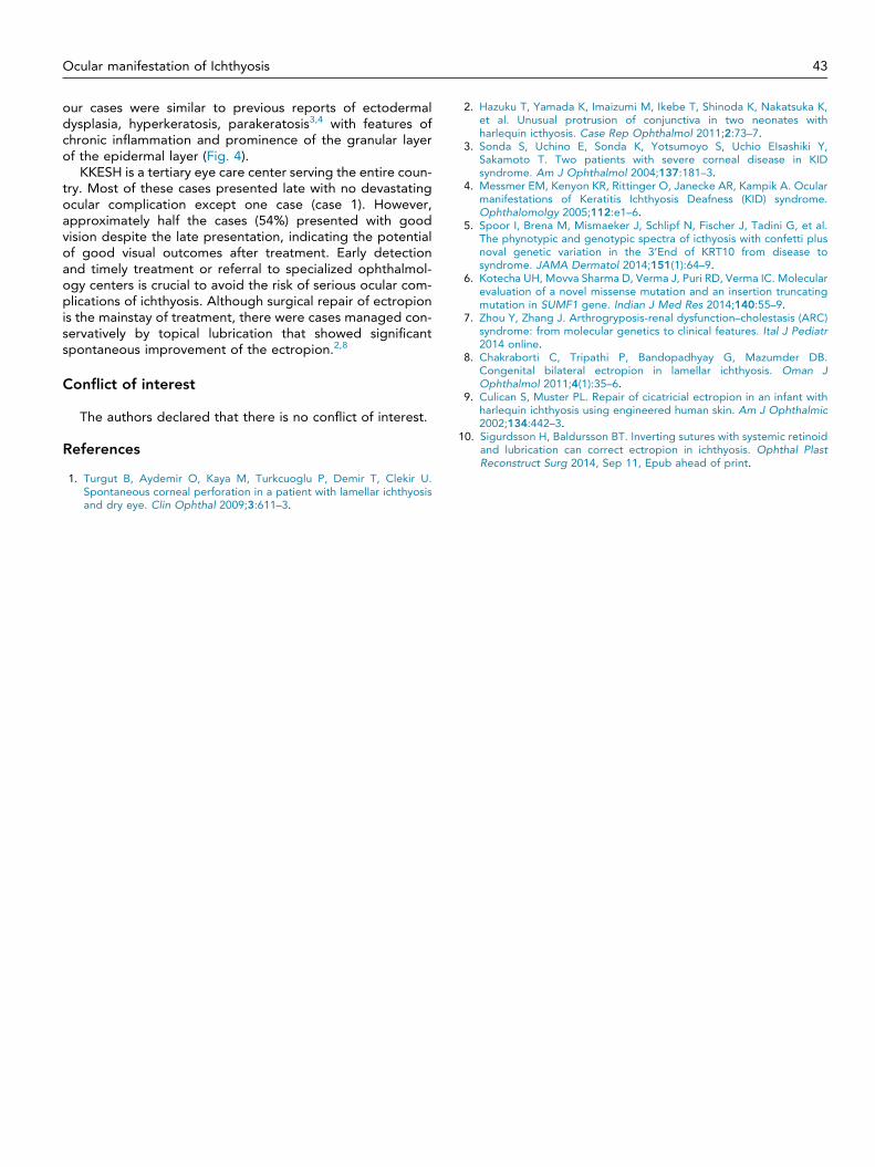
The histopathology studies were performed on excisedlid skin samples during skin grafting. The results weregenerally consistent with the classic dermatologicalchanges confirming the disease. Histopathologic featureswere, hyperkeratosis, parakeratosis and/or keratotic plug-ging and in some cases, psoriatic epidermal hyperplasiaand chronic inflammatory reaction of the dermis(Fig. 4A and B).
Discussion
In this case series of ichthyosis, we found lamellar ichthyo-sis was the most common, with ectropion of both the upperand lower eyelids and no conjunctival involvement. We foundthe ocular complications from ichthyosis were not severe inthe majority of cases. This observation concurs with previousreports of low ophthalmic morbidity due to ichthyosis.4 How-ever, severe complications such as corneal perforation didoccur in our case series. For example, there was one patient
per and the lower eyelids.
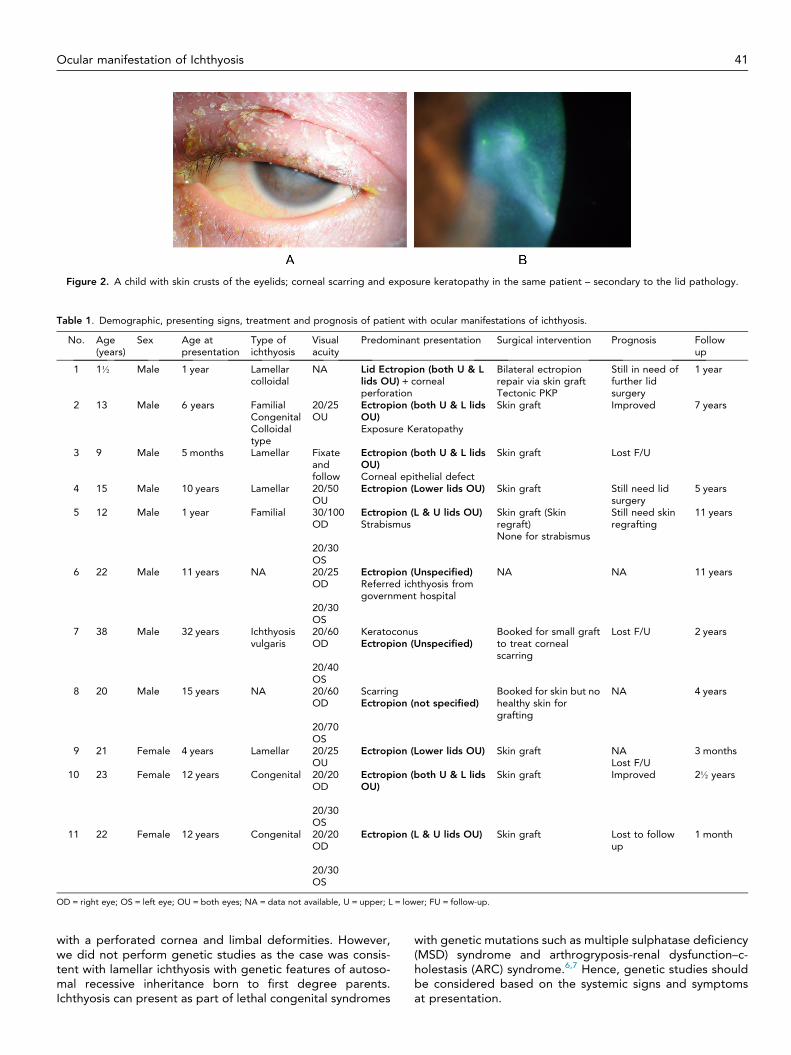
Figure 2. A child with skin crusts of the eyelids; corneal scarring and exposure keratopathy in the same patient – secondary to the lid pathology.
Table 1. Demographic, presenting signs, treatment and prognosis of patient with ocular manifestations of ichthyosis.
No. Age(years)
Sex Age atpresentation
Type ofichthyosis
Visualacuity
Predominant presentation Surgical intervention Prognosis Followup
1 1½ Male 1 year Lamellarcolloidal
NA Lid Ectropion (both U & Llids OU) + cornealperforation
Bilateral ectropionrepair via skin graftTectonic PKP
Still in need offurther lidsurgery
1 year
2 13 Male 6 years FamilialCongenitalColloidaltype
20/25OU
Ectropion (both U & L lidsOU)Exposure Keratopathy
Skin graft Improved 7 years
3 9 Male 5 months Lamellar Fixateandfollow
Ectropion (both U & L lidsOU)Corneal epithelial defect
Skin graft Lost F/U
4 15 Male 10 years Lamellar 20/50OU
Ectropion (Lower lids OU) Skin graft Still need lidsurgery
5 years
5 12 Male 1 year Familial 30/100OD
Ectropion (L & U lids OU)Strabismus
Skin graft (Skinregraft)None for strabismus
Still need skinregrafting
11 years
20/30OS
6 22 Male 11 years NA 20/25OD
Ectropion (Unspecified)Referred ichthyosis fromgovernment hospital
NA NA 11 years
20/30OS
7 38 Male 32 years Ichthyosisvulgaris
20/60OD
KeratoconusEctropion (Unspecified)
Booked for small graftto treat cornealscarring
Lost F/U 2 years
20/40OS
8 20 Male 15 years NA 20/60OD
ScarringEctropion (not specified)
Booked for skin but nohealthy skin forgrafting
NA 4 years
20/70OS
9 21 Female 4 years Lamellar 20/25OU
Ectropion (Lower lids OU) Skin graft NALost F/U
3 months
10 23 Female 12 years Congenital 20/20OD
Ectropion (both U & L lidsOU)
Skin graft Improved 2½ years
20/30OS
11 22 Female 12 years Congenital 20/20OD
Ectropion (L & U lids OU) Skin graft Lost to followup
1 month
20/30OS
OD = right eye; OS = left eye; OU = both eyes; NA = data not available, U = upper; L = lower; FU = follow-up.
Ocular manifestation of Ichthyosis 41
with a perforated cornea and limbal deformities. However,we did not perform genetic studies as the case was consis-tent with lamellar ichthyosis with genetic features of autoso-mal recessive inheritance born to first degree parents.Ichthyosis can present as part of lethal congenital syndromes
with genetic mutations such as multiple sulphatase deficiency(MSD) syndrome and arthrogryposis-renal dysfunction–c-holestasis (ARC) syndrome.6,7 Hence, genetic studies shouldbe considered based on the systemic signs and symptomsat presentation.
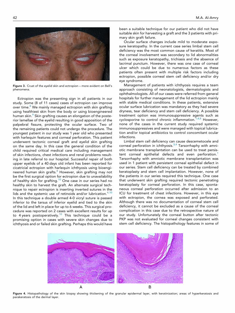
Figure 3. Crust of the eyelid skin and ectropion – more evident on Bell’sphenomena.
42 M.A. Al-Amry
Ectropion was the presenting sign in all patients in ourstudy. Some (8 of 11 cases) cases of ectropion can improveover time.8 We mainly managed ectropion with skin graftingusing healthiest skin from the body or using bioengineeredhuman skin.9 Skin grafting causes an elongation of the poste-rior lamellae of the eyelid resulting in good apposition of thepalpebral fissure, protecting the ocular surface. Two ofthe remaining patients could not undergo the procedure. Theyoungest patient in our study was 1 year old who presentedwith harlequin features and corneal perforation. This patientunderwent tectonic corneal graft and eyelid skin graftingon the same day. In this case the general condition of thechild required critical medical care including managementof skin infections, chest infections and renal problems result-ing in late referral to our hospital. Successful repair of bothupper eyelids of a 40 days old infant has been reported forcicatricial ectropion with harlequin ichthyosis using bioengi-neered human skin grafts.8 However, skin grafting may notbe the first surgical option for ectropion due to unavailabilityof healthy skin for grafting.10 One case in our series had nohealthy skin to harvest the graft. An alternate surgical tech-nique to repair ectropion is inserting inverted sutures in thelids and the systemic use of retinoids and/or lubrication.4,10
In this technique a double armed 4-0 vicryl suture is passedinferior to the tarsus of inferior eyelid and tied to the skinof the lid and left in place for up to 6 weeks. This surgical pro-cedure was reported on 2 cases with excellent results for upto 4 years postoperatively.10 This technique could be apromising option in cases with severe skin changes due toichthyosis and or failed skin grafting. Perhaps this would have
Figure 4. Histopathology of the skin biopsy showing thickening of the graparakeratosis of the dermal layer.
been a suitable technique for our patient who did not havesuitable skin for harvesting a graft and the 3 patients with pri-mary skin graft failure.
Ocular surface changes include mild to moderate expo-sure keratopathy. In the current case series limbal stem celldeficiency was the most common cause of keratitis. Most ofthe corneal involvement was secondary to lid abnormalitiessuch as exposure keratopathy, trichiasis and the absence oflacrimal punctum. However, there was one case of cornealulcer which could be due to numerous factors as thesepatients often present with multiple risk factors includingectropion, possible corneal stem cell deficiency and/or dryeye syndrome.
Management of patients with ichthyosis requires a teamapproach consisting of neonatologists, dermatologists andophthalmologists. All of our cases were referred from generalhospitals for further management of the lid ectropion mainlywith stable medical conditions. In these patients, extensiveocular surface lubrication was mandatory as they had severedryness, tear deficiency and stem cell deficiency. A possibletreatment option was immunosuppressive agents such ascyclosporine to control chronic inflammation.1,4,8 However,none of the cases in the current study required systemicimmunosuppressives and were managed with topical lubrica-tion and/or topical antibiotics to control concomitant ocularinfections.
Limbal stem cell deficiency can cause descemetocele andcorneal perforation in ichthyosis.3,4 Tarsorrhaphy with amni-otic membrane transplantation can be used to treat persis-tent corneal epithelial defects and even perforation.1
Tarsorrhaphy with amniotic membrane transplantation wasused in 1 patient with persistent corneal epithelial defect inour series. Stem cell deficiency can be treated by combinedkeratoplasty and stem cell implantation. However, none ofthe patients in our series required this technique. One casethat underwent skin grafting required tectonic penetratingkeratoplasty for corneal perforation. In this case, sponta-neous corneal perforation occurred after admission to anICU for treatment of chest infections. However, in this eyewith ectropion, the cornea was exposed and perforated.Although there was no documentation of corneal stem celldeficiency, it cannot be excluded as a cause of the cornealcomplication in this case due to the retrospective nature ofour study. Unfortunately the corneal button after tectonicPKP was not evaluated for corneal changes consistent withstem cell deficiency. The histopathology features in some of
nular epidermal layer, with keratinization; areas of hyperkeratosis and
Ocular manifestation of Ichthyosis 43
our cases were similar to previous reports of ectodermaldysplasia, hyperkeratosis, parakeratosis3,4 with features ofchronic inflammation and prominence of the granular layerof the epidermal layer (Fig. 4).
KKESH is a tertiary eye care center serving the entire coun-try. Most of these cases presented late with no devastatingocular complication except one case (case 1). However,approximately half the cases (54%) presented with goodvision despite the late presentation, indicating the potentialof good visual outcomes after treatment. Early detectionand timely treatment or referral to specialized ophthalmol-ogy centers is crucial to avoid the risk of serious ocular com-plications of ichthyosis. Although surgical repair of ectropionis the mainstay of treatment, there were cases managed con-servatively by topical lubrication that showed significantspontaneous improvement of the ectropion.2,8
Conflict of interest
The authors declared that there is no conflict of interest.
References
1. Turgut B, Aydemir O, Kaya M, Turkcuoglu P, Demir T, Clekir U.Spontaneous corneal perforation in a patient with lamellar ichthyosisand dry eye. Clin Ophthal 2009;3:611–3.
2. Hazuku T, Yamada K, Imaizumi M, Ikebe T, Shinoda K, Nakatsuka K,et al. Unusual protrusion of conjunctiva in two neonates withharlequin icthyosis. Case Rep Ophthalmol 2011;2:73–7.
3. Sonda S, Uchino E, Sonda K, Yotsumoyo S, Uchio EIsashiki Y,Sakamoto T. Two patients with severe corneal disease in KIDsyndrome. Am J Ophthalmol 2004;137:181–3.
4. Messmer EM, Kenyon KR, Rittinger O, Janecke AR, Kampik A. Ocularmanifestations of Keratitis Ichthyosis Deafness (KID) syndrome.Ophthalomolgy 2005;112:e1–6.
5. Spoor I, Brena M, Mismaeker J, Schlipf N, Fischer J, Tadini G, et al.The phynotypic and genotypic spectra of icthyosis with confetti plusnoval genetic variation in the 3’End of KRT10 from disease tosyndrome. JAMA Dermatol 2014;151(1):64–9.
6. Kotecha UH, Movva Sharma D, Verma J, Puri RD, Verma IC. Molecularevaluation of a novel missense mutation and an insertion truncatingmutation in SUMF1 gene. Indian J Med Res 2014;140:55–9.
7. Zhou Y, Zhang J. Arthrogryposis-renal dysfunction–cholestasis (ARC)syndrome: from molecular genetics to clinical features. Ital J Pediatr2014 online.
8. Chakraborti C, Tripathi P, Bandopadhyay G, Mazumder DB.Congenital bilateral ectropion in lamellar ichthyosis. Oman JOphthalmol 2011;4(1):35–6.
9. Culican S, Muster PL. Repair of cicatricial ectropion in an infant withharlequin ichthyosis using engineered human skin. Am J Ophthalmic2002;134:442–3.
10. Sigurdsson H, Baldursson BT. Inverting sutures with systemic retinoidand lubrication can correct ectropion in ichthyosis. Ophthal PlastReconstruct Surg 2014, Sep 11, Epub ahead of print.


![harlequin ichthyosis and functional recovery by · of ichthyosis, harlequin ichthyosis (HI) (Mendelian Inheritance of Man [MIM] 242500) is the most serious subtype (Figure 1); it](https://img.pdfslide.net/doc/110x75/5d4f332b88c993257d8be9c0/harlequin-ichthyosis-and-functional-recovery-by-of-ichthyosis-harlequin-ichthyosis.jpg)