Embed Size (px)
Citation preview
ASSOCIATION FOR ACADEMIC SURGERY AND SOCIETY OF UNIVERSITY SURGEONS—ABSTRACTS316
Conclusions:Wehave shown thatmechanical strain rapidly inducesa proliferative, morphological, and functional response in abdominalwall fibroblasts which is dependent upon the continued presence ofthe strain signal and quickly lost when the force is removed. Theloss of wound edge tension that occurs during laparotomy wound sep-aration and hernia formationmay contribute to impairedwound heal-ing through loss of a key stimulatory mechanical signal.
TRAUMA & CRITICAL CARE 4: TRAUMA,BURNS, & BASIC SCIENCE
44.1. Hypertonic Saline Inhibits Leukotriene B4 And Arachi-donic Acid Priming Of The Neutrophil Oxidase. L. Lee,1
M. R. Kelher,2 E. E. Moore,3 C. C. Silliman,2 J. Harr,3
D. Benson,3 A. Banerjee1; 1University of Colorado - Denver,Aurora, CO; 2Bonfils Blood Center, Denver, CO; 3DenverHealth Medical Center, Denver, CO
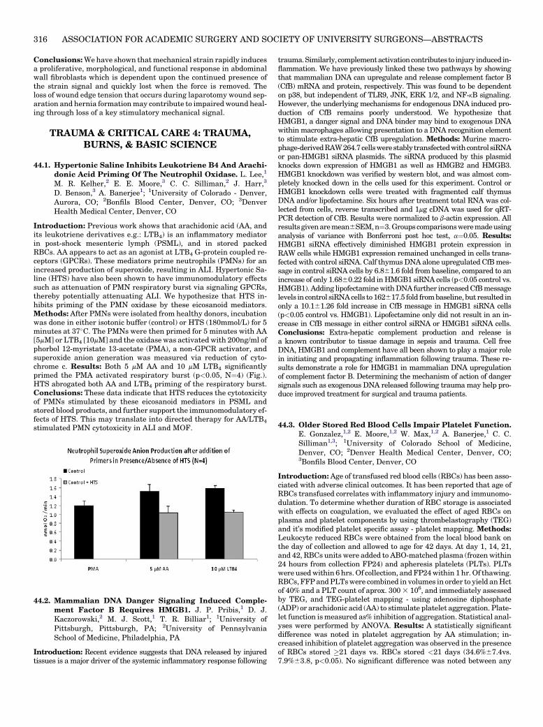
Introduction: Previous work shows that arachidonic acid (AA, andits leukotriene derivatives e.g.: LTB4) is an inflammatory mediatorin post-shock mesenteric lymph (PSML), and in stored packedRBCs. AA appears to act as an agonist at LTB4 G-protein coupled re-ceptors (GPCRs). These mediators prime neutrophils (PMNs) for anincreased production of superoxide, resulting in ALI. Hypertonic Sa-line (HTS) have also been shown to have immunomodulatory effectssuch as attenuation of PMN respiratory burst via signaling GPCRs,thereby potentially attenuating ALI. We hypothesize that HTS in-hibits priming of the PMN oxidase by these eicosanoid mediators.Methods: After PMNs were isolated from healthy donors, incubationwas done in either isotonic buffer (control) or HTS (180mmol/L) for 5minutes at 37�C. The PMNs were then primed for 5 minutes with AA[5mM] or LTB4 [10mM] and the oxidasewas activatedwith 200ng/ml ofphorbol 12-myristate 13-acetate (PMA), a non-GPCR activator, andsuperoxide anion generation was measured via reduction of cyto-chrome c. Results: Both 5 mM AA and 10 mM LTB4 significantlyprimed the PMA activated respiratory burst (p<0.05, N¼4) (Fig.).HTS abrogated both AA and LTB4 priming of the respiratory burst.Conclusions: These data indicate that HTS reduces the cytotoxicityof PMNs stimulated by these eicosanoid mediators in PSML andstored blood products, and further support the immunomodulatory ef-fects of HTS. This may translate into directed therapy for AA/LTB4
stimulated PMN cytotoxicity in ALI and MOF.
44.2. Mammalian DNA Danger Signaling Induced Comple-ment Factor B Requires HMGB1. J. P. Pribis,1 D. J.Kaczorowski,2 M. J. Scott,1 T. R. Billiar1; 1University ofPittsburgh, Pittsburgh, PA; 2University of PennsylvaniaSchool of Medicine, Philadelphia, PA
Introduction: Recent evidence suggests that DNA released by injuredtissues is a major driver of the systemic inflammatory response following
trauma.Similarly,complementactivationcontributesto injuryinducedin-flammation. We have previously linked these two pathways by showingthat mammalian DNA can upregulate and release complement factor B(CfB) mRNA and protein, respectively. This was found to be dependenton p38, but independent of TLR9, JNK, ERK 1/2, and NF-kB signaling.However, the underlying mechanisms for endogenous DNA induced pro-duction of CfB remains poorly understood. We hypothesize thatHMGB1, a danger signal and DNA binder may bind to exogenous DNAwithinmacrophages allowing presentation to a DNA recognition elementto stimulate extra-hepatic CfB upregulation. Methods: Murine macro-phage-derivedRAW264.7cellswerestablytransfectedwithcontrolsiRNAor pan-HMGB1 siRNA plasmids. The siRNA produced by this plasmidknocks down expression of HMGB1 as well as HMGB2 and HMGB3.HMGB1 knockdown was verified by western blot, and was almost com-pletely knocked down in the cells used for this experiment. Control orHMGB1 knockdown cells were treated with fragmented calf thymusDNA and/or lipofectamine. Six hours after treatment total RNA was col-lected from cells, reverse transcribed and 1mg cDNA was used for qRT-PCR detection of CfB. Results were normalized to b-actin expression. Allresultsgivenaremean6SEM,n¼3.Groupscomparisonsweremadeusinganalysis of variance with Bonferroni post hoc test, a¼0.05. Results:HMGB1 siRNA effectively diminished HMGB1 protein expression inRAW cells while HMGB1 expression remained unchanged in cells trans-fectedwith control siRNA. Calf thymusDNAalone upregulated CfBmes-sage in control siRNA cells by 6.861.6 fold from baseline, compared to anincrease of only 1.6860.22 fold inHMGB1 siRNA cells (p<0.05 control vs.HMGB1).Adding lipofectaminewithDNAfurther increasedCfBmessagelevels in control siRNAcells to162617.5 fold frombaseline, but resulted inonly a 10.161.26 fold increase in CfB message in HMGB1 siRNA cells(p<0.05 control vs. HMGB1). Lipofectamine only did not result in an in-crease in CfB message in either control siRNA or HMGB1 siRNA cells.Conclusions: Extra-hepatic complement production and release isa known contributor to tissue damage in sepsis and trauma. Cell freeDNA, HMGB1 and complement have all been shown to play a major rolein initiating and propagating inflammation following trauma. These re-sults demonstrate a role for HMGB1 in mammalian DNA upregulationof complement factor B. Determining the mechanism of action of dangersignals such as exogenous DNA released following traumamay help pro-duce improved treatment for surgical and trauma patients.
44.3. Older Stored Red Blood Cells Impair Platelet Function.E. Gonzalez,1,2 E. Moore,1,2 W. Max,1,2 A. Banerjee,1 C. C.Silliman1,3; 1University of Colorado School of Medicine,Denver, CO; 2Denver Health Medical Center, Denver, CO;3Bonfils Blood Center, Denver, CO
Introduction:Age of transfused red blood cells (RBCs) has been asso-ciated with adverse clinical outcomes. It has been reported that age ofRBCs transfused correlates with inflammatory injury and immunomo-dulation. To determine whether duration of RBC storage is associatedwith effects on coagulation, we evaluated the effect of aged RBCs onplasma and platelet components by using thrombelastography (TEG)and it’s modified platelet specific assay - platelet mapping. Methods:Leukocyte reduced RBCs were obtained from the local blood bank onthe day of collection and allowed to age for 42 days. At day 1, 14, 21,and 42, RBCs units were added to ABO-matched plasma (frozenwithin24 hours from collection FP24) and apheresis platelets (PLTs). PLTswereusedwithin 6 hrs.Of collection, andFP24within 1 hr.Of thawing.RBCs,FFPandPLTswere combined involumes in order to yield anHctof 40% and a PLT count of aprox. 3003 106, and immediately assessedby TEG, and TEG-platelet mapping - using adenosine diphosphate(ADP) or arachidonic acid (AA) to stimulate platelet aggregation. Plate-let function ismeasured as% inhibition of aggregation. Statistical anal-yses were performed by ANOVA. Results: A statistically significantdifference was noted in platelet aggregation by AA stimulation; in-creased inhibition of platelet aggregation was observed in the presenceof RBCs stored �21 days vs. RBCs stored <21 days (34.6%67.4vs.7.9%63.8, p<0.05). No significant difference was noted between any
ASSOCIATION FOR ACADEMIC SURGERY AND SOCIETY OF UNIVERSITY SURGEONS—ABSTRACTS 317
of the RBC-age groups in ADP induced aggregation. None of the TEGparameters of enzymatic clotting function (R, delta), fibrincross-linking(angle), thrombin induced platelet aggregation (MA, G), or fibrinolysis(LY30) were significantly different between RBC-age groups. Therewere no significant differences in Hct and PLT counts between the dif-ferent samples studied.Conclusions: Storage of RBCs �21 days maybe responsible for decreased platelet function when transfused. An as-sociation has been noted betweenRBC age and the AA pathway duringplatelet aggregation. Further in vivo studies are needed to determine ifplatelet dysfunction related to transfusion of RBCs�21 days is contrib-utory to poor clinical outcomes.
RBC
age
(days)
Enzymatic
Activity
(R)
Thrombin
Generation
(Delta)
Rate of
Clot
Formation
(Angle)
Maximum
Amplitude
(MA)
Clot
Strength
(G)
Lysis at
30 min.
(LY30)
ADP
inhibition
AA
inhibition
1 4.7360.9 1.060.3 51.063.4 58.764.4 8.362.1 0.560.3 5.160.9 1.461.1
14 5.061.1 0.860.5 60.765.1 61.763.7 9.761.5 0.460.3 3.961.5 7.963.8
21 4.961.3 0.860.3 62.362.9 62.463.0 10.061.7 0.160.1 5.264.4 34.667.4*,**
42 5.561.1 0.660.2 59.863.9 61.664.0 9.662.2 0.160.1 4.462.7 43.6618.0*,**
* P<0.05 vs. RBC-day 1, ** P<0.05 vs. RBC-day 14, .
44.4. A Totally Recombinant Factor XIII-SupplementedFibrin Sealant. M. A. Carlson,1 W. H. Velander,2 I. I.Pipinos,1 J. M. Johanning,1 J. Calcaterra2; 1University ofNebraska Medical Center, Omaha, NE; 2University ofNebraska Lincoln, Lincoln, NE
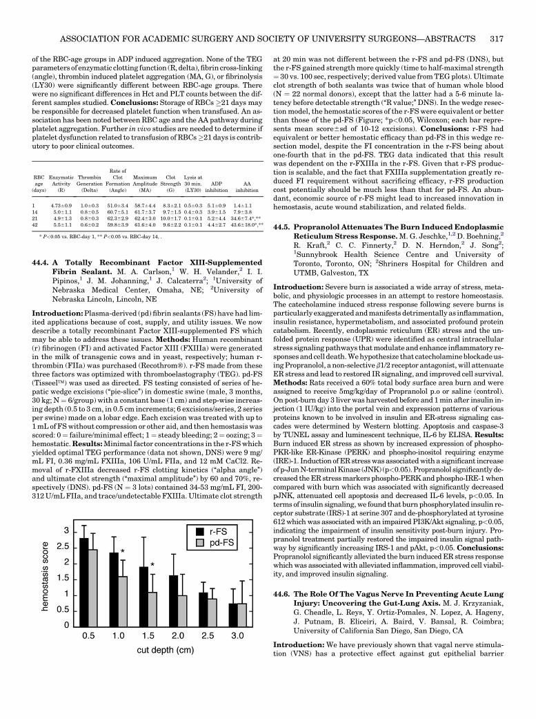
Introduction:Plasma-derived (pd) fibrin sealants (FS) have had lim-ited applications because of cost, supply, and utility issues. We nowdescribe a totally recombinant Factor XIII-supplemented FS whichmay be able to address these issues. Methods: Human recombinant(r) fibrinogen (FI) and activated Factor XIII (FXIIIa) were generatedin the milk of transgenic cows and in yeast, respectively; human r-thrombin (FIIa) was purchased (Recothrom�). r-FS made from thesethree factors was optimized with thromboelastography (TEG). pd-FS(Tisseel�) was used as directed. FS testing consisted of series of he-patic wedge excisions (‘‘pie-slice’’) in domestic swine (male, 3 months,30 kg; N¼ 6/group) with a constant base (1 cm) and step-wise increas-ing depth (0.5 to 3 cm, in 0.5 cm increments; 6 excisions/series, 2 seriesper swine) made on a lobar edge. Each excision was treated with up to1mLof FSwithout compression or other aid, and thenhemostasiswasscored: 0¼ failure/minimal effect; 1¼ steady bleeding; 2¼ oozing; 3¼hemostatic.Results:Minimal factor concentrations in the r-FSwhichyielded optimal TEG performance (data not shown, DNS) were 9 mg/mL FI, 0.36 mg/mL FXIIIa, 106 U/mL FIIa, and 12 mM CaCl2. Re-moval of r-FXIIIa decreased r-FS clotting kinetics (‘‘alpha angle’’)and ultimate clot strength (‘‘maximal amplitude’’) by 60 and 70%, re-spectively (DNS). pd-FS (N ¼ 3 lots) contained 34-53 mg/mL FI, 200-312U/mLFIIa, and trace/undetectable FXIIIa. Ultimate clot strength
at 20 min was not different between the r-FS and pd-FS (DNS), butthe r-FS gained strengthmore quickly (time to half-maximal strength¼ 30 vs. 100 sec, respectively; derived value fromTEGplots). Ultimateclot strength of both sealants was twice that of human whole blood(N ¼ 22 normal donors), except that the latter had a 5-6 minute la-tency before detectable strength (‘‘R value;’’ DNS). In the wedge resec-tionmodel, the hemostatic scores of the r-FSwere equivalent or betterthan those of the pd-FS (Figure; *p<0.05, Wilcoxon; each bar repre-sents mean score6sd of 10-12 excisions). Conclusions: r-FS hadequivalent or better hemostatic efficacy than pd-FS in this wedge re-section model, despite the FI concentration in the r-FS being aboutone-fourth that in the pd-FS. TEG data indicated that this resultwas dependent on the r-FXIIIa in the r-FS. Given that r-FS produc-tion is scalable, and the fact that FXIIIa supplementation greatly re-duced FI requirement without sacrificing efficacy, r-FS productioncost potentially should be much less than that for pd-FS. An abun-dant, economic source of r-FS might lead to increased innovation inhemostasis, acute wound stabilization, and related fields.
44.5. Propranolol Attenuates The Burn Induced EndoplasmicReticulumStress Response.M. G. Jeschke,1,2 D. Boehning,2
R. Kraft,2 C. C. Finnerty,2 D. N. Herndon,2 J. Song2;1Sunnybrook Health Science Centre and University ofToronto, Toronto, ON; 2Shriners Hospital for Children andUTMB, Galveston, TX
Introduction: Severe burn is associated a wide array of stress, meta-bolic, and physiologic processes in an attempt to restore homeostasis.The catecholamine induced stress response following severe burns isparticularlyexaggeratedandmanifests detrimentallyas inflammation,insulin resistance, hypermetabolism, and associated profound proteincatabolism. Recently, endoplasmic reticulum (ER) stress and the un-folded protein response (UPR) were identified as central intracellularstress signalingpathways thatmodulateandenhance inflammatory re-sponsesandcelldeath.Wehypothesize that catecholamineblockadeus-ingPropranolol, a non-selectiveb1/2 receptor antagonist,will attenuateER stress and lead to restored IR signaling, and improved cell survival.Methods: Rats received a 60% total body surface area burn and wereassigned to receive 5mg/kg/day of Propranolol p.o or saline (control).On post-burn day 3 liverwasharvested before and 1min after insulin in-jection (1 IU/kg) into the portal vein and expression patterns of variousproteins known to be involved in insulin and ER-stress signaling cas-cades were determined by Western blotting. Apoptosis and caspase-3by TUNEL assay and luminescent technique, IL-6 by ELISA. Results:Burn induced ER stress as shown by increased expression of phospho-PKR-like ER-Kinase (PERK) and phospho-inositol requiring enzyme(IRE)-1. Induction ofERstresswasassociatedwith a significant increaseofp-JunN-terminalKinase (JNK) (p<0.05).Propranolol significantlyde-creased theERstressmarkers phospho-PERKandphospho-IRE-1whencompared with burn which was associated with significantly decreasedpJNK, attenuated cell apoptosis and decreased IL-6 levels, p<0.05. Interms of insulin signaling,we found that burnphosphorylated insulin re-ceptor substrate (IRS)-1 at serine 307 and de-phosphorylated at tyrosine612whichwas associatedwith an impaired PI3K/Akt signaling, p<0.05,indicating the impairment of insulin sensitivity post-burn injury. Pro-pranolol treatment partially restored the impaired insulin signal path-way by significantly increasing IRS-1 and pAkt, p<0.05. Conclusions:Propranolol significantly alleviated the burn inducedER stress responsewhichwas associatedwith alleviated inflammation, improved cell viabil-ity, and improved insulin signaling.
44.6. The Role Of The Vagus Nerve In Preventing Acute LungInjury: Uncovering the Gut-Lung Axis. M. J. Krzyzaniak,G. Cheadle, L. Reys, Y. Ortiz-Pomales, N. Lopez, A. Hageny,J. Putnam, B. Eliceiri, A. Baird, V. Bansal, R. Coimbra;University of California San Diego, San Diego, CA
Introduction: We have previously shown that vagal nerve stimula-tion (VNS) has a protective effect against gut epithelial barrier