Embed Size (px)
Citation preview
Page number not for citation purposes 1
Elastofibroma dorsi: case report and review of the literature
Basma Karrakchou1,&, Youssef Yaikoubi1, Mohamed Said Chairi1, Abdelouahed Jalil1
1Surgical Oncology Department 1, National Institute of Oncology, University Mohamed V Rabat, Morocco
&Corresponding author: Basma Karrakchou, Surgical Oncology Department 1, National Institute of Oncology, University Mohamed V Rabat,
Morocco.
Key words: Elastofibroma dorsi, benign tumor, scapula, pathology, surgery
Received: 23/08/2017 - Accepted: 30/08/2017 - Published: 14/09/2017
Abstract
Elastofibroma dorsi (ED) is an uncommon benign soft tissue tumor with an uncertain pathogenesis. It mostly occurs in the infrascapular region of
elderly people with a female predominance. Typically bilateral, ED can also be unilateral. While many patients remain asymptomatic, ED can be
responsible of a periscapular arch source of ache. The diagnosis of ED is set on magnetic resonance imaging, and the pathological study ensures
the diagnosis after surgical excision and establishes the differential diagnosis with malignant neoplasic process. The prognosis is excellent with
extremely rare recurrence cases. Herein we report a case of a 54-years-old woman with a bilateral painful ED. The diagnosis was based on clinical
and MRI findings that revealed bilateral tumors. Surgery was decided due to the symptomatic nature of the tumors. Pathological study confirmed
the diagnosis. The post operative course was uncomplicated. We update through a review of the literature aspects of the diagnostic and
therapeutic care of Elastofibroma dorsi.
Pan African Medical Journal. 2017;28:34. doi:10.11604/pamj.2017.28.34.13675
This article is available online at: http://www.panafrican-med-journal.com/content/article/28/34/full/
© Basma Karrakchou et al. The Pan African Medical Journal - ISSN 1937-8688. This is an Open Access article distributed under the terms of the Creative Commons Attribution License (http://creativecommons.org/licenses/by/2.0), which permits unrestricted use, distribution, and reproduction in any medium, provided the original
work is properly cited.
Pan African Medical Journal – ISSN: 1937- 8688 (www.panafrican-med-journal.com) Published in partnership with the African Field Epidemiology Network (AFENET). (www.afenet.net)
Case report
Open Access
Page number not for citation purposes 2
Introduction
Elastofibroma dorsi (ED) was first described in 1959 by Jarvi and
Saxen and reported in 1961 in the 12thhistopathology Scandinavian
congress [1,2]. ED is an infrequent soft benign tissue tumor with a
slow growing, representing 1-2% of all primary tumors of the chest
wall [3]. It's typically located in the posterior thoracic wall regarding
the tip of the scapula, more precisely between the serratus anterior,
and the latissimus dorsi muscle [4,5]. ED is usually found in elderly
women (above 50 years) and manual workers, as a unilateral mass
of the back. Bilateral cases are although being more common than
previously. We report a case of typically located bilateral
elastofibroma dorsi in a 54-years-old woman in view of its rarity.
From this observation we present the clinical, the imaging, and the
pathological aspects of ED and we report our therapeutic strategy.
We finally provide a review of the literature.
Patient and observation
A 54-years-old woman presented with a history of right mastectomy
for a breast cancer 14 years previously, and radio-chemotherapy for
squamous cells carcinoma of the cervix 12 years ago, and a
cholecystectomy 5 years ago. She consulted for a winged right
scapula since 1 year. In recent months the lesion had increased in
size and caused discomfort and even pain in response to exercise.
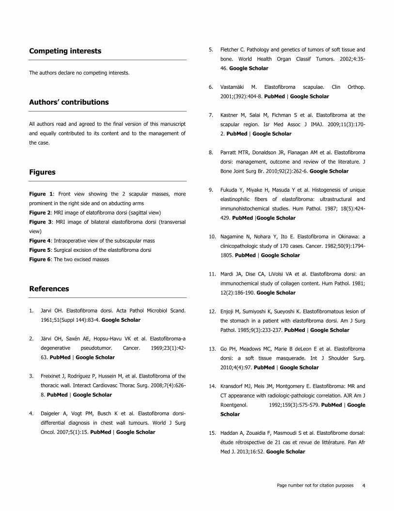
Physical examination revealed bilateral masses that became
manifest on abducting and flexing arms (Figure 1). Both lesions
were grossly rounded, of rubber-like consistency, not adhering to
the skin but adhesive to deep structures, measuring about 9 cm in
diameter, and slightly painful (the pain was reported mainly in the
right side). Ultrasound showed a homogeneous well limited
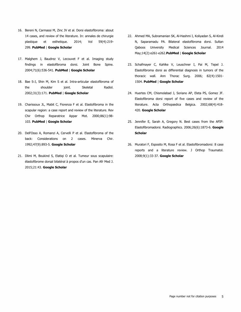
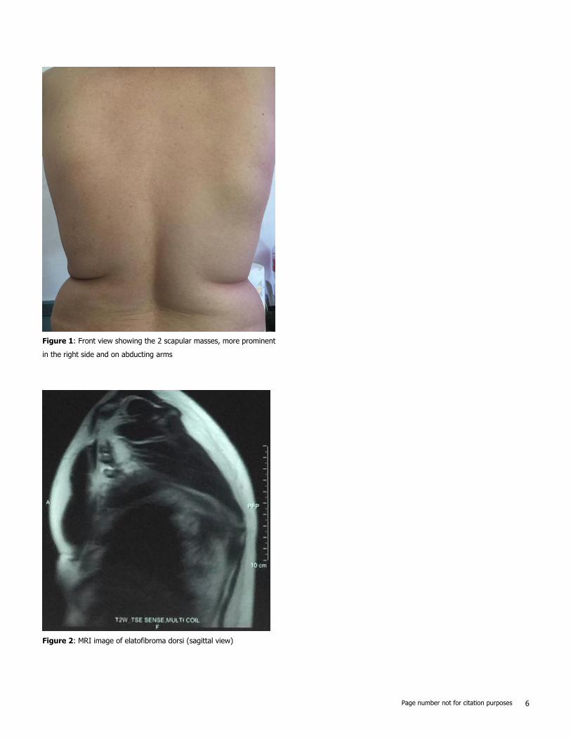
echogenic avascular structure measuring 67 x 30 mm. Magnetic
resonance imaging set the diagnosis by revealing bilateral soft
tissue tumors regarding the tip of the scapula, roughly symmetrical
and poorly circumscribed with dimensions 77 x 21 mm in the right
side and 83 x 21 mm in the left one. The masses consisted of
elements with linear fibrillary signaling which intensity was equal to
muscles in T1 and T2. Hypersignaling bands in T1 and T2 were also
dispersed among them with intensity equal to fat. No signs of
malignancy were observed, particularly absence of bone invasion.
The MRI data were compatible with elastofibroma dorsi (Figure
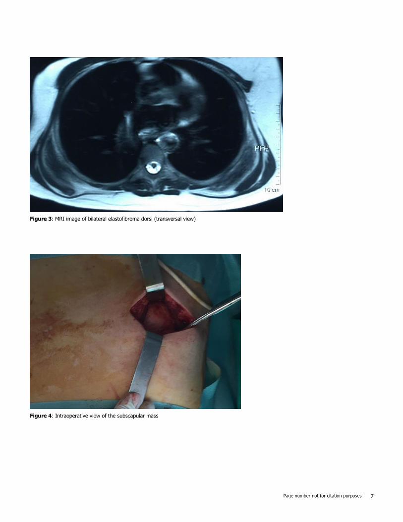
2, Figure 3). Surgical excision was performed under general
anesthesia on prone position and with abducted arms. An incision
was made over the palpable masses and the ill-defined tumors were
removed (Figure 4, Figure 5). Macroscopically, the lesions were non
encapsulated, with whitish fibrous tracts alternating with yellowish
tissue of fatty consistency (Figure 6). The histological study revealed
a benign proliferation composed of collagen and elastic fibers,
associated with some fibroblasts and fat cells. The elastic fibers
were fragmented into globules or had a linear arrangement. The
surgical excision was complete. The post operative course was
without complications, and the patient was discharged 3 days after
surgery.
Discussion
Elastofibroma dorsi (ED) is an infrequent slow growing soft tissue
benign tumor. It was defined in 2002 by the World Health
Organization (WHO) of soft tissue tumors taxonomy as a benign
fibroblastic/myofibroblastic tumor [5]. Nonetheless, most authors do
not consider ED as a real tumor, but a hyperplasia of elastic tissue
derived from fibroblasts due to chronic friction [6]. In reality, the
pathogenesis of ED remains unsolved and is still open to
controversy, but three etiological theories remain dominant. The
first one suggests that chronic and repetitive mechanical stress
leads to microtrauma, then to overproduction of elastic tissue from
the stimulated fibroblasts. And the description of heavy manual
worker men with repetitive labor was compatible with this theory
[7,8]. Thus, our clinical case seems to refute this thesis. Afterwards,
ED tends to affect elderly women like our patient, which led to the
second theory of reactive fibromatosis and secondary degeneration
of elastic fibers due to vascular insufficiency [9]. At last, a third
theory suggests a familial predisposition with an underlying
enzymatic disorder or defect, reported by Fukuda and al [4,7,9,10].
A double location of elastofibroma including the stomach and the
scapula has also been described and is in favor of this last theory
[11,12].
Elastofibroma dorsi affects primarily the elderly over 55 years of
age, with a mean age of 60 years [13]. Nevertheless, some cases of
ED have been described also in children [14]. ED is found more
frequently in women rather than men with a sex ratio Female/Man
going from 5/4 to 13/1 [15]. Although ED is infrequent and with a
slow growing, its diagnosis should be mentioned whenever a patient
presents a discomfort in response to scapula movement. Indeed,
the conception that ED is a very rare lesion seems to be unjustified
Page number not for citation purposes 3
probably because of the small size, the asymptomatic, and the
benign nature of the lesion. As consequence, ED is usually
accidentally found in CT/MRI imaging or when surgery is performed
for other reasons [16]. Regarding the localization of elastofibroma,
the vast majority of them are located in the subscapular and
infrascapular region between the thoracic wall, serratus anterior,
and latissimus dorsi muscles [1]. Unusual locations are also
described, including mediastinum, stomach, peritoneum, ischial
tuberosities, olecranon, deltoid muscle, intraspinal spaces, and foot
[17,18]. Bilateral involvement occurs only in 10% to 66% of cases,
and is usually asynchronous [3,5]. In our case, it was a typical
dorsal elastofibroma in an elderly woman, but bilateral and
asynchronous. Clinically, Elastofibroma dorsi is asymptomatic in
50% [4,5,7]. Symptoms depend on the site and the size of the
lesion. When symptoms occur, they consist in discomfort or stiffness
in shoulder abduction [19,20]. A painful scapula is only observed in
10%. And a neurological involvement of the upper limb may be
exceptionally observed, suggesting cervico-brachial neuralgia [21].
The physical examination reveals in typical ED a solid mass, of
variable size (4-12 cm), adherent to the deep layers but non
adherent to the skin, more prominent in abducting the arm,
painless, usually in the right side[15] but may be present as well on
the opposite shoulder, often smaller and silent [8,10,13,22].
Radiological investigation is performed once the diagnosis is
suspected. Chest X-ray shows unspecific images of elevation of the
scapula and an enlargement of the scapulothoracic space. An
interscapulothoracic opacity can be observed, but without bone lysis
or associated calcification [19]. Ultrasound, CT and MRI imagings
demonstrate the typical characteristics of ED which are collagen or
elastic fibers in the fatty background [6]. Indeed, ultrasound
examination shows an alternating of hypoechogenic and
hyperechogenic striations similar to muscle and parallel to its major
axis [23,24]. CT scan shows a non homogeneous mal limited mass
with density similar to muscles, including areas of lower density
secondary to fat [6]. Finally, the MRI remains the gold standard
examination for diagnosis. In fact, it shows heterogeneous well
defined mass revealing two different T1 signals, one of an
intermediate intensity equivalent to skeletal muscles signals, and the
second of high intensity representing fat imprisoned within the
mass. In T2, an increase in the signal intensity is observed.
Injection of gadolinium doesn't enhance the signal [17, 25]. MRI
imaging provides also specific features to establish differential
diagnosis between sarcoma, liposarcoma, hemangioma, hematoma,
lipoma and several other lesions. In our patient, the diagnosis was
set based on clinical and MRI findings which were compatible with
the literature's data.
Biopsy is performed only when diagnosis can't be set in front of
untypical MRI findings, it provides then confirmation of ED [26].
Fine needle aspiration can also be used but is inadequate to get a
representative tissue specimen. Macroscopically, elastofibroma is a
non-encapsulated mass, poorly defined, measuring 2 to 15 cm, with
a rubber consistency, which the cut surface exhibits white fibrous
tissue with interposing small areas of yellow fat [5]. Histological
examination describes a collagenous tissue, mixed with eosinophilic
elastic fibers fragmented into disks or globules, associated with
mature fat cells, as described in our case. It's important to notice
that there is no atypia or mitotic activity, which distinguishes ED
from other pseudotumors and neoplasms [4, 5]. Treatment of
elastofibroma dorsi is provided only in symptomatic painful forms, or
when the diagnosis is doubtful. It consists in complete surgical
excision of the mass with curative marginal resection [13]. In
asymptomatic lesions, clinical follow up proves to be sufficient. For
some authors however, surgery is needed whenever the greater
diameter is over 5 cm despite of the absence of symptoms [19, 20].
In our patient, we performed surgery due to the symptomatic
aspect of the lesion, and the excision was complete. The
postoperative course is usually simple, the most frequent
complication remains hematoma due to the hypervascularisation of
the subscapular region [4]. Thus, our patient didn't have any
complication. The prognosis is excellent, extremely low recurrence
cases have been described due to incomplete excision. But no
malignant transformation has been reported [10].
Conclusion
Elastofibroma dorsi is an infrequent benign soft tissue tumor which
frequently occurs in the subscapular region of elderly women.
Although it is frequently asymptomatic, it can be responsible of
discomfort and pain in arm mobilization. MRI is the gold standard
examination for diagnosis. Biopsy is performed only in untypical ED
to rule out a malignant tumor diagnosis. Surgical excision is the
therapeutic option for symptomatic patients only. Pathological study
confirms the diagnosis after surgery. Finally, our experience with
this case report is in accordance with the literature.
Page number not for citation purposes 4
Competing interests
The authors declare no competing interests.
Authors’ contributions
All authors read and agreed to the final version of this manuscript
and equally contributed to its content and to the management of
the case.
Figures
Figure 1: Front view showing the 2 scapular masses, more
prominent in the right side and on abducting arms
Figure 2: MRI image of elatofibroma dorsi (sagittal view)
Figure 3: MRI image of bilateral elastofibroma dorsi (transversal
view)
Figure 4: Intraoperative view of the subscapular mass
Figure 5: Surgical excision of the elastofibroma dorsi
Figure 6: The two excised masses
References
1. Jarvi OH. Elastofibroma dorsi. Acta Pathol Microbiol Scand.
1961;51(Suppl 144):83-4. Google Scholar
2. Järvi OH, Saxén AE, Hopsu-Havu VK et al. Elastofibroma-a
degenerative pseudotumor. Cancer. 1969;23(1):42-
63. PubMed | Google Scholar
3. Freixinet J, Rodríguez P, Hussein M, et al. Elastofibroma of the
thoracic wall. Interact Cardiovasc Thorac Surg. 2008;7(4):626-
8. PubMed | Google Scholar
4. Daigeler A, Vogt PM, Busch K et al. Elastofibroma dorsi-
differential diagnosis in chest wall tumours. World J Surg
Oncol. 2007;5(1):15. PubMed | Google Scholar
5. Fletcher C. Pathology and genetics of tumors of soft tissue and
bone. World Health Organ Classif Tumors. 2002;4:35-
46. Google Scholar
6. Vastamäki M. Elastofibroma scapulae. Clin Orthop.
2001;(392):404-8. PubMed | Google Scholar
7. Kastner M, Salai M, Fichman S et al. Elastofibroma at the
scapular region. Isr Med Assoc J IMAJ. 2009;11(3):170-
2. PubMed | Google Scholar
8. Parratt MTR, Donaldson JR, Flanagan AM et al. Elastofibroma
dorsi: management, outcome and review of the literature. J
Bone Joint Surg Br. 2010;92(2):262-6. Google Scholar
9. Fukuda Y, Miyake H, Masuda Y et al. Histogenesis of unique
elastinophilic fibers of elastofibroma: ultrastructural and
immunohistochemical studies. Hum Pathol. 1987; 18(5):424-
429. PubMed |Google Scholar
10. Nagamine N, Nohara Y, Ito E. Elastofibroma in Okinawa: a
clinicopathologic study of 170 cases. Cancer. 1982;50(9):1794-
1805. PubMed | Google Scholar
11. Mardi JA, Dise CA, LiVolsi VA et al. Elastofibroma dorsi: an
immunochemical study of collagen content. Hum Pathol. 1981;
12(2):186-190. Google Scholar
12. Enjoji M, Sumiyoshi K, Sueyoshi K. Elastofibromatous lesion of
the stomach in a patient with elastofibroma dorsi. Am J Surg
Pathol. 1985;9(3):233-237. PubMed | Google Scholar
13. Go PH, Meadows MC, Marie B deLeon E et al. Elastofibroma
dorsi: a soft tissue masquerade. Int J Shoulder Surg.
2010;4(4):97. PubMed | Google Scholar
14. Kransdorf MJ, Meis JM, Montgomery E. Elastofibroma: MR and
CT appearance with radiologic-pathologic correlation. AJR Am J
Roentgenol. 1992;159(3):575-579. PubMed | Google
Scholar
15. Haddan A, Zouaidia F, Masmoudi S et al. Elastofibrome dorsal:
étude rétrospective de 21 cas et revue de littérature. Pan Afr
Med J. 2013;16:52. Google Scholar
Page number not for citation purposes 5
16. Bereni N, Carmassi M, Zinc JV et al. Dorsi elastofibroma: about
14 cases, and review of the literature. In: annales de chirurgie
plastique et esthetique. 2014; Vol 59(4):219-
299. PubMed | Google Scholar
17. Malghem J, Baudrez V, Lecouvet F et al. Imaging study
findings in elastofibroma dorsi. Joint Bone Spine.
2004;71(6):536-541. PubMed | Google Scholar
18. Bae S-J, Shin M, Kim S et al. Intra-articular elastofibroma of
the shoulder joint. Skeletal Radiol.
2002;31(3):171. PubMed | Google Scholar
19. Charissoux JL, Mabit C, Fiorenza F et al. Elastofibroma in the
scapular region: a case report and review of the literature. Rev
Chir Orthop Reparatrice Appar Mot. 2000;86(1):98-
103. PubMed | Google Scholar
20. Dell'Osso A, Romanzi A, Cervelli P et al. Elastofibroma of the
back: Considerations on 2 cases. Minerva Chir.
1992;47(9):893-5. Google Scholar
21. Dlimi M, Boukind S, Elatiqi O et al. Tumeur sous scapulaire:
élastofibrome dorsal bilatéral à propos d'un cas. Pan Afr Med J.
2015;21:43. Google Scholar
22. Ahmed MA, Subramanian SK, Al-Hashmi I, Koliyadan S, Al-Kindi
N, Saparamadu PA. Bilateral elastofibroma dorsi. Sultan
Qaboos University Medical Sciences Journal. 2014
May;14(2):e261-e262.PubMed | Google Scholar
23. Schafmayer C, Kahlke V, Leuschner I, Pai M, Tepel J.
Elastofibroma dorsi as differential diagnosis in tumors of the
thoracic wall. Ann Thorac Surg. 2006; 82(4):1501-
1504. PubMed | Google Scholar
24. Huertes CM, Chismolabad J, Soriano AP, Eleta PS, Gomez JF.
Elastofibroma dorsi report of five cases and review of the
literature. Acta Orthopædica Belgica. 2002;68(4):418-
420. Google Scholar
25. Jennifer E, Sarah A, Gregory N. Best cases from the AFIP:
Elastofibromadorsi. Radiographics. 2006;26(6):1873-6. Google
Scholar
26. Muratori F, Esposito M, Rosa F et al. Elastofibromadorsi: 8 case
reports and a literature review. J Orthop Traumatol.
2008;9(1):33-37. Google Scholar
Page number not for citation purposes 6
Figure 1: Front view showing the 2 scapular masses, more prominent
in the right side and on abducting arms
Figure 2: MRI image of elatofibroma dorsi (sagittal view)
Page number not for citation purposes 7
Figure 3: MRI image of bilateral elastofibroma dorsi (transversal view)
Figure 4: Intraoperative view of the subscapular mass
Page number not for citation purposes 8
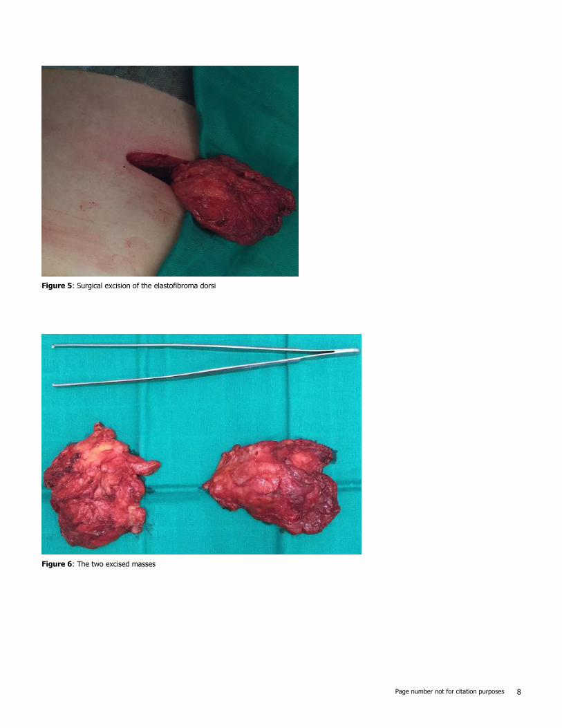
Figure 5: Surgical excision of the elastofibroma dorsi
Figure 6: The two excised masses