Embed Size (px)
Citation preview
58 The Open Toxicology Journal, 2009, 3, 58-69
1874-3404/09 2009 Bentham Open
Open Access
Genotoxic and Mutagenic Effects of Melissa officinalis (Erva Cidreira) Extracts
A. M. Alves1, L. S. Vidal1, R. M. Kuster2, C. Lage1, and A. C. Leitão*,1
1Laboratório de Radiobiologia Molecular, Programa de Biologia Molecular e Estrutural, Instituto de Biofísica Carlos Chagas Filho, UFRJ; 2Núcleo de Pesquisas de Produtos Naturais – UFRJ, Brazil
Abstract: Erva cidreira (Melissa officinalis) is a plant with sedative properties and in Brazil it has been used for insomnia and anxiety. It is also employed to stimulate liver functions and normalize menstruation and for intestinal constipation. The aim of this work was to evaluate the genotoxic and mutagenic effects of erva cidreira. Results indicate the presence of genotoxic activity in the lyophilized extract, with lysogenic induction increasing about 80-fold over the spontaneous background after treatment with lyophilized total aqueous extract. Beta-galactosidase levels raised 9-fold in indicator strains after treatment under the same conditions. A deeper look into the mechanism underlying the genotoxic action im-plicated reactive oxygen species generated by compounds retained in the ethyl-acetate, polar partition of the total aqueous extract. The spectrum of mutations revealed a great trend for base substitutions, mainly in guanines and adenines. The presence of phenolic compounds, especially caffeic acid derivatives, may be correlated to its genotoxic activity. We thus conclude that compounds present in current concentrations of erva cidreira extracts are genotoxic and mutagenic, and might have carcinogenic potential.
Keywords: Melissa officinalis, genotoxicity, reactive oxygen species, mutagenesis, caffeic acid.
INTRODUCTION
Melissa officinalis is a bush plant from the Lamiaceae family, known in Brazil as erva cidreira. Erva cidreira has many uses in ethno medicine. Infusions prepared from stem and leaves are used as tranquilizer for insomnia and anxiety. Its essential oil is indicated to normalize menstrual irregu-larities, to eliminate digestive gases and to counteract consti-pation. Besides, it has been used against viral infections such as influenza, herpes, mumps and smallpox (Dimitrova Acta Microbiol Bulg 1993) [1] (Koytchev Phytomedicine 1999) [2] (Mikus Planta Med 2000) [3]. Yamasaki et al. (Biol Pharm Bull 1998) [4] have experimentally observed the anti-viral and cytostatic actions against the Human Immunodefi-ciency Virus due the inhibition of its reverse transcriptase in vitro. A beneficial action on treatment of Alzheimer disease has also been reported (Perry J Pharmacol 1999) [5]. The use of medicinal plants is an old practice in human populations; however there are still lacking controlled stud-ies on these plants and the risks of their consumption are unknown. Moreover, plant material of non-certificated origin is often contaminated by toxic substances, and behind the standards of microbial quality control. Studies on the bio-logical effects of medicinal plant extracts are thus compli-cated because they comprise a chemical mixture of several substances (Sugimura Cancer Res 1983) [6]. The uncon-trolled use of these extracts by general population claim for studies on their side effects and toxicity.
*Address correspondence to this author at the Instituto de Biofísica Carlos Chagas Filho, Av. Carlos Chagas Filho, 373, CCS Bloco G - Cidade Uni-versitária, CEP - 21941-902, Rio de Janeiro - Brazil; Tel: +55-21-25626578; Fax: +55-21-2280-8193; E-mail: [email protected]
The absence of basic information on genotoxic effects associated to the use of Melissa officinallis has led us to use different assays to evaluate its genotoxic and mutagenic ac-tivities. Genotoxic activity of erva cidreira extract was evaluated by phage-induction assay - Inductest (Moreau PNAS 1976) [7] and SOS chromotest (Quillardet Mutat Res 1985) [8], while mutagenic activity was evaluated by differ-ent mutagenesis assays. The erva cidreira extract was shown to be genotoxic and mutagenic, probably due to the action of caffeic acid derivatives present in its composition.
MATERIAL AND METHODS
Bacterial Strains
The Escherichia coli and Salmonella typhimurium strains used in this work are listed in Tables 1 and 2, respectively.
Bacterial Media
Bacterial cells were grown overnight in a shaking incuba-tor at 37°C in LB medium (Miller Cold Spring Harbor Lab Press 1992) [9]. A starting inoculum was taken from this culture and cells were grown under the same conditions until the culture reached the exponential phase. The E. coli bacterial survival and infective center produc-tion assays were done using LB medium and LB medium containing 20 µg/ml (micrograms per ml) ampicillin (LB-amp), respectively. Both media were solidified with 1.5% Difco bacto agar. E medium (Vogel J Biol Chem 1956) [10] solidified with 1.5% Difco bacto agar was supplemented with histidine for the Ames test (Maron Mutat Res 1983) [11] or tryptophan for the WP2 mutagenicity test (Blanco Mutat Res 1998) [12] assays. A selective minimal medium
Genotoxic Effects of Cidreira Extracts The Open Toxicology Journal, 2009, Volume 3 59
containing 0.4% lactose was used for the lac+-revertant mutagenesis assay (Cupples PNAS 1989) [13] (Cupples Ge-netics 1990 [14]. Preparation of Melissa officinalis Extracts
Preparation of extracts followed the same standard pro-cedure employed by the pharmaceutical industry to obtain the phytomedicine for commercial use. Dried leaves of certified Melissa officinalis plants were kindly provided by a local pharmaceutical industry (Labo-ratório Simões Ltd., Rio de Janeiro, Brazil), and they were boiled in bidistilled water (200mg/ml) for 10min. The result-ing infusion was filtered, lyophilized, and stored at -20°C until further use. In the process of lyophilization we could retain about 11% of the original material.
HPLC
The water-reconstituted extract was analyzed by HPLC to determine its qualitative chemical composition. Ten-ml samples were injected in HPLC/DAC equipment (Shima-dzu). The stationary phase used was an RP-18 silica gel col-umn (10 mm) from Merck and the mobile phase was com-posed by a system of solvents consisting of H2O + 1% H3PO4 (solvent A), and 30% Methanol (solvent B) for 10min, 40% B for 20 min, 100% B for 60 min. The flux of mobile phase was 1 ml/min, and the chromatogram records were obtained by absorption at 254nm. Partition and Chromatography of the Reconstituted Ex-tract
The water-reconstituted extract was successively parti-tioned by an extraction method using organic solvents of inc-
Table 1. Escherichia coli Strains
Designations Relevant Genotype Reference
WP2s WP2 uvrA / pKM101 ampR Blanco et al., 1998
IC203 WP2 oxyR / pKM101 ampR Blanco et al., 1998
IC204 WP2 Δ (umuDC) catR Blanco et al., 1998
IC206 WP2 Δ (umuDC) mutY catR Blanco et al., 1998
IC208 WP2 Δ (umuDC) mutY oxyR catR Blanco et al., 1998
CC101 P90C ara Δ (lac proB)XIII(A: T C: G) Cupples and Miller, 1989
CC102 P90C ara Δ (lac proB)XIII (G:C A:T) Cupples and Miller, 1989
CC103 P90C ara Δ (lac proB)XIII (G:C C:G) Cupples and Miller, 1989
CC104 P90C ara Δ (lac proB)XIII (G:CT:A) Cupples and Miller, 1989
CC105 P90C ara Δ (lac proB)XIII (A:T T:A) Cupples and Miller, 1989
CC106 P90C ara Δ (lac proB)XIII (A:T G:C) Cupples and Miller, 1989
CC107 P90C ara Δ (lac proB)XIII (+ 1G) Cupples et al., 1990
CC108 P90C ara Δ (lac proB)XIII (-1G) Cupples et al., 1990
CC109 P90Cara Δ (lac proB)XIII (-2(CG)) Cupples et al., 1990
CC110 P90 C ara Δ (lac proB)XIII (+1A ) Cupples et al., 1990
CC111 P90C ara Δ (lac proB)XIII ( -1A) Cupples et al., 1990
WP2s(λ) WP2 uvrA (λ) trpE Our laboratory stock
RJF013 [B/r SR 714] uvrD3 trpE ampR Our laboratory stock
PQ37 uvrA rfa sfiA::lacZ Quillardet and Hofnung (1985)
Table 2. Salmonella typhimurium Bacterial Strains
Designations Relevant Genotype Reference
TA97 hisD6610/ hisO1242 - ΔuvrB rfa pKM101(ampR) Maron and Ames, 1983
TA98 hisD3052 - ΔuvrB rfa pKM101(ampR) Maron and Ames, 1983
TA100 hisG46 - ΔuvrB rfa pKM101 (ampR ) Maron and Ames, 1983
TA102 hisG428 – wild type rfa pKM101(ampR) pAQ1 (tetR ) Maron and Ames, 1983
60 The Open Toxicology Journal, 2009, Volume 3 Leitão et al.
reasing polarity (hexane, chloroform, ethyl acetate and buta-nol). Later, partitions were screened for activity by the In-ductest protocol to search for fractions with genotoxic activ-ity.
CHEMICALS
Catalase (Sigma Chemical Company - EUA) and thio-urea (Indústrias Químicas Merck S/A - Brazil) were dis-solved in bidistilled water. Dipyridyl (Sigma Chemical Com-pany - EUA) was dissolved in 10% ethanol and then diluted in bidistilled water.
GENOTOXIC STUDIES
Lysogenic Induction Assay
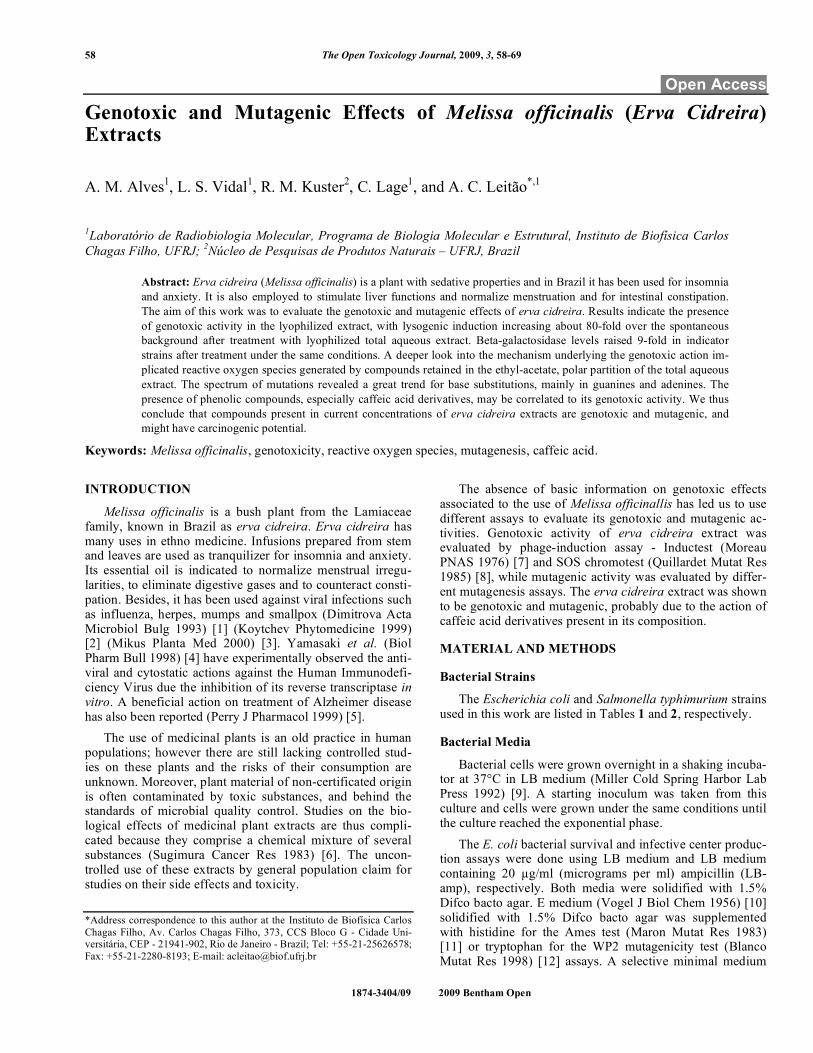
The strains WP2s(λ) and RJF013 of E. coli B/r (Table 1) were used in the lysogenic induction assay, and the protocol was similar to the quantitative Inductest developed by Mo-reau et al. in 1976 (PNAS) [7]. Lyophilized extracts were reconstituted and mixed to the melted medium at the amounts indicated in the X-axis of the induction graphs. Each experimental determination was performed in dupli-cate, and results represent the average of at least three inde-pendent experiments. The level of induction was expressed as the induction factor, which is the ratio between the num-bers of infective centers after treatment by those occurring spontaneously.
SOS Chromotest Assay
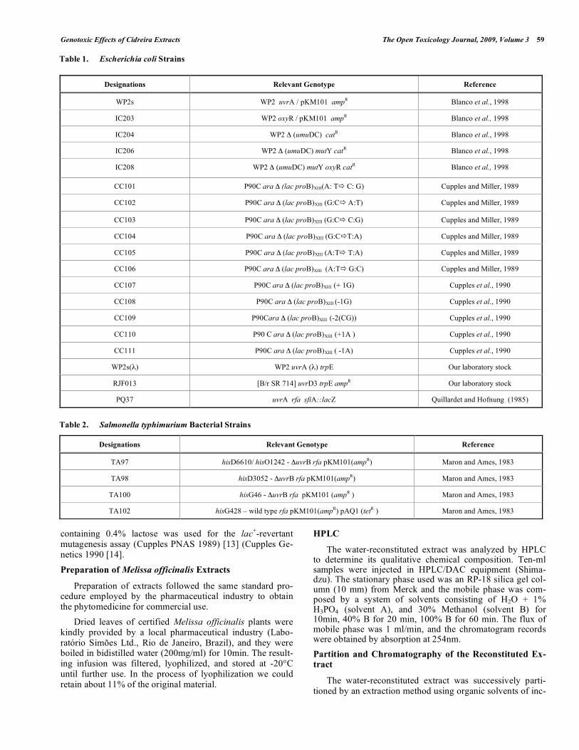
E. coli K-12 PQ37 strain (Table 1) was used in the SOS chromotest studies, according to the protocol developed by Quillardet and Hofnung (Mutat Res 1985) [8]. Results repre-sent the average of at least three experiments. The expression of β-galactosidase is shown as enzymatic units (ordinates, obtained by measurement of absorbance at 420nm immedi-ately after each treatment) as a function of the amount of extract (abscissas).
Mechanism of Action Underlying the Genotoxic Activity
The mechanism of action by which the extract may exert its genotoxic activity was investigated by experiments in which the lysogenic induction was measured after exposure to a defined amount of erva cidreira extract (150mg/plate) in the absence (control) or in the presence of anti-oxidant com-pounds. As many plant extracts are capable of generate dam-aging reactive oxygen species (ROS), this effect was deter-mined for the erva cidreira extract by the addition (or not) of increasing concentrations of: (a) catalase, (b) thiourea or (c) dipyridyl. Each experimental determination was performed in duplicate, and results represent the average of at least three independent experiments.
MUTAGENIC STUDIES
Ames Test
This assay was carried out as described by Maron and Ames (Mutat Res 1983) [11], using the histidine auxotroph strains TA97, TA98, TA100 and TA102 of S. typhimurium. Each experimental determination was performed in duplicate and results represent the average of at least three experi-
ments. Mutagenesis induced by erva cidreira in each strain was expressed as the absolute number of His+ revertants per plate (Ames Prog Clin Biol Res 1986) [15].
Tryptophan Mutagenesis Assay
This assay was carried out as described by Blanco et al. (Mutat Res 1998) [12], using the tryptophan auxotroph strains WP2s, IC203, IC204, IC206 e IC208 of E. coli (Table 1). Each experimental determination was performed in du-plicate, and results represent the average of at least three independent experiments. Mutagenesis induced by erva cidreira extract in each strain was expressed as mutants per 108 cells. The increment factor was obtained by dividing the number of induced mutants by that of the spontaneous rever-tants.
Lactose Mutagenesis Assay
This assay was carried out as described by Cupples and Miller (PNAS 1989) [13] and Cupples et al. (Genetics 1990) [14] using different strains constructed to detect specific base pair substitutions and frameshift mutations in the E. coli lacZ locus. Mutagenic activity was expressed as the number of revertants per 108 cells. The increment factor was obtained by dividing the number of induced mutants by that of the spontaneous revertants.
RESULTS
Genotoxicity of Erva Cidreira
Phage induction produced by different amounts of erva cidreira extract is shown in Fig. (1). Maximal rates of phage induction were observed at 150mg/plate (corresponding to about 80-fold increase over the spontaneous background). Positive SOS induction was detected for the uvrA-deficient strain PQ37 treated with different concentrations of erva cidreira extract. Results indicate the presence of geno-toxic activity in the lyophilized extract, and induction of en-zymatic activity reached 24 units (~9-fold increment over the spontaneous induction, as shown in Fig. (2) at the maximum amount used (150mg/ml). These results strengthen those obtained with the Inductest, suggesting that the extract bears on bacterial genotoxicity.
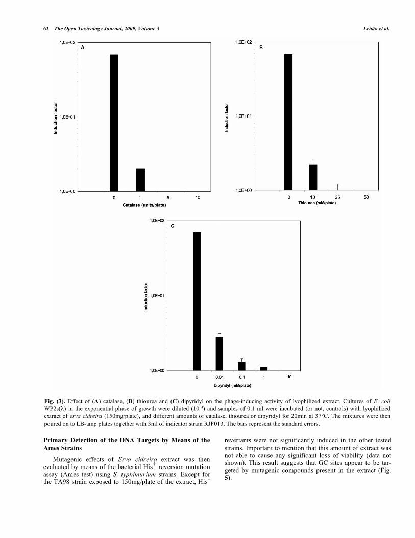
Suppression of the Genotoxicity of Erva Cidreira by Anti-oxidant Agents
Specific antioxidant agents were used simultaneously with the extract to evaluate if the lysogenic induction ob-served in Fig. (1) would be due to the production of ROS. Increasing concentrations of the different antioxidant agents were added to the Indutest experiments performed with ex-tracts at the amount of 150mg/plate. Catalase, dypiridyl and thiourea were shown to exert an inhibitory action on the genotoxic effect induced by erva cidreira extract (Fig. 3). Thus ROS may intermediate the genotoxic activity of this extract.
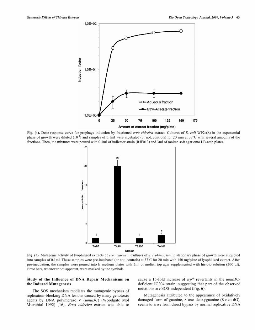
Genotoxicity of Fractionated Erva Cidreira Extracts
The aqueous fraction of erva cidreira extract was able to induce the prophage in E. coli WP2s(λ) Fig. (4) HPLC/UV
Genotoxic Effects of Cidreira Extracts The Open Toxicology Journal, 2009, Volume 3 61
analysis of this fraction indicates the presence of phenolic compounds, like benzoic acid derivatives and caffeic acid derivatives as the major components in the extract (Fig. 8). Those compounds may be thus implicated in the observed genotoxic activity present in the erva cidreira extract. Be-
sides the aqueous total extract, significant prophage induc-tion was shown to occur after treatment with the ethyl-acetate fraction, although reduced by a 10-fold factor (Fig. 4). The other non-polar fractions were not effective in induc-ing the prophage (data not shown).
Fig. (1). Dose-response curve for prophage induction by lyophilized extracts of erva cidreira. Cultures of E. coli WP2s(λ) in the exponential phase of growth were diluted (10-4) and samples of 0.1 ml were pre-incubated (or not, controls) at 37°C for 20 min with the indicated amounts of lyophilized extract. Then, the mixtures were poured with 0.3 ml of indicator strain (RJF013) and 3 ml of molten soft agar onto LB-amp plates.
Fig. (2). Induction of β-galactosidase in PQ37 strain after treatment with different concentrations of lyophilized extract of Erva Cidreira. The induction of β-galactosidase units was calculated by means of the relation defined by Quillardet and Hofnung (1985) [11] after measurement of absorbance at 420 nm.
62 The Open Toxicology Journal, 2009, Volume 3 Leitão et al.
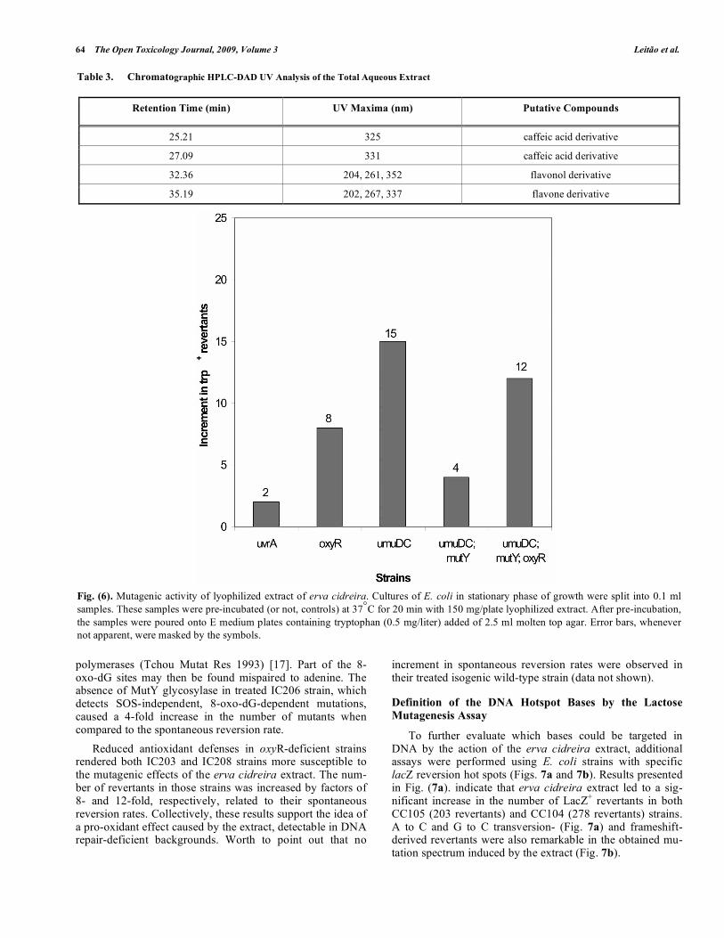
Primary Detection of the DNA Targets by Means of the Ames Strains
Mutagenic effects of Erva cidreira extract was then evaluated by means of the bacterial His+ reversion mutation assay (Ames test) using S. typhimurium strains. Except for the TA98 strain exposed to 150mg/plate of the extract, His+
revertants were not significantly induced in the other tested strains. Important to mention that this amount of extract was not able to cause any significant loss of viability (data not shown). This result suggests that GC sites appear to be tar-geted by mutagenic compounds present in the extract (Fig. 5).
Fig. (3). Effect of (A) catalase, (B) thiourea and (C) dipyridyl on the phage-inducing activity of lyophilized extract. Cultures of E. coli WP2s(λ) in the exponential phase of growth were diluted (10-4) and samples of 0.1 ml were incubated (or not, controls) with lyophilized extract of erva cidreira (150mg/plate), and different amounts of catalase, thiourea or dipyridyl for 20min at 37°C. The mixtures were then poured on to LB-amp plates together with 3ml of indicator strain RJF013. The bars represent the standard errors.
Genotoxic Effects of Cidreira Extracts The Open Toxicology Journal, 2009, Volume 3 63
Study of the Influence of DNA Repair Mechanisms on the Induced Mutagenesis
The SOS mechanism mediates the mutagenic bypass of replication-blocking DNA lesions caused by many genotoxic agents by DNA polymerase V (umuDC) (Woodgate Mol Microbiol 1992) [16]. Erva cidreira extract was able to
cause a 15-fold increase of trp+ revertants in the umuDC-deficient IC204 strain, suggesting that part of the observed mutations are SOS-independent (Fig. 6). Mutagenesis attributed to the appearance of oxidatively damaged form of guanine, 8-oxo-deoxyguanine (8-oxo-dG), seems to arise from direct bypass by normal replicative DNA
Fig. (4). Dose-response curve for prophage induction by fractioned erva cidreira extract. Cultures of E. coli WP2s(λ) in the exponential phase of growth were diluted (10-4) and samples of 0.1ml were incubated (or not, controls) for 20 min at 37°C with several amounts of the fractions. Then, the mixtures were poured with 0.3ml of indicator strain (RJF013) and 3ml of molten soft agar onto LB-amp plates.
Fig. (5). Mutagenic activity of lyophilized extracts of erva cidreira. Cultures of S. typhimurium in stationary phase of growth were aliquoted into samples of 0.1ml. These samples were pre-incubated (or not, controls) at 37°C for 20 min with 150 mg/plate of lyophilized extract. After pre-incubation, the samples were poured into E medium plates with 2ml of molten top agar supplemented with his-bio solution (200 µl). Error bars, whenever not apparent, were masked by the symbols.
64 The Open Toxicology Journal, 2009, Volume 3 Leitão et al.
polymerases (Tchou Mutat Res 1993) [17]. Part of the 8-oxo-dG sites may then be found mispaired to adenine. The absence of MutY glycosylase in treated IC206 strain, which detects SOS-independent, 8-oxo-dG-dependent mutations, caused a 4-fold increase in the number of mutants when compared to the spontaneous reversion rate. Reduced antioxidant defenses in oxyR-deficient strains rendered both IC203 and IC208 strains more susceptible to the mutagenic effects of the erva cidreira extract. The num-ber of revertants in those strains was increased by factors of 8- and 12-fold, respectively, related to their spontaneous reversion rates. Collectively, these results support the idea of a pro-oxidant effect caused by the extract, detectable in DNA repair-deficient backgrounds. Worth to point out that no
increment in spontaneous reversion rates were observed in their treated isogenic wild-type strain (data not shown).
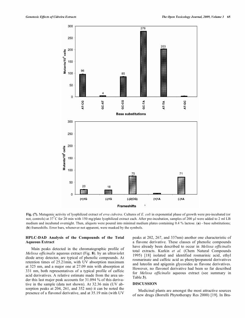
Definition of the DNA Hotspot Bases by the Lactose Mutagenesis Assay
To further evaluate which bases could be targeted in DNA by the action of the erva cidreira extract, additional assays were performed using E. coli strains with specific lacZ reversion hot spots (Figs. 7a and 7b). Results presented in Fig. (7a). indicate that erva cidreira extract led to a sig-nificant increase in the number of LacZ+ revertants in both CC105 (203 revertants) and CC104 (278 revertants) strains. A to C and G to C transversion- (Fig. 7a) and frameshift-derived revertants were also remarkable in the obtained mu-tation spectrum induced by the extract (Fig. 7b).
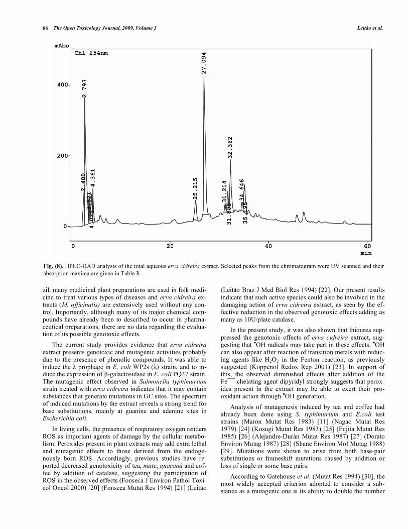
Table 3. Chromatographic HPLC-DAD UV Analysis of the Total Aqueous Extract
Retention Time (min) UV Maxima (nm) Putative Compounds
25.21 325 caffeic acid derivative
27.09 331 caffeic acid derivative
32.36 204, 261, 352 flavonol derivative
35.19 202, 267, 337 flavone derivative
Fig. (6). Mutagenic activity of lyophilized extract of erva cidreira. Cultures of E. coli in stationary phase of growth were split into 0.1 ml samples. These samples were pre-incubated (or not, controls) at 37°C for 20 min with 150 mg/plate lyophilized extract. After pre-incubation, the samples were poured onto E medium plates containing tryptophan (0.5 mg/liter) added of 2.5 ml molten top agar. Error bars, whenever not apparent, were masked by the symbols.
Genotoxic Effects of Cidreira Extracts The Open Toxicology Journal, 2009, Volume 3 65
HPLC-DAD Analysis of the Compounds of the Total Aqueous Extract
Main peaks detected in the chromatographic profile of Melissa officinalis aqueous extract (Fig. 8). by an ultraviolet diode array detector, are typical of phenolic compounds. At retention times of 25.21min, with UV absorption maximum at 325 nm, and a major one at 27.09 min with absorption at 331 nm, both representatives of a typical profile of caffeic acid derivatives. A relative estimate made from the area un-der this last major peak accounts for 31.094 % of this deriva-tive in the sample (data not shown). At 32.36 min (UV ab-sorption peaks at 204, 261, and 352 nm) it can be noted the presence of a flavonol derivative, and at 35.19 min (with UV
peaks at 202, 267, and 337nm) another one characteristic of a flavone derivative. These classes of phenolic compounds have already been described to occur in Melissa officinalis total extracts. Kurkin et al. (Chem Natural Compounds 1995) [18] isolated and identified rosmarinic acid, ethyl rosmarinate and caffeic acid as phenylpropanoid derivatives and luteolin and apigenin glycosides as flavone derivatives. However, no flavonol derivative had been so far described for Melissa officinalis aqueous extract (see summary in Table 3). DISCUSSION
Medicinal plants are amongst the most attractive sources of new drugs (Borrelli Phytotherapy Res 2000) [19]. In Bra-
Fig. (7). Mutagenic activity of lyophilized extract of erva cidreira. Cultures of E. coli in exponential phase of growth were pre-incubated (or not, controls) at 37°C for 20 min with 150 mg/plate lyophilized extract each. After pre-incubation, samples of 200 µl were added to 2 ml LB medium and incubated overnight. Then, aliquots were poured into minimal medium plates containing 0.4 % lactose. (a) - base substitutions; (b) frameshifts. Error bars, whenever not apparent, were masked by the symbols.
66 The Open Toxicology Journal, 2009, Volume 3 Leitão et al.
zil, many medicinal plant preparations are used in folk medi-cine to treat various types of diseases and erva cidreira ex-tracts (M. officinalis) are extensively used without any con-trol. Importantly, although many of its major chemical com-pounds have already been to described to occur in pharma-ceutical preparations, there are no data regarding the evalua-tion of its possible genotoxic effects. The current study provides evidence that erva cidreira extract presents genotoxic and mutagenic activities probably due to the presence of phenolic compounds. It was able to induce the λ prophage in E. coli WP2s (λ) strain, and to in-duce the expression of β-galactosidase in E. coli PQ37 strain. The mutagenic effect observed in Salmonella typhimurium strain treated with erva cidreira indicates that it may contain substances that generate mutations in GC sites. The spectrum of induced mutations by the extract reveals a strong trend for base substitutions, mainly at guanine and adenine sites in Escherichia coli. In living cells, the presence of respiratory oxygen renders ROS as important agents of damage by the cellular metabo-lism. Peroxides present in plant extracts may add extra lethal and mutagenic effects to those derived from the endoge-nously born ROS. Accordingly, previous studies have re-ported decreased genotoxicity of tea, mate, guaraná and cof-fee by addition of catalase, suggesting the participation of ROS in the observed effects (Fonseca J Environ Pathol Toxi-col Oncol 2000) [20] (Fonseca Mutat Res 1994) [21] (Leitão
(Leitão Braz J Med Biol Res 1994) [22]. Our present results indicate that such active species could also be involved in the damaging action of erva cidreira extract, as seen by the ef-fective reduction in the observed genotoxic effects adding as many as 10U/plate catalase. In the present study, it was also shown that thiourea sup-pressed the genotoxic effects of erva cidreira extract, sug-gesting that •OH radicals may take part in these effects. •OH can also appear after reaction of transition metals with reduc-ing agents like H2O2 in the Fenton reaction, as previously suggested (Koppenol Redox Rep 2001) [23]. In support of this, the observed diminished effects after addition of the Fe++ chelating agent dipyridyl strongly suggests that perox-ides present in the extract may be able to exert their pro-oxidant action through •OH generation. Analysis of mutagenesis induced by tea and coffee had already been done using S. typhimurium and E.coli test strains (Maron Mutat Res 1983) [11] (Nagao Mutat Res 1979) [24] (Kosugi Mutat Res 1983) [25] (Fujita Mutat Res 1985) [26] (Alejandre-Durán Mutat Res 1987) [27] (Dorato Environ Mutag 1987) [28] (Shane Environ Mol Mutag 1988) [29]. Mutations were shown to arise from both base-pair substitutions or frameshift mutations caused by addition or loss of single or some base pairs. According to Gatehouse et al. (Mutat Res 1994) [30], the most widely accepted criterion adopted to consider a sub-stance as a mutagenic one is its ability to double the number
Fig. (8). HPLC-DAD analysis of the total aqueous erva cidreira extract. Selected peaks from the chromatogram were UV scanned and their absorption maxima are given in Table 3.
Genotoxic Effects of Cidreira Extracts The Open Toxicology Journal, 2009, Volume 3 67
of spontaneous revertants in the Ames test. Erva cidreira extract can thus be considered a mutagenic agent and we have shown that these mutations are targeted to GC sites in the hisD locus of S. typhimurium TA98. Besides the Ames Salmonella assay, mutagenic effects of the erva cidreira extract were stressed by other bacterial assays. The first one consists in quantifying revertants to tryptophan prototrophy in treated E. coli WP2s strains to screen for the mutagenic activity of the extract. Revertants were shown to increase in the peroxide-sensitive IC203 (oxyR) strain after treatment with lyophilized extract. An increase in the number of revertants in IC204 strain (ΔumuDC), deficient in SOS mutagenic damage bypass, was also observed. It is thus suggested that erva cidreira extract induces oxidative damages in DNA, and part of them can be processed by error-prone repair system, in an SOS-independent manner (Goerlich J Bacteriol 1989) [31]. The triple umuDC, oxyR and mutY deficiency in IC208 strain did not cause reversion to exceed that observed for the single umuDC-deficient strain. Survival was reduced to up to 10% only in IC203 and IC208 strains after treatment, what rein-forces a pro-oxidant action of the extract in view of the sen-sitivity of these strains to oxidative agents (data not shown). Altogether these results forwarded the study to a deeper analysis of the mutagenic spectrum induced by treatment with the extract, since the observed SOS-independent mutagenesis might arise from direct bypass of miscoding lesions such as 8-oxo-dG. One of the main oxidative damages, induced by both endogenous and exogenous sources, is 8-oxo-dG. This dam-age is not connected with cellular lethality, instead, with mutagenic activity. Main base substitutions arising from the presence of 8-oxo-dG comprise G:C→T:A transversions. Significant levels of such transversions are attained when-ever one of three specific enzymes involved in its prevention and repair is absent in E. coli: MutT, MutM (Fpg) and MutY proteins. The mutagenic activity caused by the extract seems to target guanine bases, since the results of Ames test detected frameshift mutations in GC sites. This is also supported by the increase of revertants in IC203 and IC208 strains, both deficient in OxyR function. To address the mutational specificity of erva cidreira extract in a more direct approach, a set of E. coli strains car-rying particular point mutations in the lacZ gene (coding for β-galactosidase enzyme) were used in the lactose reversion mutation assay (Josephy Mutat Res 2000) [32]. Our results show that erva cidreira extract was able to promote G:C→T:A and A:T→T:A as major reversion events, and half-amount of A:T→C:G and G:C→C:G transversions as well. The mutation spectrum generated by the extract resem-bles, in many aspects, the one induced by ROS (Decuyper-Debergh EMBO J 1987) [33] (McBride Biochemistry 1991) [34] (McBride PNAS 1992) [35] (Retel Mutat Res 1993) [36] (Akasaka Mol Gen Genet 1994) [37]. In many cases, guanine sites are the most frequent ones to turn into abasic sites, what could account for the G→C transversions. G→C transversions are also quite frequent in the mutation spectrum induced by ROS, however, the
mechanism responsible for their appearance it is not known. At first, G→C transversions could result from the mispairing of 8-oxo-dG with guanines, since 8-oxo-dG could assume the 6-enolate-8-keto anti conformation, and pair up with guanines (Culp Chem Res Toxicol 1989) [38]. However, 8-oxo-dG is preferentially found in the tautomeric form 6,8-keto in vitro, and in its syn conformation pairs up with ade-nines leading to G→T transversions (Culp Chem Res Toxi-col 1989) [38] (Wood Biochemistry 1990) [39] (Cheng J Biol Chem 1992) [40]. In this work, A:T→T:A transversions were also observed after treatment with erva cidreira extract. Similar results were obtained by Grzesiuk and Janion (Mutat Res 1993) [41] studying mutS strains (mismatch repair deficient strains) treated with EMS. These authors observed that strains defi-cient in the mutS umuC or mutS umuD genes were more sus-ceptible to A:T→T:A transversions. Foster and Eisentadt (J Bacteriol 1985) [42] observed that A:T→T:A transversions are SOS-dependent and occur after treatment of alkA strains with MNNG. The authors considered that these transversions resulted from apurinic sites due to constitutive activity of the other 3-methyl-adenine DNA glicosilase (Tag). Translesion synthesis past abasic sites is highly dependent on the SOS functions (Schaaper PNAS 1981) [43]. In general, when SOS functions are induced, according to the “A rule”, adenine is the nucleotide that is preferentially incorporated opposite to abasic sites during bacterial DNA repair (Tessman Cold Spring Harbor Press 1976) [44] (Strauss BioEssays 1991) [45]. The appearance of A:T→T:A transversions is consis-tent with this adenine insertion during the translesion synthe-sis as shown for sites derived from depurination induced by chloroethylene oxide I (CEO) (Kunkel PNAS 1984) [46]. Barbin et al. (Mutat Res 1985) [47] observed that this agent produced A:T→T:A transversions in CEO-treated bacteria, and this could result in indirect SOS induction allowing DNA polymerase bypassing a corresponding apurinic site. In a minor scale, A:T→C:G transversions were observed after treatment with erva cidreira extract. These transver-sions were observed in bacterial strains deleted in mutT gene, with a 1000-fold increase in the frequency of A:T→C:G transversions related to the wild type strain (Tref-fers PNAS 1954) [48] (Yanofsky Cold Spring Harbor Symp Quant Biol 1966) [49]. The incorporation of 8-oxo-dGTP in DNA would lead to A:T→C:G base substitutions, as raised by Tajiri et al. (Mutat Res 1995) [50], who postulated that MutT protein could prevent mismatch errors from 8-oxo-dG:A mispairing with adenines in the template strand during replication. Later on, the activity of MutT was shown to be the dephosphorylation of 8-oxo-dGTP from the available nucleotide pool (Akiyama PNAS 1989) [51]. Results presented in this work demonstrate the presence of oxidative compounds in erva cidreira extract. Impor-tantly, it was the popularly used aqueous fraction of erva-cidreira the one implicated with the greater genotoxic activ-ity in the present study. Considering a maximum 150 mg amount of extract on 25-30 ml volume of plating medium, hence an average 5-6 mg/ml of the lyophilized material yielded the observed results. Through HPLC analysis, we found that the genotoxicity of erva cidreira extract is proba-bly due to the presence of phenolic compounds, being ben-
68 The Open Toxicology Journal, 2009, Volume 3 Leitão et al.
zoic acid and caffeic acid derivatives supposed the main pre-sent chemical species. Caffeic acid (3,4-dihydroxycinnamic acid), one of the most common phenolic acids, is frequently found in fruits, grains (Alberts Garland Publishing 1994) [52] and dietary supplements (Yamasaki Biol Pharm Bull 1998) [4] for human consumption, as simple esters with quinic acid or saccharides, and are also found in traditional Chinese herbs (Perry J Pharmacol 1999) [5]. Although behaved as a non-mutagenic compound when tested against the Ames strains in a previous report (Fung Mutat Res 1988) [53], pure caffeic acid was tested positive for carcinogenicity and was found to induce renal-cell adenomas and a high incidence of renal tubular-cell hyperplasia in mice. In rats, squamous-cell papillomas and forestomach carcinomas and a few renal-cell adenomas were found as result of oral administration of caffeic acid (Hirose Jpn J Cancer Res 1990) [54]. On the opposite, pure rosmarinic acid administered at 2-4 mg/kg to test mice was shown to be anti-anxiolytic without any detectable genotoxicity by Comet Assay (Pereira Pharmacol Res 2005) [55]. If the present results on bacterial mutagenesis cannot be attributted to caffeic acid derivatives, it means that other harmful compounds may be somehow found in the total extract made from local plant material. It thus should be taken into consideration that cummulative effects may result from its uncontrolled consume. As a future perspective, it would also be important to define if both the genotoxins and active principles remain in the same fraction or not. Such pharmacologic approach would then indicate the most proper one for popular use, as it was done for Gingko biloba (Bastianetto J Neurochem 2000) [56]. To conclude, our results indicate that erva cidreira ex-tract has genotoxic and mutagenic substances, and, therefore may be potentially carcinogenic. Further investigations are necessary to define the conditions for its proper medical use without hazard to human health.
ACKNOWLEDGEMENT
We thank J. S. Cardoso and A. B. Silva for their expert technical support. This work was supported by CNPq, CAPES and FAPERJ.
CONFLICT OF INTEREST STATEMENT
The authors declare that there are no conflicts of interest.
REFERENCES [1] Dimitrova Z, Dimov B, Manolova N, Pancheva S, Ilieva D, Shish-
kov S. Antiherpes effect of Melissa officinalis L. extracts. Acta Mi-crobiol Bulg 1993; 29: 65-72.
[2] Koytchev R, Alken RG, Dundarov S. Balm mint extract (Lo-701) for topical treatment of recurring herpes labialis. Phytomedicine 1999; 6: 225-30.
[3] Mikus J, Harkenthal M, Steverding D, Reichling J. In vitro effect of essential oils and isolated mono and sesquiterpenes on Leishma-nia major and Trypanosoma bruci. Planta Med 2000; 66(4): 366-8.
[4] Yamasaki K, Nakano M, Kawahata T, et al. Anti-HIV activity of herbs of Labiatae. Biol Pharm Bull 1998; 21: 829-33.
[5] Perry EK, Pickering AT, Wang WW, Houghton PJ, Perry NS. Medicinal plants and Alzheimer’s disease: from ethnobotany to phytotherapy. J Pharmacol 1999; 51: 527-34.
[6] Sugimura T, Sato S. Mutagens-carcinogens in foods. Cancer Res 1983; 43(Suppl): 2415s-21s.
[7] Moreau P, Bailone A, Devoret R. Prophage lambda induction in Escherichia coli K12 envA uvrB: a highly sensitive test for poten-tial carcinogens. Proc Natl Acad Sci USA 1976; 73(10): 3700-4.
[8] Quillardet P, Hofnung M. The SOS chromotest, a colorimetric bacterial assay for genotoxins: procedures. Mutat Res 1985; 147: 65-78.
[9] Miller JH. A short course in bacterial genetics. New York: Cold Spring Harbor Laboratory Press: 1992.
[10] Vogel HJ, Bonner DM. Acetylornithinase of Escherichia coli: partial purification and some properties. J Biol Chem 1956; 218: 97-106.
[11] Maron DM, Ames BN. Revised methods for the Salmonella mutagenicity test. Mutat Res 1983; 113: 173-215.
[12] Blanco M, Urios A, Martínez A. New Escherichia coli WP2 tester strain highly sensitive to reversion by oxidative mutagens. Mutat Res 1998; 413: 95-101.
[13] Cupples CG, Miller JH. A set of lacZ mutations in Escherichia coli that allow rapid detection of each of the six base substitutions. Proc Natl Acad Sci USA 1989; 86: 5345-9.
[14] Cupples CG, Cabrera M, Cruz C, Miller JH. A set of lacZ muta-tions in Escherichia coli that allow rapid detection of specific frameshift mutations. Genetics 1990; 125(2): 275-80.
[15] Ames BN. Food constituents as a source of mutagens, carcinogens and anticarcinogens. Prog Clin Biol Res 1986; 206: 3-32.
[16] Woodgate R, Sedgwick SG. Mutagenesis induced by bacterial UmuDC proteins and their plasmid homologues. Mol Microbiol 1992; 6: 2213-8.
[17] Tchou T, Grollman AP. Repair of DNA containing the oxidatively damaged base, 8-oxoguanine. Mutat Res 1993; 299: 277-87.
[18] Kurkin VA, Kurkina TV, Zapesochnaya GG, et al. Chemical inves-tigation of the herbage of Melissa officinalis. Chem Nat Compd 1995; 31(2): 266-7.
[19] Borrelli F, Izzo AA. The plant kingdom as a source of anti-ulcer remedies. Phytother Res 2000; 14: 581-91.
[20] Fonseca CAS, Otto SS, Paumgartten FJR, Leitão AC. Nontoxic mutagenic, and clastogenic activities of mate-chimarrão (Ilex para-guariesis). J Environ Pathol Toxicol Oncol 2000; 19: 333-46.
[21] Fonseca CAS, Leal J, Costa SS, Leitão AC. Genotoxic and mutagenic effects of guarana (Paullinia cupana) in prokaryotic or-ganisms. Mutat Res 1994; 321: 165-73.
[22] Leitão AC, Braga RS. Mutagenic and genotoxic effects of mate (Ilex paraguariensis) in prokaryotic organisms. Braz J Med Biol Res 1994; 27: 1517-25.
[23] Koppenol WH. The Haber-Weiss cycle - 70 years later. Redox Rep 2001; 6(4): 229-34.
[24] Nagao M, Takahashi Y, Yamanaka H, Sugimura T. Mutagens in coffee and tea. Mutat Res 1979; 68: 101-6.
[25] Kosugi A, Nagao M, Suwa Y, Wakabayashi K, Sugimura T. Roast-ing coffee beans produces compounds that induce prophage lambda in E. coli and are mutagenic in E. coli and S. typhimurium. Mutat Res 1983; 116(3-4): 179-84.
[26] Fujita Y, Wakabayashi K, Nagao M, Sugimura T. Implication of hydrogen peroxide in the mutagenicity of coffee. Mutat Res 1985; 144: 227-30.
[27] Alejandre-Durán E, Alonso-Moraga A, Pueyo C. Implications of active oxygen species in the direct-acting mutagenicity of tea. Mutat Res 1987; 188: 251-7.
[28] Dorato G, Barlanchom M, Pueyo C. Coffee is highly mutagenic in the L-arabinose resistance test in Salmonella typhimurium. Environ Mutag 1987; 9: 251-60.
[29] Shane BS, Troxclair AM, McMillin DJ, Henry CB. Comparative mutagenicity of nine brands of coffee to Salmonella typhimurium TA100, TA102 and TA104. Environ Mol Mutag 1988; 11: 195-206.
[30] Gatehouse D, Haworth S, Cebula T, et al. Recommendations for the performance of bacterial mutation assays. Mutat Res 1994; 312: 217-33.
[31] Goerlich O, Quillardet P, Hofnung M. Induction of the SOS re-sponse by hydrogen peroxide in various Escherichia coli mutants with altered protection against oxidative DNA damage. J Bacteriol 1989; 171(11): 6141-7.
[32] Josephy PD. The Escherichia coli lacZ reversion mutagenicity assay. Mutat Res 2000; 455: 71-80.
Genotoxic Effects of Cidreira Extracts The Open Toxicology Journal, 2009, Volume 3 69
[33] Decuyper-Debergh D, Piette J, van de Vorst A. Singlet oxygen-induced mutations in M13 lacZ phage DNA. EMBO J 1987; 6: 3155-61.
[34] McBride TJ, Preston BD, Loeb LA. Mutagenic spectrum resulting from DNA damage by oxygen radicals. Biochemistry 1991; 30: 207-13.
[35] McBride TJ, Schneider JE, Floyd RA, Loeb LA. Mutations induced by methylene blue plus light in single-stranded M13mp2. Proc Natl Acad Sci USA 1992; 89: 6866-70.
[36] Retel J, Hoebee B, Braun JEF, et al. Mutational specificity of oxi-dative DNA damage. Mutat Res 1993; 299: 165-82.
[37] Akasaka S, Yamamoto K. Hydrogen peroxide induces G:A to T:A and G:C to C:G transversions in the supF gene of E. coli. Mol Gen Genet 1994; 243: 500-5.
[38] Culp SJ, Cho GP, Kadlubar FF, Evans FE. Structural and confor-mational analyses of 8-hydroxy-2'-deoxyguanosine. Chem Res Toxicol 1989; 2: 416-22.
[39] Wood ML, Dizdaroglu M, Gajeweski E, Essigmann JM. Mechanis-tic studies of radiation and oxidative mutagenesis: genetic effects of a single 8-hydroxyguanine (7-hydro-8-oxoguanine) residue in-serted at a unique site in a viral genome. Biochemistry 1990; 29: 7024-32.
[40] Cheng KC, Cahill DS, Kasai H, Nishimura S, Loeb LA. 8-hydroxyguanine, an abundant form of oxidative DNA damage, causes G:C and A:C substitutions. J Biol Chem 1992; 267: 166-72.
[41] Grzesiuk E, Janion C. Some aspects of EMS-induced mutagenesis in Escherichia coli. Mutat Res 1993; 297: 313-21.
[42] Foster PL, Eisenstadt E. Induction of transversion mutations in Escherichia coli by N-methyl-N-nitro-nitrosoguanidine is SOS de-pendent. J Bacteriol 1985; 163: 213-20.
[43] Schaaper RM, Loeb LA. Depurination causes mutations in SOS-induced cells. Proc Natl Acad Sci USA 1981; 79: 1773-7.
[44] Tessman I. A mechanism of UV reactivation. Abstracts of bacteriophage meetings. In: Bukhari A, Ljungquist E, Eds. Cold Spring Harbor: New York 1976; p. 87.
[45] Strauss BS. The “A” rule of mutagen specificity: a consequence of DNA polymerase bypass of non-instructional lesions? BioEssays 1991; 13: 79-84.
[46] Kunkel TA. Mutational specificity of depurination. Proc Natl Acad Sci USA 1984; 81: 1494-8.
[47] Barbin A, Besson F, Perrard MH, et al. Induction of specific base-pair substitutions in E. coli trpA mutants by chloroethylene oxide, a carcinogenic vynil chloride metabolite. Mutat Res 1985; 152: 147-56.
[48] Treffers HP, Spinelli V, Belser NO. A factor (or mutator gene) influencing mutation rates in Escherichia coli. Proc Natl Acad Sci USA 1954; 40: 1064-71.
[49] Yanofsky C, Ito J, Horn V. Amino acid replacements and the ge-netic code. Cold Spring Harb Symp Quant Biol 1966; 31: 151-62.
[50] Tajiri T, Maki H, Sekiguchi M. Functional cooperation of MutT, MutM and MutY proteins in preventing mutations caused by spon-taneous oxidation of guanine nucleotide in Escherichia coli. Mutat Res 1995; 336: 257-67.
[51] Akiyama M, Maki H, Sekiguchi M, Horiuchi T. A specific role of MutT protein to prevent dG:dA mispairing in DNA replication. Proc Natl Acad Sci USA 1989; 86: 3949-52.
[52] Alberts A, Bray D, Lewis J, Raff M, Roberts K, Watson JD. Mo-lecular biology of the cell. 3rd ed. New York & London Garland Publishing: 1994.
[53] Fung VA, Cameron TP, Hughes TJ, Kirby PE, Dunkel VC. Mutagenic activity of some coffee flavor ingredients. Mutat Res 1988; 204(2): 219-28.
[54] Hirose M, Fukushima S, Shirai T, et al. Stomach carcinogenicity of caffeic acid, sesamol and catechol in rats and mice. Jpn J Cancer Res 1990; 81(3): 207-12.
[55] Pereira P, Tysca D, Oliveira P, da Silva Brum LF, Picada JN, Ar-denghi P. Neurobehavioral and genotoxic aspects of rosmarinic a-cid. Pharmacol Res 2005; 52(3): 199-203.
[56] Bastianetto S, Zheng WH, Quirion R. The Ginkgo biloba extract (Egb 761) protects and rescues hippocampal cells against nitric ox-ide-induced toxicity: Involvement of its flavonoid constituents and protein kinase. J Neurochem 2000; 74: 2268-77.
Received: April 3, 2009 Revised: June 2, 2009 Accepted: August 6, 2009 © Leitão et al; Licensee Bentham Open.
This is an open access article licensed under the terms of the Creative Commons Attribution Non-Commercial License (http://creativecommons.org/licenses/by-nc/3.0/) which permits unrestricted, non-commercial use, distribution and reproduction in any medium, provided the work is properly cited.