Embed Size (px)
Citation preview
Clinical StudyOptimal Keratoplasty for the Correction ofPresbyopia and Hypermetropia
Daniele Veritti,1,2 Valentina Sarao,1,2 and Paolo Lanzetta1,2
1Istituto Europeo di Microchirurgia Oculare (IEMO), Udine, Italy2Department of Medicine‐Ophthalmology, University of Udine, Udine, Italy
Correspondence should be addressed to Paolo Lanzetta; [email protected]
Received 13 November 2016; Revised 9 January 2017; Accepted 14 February 2017; Published 18 April 2017
Academic Editor: Neil Lagali
Copyright © 2017 Daniele Veritti et al. This is an open access article distributed under the Creative Commons Attribution License,which permits unrestricted use, distribution, and reproduction in any medium, provided the original work is properly cited.
Purpose. To evaluate prospectively the safety and efficacy of optimal keratoplasty for the correction of hyperopia and presbyopia.Methods. Consecutive patients undergoing bilateral optimal keratoplasty for refractive presbyopic and hypermetropic correctionswere enrolled. Each patient received a complete ophthalmologic examination at baseline, 1 hour, 1 day, 1 week, 1 month, 3 months,and 6 months after treatment. Results. The study included 40 consecutive eyes of 20 patients. All patients reached the 6-monthfollow-up. No serious intra- or postoperative complications were recorded. Monocular and binocular uncorrected near visualacuities improved significantly during the follow-up (p < 0 001). Binocular uncorrected distance visual acuity in presbyopicpatients improved from 0.28 logMAR to a maximum of 0.04 logMAR (from 20/38 to 20/22 Snellen equivalent) the day after thetreatment and remained significantly better than baseline until the end of the follow-up. A significant improvement of patientsatisfaction for near (p < 0 001) and distance (p = 0 007) activities was seen the day after treatment and was maintainedthroughout the follow-up. Conclusions. Optimal keratoplasty is a safe, noninvasive, rapid, pain-free, office-based procedure. Itoffers low to moderate hyperopes and presbyopes an improvement in uncorrected near visual acuity while maintaining orimproving their distance visual acuity.
1. Introduction
Presbyopia is the most common ocular condition associatedwith ageing of the eye. It leads to a progressively impairedability to focus on near objects, especially in the emmetropicor hyperopic eyes.
In the recent years, a series of treatments have beenproposed to avoid the use of reading glasses. Among them,conductive keratoplasty (CK), laser thermal keratoplasty(LTK), and multifocal laser in situ keratomileusis (LASIK)are those most used in clinical practice [1–4]. Theadvancements in the design of multifocal intraocularlenses have made these a valuable option too [5]. Morerecently, optimal keratoplasty (Opti-K, NTK Enterprises,Inc.) has been proposed as a new office-based laser visioncorrection procedure performed with a continuous-wavethulium fiber laser that reshapes the cornea to temporarilyimprove the symptoms of presbyopia and correct low to
moderate hyperopia similarly to LTK. Differently fromLTK, in Opti-K, laser light is transmitted through a sap-phire application window, which reduces the temperatureat the epithelial level below the threshold of thermal dam-age. Nevertheless, the rise of temperature within the ante-rior stroma is adequate to cause corneal shape changecompacting the anterior stromal lamellae. The applicationof such epithelium and basement membrane-sparing strat-egy avoids the pitfalls of LTK and CK [6]. These proce-dures produced effective refractive treatment but wereassociated with side effects due to epithelium/basementmembrane damage [7, 8].
Opti-K produces 16 spots with a diameter of 500μm at3.0 and 3.6mm from the optical center. Indeed, a minimalisttreatment is required to provide significant corneal shapechange with Opti-K. At the 4mm optical zone, the sagittaldisplacement typically required to produce a diopter (D)change is 5.6μm. Topographically, optimal keratoplasty
HindawiJournal of OphthalmologyVolume 2017, Article ID 7545687, 6 pageshttps://doi.org/10.1155/2017/7545687
produces a rosette-shaped pattern of alternating steeper andflatter sectors resulting in a sort of multifocal cornea [9].
The purpose of the present study is to evaluate the safetyand the efficacy of optimal keratoplasty treatment forcorrecting low to moderate hyperopia and for improvinguncorrected near visual acuity.
2. Methods
This safety and effectiveness study is a prospective, single-arm, nonrandomized, unmasked clinical trial. This studyfollows the tenets of the Declaration of Helsinki and wasapproved by the local institutional review board.
2.1. Patient Population and Examinations. Consecutivepatients were enrolled and underwent bilateral optimalkeratoplasty for refractive presbyopic and hypermetropiccorrections.
The inclusion criteria were (a) patient was at least 40years of age, (b) low to moderate hypermetropia (mani-fest refraction: sphere between +1 and +2.5D, absolutecylinder ≤ 1D) or presbyopia (with presbyopic adds between+1D and +2.75D), (c) documented stable refraction, and (d)corrected distance visual acuity (CDVA) of 33 Early Treat-ment Diabetic Retinopathy Study (ETDRS) letters or better.The exclusion criteria were (a) nystagmus, (b) cornealdiameter ≤ 9mm, (c) central corneal thickness ≤ 500 μm,(d) dry eye disease, (e) severe blepharitis, and (f) residual,recurrent, or active corneal disease or abnormality.
Baseline and follow-up examinations included mea-surement of uncorrected distance visual acuity (UDVA),uncorrected near visual acuity (UNVA), CDVA, manifestrefraction, corrected near visual acuity (CNVA), presbyopicadd, corneal optical coherence tomography (OCT), cornealpachymetry, slit lamp examination of the anterior segment,and dilated fundus examination.
Distance and near visual acuities were measured withETDRS charts and standardized procedures. Visual acuitiesare reported in logMAR. Patient-reported outcomes weremeasured using a visual analog scale with regard to UDVAsatisfaction, UNVA satisfaction, and global satisfaction.
All the eyes were evaluated at baseline, 1 hour, 1 day, 1week, 1 month, 3, and 6 months after treatment.
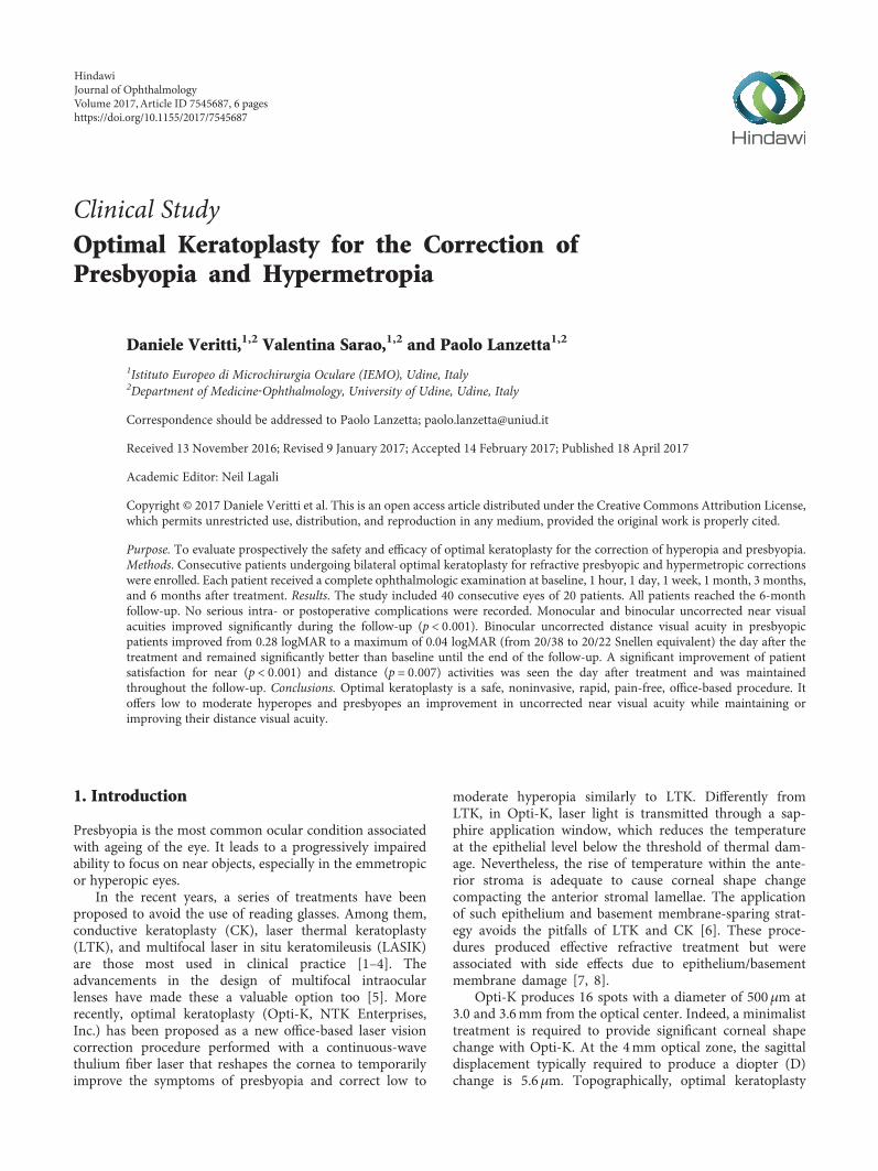
2.2. Device Description and Treatment Plan. The deviceused in the present study is the NTK Optimal Keratoplasty(Opti-K) System. It is a continuous-wave thulium fiber laserdevice for irradiation of corneal tissue. Output beam isdirected onto the cornea in a ring pattern (6 and 7.2mmdiameter) (Figure 1). Patient is lying in supine position, thenthe corneal epithelium is protected from thermal damagewith a sapphire application window/suction ring. Thereafter,laser is applied at 1.93μm wavelength. The laser is typicallyoperated with a total delivered power of 0.80–1.28W for aperiod of 150ms. The duration of the entire procedure isapproximately 10–15 minutes. Retreatment was allowed atinvestigator’s discretion based on measurement of UNVAand UDVA.
2.3. Data Analysis. The primary efficacy endpoint is meanUNVA change at 6 months. Secondary efficacy endpointsincluded UDVA in the hypemetropic/presbyopic eyes. Safetyassessments include a tabulation of complications, adverseevents, and patient symptoms and UCVA in the purepresbyopic eyes. Efficacy endpoints were analysed using ageneralized estimating equation model to account forintersubject correlation. Serial comparisons of pretreat-ment and posttreatment outcomes were performed usingpaired t-test or Wilcoxon matched pairs nonparametric testaccording to the Gaussianity or non-Gaussianity of the distri-butions. In serial comparisons, the null hypothesis wasrejected for p values <0.05.
3. Results
Forty eyes of 20 patients were included in the study. Patientreceived a mean (±SD) of 1.4± 0.5 treatments during thestudy period. Sixteen eyes (8 patients) required an additionaltreatment during the follow-up. Retreatment was performed45 days after the primary treatment, on average. Both the sin-gle- and double-treated eyes were evaluated before primarytreatment (baseline evaluation) and were then followed upfor the study period (6 months). Baseline characteristics arereported in Table 1.
3.1. Safety. No serious adverse events or complications, nopermanent loss of ≥1 line of UDVA or UDVA < 20/40, andno induced astigmatism ≥ 1D were recorded. Two patients(5%) complained of temporary glare that was resolved withinone week. No change of more than 0.1 logMAR in best-corrected distance visual acuity was recorded during thefollow-up.
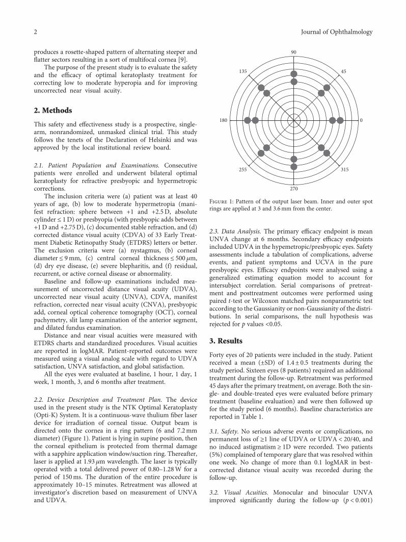
3.2. Visual Acuities. Monocular and binocular UNVAimproved significantly during the follow-up (p < 0 001)
90
45
270
315
180 0
255
135
Figure 1: Pattern of the output laser beam. Inner and outer spotrings are applied at 3 and 3.6mm from the center.
2 Journal of Ophthalmology
(Figure 2 and Table 2). Binocular UDVA in presbyopicpatients improved from 0.28 to a maximum of 0.04 logMAR(from 20/38 to 20/22 Snellen equivalent) the day after thetreatment and remained significantly better than baselineuntil the end of the follow-up (Figure 3 and Table 2).
3.3. Patient-Reported Outcomes. A significant improvementof patient satisfaction for near (p < 0 001) and distance(p = 0 007) activities was seen the day after treatment andwas maintained throughout the follow-up (Table 2).
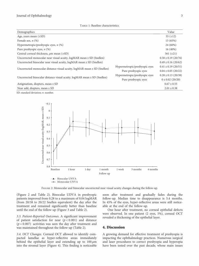
3.4. OCT Changes. Corneal OCT allowed to identify com-pacted lamellae as hyper-reflective areas immediatelybehind the epithelial layer and extending up to 100μminto the stromal layer (Figure 4). This finding is noticeable
soon after treatment and gradually fades during thefollow-up. Median time to disappearance is 5.4 months.In 45% of the eyes, hyper-reflective areas were still notice-able at the end of the follow-up.
One hour after treatment, no corneal epithelial defectswere observed. In one patient (2 eyes, 5%), corneal OCTrevealed a thickening of the epithelial layer.
4. Discussion
A growing demand for effective treatment of presbyopia isimpacting the ophthalmology practices. Numerous surgicaland laser procedures to correct presbyopia and hyperopiahave been tested over the past decade, whose main issues
Table 1: Baseline characteristics.
Demographics Value
Age, years mean (±SD) 55 (±12)Female sex, n (%) 13 (65%)
Hypermetropic/presbyopic eyes, n (%) 24 (60%)
Pure presbyopic eyes, n (%) 16 (40%)
Central corneal thickness, μm mean (±SD) 561 (±21)Uncorrected monocular near visual acuity, logMAR mean± SD (Snellen) 0.58± 0.19 (20/76)
Uncorrected binocular near visual acuity, logMAR mean± SD (Snellen) 0.49± 0.16 (20/62)
Uncorrected monocular distance visual acuity, logMAR mean± SD (Snellen)Hypermetropic/presbyopic eyes 0.41± 0.19 (20/51)
Pure presbyopic eyes 0.04± 0.05 (20/22)
Uncorrected binocular distance visual acuity, logMAR mean± SD (Snellen)Hypermetropic/presbyopic eyes 0.28± 0.13 (20/38)
Pure presbyopic eyes 0± 0.02 (20/20)
Astigmatism, diopters, mean± SD 0.67± 0.55Near add, diopters, mean± SD 2.01± 0.38SD: standard deviation; n: number.
Baseline 1 hour 1 day 1 week1 month 3 months 6 months
‒0.1
0.8
0.7
0.6
0.5
0.4
0.3
0.2
0.1
0
Follow-up
Unc
orre
cted
nea
r visu
al ac
uity
(log
MA
R, m
ean
± SD
)
Binocular UNVAMonocular UNVA
Figure 2: Monocular and binocular uncorrected near visual acuity changes during the follow-up.
3Journal of Ophthalmology
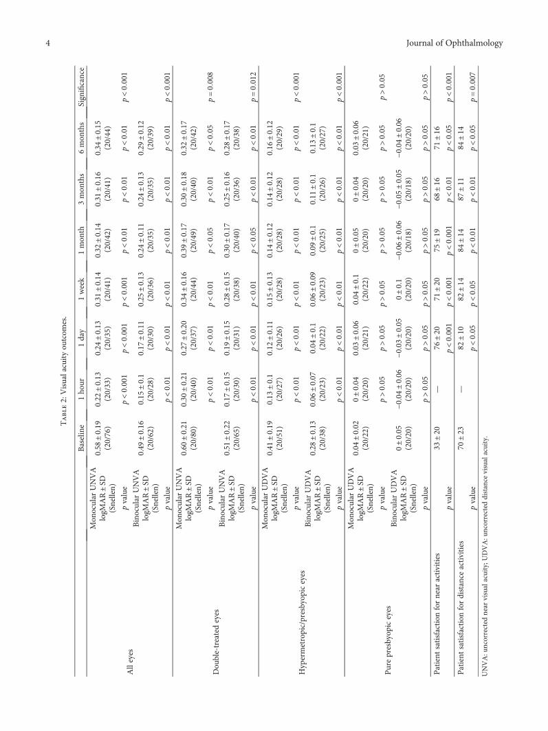
Table2:Visualacuityou
tcom
es.
Baseline
1ho
ur1day
1week
1mon
th3mon
ths
6mon
ths
Significance
Alleyes
Mon
ocular
UNVA
logM
AR±SD
(Snellen)
0.58
±0.19
(20/76)
0.22
±0.13
(20/33)
0.24
±0.13
(20/35)
0.31
±0.14
(20/41)
0.32
±0.14
(20/42)
0.31
±0.16
(20/41)
0.34
±0.15
(20/44)
pvalue
p<0 0
01p<00
01p<0001
p<001
p<00
1p<001
p<0001
Binocular
UNVA
logM
AR±SD
(Snellen)
0.49
±0.16
(20/62)
0.15
±0.1
(20/28)
0.17
±0.11
(20/30)
0.25
±0.13
(20/36)
0.24
±0.11
(20/35)
0.24
±0.13
(20/35)
0.29
±0.12
(20/39)
pvalue
p<0 0
1p<00
1p<001
p<001
p<00
1p<00
1p<0001
Dou
ble-treatedeyes
Mon
ocular
UNVA
logM
AR±SD
(Snellen)
0.60
±0.21
(20/80)
0.30
±0.21
(20/40)
0.27
±0.20
(20/37)
0.34
±0.16
(20/44)
0.39
±0.17
(20/49)
0.30
±0.18
(20/40)
0.32
±0.17
(20/42)
pvalue
p<0 0
1p<00
1p<001
p<005
p<00
1p<00
5p=0008
Binocular
UNVA
logM
AR±SD
(Snellen)
0.51
±0.22
(20/65)
0.17
±0.15
(20/30)
0.19
±0.15
(20/31)
0.28
±0.15
(20/38)
0.30
±0.17
(20/40)
0.25
±0.16
(20/36)
0.28
±0.17
(20/38)
pvalue
p<0 0
1p<00
1p<001
p<005
p<00
1p<00
1p=0012
Hypermetropic/presbyop
iceyes
Mon
ocular
UDVA
logM
AR±SD
(Snellen)
0.41
±0.19
(20/51)
0.13
±0.1
(20/27)
0.12
±0.11
(20/26)
0.15
±0.13
(20/28)
0.14
±0.12
(20/28)
0.14
±0.12
(20/28)
0.16
±0.12
(20/29)
pvalue
p<0 0
1p<00
1p<001
p<001
p<00
1p<00
1p<0001
Binocular
UDVA
logM
AR±SD
(Snellen)
0.28
±0.13
(20/38)
0.06
±0.07
(20/23)
0.04
±0.1
(20/22)
0.06
±0.09
(20/23)
0.09
±0.1
(20/25)
0.11
±0.1
(20/26)
0.13
±0.1
(20/27)
pvalue
p<0 0
1p<00
1p<001
p<001
p<00
1p<00
1p<0001
Purepresbyop
iceyes
Mon
ocular
UDVA
logM
AR±SD
(Snellen)
0.04
±0.02
(20/22)
0±0.04
(20/20)
0.03
±0.06
(20/21)
0.04
±0.1
(20/22)
0±0.05
(20/20)
0±0.04
(20/20)
0.03
±0.06
(20/21)
pvalue
p>0 0
5p>00
5p>005
p>005
p>00
5p>00
5p>005
Binocular
UDVA
logM
AR±SD
(Snellen)
0±0.05
(20/20)
−0.04±0.06
(20/20)
−0.03±0.05
(20/20)
0±0.1
(20/20)
−0.06±0.06
(20/18)
−0.05±0.05
(20/18)
−0.04±0.06
(20/20)
pvalue
p>0 0
5p>00
5p>005
p>005
p>00
5p>00
5p>005
Patient
satisfaction
fornear
activities
33±20
—76
±20
71±20
75±19
68±16
71±16
pvalue
p<0 0
01p<0001
p<00
01p<001
p<00
5p<0001
Patient
satisfaction
fordistance
activities
70±23
—82
±10
82±14
84±14
87±11
84±14
pvalue
p<0 0
5p<005
p<001
p<00
1p<00
5p=0007
UNVA:u
ncorrected
near
visualacuity;U
DVA:u
ncorrected
distance
visualacuity.
4 Journal of Ophthalmology

being unpredictable visual outcomes, irreversibility, regres-sion, and corneal damage. In 2002 and in 2004, CK has beenreported to be effective for the treatment of low to moderatehyperopia and presbyopia [1, 2]. CK and LTK efficacy lies inthe fact that heat coagulation of the corneal stroma alters thecorneal curvature. Thermal alteration of corneal collagenoccurs when the tissue is exposed to temperatures of 58° to75°C. In detail, the collagen matrix changes from the triplehelix formation to a partially coiled structure resulting inshrinkage of collagen fibers [10–14]. Opti-K, with the useof sapphire applanation ring, confines thermal elevationinto the anterior corneal stroma, which produces limitedthermally damaged zones that compact the anterior stro-mal lamellae while preserving the overlying epithelium.This is in stark contrast to antecedent technologies likeCK and LTK which both damage the epithelium and thebasement membrane causing discomfort and fibroticwound-healing response.
Optimal keratoplasty, similarly to CK, produces multipleconoids of Sturm generating useful corneal multifocality[15]. Despite the presence of multiple intervals of Sturm,patients did not report blur or decreased vision quality. Thismay be imputable to neuro-optical phenomenon for blursuppression and neuro-adaptation. The present is the firstprospective and peer-reviewed validation of optimal kerato-plasty. We reported a significant improvement in UNVA.UDVA improved in hyperopic patients and was maintainedin emmetropic patients. The mean effect duration after thefirst treatment was 45 days. The peak of efficacy was seenduring the first weeks of the follow-up, and UDVA andUNVA then progressively decline between months 3 and 6.But, a statistically significant visual benefit over baselinevalues was maintained throughout the follow-up. Longerfollow-ups are indeed needed to quantify the regression ofthe effect and the efficacy of multiple retreatments. In thepresent study, no specific intra- or postoperative complica-tions were recorded. Quality of vision, as evaluated by thepatients, was high and stable. In all cases, the postlaser recov-ery was immediate. All patients returned to their normalactivity the following day. Most patients that required arepeated treatment presented with a self-reported decline ofvisual benefit asking for undergoing a repeated procedure.
5. Conclusion
In conclusion, optimal keratoplasty is a safe, noninvasive,rapid, pain-free, office-based procedure. It offers low to mod-erate hyperopes and presbyopes a temporary improvementin uncorrected near visual acuity while maintaining orimproving their distance visual acuity. Further studies, withlonger follow-up and multiple repeated treatments, arewarranted to evaluate long-term safety.
‒0.1
0.7
0.6
0.5
0.4
0.3
0.2
0.1
0
Unc
orre
cted
dist
ance
visu
al ac
uity
(log
MA
R, m
ean
± SD
)
Baseline 1 hour 1 day 1 week1 month 3 months 6 monthsFollow-up
Binocular UNVAMonocular UNVA
Figure 3: Monocular and binocular uncorrected distance visual acuity changes in hypermetropic/presbyopic patients.
Figure 4: Corneal OCT reveals a hyper-reflective area immediatelybehind the epithelial layer in a 50-year-old patient one hour afterundergoing optimal keratoplasty.
5Journal of Ophthalmology
Conflicts of Interest
Neither of the authors has a financial or proprietary interestin any material and method mentioned.
References
[1] M. B. McDonald, P. S. Hersh, E. E. Manche et al., “Conductivekeratoplasty for the correction of low to moderate hyperopia:U.S. clinical trial 1-year results on 355 eyes,” Ophthalmology,vol. 109, no. 11, pp. 1978–1989, 2002.
[2] M. B. McDonald, D. Durrie, P. Asbell, R. Maloney, andL. Nichamin, “Treatment of presbyopia with conductivekeratoplasty: six-month results of the 1-year United StatesFDA clinical trial,” Cornea, vol. 23, no. 7, pp. 661–668,2004.
[3] U. Rehany and E. Landa, “Diode laser thermal keratoplasty tocorrect hyperopia,” Journal of Refractive Surgery, vol. 20, no. 1,pp. 53–61, 2004.
[4] D. Y. Lin and E. E. Manche, “Two-year results of conductivekeratoplasty for the correction of low to moderate hyperopia,”Journal of Cataract and Refractive Surgery, vol. 29, no. 12,pp. 2339–2350, 2003.
[5] S. R. de Silva, J. R. Evans, V. Kirthi, M. Ziaei, and M. Leyland,“Multifocal versus monofocal intraocular lenses after cataractextraction,” Cochrane Database of Systematic Reviews,vol. 12, article CD003169, 2016.
[6] E. H. Koo, H. G. Glen, M. Berry, and P. J. Botelho, “Histologiccorneal changes following optimal keratoplasty (Opti-K)treatment of cadaver eyes,” in presented at the ARVO AnnualMeeting, Fort Lauderdale, Florida, 2011.
[7] G. D. Kymionis, P. Titze, M. M. Markomanolakis, I. M.Aslanides, and I. G. Pallikaris, “Corneal perforation afterconductive keratoplasty with previous refractive surgery,”Journal of Cataract and Refractive Surgery, vol. 29, no. 12,pp. 2452–2454, 2003.
[8] I. G. Pallikaris, T. L. Naoumidi, and N. I. Astyrakakis, “Long-term results of conductive keratoplasty for low to moderatehyperopia,” Journal of Cataract and Refractive Surgery,vol. 31, no. 8, pp. 1520–1529, 2005.
[9] H. Glen, “Hyperopic presbyopia treatment: long term dis-tance and near vision improvement by noninvasive kerato-plasty,” in Presented at the American Society of Cataractand Refractive Surgery Conference, San Diego, California,May 2011.
[10] C. Baily, T. Kohnen, and M. O'Keefe, “Preloaded refractive-addition corneal inlay to compensate for presbyopia implantedusing a femtosecond laser: one-year visual outcomes andsafety,” Journal of Cataract and Refractive Surgery, vol. 40,no. 8, pp. 1341–1348, 2014.
[11] E. L. Shaw and A. R. Gasset, “Thermokeratoplasty (TKP)temperature profile,” Investigative Ophthalmology, vol. 13,no. 3, pp. 181–186, 1974.
[12] H. Stringer and J. Parr, “Shrinkage temperature of eye colla-gen,” Nature, vol. 204, p. 1307, 1964.
[13] L. Zhang and A. Aksan, “Fourier transform infrared analysis ofthe thermal modification of human cornea tissue during con-ductive keratoplasty,” Applied Spectroscopy, vol. 64, no. 1,pp. 23–29, 2010.
[14] M. B. McDonald, “Conductive keratoplasty: a radiofrequency-based technique for the correction of hyperopia,” Transac-tions of the American Ophthalmological Society, vol. 103,pp. 512–536, 2005.
[15] P. S. Hersh, “Optics of conductive keratoplasty: implicationsfor presbyopia management,” Transactions of the AmericanOphthalmological Society, vol. 103, pp. 412–456, 2005.
6 Journal of Ophthalmology

Submit your manuscripts athttps://www.hindawi.com
Stem CellsInternational
Hindawi Publishing Corporationhttp://www.hindawi.com Volume 2014
Hindawi Publishing Corporationhttp://www.hindawi.com Volume 2014
MEDIATORSINFLAMMATION
of
Hindawi Publishing Corporationhttp://www.hindawi.com Volume 2014
Behavioural Neurology
EndocrinologyInternational Journal of
Hindawi Publishing Corporationhttp://www.hindawi.com Volume 2014
Hindawi Publishing Corporationhttp://www.hindawi.com Volume 2014
Disease Markers
Hindawi Publishing Corporationhttp://www.hindawi.com Volume 2014
BioMed Research International
OncologyJournal of
Hindawi Publishing Corporationhttp://www.hindawi.com Volume 2014
Hindawi Publishing Corporationhttp://www.hindawi.com Volume 2014
Oxidative Medicine and Cellular Longevity
Hindawi Publishing Corporationhttp://www.hindawi.com Volume 2014
PPAR Research
The Scientific World JournalHindawi Publishing Corporation http://www.hindawi.com Volume 2014
Immunology ResearchHindawi Publishing Corporationhttp://www.hindawi.com Volume 2014
Journal of
ObesityJournal of
Hindawi Publishing Corporationhttp://www.hindawi.com Volume 2014
Hindawi Publishing Corporationhttp://www.hindawi.com Volume 2014
Computational and Mathematical Methods in Medicine
OphthalmologyJournal of
Hindawi Publishing Corporationhttp://www.hindawi.com Volume 2014
Diabetes ResearchJournal of
Hindawi Publishing Corporationhttp://www.hindawi.com Volume 2014
Hindawi Publishing Corporationhttp://www.hindawi.com Volume 2014
Research and TreatmentAIDS
Hindawi Publishing Corporationhttp://www.hindawi.com Volume 2014
Gastroenterology Research and Practice
Hindawi Publishing Corporationhttp://www.hindawi.com Volume 2014
Parkinson’s Disease
Evidence-Based Complementary and Alternative Medicine
Volume 2014Hindawi Publishing Corporationhttp://www.hindawi.com