Embed Size (px)
Citation preview
Sensors 2015, 15, 25015-25032; doi:10.3390/s151025015
sensors ISSN 1424-8220
www.mdpi.com/journal/sensors
Article
Optimisation and Characterisation of Anti-Fouling Ternary SAM Layers for Impedance-Based Aptasensors
Anna Miodek 1,*, Edward M. Regan 1,†, Nikhil Bhalla 1, Neal A.E. Hopkins 2, Sarah A. Goodchild 2
and Pedro Estrela 1,*
1 Department of Electronic and Electrical Engineering, University of Bath, Bath BA2 7AY, UK;
E-Mails: [email protected] (E.M.R.); [email protected] (N.B.) 2 Defence Science and Technology Laboratory, Porton Down, Salisbury, SP4 0JQ, UK;
E-Mails: [email protected] (N.A.E.H.); [email protected] (S.A.G.)
† Current address: Clarity BioSolutions Ltd., Building 114, Porton Down Science Park, Salisbury,
SP4 0JQ, UK.
* Authors to whom correspondence should be addressed; E-Mails: [email protected] (A.M.);
[email protected] (P.E.); Tel.: +44-1225-386324 (P.E.); Fax: +44-1225-386305 (P.E.).
Academic Editor: Kagan Kerman
Received: 30 July 2015 / Accepted: 18 September 2015 / Published: 29 September 2015
Abstract: An aptasensor with enhanced anti-fouling properties has been developed. As a
case study, the aptasensor was designed with specificity for human thrombin. The sensing
platform was developed on screen printed electrodes and is composed of a self-assembled
monolayer made from a ternary mixture of 15-base thiolated DNA aptamers specific for
human thrombin co-immobilised with 1,6-hexanedithiol (HDT) and further passivated with
1-mercapto-6-hexanol (MCH). HDT binds to the surface by two of its thiol groups forming
alkyl chain bridges and this architecture protects from non-specific attachment of molecules
to the electrode surface. Using Electrochemical Impedance Spectroscopy (EIS), the aptasensor
is able to detect human thrombin as variations in charge transfer resistance (Rct) upon protein
binding. After exposure to a high concentration of non-specific Bovine Serum Albumin (BSA)
solution, no changes in the Rct value were observed, highlighting the bio-fouling resistance
of the surface generated. In this paper, we present the optimisation and characterisation of
the aptasensor based on the ternary self-assembled monolayer (SAM) layer. We show that
anti-fouling properties depend on the type of gold surface used for biosensor construction,
which was also confirmed by contact angle measurements. We further studied the ratio between
OPEN ACCESS
Sensors 2015, 15 25016
aptamers and HDT, which can determine the specificity and selectivity of the sensing layer.
We also report the influence of buffer pH and temperature used for incubation of electrodes
with proteins on detection and anti-fouling properties. Finally, the stability of the aptasensor
was studied by storage of modified electrodes for up to 28 days in different buffers and
atmospheric conditions. Aptasensors based on ternary SAM layers are highly promising for
clinical applications for detection of a range of proteins in real biological samples.
Keywords: biosensor; DNA aptamers; self-assembled monolayers; antifouling; screen
printed electrodes; electrochemical detection; Thrombin
1. Introduction
There is a substantial need for the development of biosensors capable of sensitive and selective
detection, that are also low cost, easy to use and have the possibility of being integrated into portable
devices for point-of-care use with clinical or environmental samples. Electrochemical biosensors are
some of the most promising platforms with the potential to achieve these goals and have been used to
detect a number of analytes. These include, e.g., (1) disease biomarkers and pathogenic proteins in
serum [1,2], human plasma [3,4] or urine [5]; (2) Nucleic acids such as DNA samples from polymerase
chain reaction (PCR) products [6,7] and microRNAs [2,8]; (3) microorganisms in seawater medium [9]
and tap water [10]; (4) toxins in food samples [11]; and (5) drugs levels in human serum [12–14].
The applications of biosensors for complex samples, whether they be environmentally or clinically
derived, are, however, limited by their fouling properties due to non-specific binding of background
materials to the measurement surfaces. One challenge to be overcome in construction of biosensors
with broad potential for exploitation is therefore the elimination of this non-specific adsorption of
molecules to the electrode surface. Different strategies have previously been developed to prevent
non-specific binding. The most common method has been to generate binding resistant layers through
manipulation of either the components or the construction of the self-assembled monolayer. This can be
achieved by co-immobilisation of bioreceptors with alkanethiols of varying lengths of alkyl chains such as
1-mercapto-6-hexanol (MCH) [15] or 11-mercapto-1-undecanol (MCU) [16], which simultaneously can
act as spacers to avoid steric hindrances. Modifications of the self-assembled monolayer (SAM)
components can also be performed. For example, prostate specific antigen (PSA) aptamers were
immobilised on a SAM layer containing a thiolated sulfo-betaine antifouling agent [17]. Sulfo-betaine
moiety prevented any significant non-specific binding of the control protein human serum albumin
(HSA) as compared to high non-specific binding with MCH-based sensors.
Poly (ethylene glycol) (PEG) and its derivatives are the most commonly used anti-fouling materials
for biosensor construction. For example, in a study used for detecting embryonic stem cell lysate with
surface plasmon resonance imaging, the surface was modified with thiolated PEG chains or three
dimensional polymers containing PEG chains [18]. These surfaces combined with a good blocking
solution, such as amine terminated PEG, and adequate running buffer supplemented with detergent
exhibited greatly decreased non-specific binding. Thiolated oligomers with di-(ethylene glycol) [19] or
tri-(ethylene glycol) (OEG) [20] groups have also been used to generate stable non-fouling background
Sensors 2015, 15 25017
and OEG monomers constructed into polymer “brushes” have been shown to be resistant to the
adsorption of proteins from complex media such as undiluted foetal bovine serum [21]. This class of
surface has been successfully used for detection of analytes associated with viral infection in clinical
samples [22]. Among other polymers, poly-(ethyleneoxide) (PEO) derivatives were able to protect the
surface from non-specific adsorption after exposition on buffers containing either 0.1% bovine serum
albumin or 1% goat serum [23]. Low fouling background can be also obtained using zwitterionic
polymers containing groups such as phosphorylcholine (PC), sulfobetaine (SB), and carboxybetaine (CB).
These materials constitute an interesting alternative to PEG-based surfaces protecting from non-specific
interactions even in complex media [24–27]. In addition, diblock copolymer “brushes” composed of
OEG and polyCB were reported to improve fouling properties of biosensors and minimalize adsorption
from blood plasma [28]. Ionic liquid self-assembled monolayers have been investigated for lower
non-specific binding. Ratel et al. developed a SAM monolayer based on ionic liquids with low fouling in
crude serum, to a level equivalent to PEG [29]. However, the majority of these surface types are limited in
their application to optical sensors based on the surface plasmon resonance technique, because of high
molecular weight, which results in detrimental effects on electrochemical measurements and blocking
effects on charge transfer.
Much fewer examples exist concerning the development of anti-fouling materials compatible
with electrochemical sensing in clinical samples or complex media [30]. Among such materials,
hyperbranched polyester microspheres [31], hydrophilic polymer carboxymethyl-PEG-carboxymethyl
(CM-PEG-CM) [32] or zwitterionic surfaces [33,34] have been evaluated. However, due to the high
molecular weight of these compounds and potential impacts on electrochemical transfer reactions, these
materials still present serious limitations in electrochemical sensing applications.
Recently Campuzano et al. demonstrated the anti-fouling properties of a ternary self-assembled
monolayer for construction of electrochemical DNA sensors [35]. This simple layer, composed
of co-immobilised thiolated DNA probe with 1,6-hexanedithiol (HDT) and saturated with
1-mercapto-6-hexanol (MCH), was used for detection of DNA targets in serum and urine samples. The
study demonstrated improved signal-to-noise ratio with high hybridisation efficiency and detection limit
on the attomolar level. The improved antifouling properties of this surface was attributed to the
favourable surface architecture by the presence of HDT which may adopt a horizontal configuration,
acting as a bridge and providing significantly higher resistance to nonspecific adsorption of nucleic acids
and proteins. The authors showed the advantage of the ternary SAM over the commonly used binary
DNA/MCH SAM, which presents poor anti-fouling properties (as also seen in our previous studies [17]).
This can be due to the hydroxyl groups that can electrostatically bind to chemical groups of proteins.
Furthermore, the use of a ternary SAM surface improves reproducibility comparing to a DNA/MCH
layer and it was also proven that can extend storage stability of a genosensor [36].
The aim of this work is the application of the ternary SAM layer developed by Campuzano et al. in
the construction of impedimetric DNA aptamer-based biosensors. As a case study, the anti-fouling
properties of the ternary SAM layer and analytical properties of this aptasensor were assessed with low
molecular weight thrombin aptamers specific for thrombin from human serum. The G-quadruplex
structure of this aptamer system was considered likely to affect the bridge architecture obtained by using
HDT, and as such different conditions of biosensor construction were studied to ensure a well performing
system could be identified. The system was therefore optimised in terms of surface construction
Sensors 2015, 15 25018
performing an empirical assessment of the ideal ratio of aptamer to HDT within the ternary mixture. The
results clearly show the influence of the quantity of aptamer immobilised on the surface in relation to
non-specific interactions. The influence of buffer pH and temperature was also studied. The biosensor
was developed on Screen Printed Electrodes (SPE) and contrary to classic three-compartment cells an
improvement of signal-to-noise ratio was observed as compared to standard gold electrodes. Finally the
storage stability of the biosensor and influence of different storage conditions on anti-fouling properties
and performance were studied.
2. Experimental Section
2.1. Reagents
Thrombin (Th) from human plasma was purchased from Sigma-Aldrich. Thiol-modified
aptamer specific for Th (ThA aptamer) with dT3 and six carbon spacer on 5' phosphoryl terminus
(5'-HS-C6-dT3-GGT TGG TGT GGT TGG-3') were provided by Sigma-Aldrich (HPLC purified) and
dissolved in TE buffer consisting of 10 mM tris (hydroxymethyl) aminomethane (Tris) and 1 mM
ethylene di-amine tetra-acetic acid (EDTA) at pH 7.6 in ultra-pure water. 1,6-hexanedithiol (96%),
6-mercapto-1-hexanol (97%), potassium phosphate monobasic solution (1 M), potassium phosphate dibasic
solution (1 M), potassium sulphate, potassium hexacyanoferrate (III), potassium hexacyanoferrate (II)
trihydrate, ethylenediaminetetraacetic acid (EDTA, 0.5 M) and Tris were provided from Sigma Aldrich.
All buffers solutions were prepared using 18.2 MΩ cm ultra-pure water (Millipore, Billerica, MA, USA)
with a Pyrogard filter (Millipore) to remove nucleases, stored at 4 °C until use and filtered by 0.22 µm
syringe membranes prior use. Non-specific interactions were studied using Bovine Serum Albumin
(BSA) (Sigma-Aldrich).
2.2. Instrumentation
Electrochemical Impedance Spectroscopy (EIS) measurements were performed using a
μAutoLab III/FRA2 potentiostat (Metrohm, Cheshire, UK). For analysis, screen printed electrodes (SPE)
were used (DropSens C223AT). These electrodes consist of working (1.6 mm diameter) and counter
electrodes made of gold, while the reference electrode was replaced with an external Hg/Hg2SO4
(K2SO4 sat.) (IJ Cambria Scientific, Wales, UK) placed into a salt bridge. Performance of layers
generated on SPE were compared with flat gold disc macro working electrodes (1.6 mm diameter, from
BASi, Costa Mesa, CA, USA), switched to common three-electrodes cell combined with platinum mesh
as a counter electrode and Hg/Hg2SO4 as reference electrode. All impedances were carried out in 50 mM
phosphate buffer pH 7.0, containing 100 mM K2SO4, in the presence of 2.5 mM/2.5 mM
[Fe(CN)6]4−/[Fe(CN)6]3− redox probe and were obtained at 0.2 V vs. Hg/Hg2SO4 with an AC potential
of 10 mV amplitude in the frequency range from 100 kHz to 0.1 Hz.
Contact angle measurements were performed using an in-house built optical angle measurement
system. The setup consisted of 4 parts: a viewing system (camera), a stage, a dispensing system
(syringe/pipette) and a measurement system to calculate the contact angle. The electrodes (both flat and
screen printed) were placed on the stage and a 10 µL drop was dispensed on the electrode with the
dispensing system. The wetting of surface was then captured using a Nikon p520 camera. Contact angle
Sensors 2015, 15 25019
was measured using a screen protractor version 4.0 procured from Iconico. See Figures S1 and S2 in
Supporting Information for a block diagram of the experimental setup and typical images, respectively.
2.3. Preparation of Ternary Self-Assembled Monolayer Based on Thrombin Aptamers
Prior to use, screen printed electrodes were washed with acetone for 2 min, followed by sonication in
ethanol (2 min) and ultra-pure water (2 min).
Gold disc working electrodes with a diameter of 1.6 mm were polished with 50 nm aluminium oxide
particles (Buehler, Lake Bluff, IL, USA) on a polishing pad (Buehler), followed by sonication in ethanol,
polishing on a blank polishing pad, and sonication in ethanol to remove any particles. Electrodes were
subsequently electrochemically cleaned in 0.5 M H2SO4 by scanning the potential between the oxidation
and reduction of gold, −0.05 V and +1.1 V versus an Hg/Hg2SO4 reference electrode, for 50 cycles until
there was no further change in the voltammogram.
Prior to immobilisation, DNA aptamer solutions were incubated at 90 °C in a water bath and then
exposed to ice cooling to achieve the correctly unfolded structure. Electrodes were rinsed with deionised
water, dried in a stream of nitrogen, and exposed to 6 μL of mixed DNA/HDT immobilisation solution
(of variable concentration ratios) for 16 h in a humidity chamber. The DNA immobilisation buffer
consisted of 50 mM phosphate buffer (PB), 100 mM K2SO4, 1 mM ethylene diamine tetraacetic acid
(EDTA) pH 7.6 and 10% of ethanol being used to solubilise HDT. After immobilisation, electrodes were
rinsed in ultra-pure water. To ensure complete thiol coverage of the gold surface, the electrodes were
backfilled with 6-mercapto-1-hexanol (MCH) by dropping 6 μL of 1 mM MCH in PB buffer pH 7.0
containing 100 mM K2SO4 and 10% of ethanol during 50 min. Electrodes were then rinsed with ultra-pure
water and stabilised in PB buffer pH 7.0 for 2 h.
2.4. Detection of Thrombin Protein and Non-Specific Interactions
Electrodes with immobilised DNA aptamer/HDT/MCH were incubated with different concentrations
(1, 10, 100 pM, 1, 10, 25, 50, 100, 250, 500 nM, and 1 µM) of human thrombin diluted in PB buffer
pH 7.0 containing 100 mM K2SO4. Electrodes were then rinsed with the same buffer and EIS
measurements were performed. In this work, we studied EIS detection of human thrombin by successive
incubation of the biosensor with each solution of Th as well as detection of different Th concentrations
on freshly prepared surfaces.
3. Results and Discussion
3.1. Ternary SAM Layer for Anti-Fouling Aptasensor Properties
Based on the procedure of Campuzano et al. [35,36], we studied the immobilisation of thrombin DNA
aptamers (ThA) with co-immobilisation of the other components of the ternary SAM layer onto
screen-printed electrodes. In Campuzano’s work the ternary SAM layer was composed of
1,6-hexanedithiol (HDT), 1-mercapto-6-hexanol (MCH) and thiolated single-stranded DNA probes for
specific detection of DNA target. In that approach, the DNA probe was immobilised on the surface from
the mixture with 1,6-hexanedithiol. The use of HDT, which forms hydrophobic alkyl chain bridges on
the electrode surface, was effective in reducing non-specific interactions when compared with a common
Sensors 2015, 15 25020
DNA/MCH surface. The ratio of HDT and thiolated DNA probe was optimised and found to behave
most effectively at a ratio of 300/0.05 μM (HDT to DNA). To saturate all non-bonded surface sites, 1 mM
of 1-mercapto-6-hexanol was used. The composition of such layer was confirmed by studying the charge
transfer resistance and capacitance parameters on different steps of preparation. The authors showed that
this DNA sensor allows obtaining high specificity of detection, even in clinical samples.
The thrombin specific aptamer used in this work is a well characterised short DNA sequence
(15 bases) that can form a characteristic quadruplex structure. For preparation of the aptasensor using
ThA we used a similar approach to Campuzano et al.: ThA were co-immobilised with HDT in the ratio
of 0.05/300 μM, and then any exposed gold surface was saturated with 1 mM MCH. Construction of
biosensor was performed on SPEs. The presence of aptamers was confirmed by comparing such layer with
free-aptamers surface using Electrochemical Impedance Spectroscopy (see Supporting Information
Figure S3). In order to test the anti-fouling properties of the ternary SAM, electrodes were incubated
with high concentration (100 µM) Bovine Serum Albumin solution. The response of sensor was then
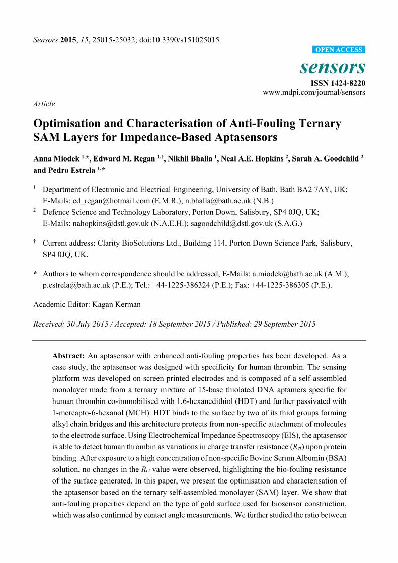
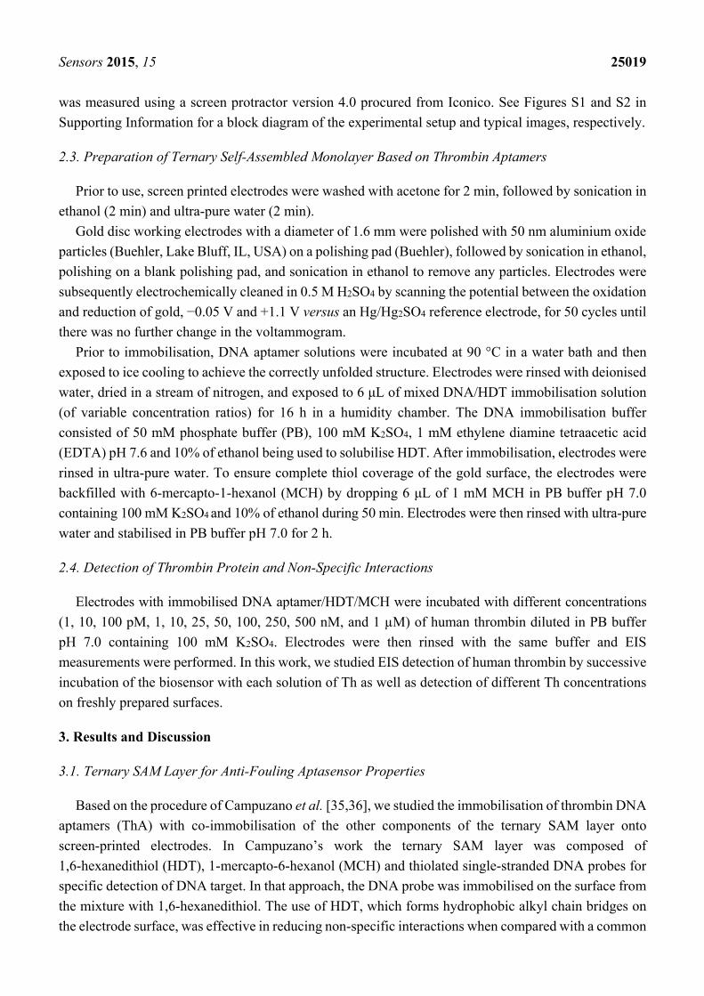
monitored by EIS in the presence of 2.5 mM of the redox species [Fe(CN)6]3−/4−. Figure 1a shows the
Nyquist plots obtained before and after incubation of the biosensor with BSA solution. The data were
fitted using a Randles equivalent circuit model (see inset Figure 1a), where Rs is the electrolyte resistance
and Rct is the electron transfer resistance; the double layer capacitance was replaced by a constant phase
element (CPE), which accounts for irregularities on the surface due to roughness of the gold substrate
and inhomogeneities on the biolayer; a Warburg element was added to account for diffusion processes
of the redox marker. The fitting results show a very slight variation in the Rct value, from 14.1 kΩ to
13.8 kΩ after BSA incubation, i.e., a −1.5% of relative change ΔRct/Rct0, where ΔRct = Rct – Rct0; Rct0 and
Rct are the charge transfer resistance obtained, respectively, before and after incubation of the aptasensor
with protein, respectively. The same behaviour was obtained when ThA was replaced with an aptamer
specific for streptavidin (SA) of higher molecular weight (60 bases) and which are characterised by
forming a secondary “loop” structure (see Supporting Information, Figure S4). The SAMs appeared very
resistant to antifouling and based on the work of Campuzano et al. where detection in urine and serum
samples was successfully performed, application of aptasensors based on a ternary SAM layer for
clinical samples is promising.
Results obtained on SPEs were compared with standard three-electrode cell with gold disc as working
electrode (macroelectrode), platinum mesh as counter electrode and Hg/HgSO4 as reference electrode.
After incubation of the electrode with 100 µM BSA, a substantial variation (60%–80%) in Rct was
observed. These seemingly contradictory results could be explained by taking into account the different
surface architectures of the SPEs in comparison to polished macroelectrodes. The topology of SPEs is
highly rough and heterogeneous which favours the formation of bridges by 1,6-hexadithiol [37], while the
macroelectrode surface is considerably flatter and may impede the attachment of HDT to the surface by
both thiol groups, favouring the formation of a compact HDT layer with exposed thiol groups. Such
planar surface with compact HDT layer was characterized by Morel et al. [38], where unbound thiol
groups were used for attachment of gold nanoparticles for biosensor construction. The schematic
explanation of the difference between SPEs and macroelectrode is presented in Figure 1c,d.
Sensors 2015, 15 25021
Figure 1. Nyquist plots showing electrochemical impedance spectroscopy responses before
(red) and after (blue) incubation of 100 μM bovine serum albumin (BSA) for 30 min on
biosensor based on (a) screen-printed electrodes (b) macroelectrodes modified with
thrombin aptamer/ternary self-assembled monolayer 0.05/300 μM and saturated with 1 mM
mercaptohexanol. Points show experimental results and solid plots show fitting. Scheme of
the surface architecture for (c) screen-printed electrodes and (d) macroelectrodes.
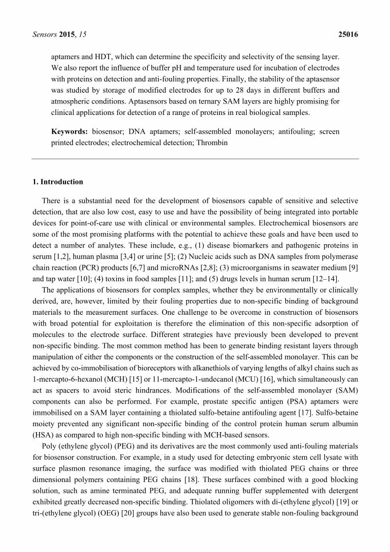
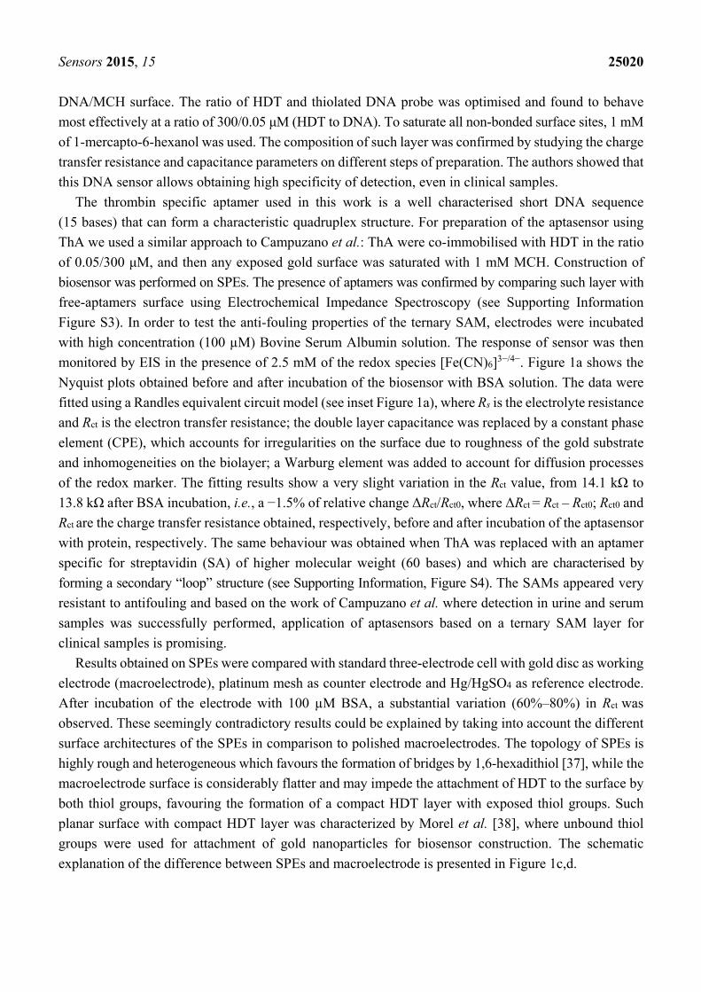
The results were confirmed by measuring the contact angle of the aptasensors constructed on SPEs
and polished gold surfaces. The contact angle was determined on blank un-modified surface, after SAM
layer preparation and after incubation with specific thrombin protein or non-specific BSA solution.
Figure 2 presents the obtained results (see Figure S2 in Supplementary Information for the contact angle
images). The contact angle of blank SPEs is 36.5° and decreases after SAM immobilisation and after
subsequent interaction with Th protein (respectively, 25.6° and 10.9°). After incubation of the aptasensor
with BSA instead of Th solution, the value remains almost unchanged (27.4°), which confirms the
anti-fouling properties of the SAM. The polished gold electrode has a similar behaviour after SAM layer
immobilisation and Th incubation (the values decrease from 70.0° to 57.7° and 30.8°, respectively).
However after exposure to non-specific BSA solution, a high variation is observed (47.4°), which
suggests that the topology of the electrode surface is crucial for anti-fouling properties of aptasensors
based on ternary SAM layers and confirms the results obtained by EIS.
0 5000 10000 15000
0
5000
10000
15000
(a)-
Z''
(Ω)
Z' (Ω)
Th aptamer/HDT/MCH + 100 μM BSA
0 5000 10000 15000
0
5000
10000
15000
-Z''
(Ω)
Z' (Ω)
Th aptamer/HDT/MCH + 100 μM BSA
60-80 %
(b)
BSA
(c) (d)BSA
Sensors 2015, 15 25022
Blank Aptamer Protein BSA
0
10
20
30
40
50
60
70
80 SPE Macroelectrode
Con
tact
an
gle
(de
gre
es)
Figure 2. Contact angle values measured for screen-printed electrodes (SPEs) and polished
gold surfaces on the different steps of aptasensor construction. All measurements were
repeated on three separate samples.
3.2. Optimisation of the Ratio Aptamer/Hexanedithiol
Aptamers, contrary to DNA probes, can form secondary (“loops”) and three-dimensional
(quadruplex) structures, which may affect analytical performances as well as anti-fouling properties of
biosensors based on ternary SAM layers. Therefore some optimisation in their construction should be
performed to ensure that the optimised conditions previously reported for DNA hybridisation, are also
valid for DNA aptamers. In particular the ratio between aptamers and HDT can have an important
effect on the ability of the DNA aptamers to form a secondary structure and bind to its target protein.
Varied aptamer concentrations (0.02, 0.05, and 0.1 µM) and HDT concentrations (100, 200, 300, 400,
and 500 µM), all backfilled with 1 mM of MCH were tested for the construction of the aptasensors.
These biolayers were incubated with 0.5 µM of human thrombin or 100 µM BSA to verify specific as well
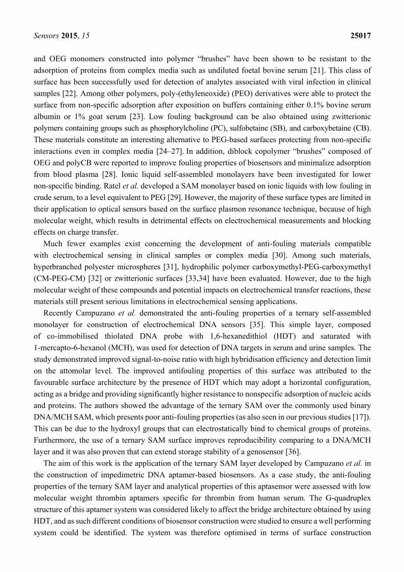
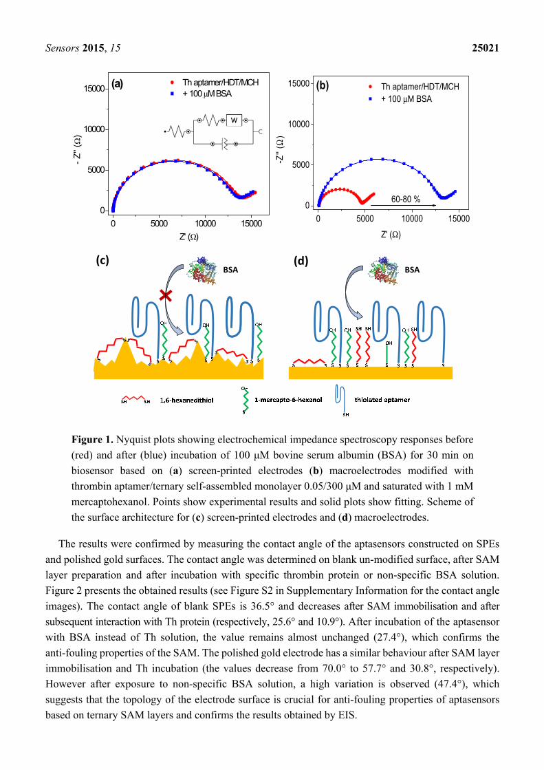
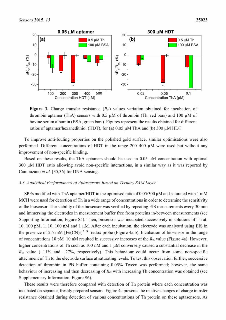
as non-specific interactions. Figure 3 shows the values obtained for the relative variations of charge
transfer resistance (ΔRct/Rct0) in the case of specific interactions with Th (red bars) and non-specific
interactions with BSA (green bars). The interactions of ThA with Th result in decreasing of the Rct value
and depend on the quantity of aptamer immobilised on the surface. The highest Rct variation was
observed for 0.05/300 µM aptamer/HDT ratio (−28%) and optimal signal-to-noise ratio was obtained for
200–300 µM HDT (Figure 3a). However, variation of aptamers concentration for optimal 300 µM HDT
did support the desired level of anti-fouling properties (Figure 3b). A 0.02 µM concentration of aptamer
resulted in lower Th binding (−10%), most likely due to the lower number of bioreceptors on the surface
and higher BSA adsorption, possibly due to larger areas containing MCH. Conversely, increasing of
quantity of aptamers on the electrode surface (0.1 µM) can cause steric hindrance effects for significant
thrombin attachment (−12%) and insufficient saturation of the surface with HDT giving high Rct value
variation after incubation of biosensor with BSA (−33%).
Sensors 2015, 15 25023
Figure 3. Charge transfer resistance (Rct) values variation obtained for incubation of
thrombin aptamer (ThA) sensors with 0.5 µM of thrombin (Th, red bars) and 100 µM of
bovine serum albumin (BSA, green bars). Figures represent the results obtained for different
ratios of aptamer/hexanedithiol (HDT), for (a) 0.05 µM ThA and (b) 300 µM HDT.
To improve anti-fouling properties on the polished gold surface, similar optimisations were also
performed. Different concentrations of HDT in the range 200–400 µM were used but without any
improvement of non-specific binding.
Based on these results, the ThA aptamers should be used in 0.05 µM concentration with optimal
300 µM HDT ratio allowing avoid non-specific interactions, in a similar way as it was reported by
Campuzano et al. [35,36] for DNA sensing.
3.3. Analytical Performances of Aptasensors Based on Ternary SAM Layer
SPEs modified with ThA aptamer/HDT in the optimised ratio of 0.05/300 μM and saturated with 1 mM
MCH were used for detection of Th in a wide range of concentrations in order to determine the sensitivity
of the biosensor. The stability of the biosensor was verified by repeating EIS measurements every 30 min
and immersing the electrodes in measurement buffer free from proteins in-between measurements (see
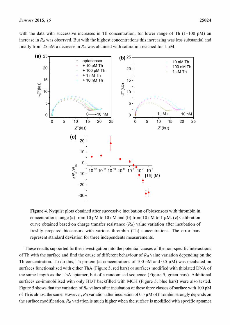
Supporting Information, Figure S5). Then, biosensor was incubated successively in solutions of Th at:
10, 100 pM, 1, 10, 100 nM and 1 μM. After each incubation, the electrode was analysed using EIS in
the presence of 2.5 mM [Fe(CN)6]3−/4− redox probe (Figure 4a,b). Incubation of biosensor in the range
of concentrations 10 pM–10 nM resulted in successive increases of the Rct value (Figure 4a). However,
higher concentrations of Th such as 100 nM and 1 μM conversely caused a substantial decrease in the
Rct value (−11% and −27%, respectively). This behaviour could occur from some non-specific
attachment of Th to the electrode surface at saturating levels. To test this observation further, successive
detection of thrombin in PB buffer containing 0.05% Tween was performed; however, the same
behaviour of increasing and then decreasing of Rct with increasing Th concentration was obtained (see
Supplementary Information, Figure S6).
These results were therefore compared with detection of Th protein where each concentration was
incubated on separate, freshly prepared sensors. Figure 4c presents the relative changes of charge transfer
resistance obtained during detection of various concentrations of Th protein on these aptasensors. As
-30
-20
-10
0
10
20
ΔRct/R
ct0
(%)
Concentration HDT (μM)
0.5 μM Th 100 μM BSA
0.05 μM aptamer
(a)
100 200 300 400 500
-30
-20
-10
0
10
20
ΔRct/R
ct0
(%)
Concentration ThA (μM)
0.5 μM Th 100 μM BSA
300 μM HDT
(b)
0.02 0.05 0.1
Sensors 2015, 15 25024
with the data with successive increases in Th concentration, for lower range of Th (1–100 pM) an
increase in Rct was observed. But with the highest concentrations this increasing was less substantial and
finally from 25 nM a decrease in Rct was obtained with saturation reached for 1 µM.
0 5 10 15 20 25
0
5
10
15
20
25 aptasensor + 10 pM Th + 100 pM Th + 1 nM Th + 10 nM Th
-Z''
(kΩ
)
Z' (kΩ)
0 10 nM
(a)
0 5 10 15 20 25
0
5
10
15
20
25 10 nM Th 100 nM Th 1 μM Th
-Z''
(kΩ
)
Z' (kΩ)
10 nM1 μM
(b)
10-12 10-11 10-10 10-9 10-8 10-7 10-6
-30
-20
-10
0
10
20
ΔRct/R
ct0
[Th] (M)
(c)
Figure 4. Nyquist plots obtained after successive incubation of biosensors with thrombin in
concentrations range (a) from 10 pM to 10 nM and (b) from 10 nM to 1 µM. (c) Calibration
curve obtained based on charge transfer resistance (Rct) value variation after incubation of
freshly prepared biosensors with various thrombin (Th) concentrations. The error bars
represent standard deviation for three independents measurements.
These results supported further investigation into the potential causes of the non-specific interactions
of Th with the surface and find the cause of different behaviour of Rct value variation depending on the
Th concentration. To do this, Th protein (at concentrations of 100 pM and 0.5 µM) was incubated on
surfaces functionalised with either ThA (Figure 5, red bars) or surfaces modified with thiolated DNA of
the same length as the ThA aptamer, but of a randomised sequence (Figure 5, green bars). Additional
surfaces co-immobilised with only HDT backfilled with MCH (Figure 5, blue bars) were also tested.
Figure 5 shows that the variation of Rct values after incubation of these three classes of surface with 100 pM
of Th is almost the same. However, Rct variation after incubation of 0.5 µM of thrombin strongly depends on
the surface modification. Rct variation is much higher when the surface is modified with specific aptamer
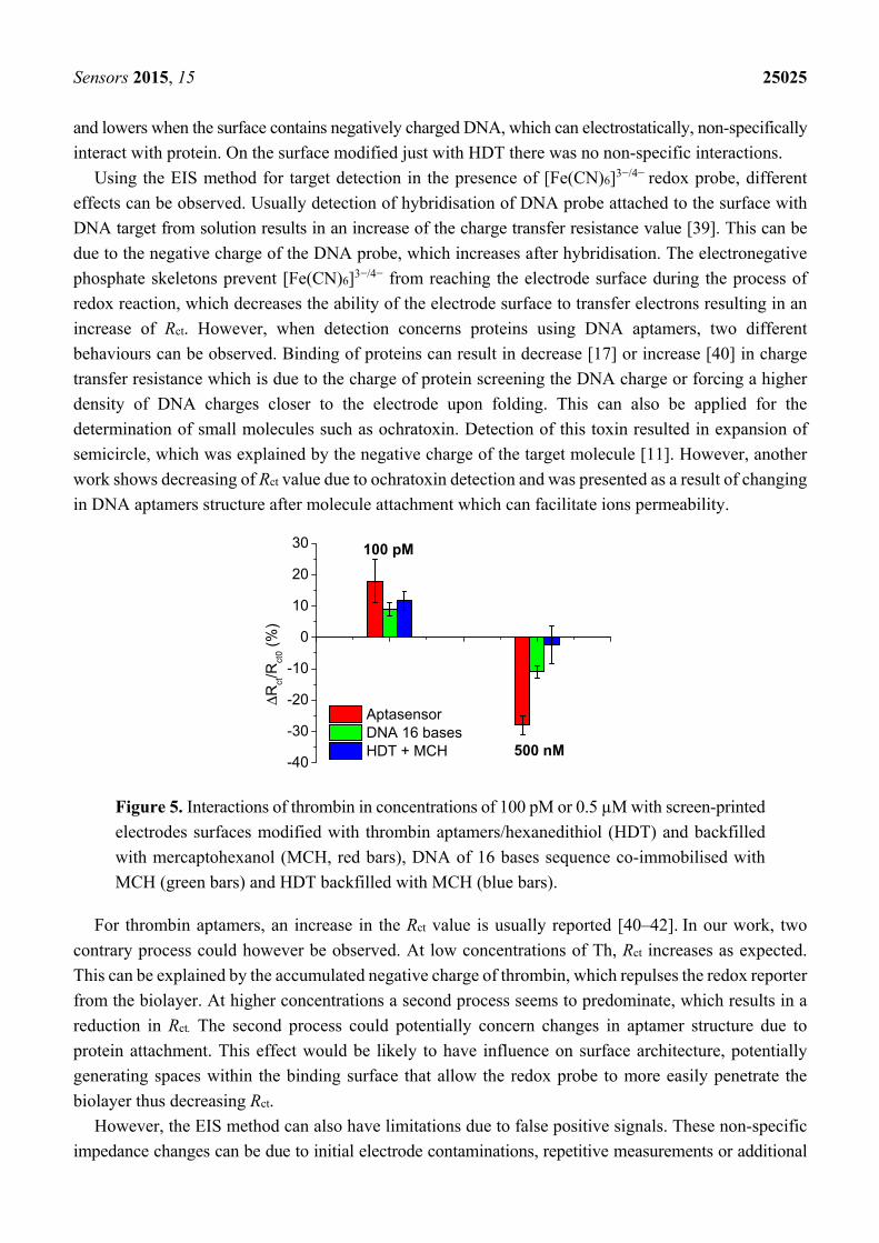
Sensors 2015, 15 25025
and lowers when the surface contains negatively charged DNA, which can electrostatically, non-specifically
interact with protein. On the surface modified just with HDT there was no non-specific interactions.
Using the EIS method for target detection in the presence of [Fe(CN)6]3−/4− redox probe, different
effects can be observed. Usually detection of hybridisation of DNA probe attached to the surface with
DNA target from solution results in an increase of the charge transfer resistance value [39]. This can be
due to the negative charge of the DNA probe, which increases after hybridisation. The electronegative
phosphate skeletons prevent [Fe(CN)6]3−/4− from reaching the electrode surface during the process of
redox reaction, which decreases the ability of the electrode surface to transfer electrons resulting in an
increase of Rct. However, when detection concerns proteins using DNA aptamers, two different
behaviours can be observed. Binding of proteins can result in decrease [17] or increase [40] in charge
transfer resistance which is due to the charge of protein screening the DNA charge or forcing a higher
density of DNA charges closer to the electrode upon folding. This can also be applied for the
determination of small molecules such as ochratoxin. Detection of this toxin resulted in expansion of
semicircle, which was explained by the negative charge of the target molecule [11]. However, another
work shows decreasing of Rct value due to ochratoxin detection and was presented as a result of changing
in DNA aptamers structure after molecule attachment which can facilitate ions permeability.
-40
-30
-20
-10
0
10
20
30
100 pM
500 nM
ΔRct/R
ct0
(%)
Aptasensor DNA 16 bases HDT + MCH
Figure 5. Interactions of thrombin in concentrations of 100 pM or 0.5 µM with screen-printed
electrodes surfaces modified with thrombin aptamers/hexanedithiol (HDT) and backfilled
with mercaptohexanol (MCH, red bars), DNA of 16 bases sequence co-immobilised with
MCH (green bars) and HDT backfilled with MCH (blue bars).
For thrombin aptamers, an increase in the Rct value is usually reported [40–42]. In our work, two
contrary process could however be observed. At low concentrations of Th, Rct increases as expected.
This can be explained by the accumulated negative charge of thrombin, which repulses the redox reporter
from the biolayer. At higher concentrations a second process seems to predominate, which results in a
reduction in Rct. The second process could potentially concern changes in aptamer structure due to
protein attachment. This effect would be likely to have influence on surface architecture, potentially
generating spaces within the binding surface that allow the redox probe to more easily penetrate the
biolayer thus decreasing Rct.
However, the EIS method can also have limitations due to false positive signals. These non-specific
impedance changes can be due to initial electrode contaminations, repetitive measurements or additional
Sensors 2015, 15 25026
incubations in the buffer between measurements and can be easily confused with a specific impedance
signal [43]. In our work, we did not exclude the possibility that the positive variation in charge transfer
resistance resulted from a false positive signal.
3.4. Influence of pH and Temperature on Thrombin Detection
pH variations can significantly affect the aptasensor’s response due to changes of the charge of Th as
well as screening of the DNA aptamer charge. Therefore, we studied the influence of different pH on
the aptasensor response after interaction with 100 pM or 0.5 µM Th (Figure 6a). Measurements and
incubation of biosensor with thrombin protein was performed in PB buffers with pH 6.6, 7.0, 7.4 and 7.8.
Interactions of aptamer/thrombin for a concentration of 100 pM resulted in positive changes in charge
transfer resistance for each of the pH values studied, while after incubation of aptasensor with 0.5 µM
Th a decrease was observed. At pH 6.6 the interaction of aptasensor with 100 pM of Th is very low, but
high Rct variation was observed after incubation with 0.5 µM of Th (green bars). The highest interaction
with 100 pM or 0.5 µM of Th was observed at pH 7.0 (red bars). Increasing of buffer pH caused lower
variation in charge transfer resistance after interactions with these two different Th concentrations.
-30
-20
-10
0
10
20
30
0.5 μM
ΔRct / R
ct0
(%
)
pH 6.6 pH 7.0 pH 7.4 pH 7.8
100 pM
0.0
0.1
0.2
0.3
0.4
0.5
ΔRct/R
ct0
Temp (oC)15 19 25 30 35 37 40
(a) (b)
Figure 6. (a) Influence of buffer pH on electrochemical response of biosensor; influence of
buffer pH was studied by interactions of aptasensor with two thrombin concentrations 100 pM
and 0.5 µM. (b) Influence of incubation temperature on biosensor response studied by
incubation of aptasensor with 0.5 µM thrombin.
Further studies concerned the influence of temperature during incubation with specific target on
aptasensors response. Freshly prepared aptasensors based on ThA aptamer/ternary SAM were incubated
with 0.5 µM of human thrombin at 15, 19, 25, 30, 35, 37 or 40 °C. After incubation of aptasensor with
human thrombin in each temperature, a decrease in Rct value was observed. Figure 6b presents the
variation of Rct values after incubation of aptasensor with Th depending for different temperatures. The
best biosensor response was observed for a temperature of 35 °C, which can signify the highest
aptamer/protein interactions. However, with further increasing of the temperature the biosensor response
resulted in lower aptamer/protein interactions, which might be due to denaturation or unfolding of the
DNA aptamers or destabilisation of the underlying thiol immobilised SAM layer. The influence of pH
Sensors 2015, 15 25027
and temperature on biosensors’ response is important to study as ternary SAM platforms can be
generalized for detection in different complex matrices such as serum, waste water or bacterial cultures,
for example. This can be obtained by immobilisation of other specific aptamers for detection of various
molecules such as drugs, toxins, microorganisms, etc.
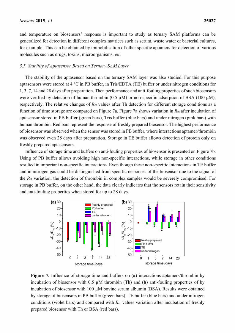
3.5. Stability of Aptasensor Based on Ternary SAM Layer
The stability of the aptasensor based on the ternary SAM layer was also studied. For this purpose
aptasensors were stored at 4 °C in PB buffer, in Tris/EDTA (TE) buffer or under nitrogen conditions for
1, 3, 7, 14 and 28 days after preparation. Then performance and anti-fouling properties of such biosensors
were verified by detection of human thrombin (0.5 µM) or non-specific adsorption of BSA (100 µM),
respectively. The relative changes of Rct values after Th detection for different storage conditions as a
function of time storage are compared on Figure 7a. Figure 7a shows variation in Rct after incubation of
aptasensor stored in PB buffer (green bars), Tris buffer (blue bars) and under nitrogen (pink bars) with
human thrombin. Red bars represent the response of freshly prepared biosensor. The highest performance
of biosensor was observed when the sensor was stored in PB buffer, where interactions aptamer/thrombin
was observed even 28 days after preparation. Storage in TE buffer allows detection of protein only on
freshly prepared aptasensors.
Influence of storage time and buffers on anti-fouling properties of biosensor is presented on Figure 7b.
Using of PB buffer allows avoiding high non-specific interactions, while storage in other conditions
resulted in important non-specific interactions. Even though these non-specific interactions in TE buffer
and in nitrogen gas could be distinguished from specific responses of the biosensor due to the signal of
the Rct variation, the detection of thrombin in complex samples would be severely compromised. For
storage in PB buffer, on the other hand, the data clearly indicates that the sensors retain their sensitivity
and anti-fouling properties when stored for up to 28 days.
Figure 7. Influence of storage time and buffers on (a) interactions aptamers/thrombin by
incubation of biosensor with 0.5 µM thrombin (Th) and (b) anti-fouling properties of by
incubation of biosensor with 100 µM bovine serum albumin (BSA). Results were obtained
by storage of biosensors in PB buffer (green bars), TE buffer (blue bars) and under nitrogen
conditions (violet bars) and compared with Rct values variation after incubation of freshly
prepared biosensor with Th or BSA (red bars).
-50
-40
-30
-20
-10
0
10
20
30 freshly prepared PB buffer TE under nitrogen
283 141 7
ΔRct/R
ct0 (%
)
storage time /days
0
(a)
-50
-40
-30
-20
-10
0
10
20
30
283 141 7
ΔRct/R
ct0 (%
)
storage time /days
freshly prepared PB buffer TE under nitrogen
0
(b)
Sensors 2015, 15 25028
4. Conclusions
We have demonstrated that antifouling properties of aptasensors (using a thrombin aptasensor as a
case study) can be achieved by means of a ternary SAM layer with the same conditions as those reported
for DNA hybridisation sensors [35]. However, one of the key aspects of this work is the observation
that, although the ternary SAM structure provides good antifouling properties when used on screen-printed
electrodes, it is rather inefficient when smoother gold surfaces are employed, presumably due to the
orientation of the hexanedithiol molecules on the surface.
For thrombin aptasensors, conflicting variations in charge transfer resistance are observed: An increase
in Rct is observed for low concentrations of thrombin target, while at higher concentrations a negative
shift is observed. Through the controls used, we speculate that the positive shifts at low concentrations
are due to non-specific interactions of thrombin with DNA, which reach saturation for concentrations
higher than 100 pM, while the negative shifts observed at higher concentrations are due to specific
binding of the thrombin to the DNA aptamer.
The pH and temperature of the buffer used during incubation of the sensor also strongly affect the
performance of the aptasensors. Optimised binding was observed for pH 7.0 and 35 °C. Stability of the
aptasensors was studied for different storage conditions and a shelf life of at least 28 days was determined
when the sensors are stored in phosphate buffer.
The results show that great care needs to be given to the characterisation of any impedance-based
aptasensor. Surface chemistries to immobilise aptamers and provide antifouling properties need to be
optimised for each aptasensor taking into account the type of electrode used, including its roughness.
Factors such as buffer, pH, temperature and storage conditions also strongly influence the performance
of the sensors. A full dose response of the sensors is required from very low to very high concentrations of
the target, as well as the use of suitable controls, to ensure that the target is binding specifically to the DNA
aptamer. Upon a full characterisation of the aptasensors, their application on clinical or environmental
samples can be envisaged using ternary SAM layers and controlled measurement conditions.
Acknowledgments
This work was funded by the Defence Science and Technology Laboratory (contract No.
Dstlx-1000074164).
Author Contributions
Anna Miodek and Edward M. Regan carried out the experimental work under the supervision of
Pedro Estrela. Nikhil Bhalla performed the contact angle experiments. Neal A.E. Hopkins and
Sarah A. Goodchild provided advice on the work. Anna Miodek and Pedro Estrela wrote the paper with
contributions from Sarah A. Goodchild and Edward M. Regan. All authors checked and approved the
final manuscript.
Sensors 2015, 15 25029
Conflicts of Interest
The authors declare no conflicts of interest.
References
1. Oh, J.; Yoo, G.; Chang, Y.W.; Kim, H.J.; Jose, J.; Kim, E.; Pyun, J.-C.; Yoo, K.-H. A carbon
nanotube metal semiconductor field effect transistor-based biosensor for detection of amyloid-beta in
human serum. Biosens. Bioelectron. 2013, 50, 345–350.
2. Tran, H.V.; Piro, B.; Reisberg, S.; Tran, L.D.; Duc, H.T.; Pham, M.C. Label-free and reagentless
electrochemical detection of microRNAs using a conducting polymer nanostructured by carbon
nanotubes: Application to prostate cancer biomarker miR-141. Biosens. Bioelectron. 2013, 49,
164–169.
3. Miodek, A.; Castillo, G.; Hianik, T.; Korri-Youssoufi, H. Electrochemical aptasensor of human
cellular prion based on multiwalled carbon nanotubes modified with dendrimers: A platform for
connecting redox markers and aptamers. Anal. Chem. 2013, 85, 7704–7712.
4. Miodek, A.; Castillo, G.; Hianik, T.; Korri-Youssoufi, H. Electrochemical aptasensor of cellular
prion protein based on modified polypyrrole with redox dendrimers. Biosens. Bioelectron. 2014,
56, 104–111.
5. Kanyong, P.; Pemberton, R.M.; Jackson, S.K.; Hart, J.P. Development of a sandwich format,
amperometric screen-printed uric acid biosensor for urine analysis. Anal. Biochem. 2012, 428,
39–43.
6. Zhang, Q.D.; March, G.; Noel, V.; Piro, B.; Reisberg, S.; Tran, L.D.; Hai, L.V.; Abadia, E.;
Nielsen, P.E.; Sola, C.; et al. Label-free and reagentless electrochemical detection of PCR fragments
using self-assembled quinone derivative monolayer: Application to Mycobacterium tuberculosis.
Biosens. Bioelectron. 2012, 32, 163–168.
7. Sun, X.; Jia, M.; Ji, J.; Guan, L.; Zhang, Y.; Tang, L.; Li, Z. Enzymatic amplification detection of
peanut allergen Ara h1 using a stem-loop DNA biosensor modified with a chitosan-mutiwalled
carbon nanotube nanocomposite and spongy gold film. Talanta. 2015, 131, 521–527.
8. Torrente-Rodríguez, R.M.; Campuzano, S.; López-Hernández, E.; Ruiz-Valdepeñas, M.V.;
Barderas, R.; Granados, R.; Sánchez-Puelles, J.M.; Pingarrón, J.M. Simultaneous detection of two
breast cancer-related miRNAs in tumor tissues using p19-based disposable amperometric
magnetobiosensing platforms. Biosens. Bioelectron. 2015, 66, 385–391.
9. Liao, J.; Lin, S.; Liu, K.; Yang, Y.; Zhang, R.R.; Du, W.; Li, X. Organic electrochemical transistor
based biosensor for detecting marine diatoms in seawater medium. Sens. Actuators B Chem. 2014,
203, 677–682.
10. Fernandes, A.M.; Abdalhai, M.H.; Ji, J.; Xi, B.W.; Xie, J.; Sun, J.; Noeline, R.; Lee, B.H.; Sun, X.
Development of highly sensitive electrochemical genosensor based on multiwalled carbon
nanotubes–chitosan–bismuth and lead sulfide nanoparticles for the detection of pathogenic
Aeromonas. Biosens. Bioelectron. 2015, 63, 399–406.
11. Castillo, G.; Lamberti, I.; Mosiello, L.; Hianik, T. Impedimetric DNA aptasensor for sensitive
detection of ochratoxin A in food. Electroanalysis 2012, 24, 512–520.
Sensors 2015, 15 25030
12. Rowe, A.A.; Miller, E.A.; Plaxco, K.W. Reagentless measurement of aminoglycoside antibiotics in
blood serum via an electrochemical, ribonucleic acid aptamer-based biosensor. Anal. Chem. 2010,
82, 7090–7095.
13. Brunetti, B.; Valdés-Ramírez, G.; Litvan, I.; Wang, J. A disposable electrochemical biosensor for
L-DOPA determination in undiluted human serum. Electrochem. Commun. 2014, 48, 28–31.
14. Ferapontova, E.E.; Olsen, E.M.; Gothelf, K.V. An RNA aptamer-based electrochemical biosensor
for detection of theophylline in serum. J. Am. Chem. Soc. 2008, 130, 4256–4258.
15. Keighley, S.D.; Li, P.; Estrela, P.; Migliorato, P. Optimization of DNA immobilization on gold
electrodes for label-free detection by electrochemical impedance spectroscopy. Biosens. Bioelectron.
2008, 23, 1291–1297.
16. Cortina-Puig, M.; Muñoz-Berbel, X.; Calas-Blanchard, C.; Marty, J.-L. Electrochemical
characterization of a superoxide biosensor based on the co-immobilization of cytochrome c and
XOD on SAM-modified gold electrodes and application to garlic samples. Talanta 2009, 79,
289–294.
17. Jolly, P.; Formisano, N.; Tkáč, J.; Kasák, P.; Frost, C.G.; Estrela, P. Label-free impedimetric
aptasensor with antifouling surface chemistry: A prostate specific antigen case study. Sens.
Actuators B Chem. 2015, 209, 306–312.
18. Tyagi, D.; Perez, J.B.; Nand, A.; Zhiqiang, C.; Wang, P.; Na, J.; Zhu, J. Detection of embryonic
stem cell lysate biomarkers by surface plasmon resonance with reduced nonspecific adsorption.
Anal. Biochem. 2015, 471, 29–37.
19. Vaisocherová, H.; Mrkvová, K.; Piliarik, M.; Jinoch, P.; Šteinbachová, M.; Homola, J. Surface
plasmon resonance biosensor for direct detection of antibody against Epstein-Barr virus. Biosens.
Bioelectron. 2007, 22, 1020–1026.
20. Lahiri, J.; Isaacs, L.; Tien, J.; Whitesides, G.M. A strategy for the generation of surfaces presenting
ligands for studies of binding based on an active ester as a common reactive intermediate: A surface
plasmon resonance study. Anal. Chem. 1999, 71, 777–790.
21. Ma, H.; Hyun, J.; Stiller, P.; Chilkoti, A. “Non-fouling” Oligo (ethylene glycol)-functionalized
polymer brushes synthesized by surface initiated atom transfer radical polymerization. Adv. Mater.
2004, 16, 338–341.
22. Riedel, T.; Rodriguez-Emmenegger, C.; de los Santos Pereira, A.; Bědajánková, A.; Jinoch, P.;
Boltovets, P.M.; Brynda, E. Diagnosis of Epstein – Barr virus infection in clinical serum samples
by an SPR biosensor assay. Biosens. Bioelectron. 2014, 55, 278–284.
23. Sonato, A.; Silvestri, D.; Ruffato, G.; Zacco, G.; Romanato, F.; Morpurgo, M. Quantitative control
of poly(ethylene oxide) surface antifouling and biodetection through azimuthally enhanced grating
coupled-surface plasmon resonance. Appl. Surf. Sci. 2013, 286, 22–30.
24. Ladd, J.; Zhang, Z.; Chen, S.; Hower, J.C.; Jiang, S. Zwitterionic polymers exhibiting high
resistance to nonspecific protein adsorption from human serum and plasma. Biomacromolecules
2008, 9, 1357–1361.
25. Brault, N.D.; White, A.D.; Taylor, A.D.; Yu, Q.; Jiang, S. Directly functionalizable surface platform
for protein arrays in undiluted human blood plasma. Anal. Chem. 2013, 85, 1447–1453.
Sensors 2015, 15 25031
26. Vaisocherová, H.; Yang, W.; Zhang, Z.; Cao, Z.; Cheng, G.; Piliarik, M.; Homola, J.; Jiang, S.
Ultralow fouling and functionalizable surface chemistry based on a zwitterionic polymer enabling
sensitive and specific protein detection in undiluted blood plasma. Anal. Chem. 2008, 80,
7894–7901.
27. Blaszykowski, C.; Sheikh, S.; Thompson, M. Surface chemistry to minimize fouling from
blood-based fluids. Chem. Soc. Rev. 2012, 41, 5599–5612.
28. de los Santos Pereira, A.; Riedel, T.; Brynda, E.; Rodriguez-Emmenegger, C. Hierarchical
antifouling brushes for biosensing applications. Sens. Actuators B Chem. 2014, 202, 1313–1321.
29. Ratel, M.; Provencher-Girard, A.; Zhao, S.S.; Breault-Turcot, J.; Labrecque-Carbonneau, J.;
Branca, M.; Pelletier, J.N.; Schmitzer, A.R.; Masson, J.-F. Imidazolium-based ionic liquid surfaces
for biosensing. Anal. Chem. 2013, 85, 5770–5777.
30. Barfidokht, A.; Gooding, J.J. Approaches toward allowing electroanalytical devices to be used in
biological fluids. Electroanalysis 2014, 26, 1182–1196.
31. Sun, C.; Han, Q.; Wang, D.; Xu, W.; Wang, W.; Zhao, W.; Zhou, M. A label-free and high sensitive
aptamer biosensor based on hyperbranched polyester microspheres for thrombin detection. Anal.
Chim. Acta 2014, 850, 33–40.
32. Sun, C.; Miao, J.; Yan, J.; Yang, K.; Mao, C.; Ju, J.; Shen, J. Applications of antibiofouling
PEG-coating in electrochemical biosensors for determination of glucose in whole blood.
Electrochim. Acta 2013, 89, 549–554.
33. Gui, A.L.; Luais, E.; Peterson, J.R.; Gooding, J.J. Zwitterionic phenyl layers: Finally, stable,
anti-biofouling coatings that do not passivate electrodes. Appl. Mater. Interfaces 2013, 5,
4827–4835.
34. Hu, Y.; Yang, G.; Liang, B.; Fang, L.; Ma, G.; Zhu, Q.; Chen, S.; Ye, X. The fabrication of superlow
protein absorption zwitterionic coating by electrochemically mediated atom transfer radical
polymerization and its application. Acta Biomater. 2015, 13, 142–149.
35. Campuzano, S.; Kuralay, F.; Lobo-Castañón, M.J.; Bartošík, M.; Vyavahare, K.; Paleček, E.;
Haake, D.A.; Wang, J. Ternary monolayers as DNA recognition interfaces for direct and sensitive
electrochemical detection in untreated clinical samples. Biosens. Bioelectron. 2011, 26, 3577–3583.
36. Kuralay, F.; Campuzano, S.; Wang, J. Greatly extended storage stability of electrochemical DNA
biosensors using ternary thiolated self-assembled monolayers. Talanta 2012, 99, 155–160.
37. Leung, T.Y.B.; Gerstenberg, M.C.; Lavrich, D.J.; Scoles, G.; Schreiber, F.; Poirier, G.E.
1,6-hexanedithiol monolayers on Au(111): A multitechnique structural study. Langmuir 2000, 16,
549–561.
38. Morel, A.-L.; Volmant, R.-M.; Méthivier, C.; Krafft, J.-M.; Boujday, S.; Pradier, C.-M. Optimized
immobilization of gold nanoparticles on planar surfaces through alkyldithiols and their use to build
3D biosensors. Coll. Surf. B Biointerfaces 2010, 81, 304–312.
39. Yang, J.; Yang, T.; Feng, Y.; Jiao, K. A DNA electrochemical sensor based on nanogold-modified
poly-2,6-pyridinedicarboxylic acid film and detection of PAT gene fragment. Anal. Biochem. 2007,
365, 24–30.
40. Castillo, G.; Trnkova, L.; Hrdy, R.; Hianik, H. Impedimetric aptasensor for thrombin recognition
based on CD support. Electroanalysis 2012, 24, 1079–1087.
Sensors 2015, 15 25032
41. Lee, J.A.; Hwang, S.; Kwak, J.; Park, S.I.; Lee, S.S.; Lee, K.-C. An electrochemical impedance
biosensor with aptamer-modified pyrolyzed carbon electrode for label-free protein detection. Sens.
Actuators B Chem. 2008, 129, 372–379.
42. Cai, H.; Lee, T.M.-H.; Hsing, I.-M. Label-free protein recognition using an aptamer-based
impedance measurement assay. Sens. Actuators B Chem. 2006, 114, 433–437.
43. Bogomolova, A.; Komarova, E.; Reber, K.; Gerasimov, T.; Yavuz, O.; Bhatt, S.; Aldissi, M.
Challenges of electrochemical impedance spectroscopy in protein biosensing. Anal. Chem. 2009,
81, 3944–3949.
© 2015 by the authors; licensee MDPI, Basel, Switzerland. This article is an open access article
distributed under the terms and conditions of the Creative Commons Attribution license
(http://creativecommons.org/licenses/by/4.0/).


![Fouling vs. Availability · PDF fileFouling vs. Availability CheMin ... Sanicro 28/ 63, Sandvik 8RE. Fouling vs. Availability CheMin ... Fouling rate...Heat Transfer [W/m2]](https://img.pdfslide.net/doc/110x75/5aadcccc7f8b9a8f498eba95/fouling-vs-availability-vs-availability-chemin-sanicro-28-63-sandvik-8re.jpg)