Embed Size (px)
Citation preview
Int J Clin Exp Med 2019;12(5):5938-5950www.ijcem.com /ISSN:1940-5901/IJCEM0085245
Original Article Clinical features, treatment, and prognosis of acute methanol poisoning: experiences in an outbreak
Maoxia Ran, Ying Li, Liling Zhang, Weihua Wu, Jiaru Lin, Qi Liu, Santao Ou
Department of Nephrology, The Affiliated Hospital of Southwest Medical University, Luzhou 646000, Sichuan, China
Received September 8, 2018; Accepted February 12, 2019; Epub May 15, 2019; Published May 30, 2019
Abstract: Aims: The aim of this study was to examine the clinical features of acute methanol poisoning to better understand its pathophysiology. Methods: A retrospective study was performed in 52 patients with acute methanol poisoning. Patient characteristics and test results were collected and analyzed. Results: A total of 52 patients, rang-ing from 20-79 years of age, consumed between 5 and 500 mL of alcohol-based fuel, equivalent to 3.3-329.5 g of methanol. Of the 52 patients, 49 were discharged without sequelae, one patient experienced decreased visual acuity, and two patients died after comprehensive treatment. Central nervous system (CNS) disorders, visual distur-bances, and gastrointestinal symptoms were found in 46 (88%), 20 (38%), and 34 (65%) patients upon admission, respectively. Univariate analysis showed that coma, dyspnea, pH, and anion gap (AG), along with calcium, potas-sium, creatinine, and blood sugar levels, were correlated with severity of methanol poisoning and associated with poor patient outcomes. Conclusion: Acute oral methanol poisoning can lead to nerve damage, metabolic acidosis, and gastrointestinal injuries. Most patients recovered after timely and effective comprehensive treatment.
Keywords: Methanol poisoning, methanol toxicity, clinical features, prognosis
Introduction
Methanol, also known as the wood alcohol or Columbian spirit, is a colorless liquid with a taste and smell similar to that of ethanol [1]. While methanol poisoning has been associated with ingestion, methanol exposure also occurs by inhalation and skin absorption. After being rapidly absorbed, methanol is oxidized, conve- rted to formaldehyde by alcohol dehydrogena- se, and subsequently metabolized into formic acid by aldehyde dehydrogenase in the liver [2]. It is a formic acid metabolite that is highly toxic to humans, as it functions as an inhibitor of mitochondrial cytochrome oxidase [3]. Formic acid is a weak inhibitor compared to cyanide, but it is strong enough to cause metabolic acidosis.
Incidence of methanol poisoning has been ris-ing, worldwide, with a number of outbreaks oc- curring in the Czech Republic, Estonia, Iran, Ke- nya, Khartoum, Libya, Norway, and other coun-tries [4-9]. From 2000-2012, there were more than 50 mass outbreaks of methanol pois- oning, worldwide, resulting in approximately
5,000 acute cases of toxicity and more than 2,000 associated deaths [10]. Outbreaks often arise from the consumption of ethanol adulter-ated with methanol (i.e., methylated spirits) [11]. In addition, several recent examples of methanol poisoning have been linked to occu-pational exposure [12]. In rare cases, methanol may be used for attempted suicide. There have been several instances of accidental transder-mal exposure [13-15]. In China, acute methanol poisoning most commonly results from the excessive consumption of liquor contaminated with methanol.
In addition to metabolic acidosis, methanol in- toxication leads to central nervous system (CNS) depression, cardiovascular disease, vi- sual disturbances, putamina hemorrhages, and even death. It has been reported that mortality rates for methanol poisoning range from 10 to 50% [6-9]. Previously, the rates of long-term visual sequelae and total sequelae (both vision and CNS) after methanol poisoning were 25- 40% and 18-40%, respectively [7, 16]. When treatment is delayed or inadequate, the mor- tality rate increases to more than 40% and
Outcomes of methanol poisoning in patients
5939 Int J Clin Exp Med 2019;12(5):5938-5950
many of these patients will experience serious long-term visual and CNS impairment [17-19]. Fomepizole, an alcohol dehydrogenase inhibi-tor, is an effective antidote for methanol poi-soning. However, this medication is currently unavailable in China. The mortality rate is 3.3% for those patients effectively treated, suggest-ing there is still room for improvement concern-ing therapies available for methanol intoxica-tion [20]. Currently, early diagnosis and rapid therapeutic intervention are essential steps for successfully treating methanol poisoning in pa- tients. However, late onset of clinical symptoms often delays diagnosis. In addition, some hospi-tals located in rural areas do not have direct access to the equipment required for methanol testing. This can also delay diagnosis and effec-tive treatment planning for some patients.
In the current study, clinical data were collected and summarized from 52 patients treated for methanol poisoning. The aim was to better un- derstand the pathophysiology and provide a basis for clinical diagnosis and treatment. All patients suffered acute methanol poisoning resulting from the ingestion of alcohol-based fuel, which was mistakenly served as ethanol at a party on February 3-4, 2017.
Materials and methods
Patients
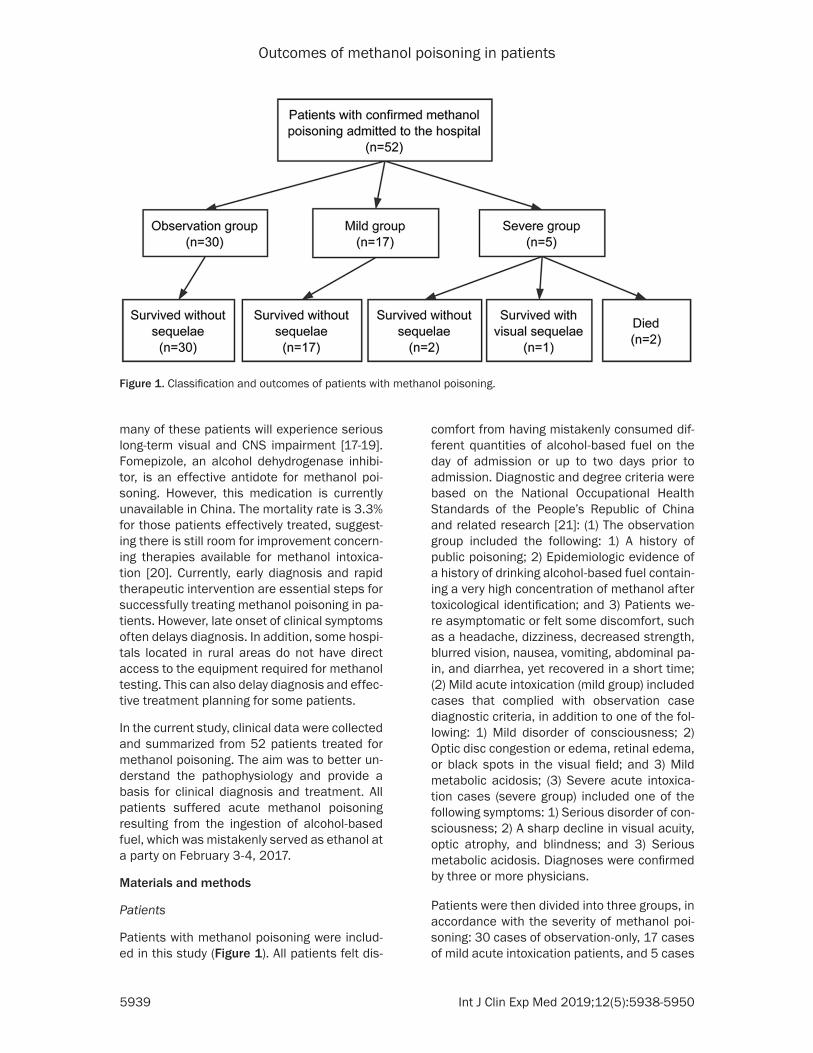
Patients with methanol poisoning were includ-ed in this study (Figure 1). All patients felt dis-
comfort from having mistakenly consumed dif-ferent quantities of alcohol-based fuel on the day of admission or up to two days prior to admission. Diagnostic and degree criteria were based on the National Occupational Health Standards of the People’s Republic of China and related research [21]: (1) The observation group included the following: 1) A history of public poisoning; 2) Epidemiologic evidence of a history of drinking alcohol-based fuel contain-ing a very high concentration of methanol after toxicological identification; and 3) Patients we- re asymptomatic or felt some discomfort, such as a headache, dizziness, decreased strength, blurred vision, nausea, vomiting, abdominal pa- in, and diarrhea, yet recovered in a short time; (2) Mild acute intoxication (mild group) included cases that complied with observation case diagnostic criteria, in addition to one of the fol-lowing: 1) Mild disorder of consciousness; 2) Optic disc congestion or edema, retinal edema, or black spots in the visual field; and 3) Mild metabolic acidosis; (3) Severe acute intoxica-tion cases (severe group) included one of the following symptoms: 1) Serious disorder of con-sciousness; 2) A sharp decline in visual acuity, optic atrophy, and blindness; and 3) Serious metabolic acidosis. Diagnoses were confirmed by three or more physicians.
Patients were then divided into three groups, in accordance with the severity of methanol poi-soning: 30 cases of observation-only, 17 cases of mild acute intoxication patients, and 5 cases
Figure 1. Classification and outcomes of patients with methanol poisoning.
Outcomes of methanol poisoning in patients
5940 Int J Clin Exp Med 2019;12(5):5938-5950
of severe acute intoxication (Figure 1). Medical histories of the patients in this study included a history of previous surgeries (n = 10), hepatitis (n = 3), and lung disease (n = 2). One patient each had hypertension, coronary heart disea- se, cerebral infarction, or cataracts. The rem- aining 34 patients had no prior medical history to report.
Collection of patient data
Patient data, including demographics, general information, and test results, were collected via an electronic medical history system. Data included hematological and biochemical test-ing, vision testing, stereoscopic fundus exami-nation, visual evoked potential (VEP) testing, electrocardiogram (ECG), computed tomogra-phy (CT) imaging, and abdominal ultrasound (US) imaging. Written informed consent was obtained from patients or families and patients did not experience increased pain or hospital-ization costs due to the study. The remaining alcohol-based fuel was sent to the National Supervision and Inspection Center of Liquor and Processed Food Quality for analysis. In the fuel, the detected methanol concentration was 659 g/L.
Treatments
Treatment protocols were formulated via dis-cussion by a multidisciplinary medical team, consisting of physicians from the Departments of Emergency Medicine, Hemopathology, Nep- hrology, Urology, Endocrinology, Gastroentero- logy, and Gerontology. There were six treatment options, including: (1) Folic acid to enhance the metabolism of formate; (2) Sodium bicarbonate for correction of metabolic acidosis; (3) Meth- ylprednisolone for patients with fundus or optic nerve damage for 2-3 days, followed by mainte-nance therapy with prednisone and precaution-ary steps to prevent direct stimulation of the fundus and/or optic nerve (i.e., eye guards); 4) Panax notoginseng saponins for reduction of oxidative stress and carithamine as an antico-agulant, along with vitamins B1, B6, and decavi-tamin for nerve recovery; (5) Blood purification to eliminate methanol and methanol metabo-lites (in serious cases), which included continu-ous renal replacement treatment (CRRT), he- modialysis (HD) [22] and plasmapheresis (PE) [23], along with continued treatment for aci- dosis; (6) Symptomatic supportive treatment,
including treatment to protect the organs, ma- intain electrolyte balance, and reduce risks of infection, along with consciousness-promoting drugs and others. Endotracheal intubation and mechanical ventilation were used in severe cases, as required, with enhanced monitoring and nursing. Other treatment options are avail-able but they were not used in this study. For example, gastrolavage was not conducted in this study due to the length of time between the ingestion of methanol and hospital admission. Also, fomepizole and ethanol, which can inhibit the alcohol dehydrogenase (ADH) enzyme, were not administered to patients in this study, as they are not yet approved in China.
Statistical analysis
All statistical analyses were performed using SPSS Version 23.0 (IBM, Chicago, IL, USA). Da- ta are expressed as median with a range and mean ± standard deviation (mean ± SD), as appropriate. For comparisons of quantitative data between groups, common statistical tests were employed, such as t-test, one-way analy-sis of variance (ANOVA), Kruskal-Wallis H-test, and Mann-Whitney U-test. Chi-squared test and Fisher’s exact test were used for qualitative data. Correlation analyses between two vari-ances were conducted using Spearman’s rank correlation analysis. P-values less than 0.05 are considered statistically significant.
Results
Demographics and general patient information
Fifty-two patients with accidental methanol poi-soning were included in the study. The mean age of all subjects was 47.67 ± 12.13 years of age (range: 20-79 years), as shown in Table 1. From the 52 recruited patients, 45 were male and seven were female. All females were classi-fied into the observation group. The average alcohol-based fuel intake was 91.73 ± 81.24 mL (range: 5-500 mL), with 1 mL of fuel con-taining 0.659 g of methanol. There was a sig-nificant difference in the average alcohol-based fuel intake between the observation group and mild toxicity group (P < 0.02), along with the observation group and severe toxicity group (P < 0.02). Symptoms appeared in all patients within 12-24 hours after ingestion of the meth-anol. The amount of time between ingestion and appearance of symptoms was not signifi-
Outcomes of methanol poisoning in patients
5941 Int J Clin Exp Med 2019;12(5):5938-5950
cantly different between the groups. There we- re no significant differences in age, history of alcohol consumption, history of cigarette smok-ing, or the time from intake to hospital admis-sion between the three groups.
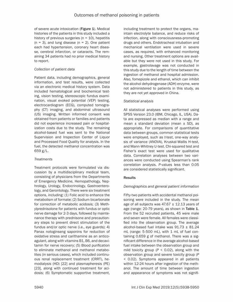
CNS symptoms were most common in patients, followed by gastrointestinal (GI) symptoms and visual disturbances. These were observed in 46 (88%), 34 (65%), and 17 (32.7%) of patients, respectively. Among CNS symptoms, dizziness was seen in 42 (80.8%) patients, cephalalgia in 10 (19.2%) patients, and a disturbance of con-sciousness in 7 (13.5%) patients. Disturbance of consciousness was only observed in patients with mild or severe methanol poisoning. So- mnolence was only noted in patients with mild methanol poisoning. More serious conscious-ness disorders were apparent in patients with severe methanol poisoning, including somno-lence, confusion, stupor, delirium, and comas.
GI symptoms, such as nausea, vomiting, ab- dominal pain, abdominal distension, and diar-rhea, were found in 16 (30.8%), 9 (17.3%), 21 (40.4%), 13 (25.0%), and 8 (15.4%) patients, respectively. Other symptoms, including chest stuffiness, palpitations, dyspnea, and general weakness, were observed in 4 (7.7%), 11 (21.2%), 5 (9.6%), and 17 (32.7%) patients, respectively (Table 1). Incidence of dyspnea and disturbances of consciousness were sig-nificantly different between the observation gr- oup and severe methanol poisoning group (P = 0.006 and P < 0.001, respectively). However, incidence of other symptoms was not signifi-cantly different between the groups. There we- re several clinical manifestations of methanol poisoning that occurred at low frequencies, not listed in Table 1, including orbital and perior-bital pain (n = 6), acanthesthesia of the throat (n = 3), numbness of limbs (n = 2), burning sen-sation of the face (n = 2), the feeling of stepping
Table 1. Demographics and general characteristics of patients with acute methanol poisoning
Characteristics Observation (n = 30)
Mild (n = 17)
Severe (n = 5)
Total (n = 52) p
Age (mean ± SD) 49.20 ± 10.87 45.76 ± 14.23 45.00 ± 12.96 47.67 ± 12.13 NSGender Male 23 (76.7) 17 (100.0) 5 (100.0) 45 (86.5) - Female 7 (23.3) 0 0 7 (13.5)Drinking Daily 5 (16.7) 6 (35.3) 0 11 (21.2) NS Not or occasionally 25 (83.3) 11 (64.7) 5 (100.0) 41 (78.8)Smoking 14 (46.7) 7 (41.2) 2 (40.0) 23 (44.2) NSIntake (range) 5-150 15-200 100-500 5-500 < 0.02#,**Latency (median; h) 12-24 12-24 12-24 12-24 NSTime from intake to admission (d) 1.84 ± 0.78 1.55 ± 0.61 1.40 ± 0.37 1.70 ± 0.71 NSSymptoms Dizzy 25 (83.3) 15 (88.2) 2 (40.0) 42 (80.8) NS Headache 5 (16.7) 4 (23.5) 1 (20.0) 10 (19.2) NS Weak 11 (36.7) 5 (29.4) 1 (20.0) 17 (32.7) NS Nausea 9 (30.0) 7 (41.2) 0 16 (30.8) NS Vomiting 3 (10.0) 4 (23.5) 2 (40.0) 9 (17.3) NS Abdominal pain 11 (36.7) 7 (41.2) 1 (20.0) 21 (40.4) NS Abdominal distension 10 (33.3) 3(17.6) 0 13 (25.0) NS Diarrhea 6 (20.0) 2 (11.8) 0 8 (15.4) NS Blurred vision 10 (33.3) 4 (23.5) 3 (60.0) 17 (32.7) NS Chest stuffiness 1 (3.3) 2 (11.8) 1 (20.0) 4 (7.7) NS Palpitation 6 (20.0) 2 (11.8) 3 (60.0) 11 (21.2) NS Dyspnea 1 (3.3) 1 (5.9) 3 (60.0) 5 (9.6) 0.006** Disturbance of consciousness 0 3 (17.6) 4 (80.0) 7 (13.5) < 0.001**#Observation group vs Mild group; **Observation group VS Severe group; *Mild group VS Severe group.
Outcomes of methanol poisoning in patients
5942 Int J Clin Exp Med 2019;12(5):5938-5950
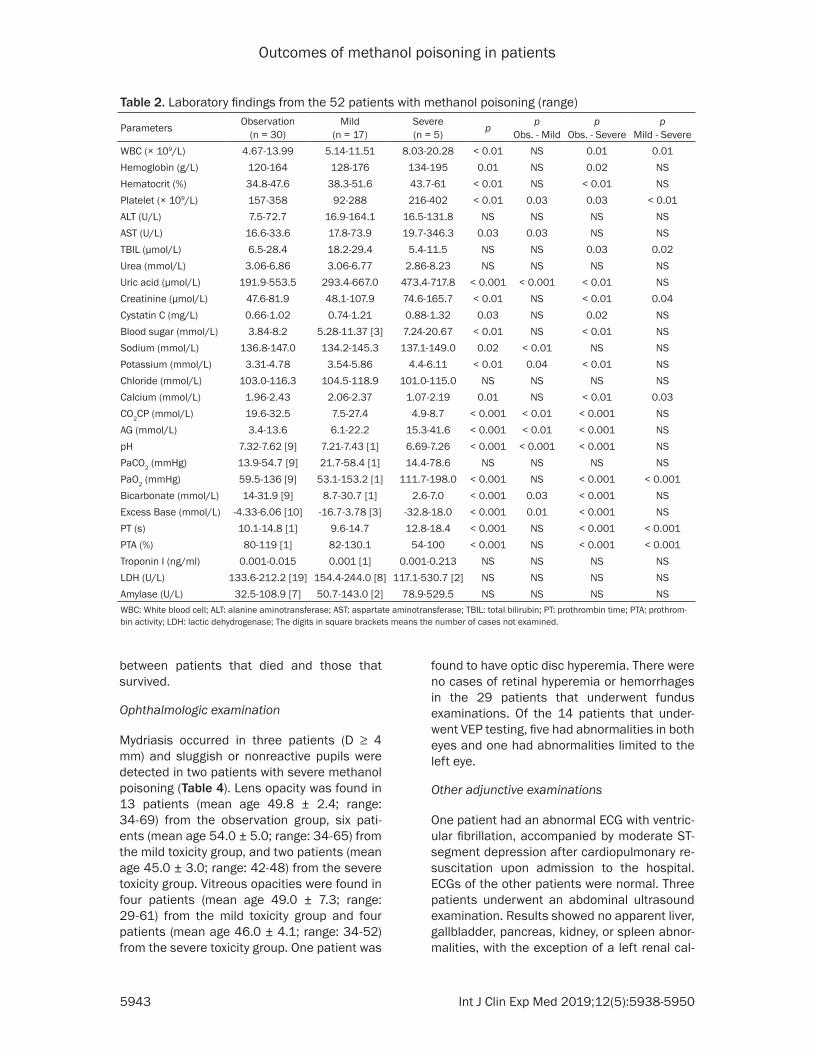
Despite some differences in hemoglobin and platelet levels between the groups, there were no apparent abnormalities in any patients from the three groups. However, WBC and hemato-crit levels were higher in severe cases of meth-anol poisoning, compared with observation and mild toxicity groups. Abnormal liver function, as indicated by increased transaminase without a change in bilirubin, was detected in varying degrees among the patients. Patients with severe poisoning had significantly elevated lev-els of creatinine, compared to patients from the observation and mild toxicity groups. Uric acid levels were increased in 20 patients and were remarkably different between the severe toxic-ity group and other groups (observation and mild toxicity). Urea levels were within the nor-mal range in all patients and were not signifi-cantly different between any of the three groups. Patients with severe poisoning suffered from myocardial damage, evidenced by highly increased Troponin I levels and abnormal coag-ulation functioning, as indicated by the pro-longed prothrombin time and decreased pro-thrombin activity. There was no difference in amylopsin between the three groups. In addi-tion, random blood sugar levels were higher in the severe group, compared with the observa-tion group. Routine feces examinations and fecal occult blood tests were performed in 31 of the patients. Of these, one patient had a positive fecal occult blood test. In addition, urine tests were conducted in 41 of the pa- tients, Three patients were positive for urine occult blood and one patient was positive for urine protein.
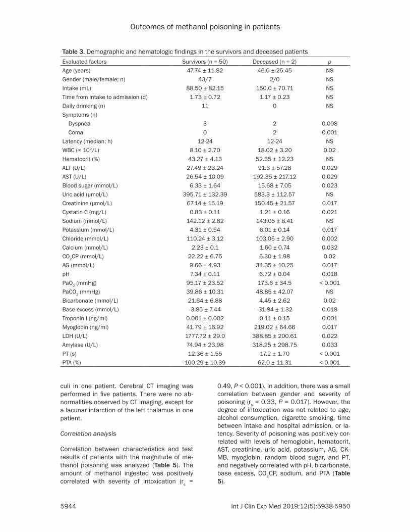
Demographic and hematologic findings were compared between the survivors and patients that died. Incidence of dyspnea and coma was higher in patients that died, compared to tho- se individuals that survived (P = 0.008 and P = 0.001, respectively), as shown in Table 3. Similarly, WBC count, ALT, AST, random blood sugar, creatinine, cystatin C, potassium, anion gap, PaO2, troponin I, myoglobin, LDH, amylase, and PT levels were higher in patients that died, while levels of calcium, CO2CP, pH, bicarbonate, base excess, and PTA were lower in patients that died when, compared with survivors. However, there was no difference in age, gen-der, amount of methanol intake, time between intake to hospital admission, drinking history, latency, hematocrit, uric acid, or PaCO2 levels
on cotton (n = 1), photophobia and tearing (n = 1), ocular foreign-body sensation (n = 1), amau-rosis (n = 1), hoarseness (n = 1), odynophagia (n = 1), and fevers (n = 1).
Interestingly, 31 of the 52 (59.62%) patients showed no distinctly positive clinical signs of methanol poisoning. Abdominal tenderness was confirmed in 13 patients, though only one patient experienced rebound tenderness. Th- ree patients were found to have a flushed face during intake and two patients displayed pha-ryngeal hyperemia. Coma, cyanosis, rapid and shallow breathing, Kussmaul breathing, crack-ling in the lungs, arrhythmias, and weak pulse or pulselessness were found among individual patients with severe methanol poisoning.
Blood testing and evaluation parameters
Blood gas analysis was performed in 42 pa- tients. There were 21 in the observation group, 16 in the mild toxicity group, and 5 in the severe toxicity group. Among the 21 patients in the observation group that received blood work, one patient had respiratory acidosis and anoth-er patient had respiratory alkalosis. Among the 16 patients with mild poisoning, 3 had incr- eased AG metabolic acidosis, and 13 had nor-mal AG metabolic acidosis. Increased AG meta-bolic acidosis was observed in four patients with severe poisoning, one of which also had respiratory acidosis. One patient with severe poisoning had normal AG metabolic acidosis. Furthermore, patients with severe poisoning had lower carbon dioxide combining power (CO2CP), pH, bicarbonate, base excess levels, and higher AG levels than patients in the obser-vation and mild poisoning groups, as illustrated in Table 2. There were no significant differenc-es in PaCO2 between the three groups. However, PaO2 in the observation group was notably lower in mild and severe toxicity groups. In addi-tion, sodium, potassium, and calcium levels were significantly different, while there was no difference in chloride levels among the three groups. After hospital admission, one patient had mild hyponatremia, four had hyperkalemia, and four had hypokalemia. It is worth noting that 10 patients became hypokalemic during treatment, with three of the 10 patients being hyperkalemic prior to admission. The other seven patients had normal potassium levels at admission.
Outcomes of methanol poisoning in patients
5943 Int J Clin Exp Med 2019;12(5):5938-5950
between patients that died and those that survived.
Ophthalmologic examination
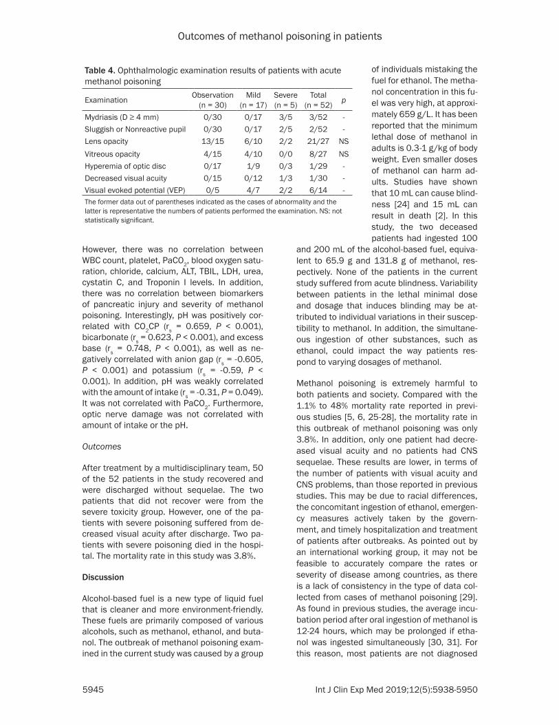
Mydriasis occurred in three patients (D ≥ 4 mm) and sluggish or nonreactive pupils were detected in two patients with severe methanol poisoning (Table 4). Lens opacity was found in 13 patients (mean age 49.8 ± 2.4; range: 34-69) from the observation group, six pati- ents (mean age 54.0 ± 5.0; range: 34-65) from the mild toxicity group, and two patients (mean age 45.0 ± 3.0; range: 42-48) from the severe toxicity group. Vitreous opacities were found in four patients (mean age 49.0 ± 7.3; range: 29-61) from the mild toxicity group and four patients (mean age 46.0 ± 4.1; range: 34-52) from the severe toxicity group. One patient was
found to have optic disc hyperemia. There were no cases of retinal hyperemia or hemorrhages in the 29 patients that underwent fundus examinations. Of the 14 patients that under-went VEP testing, five had abnormalities in both eyes and one had abnormalities limited to the left eye.
Other adjunctive examinations
One patient had an abnormal ECG with ventric-ular fibrillation, accompanied by moderate ST- segment depression after cardiopulmonary re- suscitation upon admission to the hospital. ECGs of the other patients were normal. Three patients underwent an abdominal ultrasound examination. Results showed no apparent liver, gallbladder, pancreas, kidney, or spleen abnor-malities, with the exception of a left renal cal-
Table 2. Laboratory findings from the 52 patients with methanol poisoning (range)
Parameters Observation (n = 30)
Mild (n = 17)
Severe (n = 5) p p
Obs. - Mildp
Obs. - Severep
Mild - SevereWBC (× 109/L) 4.67-13.99 5.14-11.51 8.03-20.28 < 0.01 NS 0.01 0.01Hemoglobin (g/L) 120-164 128-176 134-195 0.01 NS 0.02 NSHematocrit (%) 34.8-47.6 38.3-51.6 43.7-61 < 0.01 NS < 0.01 NSPlatelet (× 109/L) 157-358 92-288 216-402 < 0.01 0.03 0.03 < 0.01ALT (U/L) 7.5-72.7 16.9-164.1 16.5-131.8 NS NS NS NSAST (U/L) 16.6-33.6 17.8-73.9 19.7-346.3 0.03 0.03 NS NSTBIL (µmol/L) 6.5-28.4 18.2-29.4 5.4-11.5 NS NS 0.03 0.02Urea (mmol/L) 3.06-6.86 3.06-6.77 2.86-8.23 NS NS NS NSUric acid (µmol/L) 191.9-553.5 293.4-667.0 473.4-717.8 < 0.001 < 0.001 < 0.01 NSCreatinine (µmol/L) 47.6-81.9 48.1-107.9 74.6-165.7 < 0.01 NS < 0.01 0.04Cystatin C (mg/L) 0.66-1.02 0.74-1.21 0.88-1.32 0.03 NS 0.02 NSBlood sugar (mmol/L) 3.84-8.2 5.28-11.37 [3] 7.24-20.67 < 0.01 NS < 0.01 NSSodium (mmol/L) 136.8-147.0 134.2-145.3 137.1-149.0 0.02 < 0.01 NS NSPotassium (mmol/L) 3.31-4.78 3.54-5.86 4.4-6.11 < 0.01 0.04 < 0.01 NSChloride (mmol/L) 103.0-116.3 104.5-118.9 101.0-115.0 NS NS NS NSCalcium (mmol/L) 1.96-2.43 2.06-2.37 1.07-2.19 0.01 NS < 0.01 0.03CO2CP (mmol/L) 19.6-32.5 7.5-27.4 4.9-8.7 < 0.001 < 0.01 < 0.001 NSAG (mmol/L) 3.4-13.6 6.1-22.2 15.3-41.6 < 0.001 < 0.01 < 0.001 NSpH 7.32-7.62 [9] 7.21-7.43 [1] 6.69-7.26 < 0.001 < 0.001 < 0.001 NSPaCO2 (mmHg) 13.9-54.7 [9] 21.7-58.4 [1] 14.4-78.6 NS NS NS NSPaO2 (mmHg) 59.5-136 [9] 53.1-153.2 [1] 111.7-198.0 < 0.001 NS < 0.001 < 0.001Bicarbonate (mmol/L) 14-31.9 [9] 8.7-30.7 [1] 2.6-7.0 < 0.001 0.03 < 0.001 NSExcess Base (mmol/L) -4.33-6.06 [10] -16.7-3.78 [3] -32.8-18.0 < 0.001 0.01 < 0.001 NSPT (s) 10.1-14.8 [1] 9.6-14.7 12.8-18.4 < 0.001 NS < 0.001 < 0.001PTA (%) 80-119 [1] 82-130.1 54-100 < 0.001 NS < 0.001 < 0.001Troponin I (ng/ml) 0.001-0.015 0.001 [1] 0.001-0.213 NS NS NS NSLDH (U/L) 133.6-212.2 [19] 154.4-244.0 [8] 117.1-530.7 [2] NS NS NS NSAmylase (U/L) 32.5-108.9 [7] 50.7-143.0 [2] 78.9-529.5 NS NS NS NSWBC: White blood cell; ALT: alanine aminotransferase; AST: aspartate aminotransferase; TBIL: total bilirubin; PT: prothrombin time; PTA: prothrom-bin activity; LDH: lactic dehydrogenase; The digits in square brackets means the number of cases not examined.
Outcomes of methanol poisoning in patients
5944 Int J Clin Exp Med 2019;12(5):5938-5950
culi in one patient. Cerebral CT imaging was performed in five patients. There were no ab- normalities observed by CT imaging, except for a lacunar infarction of the left thalamus in one patient.
Correlation analysis
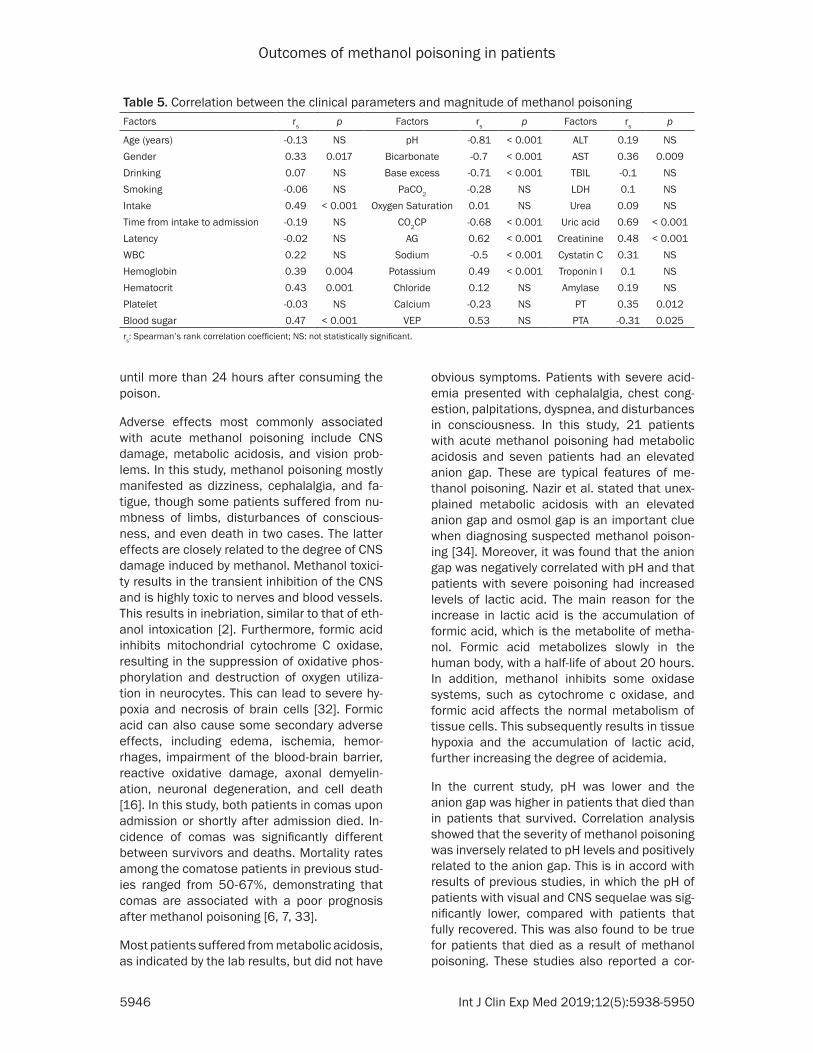
Correlation between characteristics and test results of patients with the magnitude of me- thanol poisoning was analyzed (Table 5). The amount of methanol ingested was positively correlated with severity of intoxication (rs =
0.49, P < 0.001). In addition, there was a small correlation between gender and severity of poisoning (rs = 0.33, P = 0.017). However, the degree of intoxication was not related to age, alcohol consumption, cigarette smoking, time between intake and hospital admission, or la- tency. Severity of poisoning was positively cor-related with levels of hemoglobin, hematocrit, AST, creatinine, uric acid, potassium, AG, CK- MB, myoglobin, random blood sugar, and PT, and negatively correlated with pH, bicarbonate, base excess, CO2CP, sodium, and PTA (Table 5).
Table 3. Demographic and hematologic findings in the survivors and deceased patients Evaluated factors Survivors (n = 50) Deceased (n = 2) pAge (years) 47.74 ± 11.82 46.0 ± 25.45 NSGender (male/female; n) 43/7 2/0 NSIntake (mL) 88.50 ± 82.15 150.0 ± 70.71 NSTime from intake to admission (d) 1.73 ± 0.72 1.17 ± 0.23 NSDaily drinking (n) 11 0 NSSymptoms (n) Dyspnea 3 2 0.008 Coma 0 2 0.001Latency (median; h) 12-24 12-24 NSWBC (× 109/L) 8.10 ± 2.70 18.02 ± 3.20 0.02Hematocrit (%) 43.27 ± 4.13 52.35 ± 12.23 NSALT (U/L) 27.49 ± 23.24 91.3 ± 57.28 0.029AST (U/L) 26.54 ± 10.09 192.35 ± 217.12 0.029Blood sugar (mmol/L) 6.33 ± 1.64 15.68 ± 7.05 0.023Uric acid (µmol/L) 395.71 ± 132.39 583.3 ± 112.57 NSCreatinine (µmol/L) 67.14 ± 15.19 150.45 ± 21.57 0.017Cystatin C (mg/L) 0.83 ± 0.11 1.21 ± 0.16 0.021Sodium (mmol/L) 142.12 ± 2.82 143.05 ± 8.41 NSPotassium (mmol/L) 4.31 ± 0.54 6.01 ± 0.14 0.017Chloride (mmol/L) 110.24 ± 3.12 103.05 ± 2.90 0.002Calcium (mmol/L) 2.23 ± 0.1 1.60 ± 0.74 0.032CO2CP (mmol/L) 22.22 ± 6.75 6.30 ± 1.98 0.02AG (mmol/L) 9.66 ± 4.93 34.35 ± 10.25 0.017pH 7.34 ± 0.11 6.72 ± 0.04 0.018PaO2 (mmHg) 95.17 ± 23.52 173.6 ± 34.5 < 0.001PaCO2 (mmHg) 39.86 ± 10.31 48.85 ± 42.07 NSBicarbonate (mmol/L) 21.64 ± 6.88 4.45 ± 2.62 0.02Base excess (mmol/L) -3.85 ± 7.44 -31.84 ± 1.32 0.018Troponin I (ng/ml) 0.001 ± 0.002 0.11 ± 0.15 0.001Myoglobin (ng/ml) 41.79 ± 16.92 219.02 ± 64.66 0.017LDH (U/L) 1777.72 ± 29.0 388.85 ± 200.61 0.022Amylase (U/L) 74.94 ± 23.98 318.25 ± 298.75 0.033PT (s) 12.36 ± 1.55 17.2 ± 1.70 < 0.001PTA (%) 100.29 ± 10.39 62.0 ± 11.31 < 0.001
Outcomes of methanol poisoning in patients
5945 Int J Clin Exp Med 2019;12(5):5938-5950
of individuals mistaking the fuel for ethanol. The metha-nol concentration in this fu- el was very high, at approxi-mately 659 g/L. It has been reported that the minimum lethal dose of methanol in adults is 0.3-1 g/kg of body weight. Even smaller doses of methanol can harm ad- ults. Studies have shown that 10 mL can cause blind-ness [24] and 15 mL can result in death [2]. In this study, the two deceased patients had ingested 100
However, there was no correlation between WBC count, platelet, PaCO2, blood oxygen satu-ration, chloride, calcium, ALT, TBIL, LDH, urea, cystatin C, and Troponin I levels. In addition, there was no correlation between biomarkers of pancreatic injury and severity of methanol poisoning. Interestingly, pH was positively cor-related with CO2CP (rs = 0.659, P < 0.001), bicarbonate (rs = 0.623, P < 0.001), and excess base (rs = 0.748, P < 0.001), as well as ne- gatively correlated with anion gap (rs = -0.605, P < 0.001) and potassium (rs = -0.59, P < 0.001). In addition, pH was weakly correlated with the amount of intake (rs = -0.31, P = 0.049). It was not correlated with PaCO2. Furthermore, optic nerve damage was not correlated with amount of intake or the pH.
Outcomes
After treatment by a multidisciplinary team, 50 of the 52 patients in the study recovered and were discharged without sequelae. The two patients that did not recover were from the severe toxicity group. However, one of the pa- tients with severe poisoning suffered from de- creased visual acuity after discharge. Two pa- tients with severe poisoning died in the hospi-tal. The mortality rate in this study was 3.8%.
Discussion
Alcohol-based fuel is a new type of liquid fuel that is cleaner and more environment-friendly. These fuels are primarily composed of various alcohols, such as methanol, ethanol, and buta-nol. The outbreak of methanol poisoning exam-ined in the current study was caused by a group
and 200 mL of the alcohol-based fuel, equiva-lent to 65.9 g and 131.8 g of methanol, res- pectively. None of the patients in the current study suffered from acute blindness. Variability between patients in the lethal minimal dose and dosage that induces blinding may be at- tributed to individual variations in their suscep-tibility to methanol. In addition, the simultane-ous ingestion of other substances, such as ethanol, could impact the way patients res- pond to varying dosages of methanol.
Methanol poisoning is extremely harmful to both patients and society. Compared with the 1.1% to 48% mortality rate reported in previ- ous studies [5, 6, 25-28], the mortality rate in this outbreak of methanol poisoning was only 3.8%. In addition, only one patient had decre- ased visual acuity and no patients had CNS sequelae. These results are lower, in terms of the number of patients with visual acuity and CNS problems, than those reported in previous studies. This may be due to racial differences, the concomitant ingestion of ethanol, emergen-cy measures actively taken by the govern- ment, and timely hospitalization and treatment of patients after outbreaks. As pointed out by an international working group, it may not be feasible to accurately compare the rates or severity of disease among countries, as there is a lack of consistency in the type of data col-lected from cases of methanol poisoning [29]. As found in previous studies, the average incu-bation period after oral ingestion of methanol is 12-24 hours, which may be prolonged if etha-nol was ingested simultaneously [30, 31]. For this reason, most patients are not diagnosed
Table 4. Ophthalmologic examination results of patients with acute methanol poisoning
Examination Observation (n = 30)
Mild (n = 17)
Severe (n = 5)
Total (n = 52) p
Mydriasis (D ≥ 4 mm) 0/30 0/17 3/5 3/52 -Sluggish or Nonreactive pupil 0/30 0/17 2/5 2/52 -Lens opacity 13/15 6/10 2/2 21/27 NSVitreous opacity 4/15 4/10 0/0 8/27 NSHyperemia of optic disc 0/17 1/9 0/3 1/29 -Decreased visual acuity 0/15 0/12 1/3 1/30 -Visual evoked potential (VEP) 0/5 4/7 2/2 6/14 -The former data out of parentheses indicated as the cases of abnormality and the latter is representative the numbers of patients performed the examination. NS: not statistically significant.
Outcomes of methanol poisoning in patients
5946 Int J Clin Exp Med 2019;12(5):5938-5950
until more than 24 hours after consuming the poison.
Adverse effects most commonly associated with acute methanol poisoning include CNS damage, metabolic acidosis, and vision prob-lems. In this study, methanol poisoning mostly manifested as dizziness, cephalalgia, and fa- tigue, though some patients suffered from nu- mbness of limbs, disturbances of conscious-ness, and even death in two cases. The latter effects are closely related to the degree of CNS damage induced by methanol. Methanol toxici-ty results in the transient inhibition of the CNS and is highly toxic to nerves and blood vessels. This results in inebriation, similar to that of eth-anol intoxication [2]. Furthermore, formic acid inhibits mitochondrial cytochrome C oxidase, resulting in the suppression of oxidative phos-phorylation and destruction of oxygen utiliza-tion in neurocytes. This can lead to severe hy- poxia and necrosis of brain cells [32]. Formic acid can also cause some secondary adverse effects, including edema, ischemia, hemor-rhages, impairment of the blood-brain barrier, reactive oxidative damage, axonal demyelin-ation, neuronal degeneration, and cell death [16]. In this study, both patients in comas upon admission or shortly after admission died. In- cidence of comas was significantly different between survivors and deaths. Mortality rates among the comatose patients in previous stud-ies ranged from 50-67%, demonstrating that comas are associated with a poor prognosis after methanol poisoning [6, 7, 33].
Most patients suffered from metabolic acidosis, as indicated by the lab results, but did not have
obvious symptoms. Patients with severe acid- emia presented with cephalalgia, chest cong- estion, palpitations, dyspnea, and disturbances in consciousness. In this study, 21 patients with acute methanol poisoning had metabolic acidosis and seven patients had an elevated anion gap. These are typical features of me- thanol poisoning. Nazir et al. stated that unex-plained metabolic acidosis with an elevated anion gap and osmol gap is an important clue when diagnosing suspected methanol poison-ing [34]. Moreover, it was found that the anion gap was negatively correlated with pH and that patients with severe poisoning had increased levels of lactic acid. The main reason for the increase in lactic acid is the accumulation of formic acid, which is the metabolite of metha-nol. Formic acid metabolizes slowly in the human body, with a half-life of about 20 hours. In addition, methanol inhibits some oxidase systems, such as cytochrome c oxidase, and formic acid affects the normal metabolism of tissue cells. This subsequently results in tissue hypoxia and the accumulation of lactic acid, further increasing the degree of acidemia.
In the current study, pH was lower and the anion gap was higher in patients that died than in patients that survived. Correlation analysis showed that the severity of methanol poisoning was inversely related to pH levels and positively related to the anion gap. This is in accord with results of previous studies, in which the pH of patients with visual and CNS sequelae was sig-nificantly lower, compared with patients that fully recovered. This was also found to be true for patients that died as a result of methanol poisoning. These studies also reported a cor-
Table 5. Correlation between the clinical parameters and magnitude of methanol poisoning Factors rs p Factors rs p Factors rs p
Age (years) -0.13 NS pH -0.81 < 0.001 ALT 0.19 NSGender 0.33 0.017 Bicarbonate -0.7 < 0.001 AST 0.36 0.009Drinking 0.07 NS Base excess -0.71 < 0.001 TBIL -0.1 NSSmoking -0.06 NS PaCO2 -0.28 NS LDH 0.1 NSIntake 0.49 < 0.001 Oxygen Saturation 0.01 NS Urea 0.09 NSTime from intake to admission -0.19 NS CO2CP -0.68 < 0.001 Uric acid 0.69 < 0.001Latency -0.02 NS AG 0.62 < 0.001 Creatinine 0.48 < 0.001WBC 0.22 NS Sodium -0.5 < 0.001 Cystatin C 0.31 NSHemoglobin 0.39 0.004 Potassium 0.49 < 0.001 Troponin I 0.1 NSHematocrit 0.43 0.001 Chloride 0.12 NS Amylase 0.19 NSPlatelet -0.03 NS Calcium -0.23 NS PT 0.35 0.012Blood sugar 0.47 < 0.001 VEP 0.53 NS PTA -0.31 0.025rs: Spearman’s rank correlation coefficient; NS: not statistically significant.
Outcomes of methanol poisoning in patients
5947 Int J Clin Exp Med 2019;12(5):5938-5950
relation between anion gap, levels of formic acid, and levels of lactic acid, finding that severe metabolic acidosis, with an increased anion gap, was an indicator of poor prognoses after methanol poisoning. Also, they suggest- ed that pH may serve as an independent pre-dictor of death in patients with methanol poi-soning [8, 35]. Hovda et al. discovered that PaCO2 decreased when pH decreased among the survivors, but the opposite was true for deceased patients. This suggests that higher PaCO2 may reflect insufficient respiratory com-pensation and CNS respiration depression [6]. However, there was no difference in PaCO2 lev-els between the patients that survived and patients that died in the current study. In con-trast, PaO2 was higher in patients that died than in those that survived. One explanation for this may be the oxygen therapy administered via mechanical ventilation or nasal catheter oxygen inhalation to patients after admission.
Electrolyte metabolic disorders, particularly th- ose affecting potassium, occur in most patients with methanol poisoning. In this study, some patients suffered from hyperkalemia. Hyper- kalemia may be caused by a compensatory mechanism used to restore the acid-base bal-ance in cases like metabolic acidosis and hy- pokalemia. This may be attributed to the dec- reased intake and increased loss of nutrients caused by gastrointestinal problems caused by methanol poisoning, such as nausea and vom-iting. Levels of blood calcium among patients with severe toxicity and deceased patients were lower than in individuals in the observa-tion and mild toxicity groups, suggesting that calcium levels are significantly correlated with severity of poisoning. Although detailed mecha-nisms related to hypocalcemia remain to be elucidated, it is often seen in critically ill pa- tients. It was previously shown that hypocalce-mia (0.81-0.90 mmol/L) is positively associ- ated with increased mortality in critically ill patients and that it is independently associat- ed with persistent organ failure in patients with acute pancreatitis [36, 37].
Ophthalmologic symptoms observed in this st- udy included blurred vision, decreased visual acuity, photophobia, tearing, dilated pupils, no- nreactive pupils, hyperemia of the optic disc, and lens or vitreous opacity. Moreover, VEP was abnormal in 6 of the 14 (43%) patients. Formic acid or formate can inhibit oxidative phosph-
orylation of the retina and optic nerves, result- ing in optic atrophy and toxic optic neuropathy. This involves both eyes, as was observed in this study. The optic nerve, retina, and basal ganglia are tissues most at risk from methanol poison-ing [24]. Although the specific reasons for this are not yet fully understood, some research has suggested that damage to these tissues may be associated with their vulnerability to histo-toxic hypoxia, possibly due to their relatively fast metabolic rates and high energy depen-dence. These are inhibited by formic acid [24, 38]. Eells et al. demonstrated that the accumu-lation of formic acid was much higher in the eyes than in the brain. This was attributed to the slower oxidation of formic acid in the eyes [39]. Furthermore, a series of clinical studies showed that patients with visual sequelae after methanol poisoning had a much lower pH than patients that had fully recovered. Results sug-gest a strong connection between the probabil-ity of long-term visual sequelae and the degree of acidemia, though this was not verified in the current study [6-8, 38-40]. However, early inter-vention is critical in correcting metabolic acido-sis, eliminating toxicants via blood purification, nourishing and supporting the nerves, and pro-moting the circulation of blood for repair of nerve injuries.
High concentrations of methanol can directly stimulate mucosal membranes of the digestive and upper respiratory tracts, leading to gastro-intestinal and respiratory problems. Gastroi- ntestinal symptoms, such as nausea, vomiting, abdominal pain, and diarrhea, were the most common in acute methanol poisoning, occur-ring in 65% of patients in this study. This is in agreement with previous studies that have sug-gested that these symptoms occur in 18-67% of cases [6, 7]. Cascallana et al. reported a case of a 67-year-old woman that committed suicide by ingesting 500 mL of absolute metha-nol [13]. The autopsy revealed complete de- tachment of the esophagus mucosa, brownish discoloration of the gastric mucosa, and app- roximately 200 mL of hemorrhagic liquid in the stomach. Histological findings showed diffuse hemorrhagic necrosis and intense acute in- flammatory infiltration of the lamina propria [13].
In this study, some patients may have devel-oped hemorrhages in gastrointestinal or uri-nary tracts, as evidenced by positive tests for
Outcomes of methanol poisoning in patients
5948 Int J Clin Exp Med 2019;12(5):5938-5950
fecal and urine occult blood. Mostafazadeh et al. reported upper gastrointestinal bleeding and hematuria, after dialysis treatment, in one patient with methanol poisoning, suggesting that it could have been related to the use of anticoagulants for dialysis treatment [41]. High doses of heparin or low molecular weight he- parin can facilitate bleeding in necrotic areas of the brain [42]. However, some studies have reported no remarkable connection between hemorrhagic brain lesions and the use of sys-temic anticoagulation during hemodialysis [22, 43, 44]. Thus, more studies are necessary to elucidate the mechanisms of methanol-mediat-ed hemorrhages and the effects of anticoagu- lation on bleeding during dialysis. It should also be noted that pancreatic injury markers were significantly increased in the two patients that died during the current study, suggesting that methanol may have induced concurrent pan-creatitis. A previous study reported that acute pancreatitis is a complication of methanol poi-soning [42]. In addition, Hantson and Mahieu [45] reported that a 54-year-old woman devel-oped acute necrotizing pancreatitis following acute methanol poisoning. They also retrospec-tively examined 11 cases of acute pancreatitis in 22 patients with methanol poisoning. They discovered an association between acute pan-creatic injury and the magnitude of metabolic acidosis. However, it was not associated with alcohol abuse, suggesting that methanol likely causes pancreas injury.
As with pH and anion gap, blood parameters, such as creatinine and blood sugar, were also correlated with severity of symptoms and death in poisoned patients. Many studies have report-ed that hyperglycemia is a strong prognostic factor for death from methanol poisoning [8, 42, 46]. Similarly, blood sugar was higher in patients that died from methanol poisoning than survivors in the current study. Previously, Morteza et al. showed that an increased creati-nine level was an independent risk factor for alcohol-related death [25]. In the current study, multivariate logistic regression did not show any independent risk factors of death among poisoned patients, possibly due to the small numbers of patients. Hence, the severity and prognosis of methanol poisoning should be evaluated comprehensively using a combina-tion of symptoms, examinations, and blood tests. More clinical studies with larger sample sizes are necessary to assess prognostic, risk factors, and lethality of methanol poisoning.
Strengths and limitations
There were several limitations to the current study. First, this was a retrospective study th- at was not randomized. Hence, certain con-founders were inevitable (recall bias could not be completely removed). Second, the relatively small sample size of the groups provided insuf-ficient data confirming the relationship between the severity of methanol poisoning and certain laboratory and clinic parameters. In addition, the hospital was unable to collect all data for each patient upon admission. For instance, the two deceased patients did not receive oph- thalmologic examinations. Furthermore, con-centrations of other components in the alcohol-based fuels were not detected and blood levels of methanol, formic acid, and ethanol were not measured, due to a lack of available laboratory equipment. For this reason, poisoning from methanol could not be accurately distinguished from poisoning by other chemicals, such as ethanol or butanol. Despite these limitations, this study does summarize the clinic features of methanol poisoning. Consistent with previous studies, this study demonstrated associations between coma, dyspnea, pH, anion gap, blood sugar, and increased creatinine on admission with poor outcomes after methanol poisoning. In addition, the current study found that levels of calcium, potassium, amylase, and troponin I, as well as abnormal coagulation function, were related to methanol-induced death.
Conclusion
Acute oral methanol poisoning can lead to CNS damage, metabolic acidosis, visual disturban- ces, and gastrointestinal injuries. Association between dyspnea, coma, decreased pH, incr- eased anion gap, and increased levels of potas-sium, creatinine, and blood sugar were corre-lated with poor patient outcomes after metha-nol poisoning. Most patients were discharged without sequelae after timely and effective tre- atment. Early and effective treatment of metha-nol poisoning is vitally important for patient survival.
Acknowledgements
This research received no specific grants from any funding agency in the public, commercial, or not-for-profit sectors.
Outcomes of methanol poisoning in patients
5949 Int J Clin Exp Med 2019;12(5):5938-5950
Disclosure of conflict of interest
None.
Address correspondence to: Santao Ou, Depart- ment of Nephrology, The Affiliated Hospital of Sou- thwest Medical University, The 25th Taiping Street, Luzhou 646000, Sichuan, China. Tel: +86-830-3165340; Fax: +86-830-2392752; E-mail: [email protected]
References
[1] Karaduman F, Asil T, Balci K, Temizoz O, Unlu E, Yilmaz A, Utku U. Bilateral basal ganglionic lesions due to transdermal methanol intoxica-tion. J Clin Neurosci 2009; 16: 1504-1506.
[2] Rauber-Luthy C, Kupferschmidt H. Household chemicals: management of intoxication and antidotes. EXS 2010; 100: 339-363.
[3] Pohanka M. Toxicology and the biological role of methanol and ethanol: current view. Biomed Pap Med Fac Univ Palacky Olomouc Czech Repub 2016; 160: 54-63.
[4] Abdulrahi FA SA. Substance abuse and home-lessness: mass methanol poisoning in Kha- rtoum.
[5] Hassanian-Moghaddam H, Nikfarjam A, Mir- afzal A, Saberinia A, Nasehi AA, Masoumi Asl H, Memaryan N. Methanol mass poisoning in Iran: role of case finding in outbreak manage-ment. J Public Health (Oxf) 2015; 37: 354-359.
[6] Hovda KE, Hunderi OH, Tafjord AB, Dunlop O, Rudberg N, Jacobsen D. Methanol outbreak in Norway 2002-2004: epidemiology, clinical fea-tures and prognostic signs. J Intern Med 2005; 258: 181-190.
[7] Paasma R, Hovda KE, Tikkerberi A, Jacobsen D. Methanol mass poisoning in Estonia: out-break in 154 patients. Clin Toxicol (Phila) 2007; 45: 152-157.
[8] Zakharov S, Pelclova D, Urban P, Navratil T, Diblik P, Kuthan P, Hubacek JA, Miovsky M, Klempir J, Vaneckova M, Seidl Z, Pilin A, Fe- nclova Z, Petrik V, Kotikova K, Nurieva O, Ri- dzon P, Rulisek J, Komarc M, Hovda KE. Czech mass methanol outbreak 2012: epidemiology, challenges and clinical features. Clin Toxicol (Phila) 2014; 52: 1013-1024.
[9] Rostrup M, Edwards JK, Abukalish M, Ezzabi M, Some D, Ritter H, Menge T, Abdelrahman A, Rootwelt R, Janssens B, Lind K, Paasma R, Hovda KE. The methanol poisoning outbreaks in Libya 2013 and Kenya 2014. PLoS One 2016; 11: e0152676.
[10] Nurieva O KK, Urban P. Prevalence, dynamics, and biochemical predictors of optic nerve re-myelination after methanol-induced acute op-tic neuropathy: a 2-year prospective study in
54 patient. Monatshefte für Chemie - Chemical Monthly 2016; 147: 239-249.
[11] Collister D, Duff G, Palatnick W, Komenda P, Tangri N, Hingwala J. A methanol intoxication outbreak from recreational ingestion of frack-ing fluid. Am J Kidney Dis 2017; 69: 696-700.
[12] Choi JH, Lee SK, Gil YE, Ryu J, Jung-Choi K, Kim H, Choi JY, Park SA, Lee HW, Yun JY. Neurological complications resulting from non-oral occupa-tional methanol poisoning. J Korean Med Sci 2017; 32: 371-376.
[13] Cascallana JL, Gordo V, Montes R. Severe ne-crosis of oesophageal and gastric mucosa in fatal methanol poisoning. Forensic Sci Int 2012; 220: e9-12.
[14] Hsiao PJ, Chen TY, Chiu CC, Wu TJ, Chan JS, Wu CC, Chen JS. Delayed high anion gap metabolic acidosis after a suicide attempt: case report. Clin Chim Acta 2014; 436: 329-331.
[15] Dogan H, Yilmaz Karakus B, Serefoglu Cabuk K, Uzun O, Yenice H, Orucoglu A. Transdermal spirit (methanol) poisoning: a case report. Iran Red Crescent Med J 2016; 18: e23767.
[16] Zakharov S, Kotikova K, Nurieva O, Hlusicka J, Kacer P, Urban P, Vaneckova M, Seidl Z, Diblik P, Kuthan P, Navratil T, Pelclova D. Leukotriene-mediated neuroinflammation, toxic brain dam-age, and neurodegeneration in acute metha-nol poisoning. Clin Toxicol (Phila) 2017; 55: 249-259.
[17] Kraut JA. Approach to the treatment of metha-nol intoxication. Am J Kidney Dis 2016; 68: 161-167.
[18] Sanaei-Zadeh H, Zamani N, Shadnia S. Out- comes of visual disturbances after methanol poisoning. Clin Toxicol (Phila) 2011; 49: 102-107.
[19] Vaneckova M, Zakharov S, Klempir J, Ruzicka E, Bezdicek O, Brozova H, Diblik P, Miovsky M, Hubacek JA, Urban P, Ridzon P, Pelclova D, Burgetova A, Masek M, Kotikova K, Peterova K, Liskova I, Hamplova L, Seidl Z. Imaging find-ings after methanol intoxication (cohort of 46 patients). Neuro Endocrinol Lett 2015; 36: 737-744.
[20] Kute VB, Godara SM, Shah PR, Gumber MR, Goplani KR, Vanikar AV, Munjappa BC, Patel HV, Trivedi HL. Hemodialysis for methyl alcohol poisoning: a single-center experience. Saudi J Kidney Dis Transpl 2012; 23: 37-43.
[21] Cheng JX ZS, Jiang ZQ. The automated thresh-old perimertry and P-VEP in diagnostic classifi-cation of acute methanol posisoning. Chin J Pract Ophthalmol 2006; 24: 585-588.
[22] Hassanian-Moghaddam H, Bahrami-Motlagh H, Zamani N, Fazeli SA, Behnam B. Intracranial hemorrhage in methanol toxicity: challenging the probable heparin effect during hemodialy-sis. J Res Pharm Pract 2017; 6: 186-189.
Outcomes of methanol poisoning in patients
5950 Int J Clin Exp Med 2019;12(5):5938-5950
[23] Liu Z, Sun M, Zhao H, Zhao M. Acute self-in-duced poisoning with sodium ferrocyanide and methanol treated with plasmapheresis and continuous renal replacement therapy suc-cessfully: a case report. Medicine (Baltimore) 2015; 94: e890.
[24] Galvez-Ruiz A, Elkhamary SM, Asghar N, Bosley TM. Visual and neurologic sequelae of metha-nol poisoning in Saudi Arabia. Saudi Med J 2015; 36: 568-574.
[25] Morteza Bagi HR, Tagizadieh M, Moharamzadeh P, Pouraghaei M, Kahvareh Barhagi A, Shah- savari Nia K. Epidemiology of alcohol poison-ing and its outcome in the North-West of Iran. Emerg (Tehran) 2015; 3: 27-32.
[26] Rulisek J, Balik M, Polak F, Waldauf P, Pelclova D, Belohlavek J, Zakharov S. Cost-effectiveness of hospital treatment and outcomes of acute methanol poisoning during the Czech Republic mass poisoning outbreak. J Crit Care 2017; 39: 190-198.
[27] P A. Prognostic factors including clinical mani-festation and paraclinic finding in sever metha-nol toxicity. J Alcoh Drug Depend 2013.
[28] Ghannoum M, Hoffman RS, Mowry JB, Laver- gne V. Trends in toxic alcohol exposures in the United States from 2000 to 2013: a focus on the use of antidotes and extracorporeal treat-ments. Semin Dial 2014; 27: 395-401.
[29] Zyoud SH, Al-Jabi SW, Sweileh WM, Awang R, Waring WS. Bibliometric profile of the global scientific research on methanol poisoning (1902-2012). J Occup Med Toxicol 2015; 10: 17.
[30] Beauchamp GA, Valento M. Toxic alcohol in-gestion: prompt recognition and management in the emergency department. Emerg Med Pract 2016; 18: 1-20.
[31] Vakil A, Upadhyay H, Sherani K, Cervellione K, Trepeta S, Patel MC. A 44-year-old woman with metabolic acidosis, high anion gap, and de-layed neurologic deterioration. Chest 2015; 147: e18-e21.
[32] Hubacek JA, Pelclova D, Seidl Z, Vaneckova M, Klempir J, Ruzicka E, Ridzon P, Urban P, Fen- clova Z, Petrik V, Diblik P, Kuthan P, Miovsky M, Janikova B, Adamkova V, Zakharov S. Rare al-leles within the CYP2E1 (MEOS system) could be associated with better short-term health outcome after acute methanol poisoning. Ba- sic Clin Pharmacol Toxicol 2015; 116: 168-172.
[33] Paasma R, Hovda KE, Hassanian-Moghaddam H, Brahmi N, Afshari R, Sandvik L, Jacobsen D. Risk factors related to poor outcome after methanol poisoning and the relation between outcome and antidotes--a multicenter study. Clin Toxicol (Phila) 2012; 50: 823-831.
[34] Nazir S, Melnick S, Ansari S, Kanneh HT. Mind the gap: a case of severe methanol intoxica-tion. BMJ Case Rep 2016; 2016.
[35] Zakharov S NT, Pelclova D. Analysis of serum anion gap and osmolal gap in diagnosis and prognosis of acute methanol poisoning: clini-cal study in 86 patients. Chem Monthly 2015; 146: 787-794.
[36] Dey S, Karim HMR, Yunus M, Barman A, Bha- ttacharyya P, Borthakur MP. Relationship of on admission hypocalcaemia and illness severity as measured by APACHE-II and SOFA score in intensive care patients’. J Clin Diagn Res 2017; 11: UC01-UC03.
[37] Peng T, Peng X, Huang M, Cui J, Zhang Y, Wu H, Wang C. Serum calcium as an indicator of per-sistent organ failure in acute pancreatitis. Am J Emerg Med 2017; 35: 978-982.
[38] Zakharov S, Pelclova D, Diblik P, Urban P, Kuthan P, Nurieva O, Kotikova K, Navratil T, Komarc M, Belacek J, Seidl Z, Vaneckova M, Hubacek JA, Bezdicek O, Klempir J, Yurchenko M, Ruzicka E, Miovsky M, Janikova B, Hovda KE. Long-term visual damage after acute me- thanol poisonings: longitudinal cross-sectional study in 50 patients. Clin Toxicol (Phila) 2015; 53: 884-892.
[39] Eells JT, Salzman MM, Lewandowski MF, Murray TG. Formate-induced alterations in reti-nal function in methanol-intoxicated rats. Toxi- col Appl Pharmacol 1996; 140: 58-69.
[40] Desai T, Sudhalkar A, Vyas U, Khamar B. Me- thanol poisoning: predictors of visual outco- mes. JAMA Ophthalmol 2013; 131: 358-364.
[41] Mostafazadeh B, Talaie H, Mahdavinejad A, Mesri M, Emanhadi M. Gastrointestinal and urinary tract bleeding in methanol toxicity. BMJ Case Rep 2008; 2008: bcr0820080619.
[42] Zakharov S, Kotikova K, Vaneckova M, Seidl Z, Nurieva O, Navratil T, Caganova B, Pelclova D. Acute methanol poisoning: prevalence and predisposing factors of haemorrhagic and non-haemorrhagic brain lesions. Basic Clin Phar- macol Toxicol 2016; 119: 228-238.
[43] Aisa TM, Ballut OM. Methanol intoxication with cerebral hemorrhage. Neurosciences (Riyadh) 2016; 21: 275-277.
[44] Zakharov S, Rulisek J, Nurieva O, Kotikova K, Navratil T, Komarc M, Pelclova D, Hovda KE. Intermittent versus continuous renal replace-ment therapy in acute methanol poisoning: comparison of clinical effectiveness in mass poisoning outbreaks. Ann Intensive Care 2017; 7: 77.
[45] Hantson P, Mahieu P. Pancreatic injury follow-ing acute methanol poisoning. J Toxicol Clin Toxicol 2000; 38: 297-303.
[46] Sanaei-Zadeh H, Esfeh SK, Zamani N, Jamshidi F, Shadnia S. Hyperglycemia is a strong prog-nostic factor of lethality in methanol poisoning. J Med Toxicol 2011; 7: 189-194.