Embed Size (px)
Citation preview
Int J Clin Exp Med 2015;8(11):20988-21000www.ijcem.com /ISSN:1940-5901/IJCEM0013509
Original ArticleImmunohistochemistry and microsatellite instability analysis in molecular subtyping of colorectal carcinoma based on mismatch repair competency
Lin Yuan1,2,3, Yayun Chi2,4, Weixiang Chen1,2, Xiaochen Chen1,2, Ping Wei1,2, Weiqi Sheng1,2, Xiaoyan Zhou1,2, Daren Shi1,2
1Department of Pathology, Fudan University Shanghai Cancer Center, Shanghai 200032, China; 2Department of Oncology, Shanghai Medical College, Fudan University, Shanghai 200032, China; 3Pathology Center, Shanghai General Hospital, School of Medicine, Shanghai Jiao Tong University, Shanghai 200080, China; 4Breast Cancer Institute, Fudan University Shanghai Cancer Center, Shanghai 200032, China
Received July 27, 2015; Accepted October 28, 2015; Epub November 15, 2015; Published November 30, 2015
Abstract: Mismatch repair defective (MMRd) colorectal carcinoma (CRC) is a distinct molecular phenotype of colorectal cancer, including 12% of sporadic CRC and 3% of Lynch Syndrome. In order to investigate the clinico-pathological characteristics of MMRd colorectal carcinoma, and to find the most effective method for preliminary screening, 296 CRC fulfilled revised Bethesda Guideline (RB) were selected from 1450 CRCs to perform both IHC staining for MLH1, MSH2, MSH6, PMS2 and MSI analysis. Sixty-eight tumors were classified as MSI-H by MSI test. Colorectal carcinomas with MSI-H were prone to be proximal located, poorly differentiated, and relatively early staged, with infrequent metastasis to lymph node as well as to distant organs, compared with MSS ones. All of the 68 MMRd CRCs presented abnormal expression of at least one mismatch repair protein (MMRP), with 48 concur-rent negative of MLH1 and PMS2, 14 concurrent negative of MSH2 and MSH6, 4 isolated negative of MSH6, 1 isolated negative of PMS2, and 1 concurrent negative of 4 MMRPs. All of the MLH1 negative tumors also showed abnormal expression of PMS2. All of the MSH2 negative cases also presented negative expression of MSH6. The sensitivity and specificity of the 2-antibody IHC test contained only PMS2 and MSH6 for screening for MMRd CRC were 100% and 98.2% respectively, exactly the same as that of the 4-antibody IHC test with all of the 4 MMRPs. The diagnostic accordance rate of the 2-antibody approach and MSI analysis was 98.6%. In conclusion, MMRd CRC has characteristic clinicopathological features different from MSS CRCs. The 2-antibody IHC approach containing MSH6 and PMS2 is the most easy and effective way to detecting MMR deficiency in CRC.
Keywords: Mismatch repair, microsatellite instability, Lynch syndrome, immunohistochemistry, molecular pheno-type, CRC
Introduction
Colorectal carcinoma (CRC) is the third most common cancer and the fourth leading cause of cancer death in the world, according to the GLOBOCAN 2008 estimates [1]. Cases and deaths occurred in China are increasing rapidly in recent years on account of the so called “westernization” [2, 3]. The pathogenesis of colorectal cancer is still unclear, but it could be generally defined in two molecular pathways of genomic instability. One is chromosome insta-bility, which is involved in certain oncogenes and cancer suppressor genes, such as APC, KRAS, and P53 [4]. Another way is microsatel-
lite instability (MSI), which is due to mismatch repair system defect, and account for approxi-mately 15% of all colorectal carcinomas. Most of the MSI colorectal carcinomas (12%) are spo-radic MSI, which is attributed to acquired hyper-methylation of MLH1 gene promoter, accompa-nied by the CpG island methylation phenotype. And 3% of the MSI CRCs are Lynch syndrome related colorectal carcinomas, once defined as HNPCC (hereditary non-polyposis colorectal cancer), a classic model of hereditary colorec-tal cancer [5]. The mismatch repair deficiency of Lynch syndrome is caused mostly by germ-line mutation of certain DNA mismatch repair gene. Thus, colorectal cancers could be classi-
IHC and MSI analysis in CRC subtyping
20989 Int J Clin Exp Med 2015;8(11):20988-21000
fied under two molecular phenotypes, micro-satellite instable and microsatellite stable (MSS) phenotype. It is more and more im- portant to reveal the molecular phenotype of colorectal cancer, as different phenotype means different pathogenesis, prognosis, even treatment response. Mismatch repair-deficient (MMRd) colorectal cancer has distinct clini- copathological characteristics, including early onset, proximal localization, Crohn’s-like lym-phocytic reaction, mucinous/signet-ring differ-entiation, and medullary growth pattern [6-9]. Furthermore, the clinical prognosis and treat-ment response of patient are closely related to the molecular mechanism underlying cancer development. Patients with MSI-H colorectal carcinoma have different outcome and chemo-therapy response from those with microsatel-lite stable (MSS) colorectal cancer [12-14]. So, it has great significance to identify MSI in colorectal carcinoma, not only because MSI is a critical DNA marker for screening for Lynch syndrome, but also because it can help to dif-ferentiate MMRd colorectal cancers from MSS ones, which will provide valuable information for prognosis estimation and treatment indivi- dualization.
Previously, immunohistochemistry (IHC) of mis-match repair protein (MMRP) and MSI test were widely used for screening for HNPCC. And the MSI analysis based on PCR is the gold standard approach for detecting microsatellite status. American National Cancer Institute (NCI) rec-ommended five DNA markers for MSI test. These markers are five microsatellite sequenc-es including BAT25, BAT26, D2S123, D5S346 and D17S250. It is defined as high frequency of microsatellite instability (MSI-H) when two or more of the five markers in the tumor DNA were positive. If only one marker was positive, the tumor is termed as low frequency of MSI (MSI-L). And MSS is determined when all of the five markers were negative [7]. Another sensitive and specific method for MSI detection is IHC for MMRPs, which often involves in a traditional panel including MLH1 and MSH2. Absent expression of any one of the MMRPs indicates defective mismatch repair system in the tumor detected. It is considered that adding PMS2 can elevate the sensitivity of screening for HNPCC [15]. In the past decade, a four-anti-body panel including MLH1, MSH2, MSH6, and PMS2 was becoming popular for screening for HNPCC and mismatch repair deficiency of
CRC. Recent data suggest that a two-antibody panel approach using only PMS2 and MSH6 is an effective screening protocol for HNPCC related colorectal carcinoma, and even extrain-testinal tumors [16-18]. In this study, the rela-tionship between clinicopathological features and mismatch repair status was further asse- ssed, and IHC test for mismatch repair gene proteins and MSI analysis were compared for subtyping of colorectal carcinoma by mismatch repair competency.
Materials and methods
Patients
A total of 1450 colorectal carcinomas were recruited from Fu Dan University Shanghai Cancer Center between January 2007 and March 2010. Clinical and family history of these patients was reviewed, and clinical-pathologi-cal features were examined based on the revised Bethesda Guideline [7]. Finally, 296 cases were selected for analysis, which ful- filled at least one of the following criteria: (1) Colorectal cancer diagnosed in a patient who is less than 50 years of age; (2) Presence of syn-chronous, metachronous colorectal, or other HNPCC-associated tumors [including colorec-tal, endometrial, stomach, ovarian, pancreas, ureter and renal pelvis, biliary tract, and brain (usually glioblastoma as seen in Turcot syn-drome) tumors, sebaceous gland adenomas and keratoacanthoma in Muir-Torre syndrome, and carcinoma of the small bowel], regardless of age; (3) Colorectal cancer with the MSI-H his-tology (presence of tumor infiltrating lympho-cytes, Crohn’s-like lymphocytic reaction, muci-nous/signet-ring differentiation, or medullary growth pattern) diagnosed in a patient who is less than 60 years of age; (4) Colorectal cancer diagnosed in one or more first-degree relatives with an HNPCC-related tumors, with one of the cancers being diagnosed under age 50 years; (5) Colorectal cancer diagnosed in two or more first- or second-degree relatives with HNPCC-related tumors, regardless of age [7]. The in- formed consents of all patients and approval of Medical Ethical Committee of Shanghai Cancer Center, Fudan University were obtained at the beginning of the study.
MMRP immunohistochemistry
Immunohistochemistry for the four most com-mon mismatch repair proteins were performed
IHC and MSI analysis in CRC subtyping
20990 Int J Clin Exp Med 2015;8(11):20988-21000
in all of the 296 cases using the standard Envision two-step procedure. Tumor represen-tative blocks were carefully selected for analy-sis with normal-tumor junction in order to assess staining result properly. Primary mono-clonal antibodies against MLH1 (clone ES05, Novocastra, Leica Biosystems Newcastle Ltd, Newcastle Upon Tyne, UK, 1:30), MSH2 (clone FE11, Calbiochem, Merck KGaA, Darmstadt, Germany, 1:50), MSH6 (clone EPR3945, Epito- mics Inc, Burlingame, California, USA, 1:200), and PMS2 (clone EPR3947, Epitomics Inc, Bur- lingame, California, USA, 1:200) were applied to 4-μm-thick 10% formalin-fixed, paraffin-embedded tissue sections. The sections were deparaffinized in xylene for three times, every time for 10 minutes, and subsequently rehy-drated through graded alcohols to distilled water. Antigen heat retrieval was performed in 1 mM EDTA (pH 9.0) for 10 minutes (PMS2 15 minutes) using a microwave oven, after that the sections were cooled down in room tempera-ture for 1.5 hours. After rinsing in distilled water and TBS successively, sections were incubated with specific monoclonal antigen at 4°C over-night. The Dako REAL™ EnVision™ Detection System (Dako, Shanghai, China) was used as the secondary detection system according to manufacturer’s instructions. The reaction is visualized by Dako REAL™ DAB + Chromogen, and slides were counterstained with hemato- xylin.
Non-neoplastic colonic mucosa, stromal cells, infiltrating lymphocytes or the centers of lym-phoid follicles were used as internal positive controls. And the known MMR deficient colorec-tal carcinomas served as external negative controls. Two experienced pathologists evalu-ated the staining results independently and blindly to the MSI status. Normal expression was defined as nuclear staining within tumor cells, while negative protein expression was defined as complete absence of nuclear stain-ing within tumor cells with concurrent internal positive controls. If internal non-neoplastic tis-sues showed invalid negative staining, proce-dure was routinely repeated.
Microsatellite instability analysis
Genomic DNA of matched tumor and non-neo-plastic tissue was extracted from the paraffin-embedded tissue sections by manual micro-
dissection on the basis of a HE-stained slide, using the QIAamp DNA Mini Kit (QIAGEN China (Shanghai) Co., Ltd., Shanghai, China). For mic-rosatellite instability analysis, the panel of 5 markers BAT26, BAT25, D5S346, D2S123, and D17S250, recommended by NCI [19], was em- ployed to test all of the 296 paired DNA sam-ples. This panel composed of 2 mononucleo-tide repeat sequences BAT26, BAT25, and 3 dinucleotide repeat markers, D5S346, D2S- 123, and D17S250. The primers used for ampli-fication were just as described previously [20]. The forward primers (Applied Biosystems, Life Technologies Corporation, California, USA) were marked with a fluorescent tag (FAM-BAT25/D2S123/D17S250, NED-BAT26, PET-D5S346) at the 5’ end. Fluorescence multiplex poly-merase chain reaction (PCR) was performed with a 20 μL reaction system consisted of 10 μL 2× Taq PCR premix (containing Taq Poly- merase, magnesium chloride and dNTPs) (San- gon Biotech, Shanghai, China), 2 μL of mixed primers, 1 μL genomic DNA (100 ng) and 7 μL DNASE-free H2O. After initial pre-denature at 95°C for 9 minutes, 35 cycles of: denaturation at 95°C for 30 seconds, annealing at 53°C for 30 seconds and extension at 72°C for 1 minute were performed. Final extension was at 60°C for 45 minutes. PCR reaction product (1 μL) was then mixed with 0.1 μL LIZ (internal size standard) (Applied Biosystems) and 9 μl of Hi-Di formamide (denaturant) (Applied Biosystems). Heat pre-denaturation was performed on the mixture at 95°C for 5 minutes, and the sample was kept at 4°C for 5 minutes. Then the prod-uct was put in a 96 well plate, and capillary electrophoresis was performed with an ABI 3500 Genetic Analyzer (Applied Biosystem Shanghai Division, Shanghai, China) for 45 minutes. Data was automatically collected and then analyzed with Genemapper v4.1 (Applied Biosystems). The presence of peaks in the fluo-rescence profile of the amplified microsatellite DNA that were absent in a corresponding pro-file derived from the matched normal muco- sa was interpreted as microsatellite instability. If there were two or more of the 5 markers showed instability, tumors were defined as high frequency microsatellite instability (MSI-H); instability at a single locus was defined as low frequency microsatellite instability (MSI-L); and if none of the markers showed instability, the tumor was considered to be microsatellite stable (MSS).
IHC and MSI analysis in CRC subtyping
20991 Int J Clin Exp Med 2015;8(11):20988-21000
Statistical analysis
The correlation between clinicopathological features and microsatellite status were ana-lyzed using χ2 test. Sensitivity was defined as the IHC negative expression of mismatch re- pair gene products (MLH1, MSH2, MSH6, and PMS2) in screening for MMRd, with MSI test as gold standard. And specificity was the normal IHC result of MMRPs in MSI-L/MSS colorectal cancer. P values were reported correspond to two-sided test. And the statistical significance
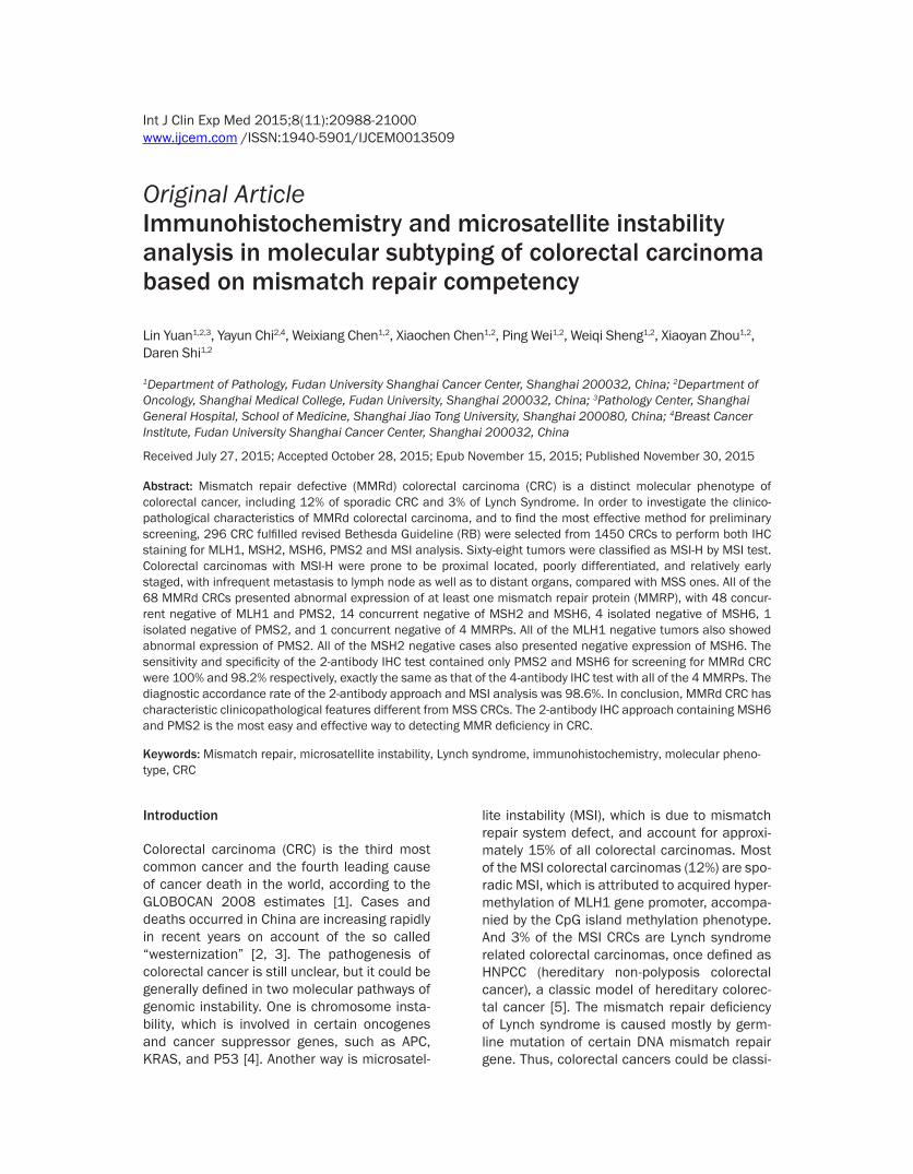
When it came to IHC test of MMRPs, there were 72 (24.3%, 72/296) tumors showed absence expression of at least one MMRP, with 68 MSI-H, 2 MSI-L, and 2 MSS. Of the 72 cases, PMS2 was negative in 52 (17.6%, 52/296) tumors, 50 of which were MSI-H, 2 were MSS. And MLH1 was negative in 51 (17.2%, 51/296) tumors, 49 of that were MSI-H, and 2 were MSS. Fifty-one tumors displayed absent co-expression of PMS2 and MLH1. All MLH1 negative tumors also showed absent expression of PMS2, with only one PMS2 negative tumor displaying intact
Table 1. Clinicopathological features and microsatellite status
Clinicopathological features MSI-H
MSI-L MSS P-
HvsL*P-
HvsSP-
LvsSSex Male 41 5 115 1.000 0.26 1.000 Female 27 4 104Age <50 41 4 104 0.585 0.065 1.000 ≥50 27 5 115Tumor site Right colon 35 1 64 0.037 0.002 0.228 Left colon 19 4 107 Rectum 14 4 48Tumor grade I/II 46 7 176 0.815 0.029 0.261 III 22 2 43Mucinous/signet ring differentiation Yes 23 4 61 0.798 0.345 0.482 No 45 5 158Lymphocytic infiltration† Yes 14 0 29 0.198 0.138 0.608 No 54 9 190TNM stage I/II 46 5 104 0.729 0.004 0.893 III 22 4 115Lymph nodes metastasis Yes 21 4 112 0.662 0.003 0.957 No 47 5 107Distant metastasis Yes 2 2 34 0.099 0.011 0.941 No 66 7 185N 68 9 219*P-HvsL, P-HvsS, P-LvsS refers to P value of Pearson’s test or Fischer’s exact test for the comparison of clinicopathological features of CRC with different microsatellite status, MSI-H group with MSI-L group, MSI-H group with MSS group, and MSI-L group with MSS group; right colon including cecum, ascending colon, and hepatic flexure; left colon including splenic flexure, decending colon, and sigmoid colon; Tumor grade I/II/III correspond to Well/moderately/poor differentiation.
was defined as the P value <0.05. All data were pro-cessed using SPSS 16.0 (SPSS, Chicago, IL, USA).
Results
Of the 296 cases match- ed RB criteria, 68 (23.0%, 68/296) were classified as MSI-H, 9 (3.0%, 9/296) were MSI-L, and 219 (74.0%, 219/296) were MSS by MSI analysis. The clinicopatho-logical characteristics of MSI colorectal carcinomas were different from MSS ones. Especially for tumor location, tumor grade, TNM stage, lymph nodes metas-tasis, and distant metasta-sis, the differences were statistically significant as showed in Table 1. Com- pared with MSS CRC, MSI-H CRC were more frequently located in right colon, poorly differentiated, at relatively early TNM stage, less lym- ph node metastasis as well as infrequent distant meta- stasis. Although the clini- copathological features of MSI-L tumors had no signifi-cant differences compared either to MSI-H CRC or to MSS CRC, except tumor site, the clinicopathological char-acteristics of MSI-L group was more close to that of MSS group.
IHC and MSI analysis in CRC subtyping
20992 Int J Clin Exp Med 2015;8(11):20988-21000
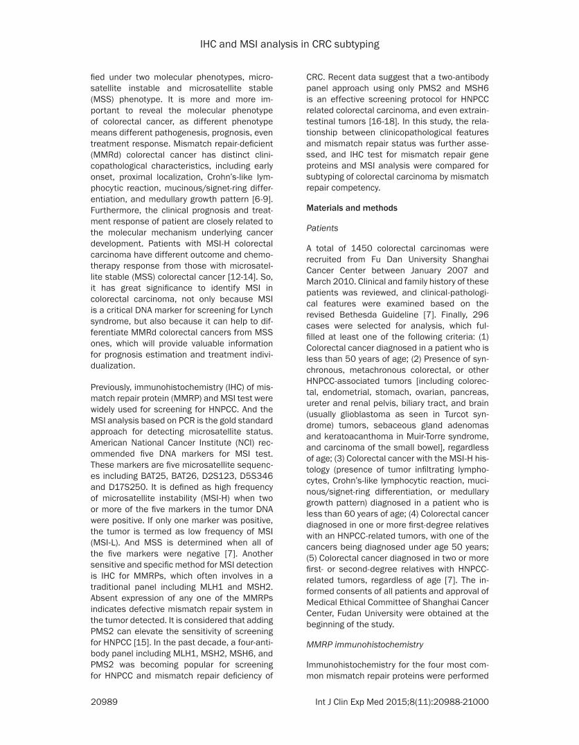
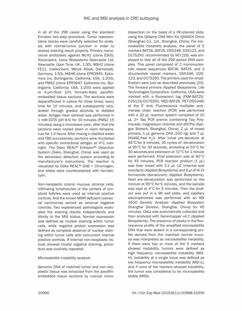
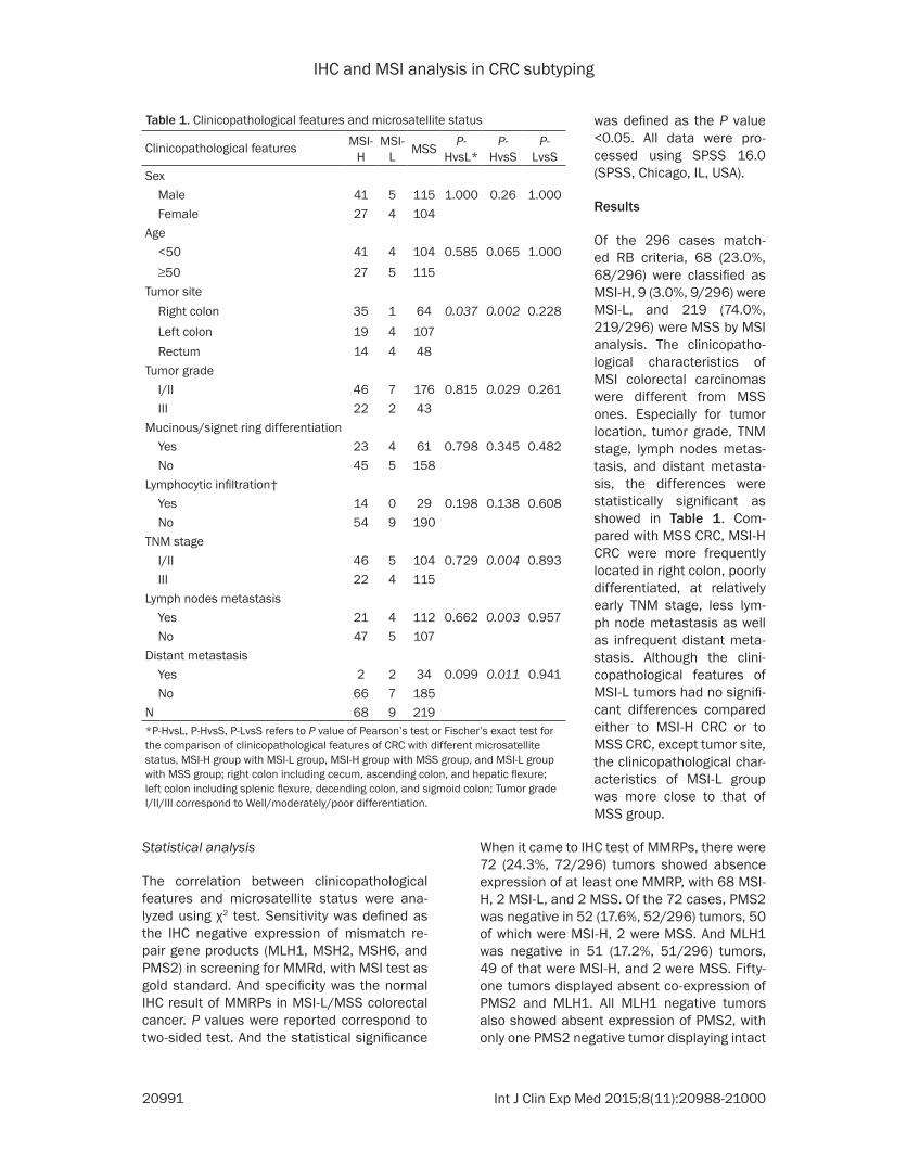
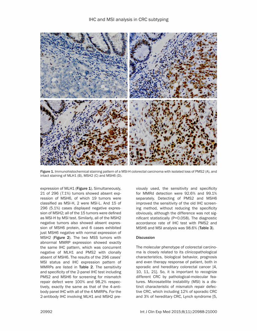
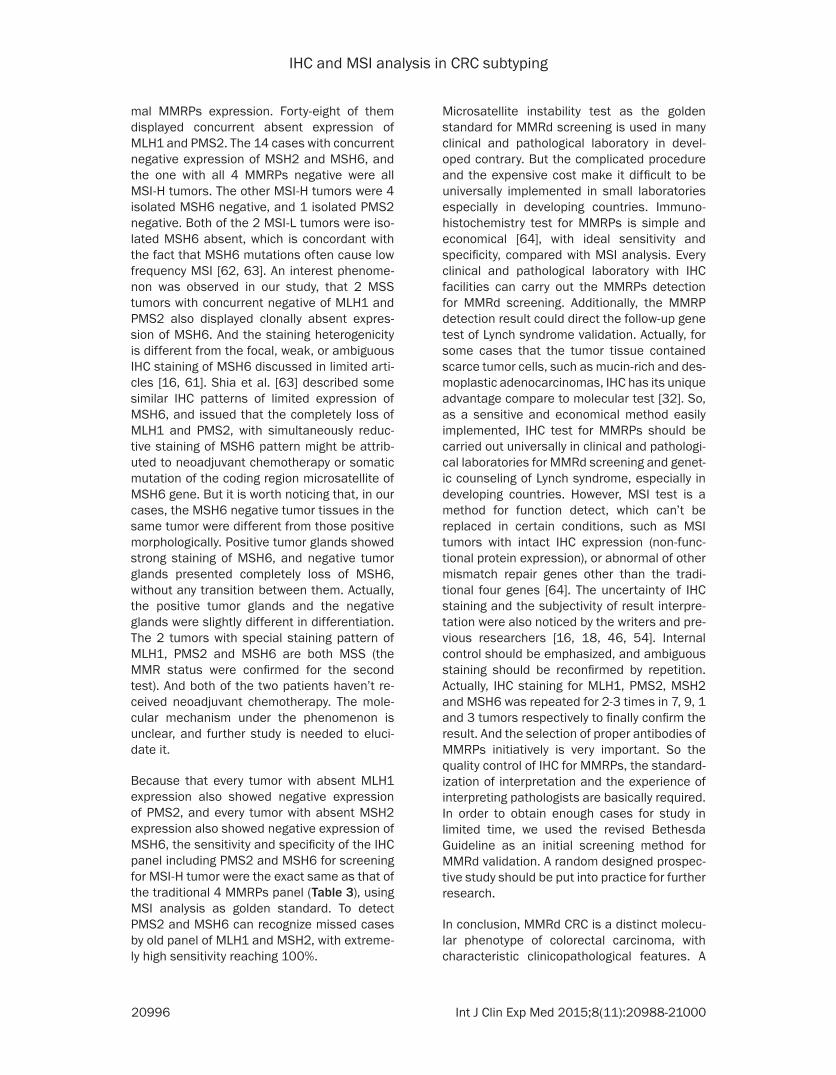
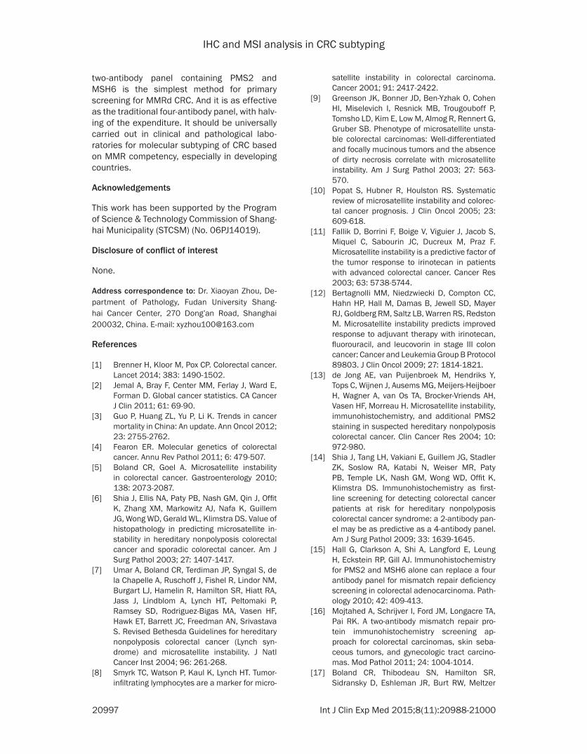
expression of MLH1 (Figure 1). Simultaneously, 21 of 296 (7.1%) tumors showed absent exp- ression of MSH6, of which 19 tumors were classified as MSI-H, 2 were MSI-L. And 15 of 296 (5.1%) cases displayed negative expres-sion of MSH2; all of the 15 tumors were defined as MSI-H by MSI test. Similarly, all of the MSH2 negative tumors also showed absent expres-sion of MSH6 protein, and 6 cases exhibited just MSH6 negative with normal expression of MSH2 (Figure 2). The two MSS tumors with abnormal MMRP expression showed exactly the same IHC pattern, which was concurrent negative of MLH1 and PMS2 with clonally absent of MSH6. The results of the 296 cases’ MSI status and IHC expression pattern of MMRPs are listed in Table 2. The sensitivity and specificity of the 2-panel IHC test including PMS2 and MSH6 for screening for mismatch repair defect were 100% and 98.2% respec-tively, exactly the same as that of the 4-anti-body panel IHC with all of the 4 MMRPs. For the 2-antibody IHC involving MLH1 and MSH2 pre-
viously used, the sensitivity and specificity for MMRd detection were 92.6% and 99.1% separately. Detecting of PMS2 and MSH6 improved the sensitivity of the old IHC screen-ing method, without reducing the specificity obviously, although the difference was not sig-nificant statistically (P=0.058). The diagnostic accordance rate of IHC test with PMS2 and MSH6 and MSI analysis was 98.6% (Table 3).
Discussion
The molecular phenotype of colorectal carcino-ma is closely related to its clinicopathological characteristics, biological behavior, prognosis and even therapy response of patient, both in sporadic and hereditary colorectal cancer [4, 10, 11, 21]. So, it is important to recognize different CRC by pathological-molecular fea-tures. Microsatellite instability (MSI) is a dis-tinct characteristic of mismatch repair defec-tive CRC, which including 12% of sporadic CRC and 3% of hereditary CRC, Lynch syndrome [5,
Figure 1. Immunohistochemical staining pattern of a MSI-H colorectal carcinoma with isolated loss of PMS2 (A), and intact staining of MLH1 (B), MSH2 (C) and MSH6 (D).
IHC and MSI analysis in CRC subtyping
20993 Int J Clin Exp Med 2015;8(11):20988-21000
22]. Microsatellite instability was well defined in Lynch syndrome, and then recommended to be screened with Bethesda guidelines, which was revised in 2003 [7]. It was issued that colorectal carcinoma with MSI-H (MMRd CRC) has characteristic clinical and pathological fea-tures different from MSS CRC, even including
different microsatellite status showed that MMRd CRCs truly have distinct clinical and pathological features. Colorectal carcinomas with MSI-H in this study display a predilection for right colon, with 51.5% of MMRd CRCs locat-ed in proximal colon, which is similar to the result of previous studies [27-30]. Compared to
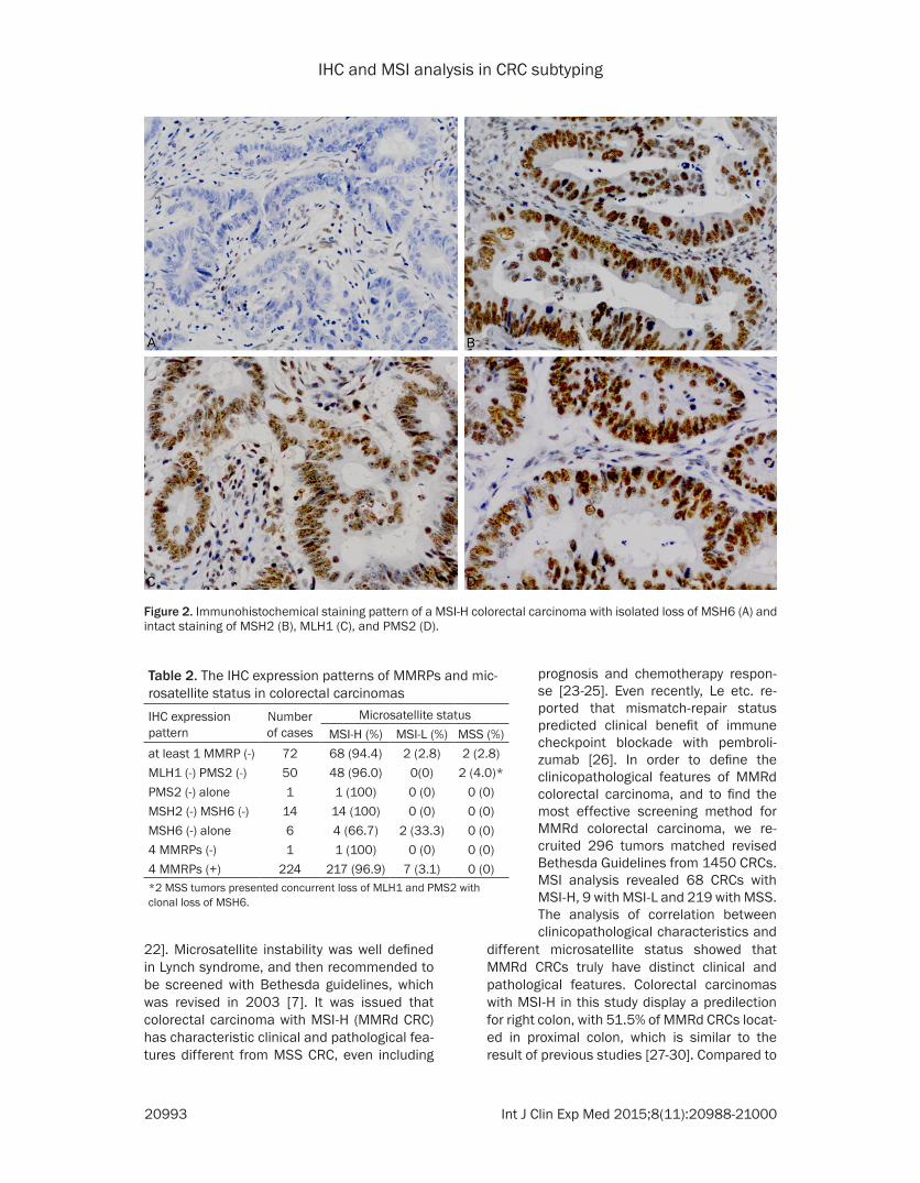
Figure 2. Immunohistochemical staining pattern of a MSI-H colorectal carcinoma with isolated loss of MSH6 (A) and intact staining of MSH2 (B), MLH1 (C), and PMS2 (D).
Table 2. The IHC expression patterns of MMRPs and mic-rosatellite status in colorectal carcinomasIHC expression pattern
Number of cases
Microsatellite statusMSI-H (%) MSI-L (%) MSS (%)
at least 1 MMRP (-) 72 68 (94.4) 2 (2.8) 2 (2.8)MLH1 (-) PMS2 (-) 50 48 (96.0) 0(0) 2 (4.0)*PMS2 (-) alone 1 1 (100) 0 (0) 0 (0)MSH2 (-) MSH6 (-) 14 14 (100) 0 (0) 0 (0)MSH6 (-) alone 6 4 (66.7) 2 (33.3) 0 (0)4 MMRPs (-) 1 1 (100) 0 (0) 0 (0)4 MMRPs (+) 224 217 (96.9) 7 (3.1) 0 (0)*2 MSS tumors presented concurrent loss of MLH1 and PMS2 with clonal loss of MSH6.
prognosis and chemotherapy respon- se [23-25]. Even recently, Le etc. re- ported that mismatch-repair status predicted clinical benefit of immune checkpoint blockade with pembroli-zumab [26]. In order to define the clinicopathological features of MMRd colorectal carcinoma, and to find the most effective screening method for MMRd colorectal carcinoma, we re- cruited 296 tumors matched revised Bethesda Guidelines from 1450 CRCs. MSI analysis revealed 68 CRCs with MSI-H, 9 with MSI-L and 219 with MSS. The analysis of correlation between clinicopathological characteristics and
IHC and MSI analysis in CRC subtyping
20994 Int J Clin Exp Med 2015;8(11):20988-21000
MSS CRCs, 32.3% of CRCs with MSI-H showed poor differentiation, which is significantly differ-ent from MSS CRCs (19.6%). But when it came to mucinous or signet ring differentiation, the difference between MMRd (51.1%) and MSS (38.6%) colorectal carcinomas was not so sig-nificant as the results of other studies [6, 9, 29, 31-35]. That is partially in line with these previ-ous researches. Lymphocytic infiltration is gen-erally recognized as a striking characteristic of MMRd CRCs, as many studies issued [6, 29, 31, 33, 36-38]. In our study, 20.6% of MMRd CRCs presented Crohn-like lymphoid reaction, peritumoral lymphocytes, or tumor-infiltrating lymphocytes, which is relatively higher than MSS CRCs (13.2%), but with no statistic differ-ences. Patients with MSI-H colorectal carcino-ma in our series have relatively lower stage compared with patients with MSS tumor, with 32.4% of MMRd CRCs and 52.5% of MSS CRCs at Ⅲ/Ⅳ TNM stage. And MMRd tumors less prone to metastasize to lymph nodes or distant organs compared to MSS ones, with only 30.9% and 2.9% of tumors have lymph nodes and distant metastasis respectively. That is similar to the results of previous researches [39-41], and can possibly explain the relative better prognosis of MMRd colorectal cancers com-pared with MSS ones [42, 43]. Additionally, our study suggest that colorectal carcinomas with MSI-L in this series are more close to MSS ones morphologically and biologically, as previ-ous studies issued [44, 45]. Although neither the differences between the MSI-L group and MSI-H group, nor the differences between MSI-L group and MSS group are statistically significant.
Microsatellite instability is caused by mismatch repair deficiency, presenting accumulation of insertion or deletion mutations at microsatel-lite across entire genome. The most reliable method for MMRd detecting is microsatellite segments analysis with fluorescent multiplex
match repair deficiency of the tumor. Most researches around 2000 recommended the classical panel containing MLH1 and MSH2, with considerable predictive value for a MSI-H phenotype or germline mutation of MMR gene [46-49], but always missed cases with abnor-mal MSH6 or PMS2, or protein intact missense mutation of MLH1 [50-52].
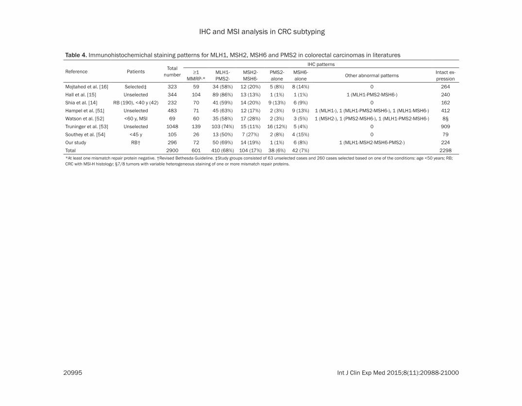
We used all of the four MMRPs including MLH1, MSH2, MSH6 and PMS2 for IHC detecting, to select the most effective panel for MMRd screening. In the 72 tumors with at least one MMRP negative, the most frequent expres- sion pattern of MMRPs was concurrent lost of MLH1 and PMS2, account for 69.4% (50) of all cases. And the concurrent negative expression of MSH2 and MSH6 was the second common pattern, with 19.4% (14) of tumors showed as this. Isolated MSH6 lost was presented in 6 cases (8.3%), followed by isolated PMS2 nega-tive in 1 case (1.4%). That is similar to the result of the previous related studies list in Table 4 [14-16, 51-54]. And one tumor displayed nega-tive expression of all of the four MMRPs (1.4%). This pattern as well as some staining variants listed in Table 4 is rare, and the mechanism is still unclear. Neither isolated MLH1 nor isolated MSH2 lost was found in our series. The result is concordant to the molecular characteristics of MMRPs. As researches in vitro and in vivo proved [13, 55-61], that mismatch repair gene products existing in cells are always stay as heterodimers complex. And MLH1 and MSH2 are obligatory partners, combined with their secondary partners PMS2 and MSH6 respec-tively. If degradation of the former partners occurs, caused by the mutation of respective MMR gene, the later partners will not exist any-more. But the opposite situation is not true. Of the 72 tumors with abnormal MMRPs expres-sion, 68 were classified as MSI-H, 2 were MSI-L and 2 were MSS by MSI analysis. That is all of the 68 MSI-H colorectal cancers present abnor-
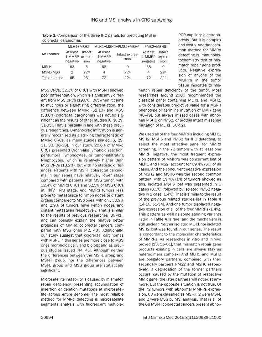
Table 3. Comparison of the three IHC panels for predicting MSI in colorectal carcinomas
MSI status
MLH1+MSH2 MLH1+MSH2+PMS2+MSH6 PMS2+MSH6At least 1 MMRP negative
Intact expres-
sion
At least 1 MMRP negative
Intact expres-sion
At least 1 MMRP negative
Intact expres-
sionMSI-H 63 5 68 0 68 0MSI-L/MSS 2 226 4 224 4 224Total number 65 231 72 224 72 224
PCR-capillary electroph- oresis. But it is complex and costly. Another com-mon method for MMRd detecting is immunohis-tochemistry test of mis-match repair gene prod-ucts. Negative expres-sion of anyone of the MMRPs in the tumor tissue indicates to mis-
IHC and MSI analysis in CRC subtyping
20995 Int J Clin Exp Med 2015;8(11):20988-21000
Table 4. Immunohistochemichal staining patterns for MLH1, MSH2, MSH6 and PMS2 in colorectal carcinomas in literatures
Reference Patients Total number
IHC patterns≥1
MMRP-*MLH1-PMS2-
MSH2-MSH6-
PMS2-alone
MSH6-alone Other abnormal patterns Intact ex-
pressionMojtahed et al. [16] Selected‡ 323 59 34 (58%) 12 (20%) 5 (8%) 8 (14%) 0 264Hall et al. [15] Unselected 344 104 89 (86%) 13 (13%) 1 (1%) 1 (1%) 1 (MLH1-PMS2-MSH6-) 240Shia et al. [14] RB (190), <40 y (42) 232 70 41 (59%) 14 (20%) 9 (13%) 6 (9%) 0 162Hampel et al. [51] Unselected 483 71 45 (63%) 12 (17%) 2 (3%) 9 (13%) 1 (MLH1-), 1 (MLH1-PMS2-MSH6-), 1 (MLH1-MSH6-) 412Watson et al. [52] <60 y, MSI 69 60 35 (58%) 17 (28%) 2 (3%) 3 (5%) 1 (MSH2-), 1 (PMS2-MSH6-), 1 (MLH1-PMS2-MSH6-) 8§Truninger et al. [53] Unselected 1048 139 103 (74%) 15 (11%) 16 (12%) 5 (4%) 0 909Southey et al. [54] <45 y 105 26 13 (50%) 7 (27%) 2 (8%) 4 (15%) 0 79Our study RB† 296 72 50 (69%) 14 (19%) 1 (1%) 6 (8%) 1 (MLH1-MSH2-MSH6-PMS2-) 224Total 2900 601 410 (68%) 104 (17%) 38 (6%) 42 (7%) 2298*At least one mismatch repair protein negative. †Revised Bethesda Guideline. ‡Study groups consisted of 63 unselected cases and 260 cases selected based on one of the conditions: age <50 years; RB; CRC with MSI-H histology; §7/8 tumors with variable heterogeneous staining of one or more mismatch repair proteins.
IHC and MSI analysis in CRC subtyping
20996 Int J Clin Exp Med 2015;8(11):20988-21000
mal MMRPs expression. Forty-eight of them displayed concurrent absent expression of MLH1 and PMS2. The 14 cases with concurrent negative expression of MSH2 and MSH6, and the one with all 4 MMRPs negative were all MSI-H tumors. The other MSI-H tumors were 4 isolated MSH6 negative, and 1 isolated PMS2 negative. Both of the 2 MSI-L tumors were iso-lated MSH6 absent, which is concordant with the fact that MSH6 mutations often cause low frequency MSI [62, 63]. An interest phenome-non was observed in our study, that 2 MSS tumors with concurrent negative of MLH1 and PMS2 also displayed clonally absent expres-sion of MSH6. And the staining heterogenicity is different from the focal, weak, or ambiguous IHC staining of MSH6 discussed in limited arti-cles [16, 61]. Shia et al. [63] described some similar IHC patterns of limited expression of MSH6, and issued that the completely loss of MLH1 and PMS2, with simultaneously reduc-tive staining of MSH6 pattern might be attrib-uted to neoadjuvant chemotherapy or somatic mutation of the coding region microsatellite of MSH6 gene. But it is worth noticing that, in our cases, the MSH6 negative tumor tissues in the same tumor were different from those positive morphologically. Positive tumor glands showed strong staining of MSH6, and negative tumor glands presented completely loss of MSH6, without any transition between them. Actually, the positive tumor glands and the negative glands were slightly different in differentiation. The 2 tumors with special staining pattern of MLH1, PMS2 and MSH6 are both MSS (the MMR status were confirmed for the second test). And both of the two patients haven’t re- ceived neoadjuvant chemotherapy. The mole- cular mechanism under the phenomenon is unclear, and further study is needed to eluci-date it.
Because that every tumor with absent MLH1 expression also showed negative expression of PMS2, and every tumor with absent MSH2 expression also showed negative expression of MSH6, the sensitivity and specificity of the IHC panel including PMS2 and MSH6 for screening for MSI-H tumor were the exact same as that of the traditional 4 MMRPs panel (Table 3), using MSI analysis as golden standard. To detect PMS2 and MSH6 can recognize missed cases by old panel of MLH1 and MSH2, with extreme-ly high sensitivity reaching 100%.
Microsatellite instability test as the golden standard for MMRd screening is used in many clinical and pathological laboratory in devel-oped contrary. But the complicated procedure and the expensive cost make it difficult to be universally implemented in small laboratories especially in developing countries. Immuno- histochemistry test for MMRPs is simple and economical [64], with ideal sensitivity and specificity, compared with MSI analysis. Every clinical and pathological laboratory with IHC facilities can carry out the MMRPs detection for MMRd screening. Additionally, the MMRP detection result could direct the follow-up gene test of Lynch syndrome validation. Actually, for some cases that the tumor tissue contained scarce tumor cells, such as mucin-rich and des-moplastic adenocarcinomas, IHC has its unique advantage compare to molecular test [32]. So, as a sensitive and economical method easily implemented, IHC test for MMRPs should be carried out universally in clinical and pathologi-cal laboratories for MMRd screening and genet-ic counseling of Lynch syndrome, especially in developing countries. However, MSI test is a method for function detect, which can’t be replaced in certain conditions, such as MSI tumors with intact IHC expression (non-func-tional protein expression), or abnormal of other mismatch repair genes other than the tradi- tional four genes [64]. The uncertainty of IHC staining and the subjectivity of result interpre-tation were also noticed by the writers and pre-vious researchers [16, 18, 46, 54]. Internal control should be emphasized, and ambiguous staining should be reconfirmed by repetition. Actually, IHC staining for MLH1, PMS2, MSH2 and MSH6 was repeated for 2-3 times in 7, 9, 1 and 3 tumors respectively to finally confirm the result. And the selection of proper antibodies of MMRPs initiatively is very important. So the quality control of IHC for MMRPs, the standard-ization of interpretation and the experience of interpreting pathologists are basically required. In order to obtain enough cases for study in limited time, we used the revised Bethesda Guideline as an initial screening method for MMRd validation. A random designed prospec-tive study should be put into practice for further research.
In conclusion, MMRd CRC is a distinct molecu-lar phenotype of colorectal carcinoma, with characteristic clinicopathological features. A
IHC and MSI analysis in CRC subtyping
20997 Int J Clin Exp Med 2015;8(11):20988-21000
two-antibody panel containing PMS2 and MSH6 is the simplest method for primary screening for MMRd CRC. And it is as effective as the traditional four-antibody panel, with halv-ing of the expenditure. It should be universally carried out in clinical and pathological labo- ratories for molecular subtyping of CRC based on MMR competency, especially in developing countries.
Acknowledgements
This work has been supported by the Program of Science & Technology Commission of Shang- hai Municipality (STCSM) (No. 06PJ14019).
Disclosure of conflict of interest
None.
Address correspondence to: Dr. Xiaoyan Zhou, De- partment of Pathology, Fudan University Shang- hai Cancer Center, 270 Dong’an Road, Shanghai 200032, China. E-mail: [email protected]
References
[1] Brenner H, Kloor M, Pox CP. Colorectal cancer. Lancet 2014; 383: 1490-1502.
[2] Jemal A, Bray F, Center MM, Ferlay J, Ward E, Forman D. Global cancer statistics. CA Cancer J Clin 2011; 61: 69-90.
[3] Guo P, Huang ZL, Yu P, Li K. Trends in cancer mortality in China: An update. Ann Oncol 2012; 23: 2755-2762.
[4] Fearon ER. Molecular genetics of colorectal cancer. Annu Rev Pathol 2011; 6: 479-507.
[5] Boland CR, Goel A. Microsatellite instability in colorectal cancer. Gastroenterology 2010; 138: 2073-2087.
[6] Shia J, Ellis NA, Paty PB, Nash GM, Qin J, Offit K, Zhang XM, Markowitz AJ, Nafa K, Guillem JG, Wong WD, Gerald WL, Klimstra DS. Value of histopathology in predicting microsatellite in-stability in hereditary nonpolyposis colorectal cancer and sporadic colorectal cancer. Am J Surg Pathol 2003; 27: 1407-1417.
[7] Umar A, Boland CR, Terdiman JP, Syngal S, de la Chapelle A, Ruschoff J, Fishel R, Lindor NM, Burgart LJ, Hamelin R, Hamilton SR, Hiatt RA, Jass J, Lindblom A, Lynch HT, Peltomaki P, Ramsey SD, Rodriguez-Bigas MA, Vasen HF, Hawk ET, Barrett JC, Freedman AN, Srivastava S. Revised Bethesda Guidelines for hereditary nonpolyposis colorectal cancer (Lynch syn-drome) and microsatellite instability. J Natl Cancer Inst 2004; 96: 261-268.
[8] Smyrk TC, Watson P, Kaul K, Lynch HT. Tumor-infiltrating lymphocytes are a marker for micro-
satellite instability in colorectal carcinoma. Cancer 2001; 91: 2417-2422.
[9] Greenson JK, Bonner JD, Ben-Yzhak O, Cohen HI, Miselevich I, Resnick MB, Trougouboff P, Tomsho LD, Kim E, Low M, Almog R, Rennert G, Gruber SB. Phenotype of microsatellite unsta-ble colorectal carcinomas: Well-differentiated and focally mucinous tumors and the absence of dirty necrosis correlate with microsatellite instability. Am J Surg Pathol 2003; 27: 563-570.
[10] Popat S, Hubner R, Houlston RS. Systematic review of microsatellite instability and colorec-tal cancer prognosis. J Clin Oncol 2005; 23: 609-618.
[11] Fallik D, Borrini F, Boige V, Viguier J, Jacob S, Miquel C, Sabourin JC, Ducreux M, Praz F. Microsatellite instability is a predictive factor of the tumor response to irinotecan in patients with advanced colorectal cancer. Cancer Res 2003; 63: 5738-5744.
[12] Bertagnolli MM, Niedzwiecki D, Compton CC, Hahn HP, Hall M, Damas B, Jewell SD, Mayer RJ, Goldberg RM, Saltz LB, Warren RS, Redston M. Microsatellite instability predicts improved response to adjuvant therapy with irinotecan, fluorouracil, and leucovorin in stage III colon cancer: Cancer and Leukemia Group B Protocol 89803. J Clin Oncol 2009; 27: 1814-1821.
[13] de Jong AE, van Puijenbroek M, Hendriks Y, Tops C, Wijnen J, Ausems MG, Meijers-Heijboer H, Wagner A, van Os TA, Brocker-Vriends AH, Vasen HF, Morreau H. Microsatellite instability, immunohistochemistry, and additional PMS2 staining in suspected hereditary nonpolyposis colorectal cancer. Clin Cancer Res 2004; 10: 972-980.
[14] Shia J, Tang LH, Vakiani E, Guillem JG, Stadler ZK, Soslow RA, Katabi N, Weiser MR, Paty PB, Temple LK, Nash GM, Wong WD, Offit K, Klimstra DS. Immunohistochemistry as first-line screening for detecting colorectal cancer patients at risk for hereditary nonpolyposis colorectal cancer syndrome: a 2-antibody pan-el may be as predictive as a 4-antibody panel. Am J Surg Pathol 2009; 33: 1639-1645.
[15] Hall G, Clarkson A, Shi A, Langford E, Leung H, Eckstein RP, Gill AJ. Immunohistochemistry for PMS2 and MSH6 alone can replace a four antibody panel for mismatch repair deficiency screening in colorectal adenocarcinoma. Path- ology 2010; 42: 409-413.
[16] Mojtahed A, Schrijver I, Ford JM, Longacre TA, Pai RK. A two-antibody mismatch repair pro-tein immunohistochemistry screening ap-proach for colorectal carcinomas, skin seba-ceous tumors, and gynecologic tract carcino-mas. Mod Pathol 2011; 24: 1004-1014.
[17] Boland CR, Thibodeau SN, Hamilton SR, Sidransky D, Eshleman JR, Burt RW, Meltzer
IHC and MSI analysis in CRC subtyping
20998 Int J Clin Exp Med 2015;8(11):20988-21000
SJ, Rodriguez-Bigas MA, Fodde R, Ranzani GN, Srivastava S. A National Cancer Institute Workshop on Microsatellite Instability for can-cer detection and familial predisposition: Development of international criteria for the determination of microsatellite instability in colorectal cancer. Cancer Res 1998; 58: 5248-5257.
[18] Dietmaier W, Wallinger S, Bocker T, Kullmann F, Fishel R, Ruschoff J. Diagnostic microsatel-lite instability: definition and correlation with mismatch repair protein expression. Cancer Res 1997; 57: 4749-4756.
[19] Jass JR. Classification of colorectal cancer based on correlation of clinical, morphological and molecular features. Histopathology 2007; 50: 113-130.
[20] Lynch HT, de la Chapelle A. Hereditary colorec-tal cancer. N Engl J Med 2003; 348: 919-932.
[21] Ionov Y, Peinado MA, Malkhosyan S, Shibata D, Perucho M. Ubiquitous somatic mutations in simple repeated sequences reveal a new mechanism for colonic carcinogenesis. Nature 1993; 363: 558-561.
[22] Blake C, Tsao JL, Wu A, Shibata D. Stepwise deletions of polyA sequences in mismatch re-pair-deficient colorectal cancers. Am J Pathol 2001; 158: 1867-1870.
[23] Thibodeau SN, Bren G, Schaid D. Microsatellite instability in cancer of the proximal colon. Science 1993; 260: 816-819.
[24] Ribic CM, Sargent DJ, Moore MJ, Thibodeau SN, French AJ, Goldberg RM, Hamilton SR, Laurent-Puig P, Gryfe R, Shepherd LE, Tu D, Redston M, Gallinger S. Tumor microsatellite-instability status as a predictor of benefit from fluorouracil-based adjuvant chemotherapy for colon cancer. N Engl J Med 2003; 349: 247-257.
[25] Carethers JM, Smith EJ, Behling CA, Nguyen L, Tajima A, Doctolero RT, Cabrera BL, Goel A, Arnold CA, Miyai K, Boland CR. Use of 5-fluo-rouracil and survival in patients with microsat-ellite-unstable colorectal cancer. Gastroenter- ology 2004; 126: 394-401.
[26] Le DT, Uram JN, Wang H, Bartlett BR, Kem- berling H, Eyring AD, Skora AD, Luber BS, Azad NS, Laheru D, Biedrzycki B, Donehower RC, Zaheer A, Fisher GA, Crocenzi TS, Lee JJ, Duffy SM, Goldberg RM, de la Chapelle A, Koshiji M, Bhaijee F, Huebner T, Hruban RH, Wood LD, Cuka N, Pardoll DM, Papadopoulos N, Kinzler KW, Zhou S, Cornish TC, Taube JM, Anders RA, Eshleman JR, Vogelstein B, Diaz LJ. PD-1 Blockade in Tumors with Mismatch-Repair Deficiency. N Engl J Med 2015; 372: 2509-2520.
[27] Ward R, Meagher A, Tomlinson I, O’Connor T, Norrie M, Wu R, Hawkins N. Microsatellite in-
stability and the clinicopathological features of sporadic colorectal cancer. Gut 2001; 48: 821-829.
[28] Young J, Simms LA, Biden KG, Wynter C, Whitehall V, Karamatic R, George J, Goldblatt J, Walpole I, Robin SA, Borten MM, Stitz R, Searle J, McKeone D, Fraser L, Purdie DR, Podger K, Price R, Buttenshaw R, Walsh MD, Barker M, Leggett BA, Jass JR. Features of colorectal cancers with high-level microsatel-lite instability occurring in familial and sporad-ic settings: parallel pathways of tumorigenesis. Am J Pathol 2001; 159: 2107-2116.
[29] Halvarsson B, Anderson H, Domanska K, Lindmark G, Nilbert M. Clinicopathologic fac-tors identify sporadic mismatch repair-defec-tive colon cancers. Am J Clin Pathol 2008; 129: 238-244.
[30] Jover R, Paya A, Alenda C, Poveda MJ, Peiro G, Aranda FI, Perez-Mateo M. Defective mis-match-repair colorectal cancer: clinicopatho-logic characteristics and usefulness of immu-nohistochemical analysis for diagnosis. Am J Clin Pathol 2004; 122: 389-394.
[31] Jass JR, Do KA, Simms LA, Iino H, Wynter C, Pillay SP, Searle J, Radford-Smith G, Young J, Leggett B. Morphology of sporadic colorectal cancer with DNA replication errors. Gut 1998; 42: 673-679.
[32] Kakar S, Aksoy S, Burgart LJ, Smyrk TC. Muci- nous carcinoma of the colon: Correlation of loss of mismatch repair enzymes with clinico-pathologic features and survival. Mod Pathol 2004; 17: 696-700.
[33] Wright CL, Stewart ID. Histopathology and mismatch repair status of 458 consecutive colorectal carcinomas. Am J Surg Pathol 2003; 27: 1393-1406.
[34] Yearsley M, Hampel H, Lehman A, Nakagawa H, de la Chapelle A, Frankel WL. Histologic fea-tures distinguish microsatellite-high from mic-rosatellite-low and microsatellite-stable colo- rectal carcinomas, but do not differentiate germline mutations from methylation of the MLH1 promoter. Hum Pathol 2006; 37: 831-838.
[35] Alexander J, Watanabe T, Wu TT, Rashid A, Li S, Hamilton SR. Histopathological identification of colon cancer with microsatellite instability. Am J Pathol 2001; 158: 527-535.
[36] Jenkins MA, Hayashi S, O’Shea AM, Burgart LJ, Smyrk TC, Shimizu D, Waring PM, Ruszkiewicz AR, Pollett AF, Redston M, Barker MA, Baron JA, Casey GR, Dowty JG, Giles GG, Limburg P, Newcomb P, Young JP, Walsh MD, Thibo- deau SN, Lindor NM, Lemarchand L, Gallinger S, Haile RW, Potter JD, Hopper JL, Jass JR. Pathology features in Bethesda guidelines pre-dict colorectal cancer microsatellite instability:
IHC and MSI analysis in CRC subtyping
20999 Int J Clin Exp Med 2015;8(11):20988-21000
A population-based study. Gastroenterology 2007; 133: 48-56.
[37] Watson P, Lin KM, Rodriguez-Bigas MA, Smyrk T, Lemon S, Shashidharan M, Franklin B, Karr B, Thorson A, Lynch HT. Colorectal carcino- ma survival among hereditary nonpolyposis colorectal carcinoma family members. Cancer 1998; 83: 259-266.
[38] Aarnio M, Mustonen H, Mecklin JP, Jarvinen HJ. Prognosis of colorectal cancer varies in dif-ferent high-risk conditions. Ann Med 1998; 30: 75-80.
[39] Malesci A, Laghi L, Bianchi P, Delconte G, Randolph A, Torri V, Carnaghi C, Doci R, Rosati R, Montorsi M, Roncalli M, Gennari L, Santoro A. Reduced likelihood of metastases in pa-tients with microsatellite-unstable colorectal cancer. Clin Cancer Res 2007; 13: 3831-3839.
[40] Ogino S, Nosho K, Kirkner GJ, Kawasaki T, Meyerhardt JA, Loda M, Giovannucci EL, Fuchs CS. CpG island methylator phenotype, micro-satellite instability, BRAF mutation and clinical outcome in colon cancer. Gut 2009; 58: 90-96.
[41] Gryfe R, Swallow C, Bapat B, Redston M, Gallinger S, Couture J. Molecular biology of colorectal cancer. Curr Probl Cancer 1997; 21: 233-300.
[42] Laiho P, Launonen V, Lahermo P, Esteller M, Guo M, Herman JG, Mecklin JP, Jarvinen H, Sistonen P, Kim KM, Shibata D, Houlston RS, Aaltonen LA. Low-level microsatellite instability in most colorectal carcinomas. Cancer Res 2002; 62: 1166-1170.
[43] Halford SE, Sawyer EJ, Lambros MB, Gorman P, Macdonald ND, Talbot IC, Foulkes WD, Gillett CE, Barnes DM, Akslen LA, Lee K, Jacobs IJ, Hanby AM, Ganesan TS, Salvesen HB, Bodmer WF, Tomlinson IP, Roylance RR. MSI-low, a real phenomenon which varies in frequency among cancer types. J Pathol 2003; 201: 389-394.
[44] Lindor NM, Burgart LJ, Leontovich O, Goldberg RM, Cunningham JM, Sargent DJ, Walsh-Vockley C, Petersen GM, Walsh MD, Leggett BA, Young JP, Barker MA, Jass JR, Hopper J, Gallinger S, Bapat B, Redston M, Thibodeau SN. Immunohistochemistry versus microsatel-lite instability testing in phenotyping colorectal tumors. J Clin Oncol 2002; 20: 1043-1048.
[45] Dieumegard B, Grandjouan S, Sabourin JC, Le Bihan ML, Lefrere I, Bellefqih, Pignon JP, Rougier P, Lasser P, Benard J, Couturier D, Bressac-de PB. Extensive molecular screening for hereditary non-polyposis colorectal cancer. Br J Cancer 2000; 82: 871-880.
[46] Cawkwell L, Gray S, Murgatroyd H, Sutherland F, Haine L, Longfellow M, O’Loughlin S, Cross D, Kronborg O, Fenger C, Mapstone N, Dixon M, Quirke P. Choice of management strategy
for colorectal cancer based on a diagnostic im-munohistochemical test for defective mis-match repair. Gut 1999; 45: 409-415.
[47] Muller W, Burgart LJ, Krause-Paulus R, Thibo- deau SN, Almeida M, Edmonston TB, Boland CR, Sutter C, Jass JR, Lindblom A, Lubinski J, MacDermot K, Sanders DS, Morreau H, Muller A, Oliani C, Orntoft T, Ponz DLM, Rosty C, Rodriguez-Bigas M, Ruschoff J, Ruszkiewicz A, Sabourin J, Salovaara R, Moslein G. The reli-ability of immunohistochemistry as a pre-screening method for the diagnosis of heredi-tary nonpolyposis colorectal cancer (HNPCC)--results of an international collaborative study. Fam Cancer 2001; 1: 87-92.
[48] Christensen M, Katballe N, Wikman F, Prim- dahl H, Sorensen FB, Laurberg S, Orntoft TF. Antibody-based screening for hereditary non-polyposis colorectal carcinoma compared with microsatellite analysis and sequencing. Cancer 2002; 95: 2422-2430.
[49] Mangold E, Pagenstecher C, Friedl W, Fischer HP, Merkelbach-Bruse S, Ohlendorf M, Fried- richs N, Aretz S, Buettner R, Propping P, Mathiak M. Tumours from MSH2 mutation car-riers show loss of MSH2 expression but many tumours from MLH1 mutation carriers exhibit weak positive MLH1 staining. J Pathol 2005; 207: 385-395.
[50] Wahlberg SS, Schmeits J, Thomas G, Loda M, Garber J, Syngal S, Kolodner RD, Fox E. Evaluation of microsatellite instability and immunohistochemistry for the prediction of germ-line MSH2 and MLH1 mutations in he-reditary nonpolyposis colon cancer families. Cancer Res 2002; 62: 3485-3492.
[51] Hampel H, Frankel WL, Martin E, Arnold M, Khanduja K, Kuebler P, Clendenning M, Sota- maa K, Prior T, Westman JA, Panescu J, Fix D, Lockman J, LaJeunesse J, Comeras I, de la Chapelle A. Feasibility of screening for Lynch syndrome among patients with colorectal can-cer. J Clin Oncol 2008; 26: 5783-5788.
[52] Watson N, Grieu F, Morris M, Harvey J, Stewart C, Schofield L, Goldblatt J, Iacopetta B. Hetero- geneous staining for mismatch repair proteins during population-based prescreening for he-reditary nonpolyposis colorectal cancer. J Mol Diagn 2007; 9: 472-478.
[53] Truninger K, Menigatti M, Luz J, Russell A, Haider R, Gebbers JO, Bannwart F, Yurtsever H, Neuweiler J, Riehle HM, Cattaruzza MS, Heinimann K, Schar P, Jiricny J, Marra G. Im- munohistochemical analysis reveals high fre-quency of PMS2 defects in colorectal cancer. Gastroenterology 2005; 128: 1160-1171.
[54] Southey MC, Jenkins MA, Mead L, Whitty J, Trivett M, Tesoriero AA, Smith LD, Jennings K, Grubb G, Royce SG, Walsh MD, Barker MA,
IHC and MSI analysis in CRC subtyping
21000 Int J Clin Exp Med 2015;8(11):20988-21000
Young JP, Jass JR, St JD, Macrae FA, Giles GG, Hopper JL. Use of molecular tumor characteris-tics to prioritize mismatch repair gene testing in early-onset colorectal cancer. J Clin Oncol 2005; 23: 6524-6532.
[55] Koi M, Umar A, Chauhan DP, Cherian SP, Carethers JM, Kunkel TA, Boland CR. Human chromosome 3 corrects mismatch repair defi-ciency and microsatellite instability and reduc-es N-methyl-N’-nitro-N-nitrosoguanidine toler-ance in colon tumor cells with homozygous hMLH1 mutation. Cancer Res 1994; 54: 4308-4312.
[56] Umar A, Koi M, Risinger JI, Glaab WE, Tindall KR, Kolodner RD, Boland CR, Barrett JC, Kunkel TA. Correction of hypermutability, N- methyl-N’-nitro-N-nitrosoguanidine resistance, and defective DNA mismatch repair by intro-ducing chromosome 2 into human tumor cells with mutations in MSH2 and MSH6. Cancer Res 1997; 57: 3949-3955.
[57] Watanabe Y, Haugen-Strano A, Umar A, Yama- da K, Hemmi H, Kikuchi Y, Takano S, Shibata Y, Barrett JC, Kunkel TA, Koi M. Complementation of an hMSH2 defect in human colorectal carci-noma cells by human chromosome 2 transfer. Mol Carcinog 2000; 29: 37-49.
[58] Acharya S, Wilson T, Gradia S, Kane MF, Guerrette S, Marsischky GT, Kolodner R, Fishel R. hMSH2 forms specific mispair-binding com-plexes with hMSH3 and hMSH6. Proc Natl Acad Sci U S A 1996; 93: 13629-13634.
[59] Boland CR, Koi M, Chang DK, Carethers JM. The biochemical basis of microsatellite insta-bility and abnormal immunohistochemistry and clinical behavior in Lynch syndrome: from bench to bedside. Fam Cancer 2008; 7: 41-52.
[60] Berends MJ, Wu Y, Sijmons RH, Mensink RG, van der Sluis T, Hordijk-Hos JM, de Vries EG, Hollema H, Karrenbeld A, Buys CH, van der Zee AG, Hofstra RM, Kleibeuker JH. Molecular and clinical characteristics of MSH6 variants: An analysis of 25 index carriers of a germline vari-ant. Am J Hum Genet 2002; 70: 26-37.
[61] Buttin BM, Powell MA, Mutch DG, Babb SA, Huettner PC, Edmonston TB, Herzog TJ, Rader JS, Gibb RK, Whelan AJ, Goodfellow PJ. Pene- trance and expressivity of MSH6 germline mu-tations in seven kindreds not ascertained by family history. Am J Hum Genet 2004; 74: 1262-1269.
[62] Shia J, Klimstra DS, Nafa K, Offit K, Guillem JG, Markowitz AJ, Gerald WL, Ellis NA. Value of immunohistochemical detection of DNA mis-match repair proteins in predicting germline mutation in hereditary colorectal neoplasms. Am J Surg Pathol 2005; 29: 96-104.
[63] Shia J, Zhang L, Shike M, Guo M, Stadler Z, Xiong X, Tang LH, Vakiani E, Katabi N, Wang H, Bacares R, Ruggeri J, Boland CR, Ladanyi M, Klimstra DS. Secondary mutation in a coding mononucleotide tract in MSH6 causes loss of immunoexpression of MSH6 in colorectal car-cinomas with MLH1/PMS2 deficiency. Mod Pathol 2013; 26: 131-138.
[64] Debniak T, Kurzawski G, Gorski B, Kladny J, Domagala W, Lubinski J. Value of pedigree/clinical data, immunohistochemistry and mic-rosatellite instability analyses in reducing the cost of determining hMLH1 and hMSH2 gene mutations in patients with colorectal cancer. Eur J Cancer 2000; 36: 49-54.