Embed Size (px)
Citation preview
Am J Cancer Res 2019;9(4):779-790www.ajcr.us /ISSN:2156-6976/ajcr0089166
Original ArticleLong non-coding RNA LINC01133 mediates nasopharyngeal carcinoma tumorigenesis by binding to YBX1
Wenjun Zhang1*, Mingyu Du1*, Tingting Wang1*, Wei Chen1, Jing Wu1, Qian Li1, Xiaokang Tian1,2, Luxi Qian1, Yan Wang1, Fanyu Peng1, Qian Fei1, Jie Chen1,2, Xia He1, Li Yin1
1The Affiliated Cancer Hospital of Nanjing Medical University, Jiangsu Cancer Hospital, Jiangsu Institute of Cancer Research, 42 Bai Zi Ting Road, Nanjing, Jiangsu, China; 2Xuzhou Medical University, 209 Tong-Shan Road, Xu-zhou, Jiangsu, China. *Equal contributors.
Received November 29, 2018; Accepted March 7, 2019; Epub April 1, 2019; Published April 15, 2019
Abstract: Recently, long non-coding RNAs (lncRNAs) have been reported as the vital regulators of various cancers in-cluding nasopharyngeal carcinoma (NPC). An increasing number of studies have suggested that lncRNA LINC01133 is dysregulated and involved in human carcinogenesis. However, the roles of LINC01133 in NPC remain largely unknown. In this work, we demonstrated that LINC01133 was significantly downregulated in NPC tissues and cell lines. Loss and gain of function experiments provided evidence that LINC01133 inhibited NPC cell proliferation, invasion and migration both in vitro and in vivo. Besides, Fluorescence in situ hybridization (FISH) assay was per-formed to determine the localization of LINC01133 and LINC01133 was observed mainly distributed in the nucleus. Importantly, RNA pull-down and RIP assays showed that LINC01133 directly combined with YBX1, and YBX1 can partly reverse the repression of NPC cell proliferation, migration, and invasion caused by LINC01133. Collectively, our exploration indicate that LINC01133 inhibits the malignant-biological behavior of NPC cells by binding to YBX1, thereby suggesting a novel biomarker for the NPC prognosis and treatment.
Keywords: LINC01133, nasopharyngeal carcinoma, YBX1, EMT
Introduction
Nasopharyngeal carcinoma (NPC) is a preva-lent malignancy with geographic features and different etiopathogenesis in Southeast Asia and North Africa [1]. Although intensity-modu-lated radiation therapy and other combining treatments provide locoregional control for most NPCs, tumor relapse and distant metas-tasis remain a huge problem to patient survival [2, 3]. Thus, elucidating the molecular mecha-nisms and developing effective therapeutic tar-gets for these patients are necessary.
Long non-coding RNAs (lncRNAs) are the RNA transcripts that are > 200 nucleotides in leng- th with limited protein coding potential [4]. Increasing evidence has indicated that lncRNAs can play significant roles in a wide variety of cancer pathogenesis [5-7]. The aberrant ex- pression of lncRNAs can also contribute to tumor initiation and progression by different
mechanisms, ranging from chromatin modifica-tion, transcriptional and post-transcriptional regulation [8]. Although several lncRNAs, includ-ing HOTAIR, MALAT1, and ANRIL are involved in NPC [9-11], the clinical significance and func-tions of most dysregulated lncRNAs in NPC are not well characterized.
The lncRNA LINC01133, which is located in chromosome 1q23.2, is downregulated in colorectal cancer and suppress epithelial-mes-enchymal transition (EMT) and metastasis through interaction with SRSF6 [12]. Similarly, LINC01133 shows low expression in oral squa-mous cell carcinoma and gastric cancer [13, 14]. However, studies on other malignant tu- mors indicate opposite oncogenic roles and dif-ferent molecular mechanisms of LINC01133, thereby suggesting the tissue-specific regula-tion of LINC01133 expression [15-17]. Neve- rtheless, information on the role of LINC01133 in NPC progression is scarce.
LINC01133 mediates nasopharyngeal carcinoma tumorigenesis
780 Am J Cancer Res 2019;9(4):779-790
In this work, we focused on exploring LIN- C01133, which was downregulated in NPC tumor tissues and cell lines. Several attempts were carried out to investigate whether LIN- C01133 can repress NPC cell proliferation, invasion and migration in vitro and in vivo. We demonstrated that the LINC01133 function was also partly mediated by binding to Y-box binding protein 1 (YBX1). Overall, this study pro-vided an insight into the biological function and potential mechanisms exerted by LINC01133 in NPC development and progression.
Materials and methods
Cell culture and clinical specimens
The human immortalized nasopharyngeal epi-thelial cell line (NP69) was cultured in keratino-cyte/serum-free medium covered in growth fac-tors (Gibco, Grand Island, NY, USA). Five human NPC cell lines (i.e., CNE-1, CNE-2, 5-8F, 6-10B, and SUNE-1) were grown in RPMI-1640 (Co- rning, Manassas, VA, USA) supplemented with 5% FBS (Gibco, Grand Island, USA) in the pres-ence of 5% CO2 at 37°C. Fifteen frozen NPC tis-sues and six normal nasopharyngeal epitheli-um tissues were obtained from Jiangsu Cancer Hospital (Nanjing, China). All tissue samples were confirmed by pathologists. This study was approved by the Institutional Ethical Review Board of Jiangsu Cancer Hospital, and each patient provided written informed consent. The expression profiles of LINC01133 were also detected in NPC samples provided by Gene Expression Omnibus (GEO, http://www.ncbi.nlm.nih.gov/geo).
RNA extraction and quantitative real-time PCR (qRT-PCR)
The total RNA from NPC cells or clinical sam-ples was isolated using TRIzol reagent (Invi- trogen) following the manufacturer’s instruc-tions. SYBR Green PCR Master Mix was used to perform qRT-PCR reactions on an ABI7300 real-time PCR machine (Applied Biosystems). U6 and β-actin were used as normalized con-trols, and the sequences of specific primers used are listed in Table S1.
Western blot analysis
Proteins were extracted from the transfected cell lines by using modified RIPA buffer and PMSF (Beyotime, Shanghai, China) following
the manufacturer’s protocols. BCA protein assay kit (Beyotime, Shanghai, China) was used to quantify the protein concentration. The pro-teins from each sample were separated by sodium dodecyl sulfate-polyacrylamide gel electrophoresis (SDS-PAGE) and then trans-ferred into a special nitrocellulose membrane. After being blocked with bovine serum albumin, all membranes were incubated with specific antibodies for β-actin (Cell Signaling Technology, USA), YBX1 (Abcam, Hong Kong, China), E-ca- dherin (Cell Signaling Technology), N-cadherin (Cell Signaling Technology), Vimentin (Cell Signaling Technology), or Snail (Cell Signaling Technology). ECL detection reagent (Millipore, Billerica, MA, USA) was used to detect immuno-reactive bands.
LINC01133 overexpression and RNA interfer-ence
For LINC01133 overexpression experiments, CNE-1 and CNE-2 cells were infected with lenti-virus containing the LINC01133-GV367 plas-mid synthesized by GeneChem (Shanghai, China). Smart Silencer-LINC01133 and Smart Silencer-NC were obtained from RiboBio (Gu- angzhou, China). Three specific siRNA targeting human YBX1 along with control-siRNA were purchased from RiboBio (Guangzhou, Guang- dong, China). All transfections were conducted using Lipofectamine 2000 (Invitrogen) follow-ing the manufacturer’s instructions, and the siRNA sequences are listed in Table S1.
Colony formation assay
To measure the colony-forming activity, were seeded the CNE-1, CNE-2 and SUNE-1 cells in six-well plates (1×10³ cells/well) after transfec-tion and then incubated for 10 days. After being fixed with paraformaldehyde and stained with 0.1% crystal violet, the number of colonies was counted under an inverted microscope.
Cell Counting Kit-8 (CCK8) assay
Cell proliferation was measured via CCK8 assay (Promega) every 24 h. After transfection, the cells were seeded in 96-well plates (3000 cells/well). Then, the CCK8 solution was added into these cultured cells. Each well was detect-ed spectrophotometrically at 450 nm after being cultured for 2 h. This experiment included three independent replications, and the results were all averaged.
LINC01133 mediates nasopharyngeal carcinoma tumorigenesis
781 Am J Cancer Res 2019;9(4):779-790
Wound healing assay
Wound healing assays were performed to detect the migration capacity of NPC cells. Transfected CNE-1, CNE-2 and SUNE-1 cells were placed in six-well plates and cultured with serum starvation for 24 h. A wound was creat-ed artificially by a 200-µL pipette tube. Cell migration was measured under an optical microscope at 0 and 24 h.
Transwell migration and invasion assay
To evaluate the cell invasion potential, we used Transwell chambers (Corning, NY, USA) covered with Matrigel (BD Biosciences). The transfected cells were collected after being cultured for 48 h. Then, these cells were resuspended (2×10⁴ cells per well) in 200 µL of serum-free media and plated in the upper chamber. The lower compartment was added with 20% FBS and 500 µL of RPMI-1640. After 36 h of incubation, the membranes were fixed and stained. An inverted microscope was used to count the invaded cells. Transwell migration assays were performed similarly without the Matrigel cover-ing the upper chamber.
Subcellular fractionation location
The nuclear and cytosolic fractions were sepa-rated using the PARIS Kit (Invitrogen) according to the manufacturer’s instructions.
Fluorescence in situ hybridization (FISH)
SUNE-1 cells were fixed in 4% formaldehyde for 20 min and subsequently permeabilized with Triton X-100. Cells were incubated with hybrid-ization buffer supplemented with FISH probe and washed with 2× saline-sodium citrate. Horseradish peroxidase conjugated anti-DIG secondary antibodies (Jackson, West Grove, PA, USA) were used to detect the signals, and DAPI was applied to stain the nuclei. Representative pictures were taken using an Olympus confocal laser scanning microscope.
RNA pull-down assay
LINC01133 RNAs were transcribed in vitro and biotin-labeled with Biotin RNA Labeling Mix (Roche). Biotinylated RNAs were incubated with protein extracted from SUNE-1 cells. Each bind-ing reaction was mixed with magnetic beads and then washed with washing buffer. The
lncRNA-interacting proteins were separated by SDS-PAGE. Afterward, the gels were silver stained, and the proteins were identified by mass spectrometry (MS) or Western blot analysis.
RNA immunoprecipitation (RIP)
RIP assay was conducted using a Magna RIP Kit (Cat. 17-701, Millipore, USA) according to the manufacturer’s instructions. YBX1 antibod-ies were purchased from Abcam (Hong Kong, China).
In vivo tumor growth and metastasis assays
Six-week-old immunodeficient male BALB/c nu- de mice were obtained from Nanjing Univer- sity (Nanjing, China). For the in vivo tumor growth assay, 2×10⁶ CNE-1 cells in 20 µL of cell suspension were separately subcutaneously injected into the nude mice. The cells men-tioned above were infected with lentivirus con-taining LINC01133 expression vector or empty vector. The volume of tumor was measured and recorded every 3-4 days. At 6 weeks after injec-tion, all mice were sacrificed and dissected. Afterward, these xenograft tumors were weighed. Tumor metastasis experiment was performed similarly except that the infected cells were injected into the plantar of each mouse. After 6 weeks, all mice were euthanized and the presence of popliteal and groin lymph node metastasis was observed. All mouse experiments were carried out under the approv-al of the Animal Committee of Nanjing Origin Biosciences, China.
Statistical analysis
All statistical analysis was carried out by SPSS 23.0. Student’s t-test was conducted to analyze the significance of mean values between the two groups. P values < 0.05 was considered significant (*P < 0.05, **P < 0.01, and ***P < 0.001). The results are presented as mean ± standard deviation from at least three separate experiments.
Results
LINC01133 downregulation in NPC cell lines and tissues
The relative LINC01133 expression was evalu-ated by qPCR in both clinical samples and NPC
LINC01133 mediates nasopharyngeal carcinoma tumorigenesis
782 Am J Cancer Res 2019;9(4):779-790
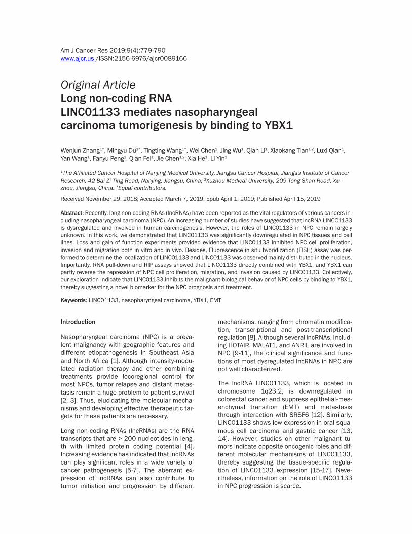
cell lines. Our data showed that LINC01133 expression was significantly downregulated in NPC tissues as compared with normal na- sopharyngeal epithelial tissues (Figure 1B, P < 0.01). LINC01133 expression was also de- creased in NPC cell lines compared with NP- 69, which was the immortalized nasopharyn-geal epithelial cell line [18] (Figure 1A). Moreover, the GEO database confirmed that LINC01133 showed lower expression in NPC (n = 31) than the normal nasopharyngeal samples (n = 10, GSE12452, Figure 1C, P = 0.0375). These results suggested that LIN- C01133 may perform tumor suppressor func-tion in NPC.
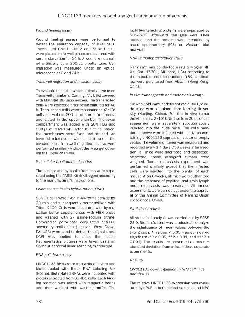
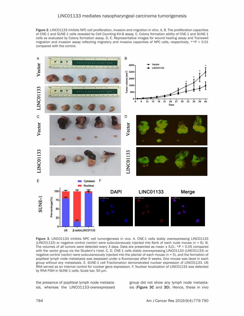
liferation in vivo, we subcutaneously injected LINC01133-overexpressed or control CNE-1 cells into nude mice. At 16 days after tumor for-mation, the tumors formed in the LINC01133-overexpressed group were substantially small-er than those in the control group (Figure 3A and 3B). Meanwhile, a new model of spon- taneous lymph node metastasis was also established using CNE-1 cells. LINC01133 expression vector or control vector was inject-ed into the plantar of each mouse twice a week. After six weeks of injection, the mice were sac-rificed, and popliteal and groin lymph node metastasis was observed after necropsy. The nude mouse in the control vector group showed
Figure 1. LINC01133 is downregulated in NPC cell lines and tissue samples. A. Relative LINC01133 expression in NP69 and NPC cell lines analyzed by real-time PCR and normalized to β-actin expression. B. Relative LINC01133 expression in NPC tissues (n = 15) compared with normal tissues (n = 6). C. LINC01133 expression profile in 31 NPC and 10 normal nasopharyn-geal epithelial samples by GEO datasets GSE 12452. D, E. The LINC01133 expression in stable CNE-1 cell clones infected with lentiviruses encoding LINC01133. Relative levels of LINC01133 in the SUNE-1 cells after trans-fection with siRNA pool against LINC01133. Each experiment was indepen-dently repeated for at least three times. Values are represented as mean ± standard deviation (n = 3). **P < 0.01 and ***P < 0.001.
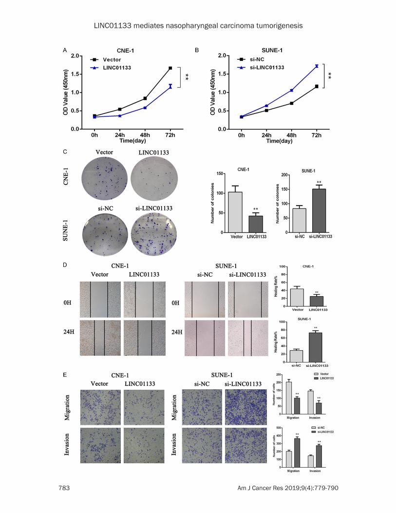
NPC cell proliferation, inva-sion, and migration regula-tion by LINC01133 in vitro
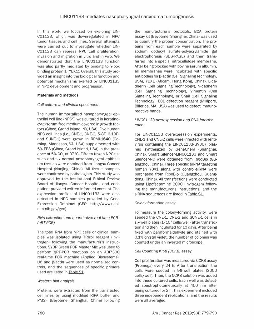
As shown in Figure 1A, CNE-1, CNE-2 and SUNE-1 were cho-sen as three representative NPC cell lines to explore the role of LINC01133 in NPC tumorigenesis. CNE-1 and CNE-2 cells were infected with lentivirus containing the LIN- C01133 expression vector while Smart Silencer-LIN- C01133-mediated knockdo- wns were used in SUNE-1 cells (Figures 1D, 1E and S1A). As shown in Figures 2 and S1, exotic expression of LINC01133 significantly sup-pressed NPC cell prolifera-tion, invasion and migration in vitro. In addition, the co- lony formation ability of CNE-1 and CNE-2 was markedly decreased compared with the control groups (Figures 2C and S1C). However, LINC01- 133 silencing increased the proliferation, invasion, and migration abilities of SUNE1 cells, as well as colony forma-tion (Figure 2A-E).
Tumor growth and metastasis regulation by LINC01133 in NPC in vivo
To investigate whether LIN- C01133 affects NPC cell pro-
LINC01133 mediates nasopharyngeal carcinoma tumorigenesis
783 Am J Cancer Res 2019;9(4):779-790
LINC01133 mediates nasopharyngeal carcinoma tumorigenesis
784 Am J Cancer Res 2019;9(4):779-790
the presence of popliteal lymph node metasta-sis, whereas the LINC01133-overexpressed
group did not show any lymph node metasta- sis (Figure 3C and 3D). Hence, these in vivo
Figure 2. LINC01133 inhibits NPC cell proliferation, invasion and migration in vitro. A, B. The proliferation capacities of CNE-1 and SUNE-1 cells revealed by Cell Counting Kit-8 assay. C. Colony formation ability of CNE-1 and SUNE-1 cells as evaluated by Colony formation assay. D, E. Representative images for wound healing assay and Transwell migration and invasion assay reflecting migratory and invasive capacities of NPC cells, respectively. **P < 0.01 compared with the control.
Figure 3. LINC01133 inhibits NPC cell tumorigenesis in vivo. A. CNE-1 cells stably overexpressing LINC01133 (LINC01133) or negative control (vector) were subcutaneously injected into flank of each nude mouse (n = 6). B. The volumes of all tumors were detected every 3 days. Data are presented as mean ± S.D.; *P < 0.05 compared with the vector group via the Student’s t-test. C, D. CNE-1 cells stably overexpressing LINC01133 (LINC01133) or negative control (vector) were subcutaneously injected into the plantar of each mouse (n = 5), and the formation of popliteal lymph node metastasis was assessed under a fluoroscope after 6 weeks. One mouse was dead in each group without any metastasis. E. SUNE-1 cell Fractionation demonstrated nuclear expression of LINC01133. U6 RNA served as an internal control for nuclear gene expression. F. Nuclear localization of LINC01133 was detected by RNA FISH in SUNE-1 cells. Scale bar, 50 μm.
LINC01133 mediates nasopharyngeal carcinoma tumorigenesis
785 Am J Cancer Res 2019;9(4):779-790
data indicated the functional significance of LINC01133 in NPC cells.
Interaction of LINC01133 with YBX1
To explore the possible mechanism underlying the biological function of LINC01133, we per-formed subcellular fractionation location as- say. Then, LINC01133 showed substantial expression in the nucleus versus the cytosol, also confirmed by FISH (Figure 3E and 3F). These data indicated that LINC01133 may play
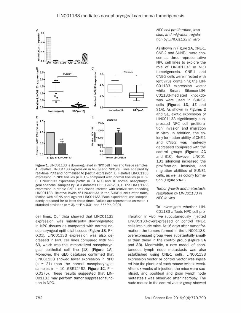
a regulatory function in interacting with nucleus proteins or molecules at the transcriptional level. Considering that several studies have demonstrated that many lncRNAs are involved in diverse regulation pathways through their association with proteins [19-21], we carried out RNA pull-down assays to identify whether LINC01133 may affect cellular function in a similar manner. The bands specific to LIN- C01133 was subjected to MS. As a result, EEF1A1P5, HNRNPF, and YBX1 were the repre-sentative proteins detected by MS (Figure 4A
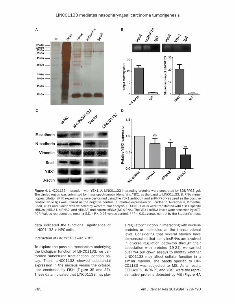
Figure 4. LINC01133 interaction with YBX1. A. LINC01133-interacting proteins were separated by SDS-PAGE gel. The circled region was submitted for mass spectrometry identifying YBX1 as the band to LINC01133. B. RNA immu-noprecipitation (RIP) experiments were performed using the YBX1 antibody, and snRNP70 was used as the positive control, while IgG was utilized as the negative control. C. Relative expression of E-cadherin, N-cadherin, Vimentin, Snail, YBX1 and β-actin was detected by Western blot analysis. D. SUNE-1 cells were transfected with YBX1-specifc siRNAs (siRNA1, siRNA2, and siRNA3) and control siRNA (NC-siRNA). The YBX1 mRNA levels were assessed by qRT-PCR. Values represent the mean ± S.D. *P < 0.05 versus control, **P < 0.01 versus control by the Student’s t-test.
LINC01133 mediates nasopharyngeal carcinoma tumorigenesis
786 Am J Cancer Res 2019;9(4):779-790
and Table S2). Among these primary proteins, YBX1, which is a well-known RNA and DNA bind-ing protein, caught our attention. Existing stud-ies have indicated the specific roles performed by YBX1 in the lncRNA-mediated biological reg-ulation [22, 23]. Therefore, we chose YBX1 for follow-up study, and RIP assay with a YBX1 spe-cific antibody showed that LINC01133 could bind to YBX1 protein (Figure 4B). However, change in the protein level of YBX1 in LINC01133-overexpressed CNE-1 cells and LINC01133-depleted SUNE-1 cells was insig-nificant (Figure 4C). These results suggested that LINC01133 can bind to YBX1 but cannot change the protein expression of YBX1.
EMT process inhibition by LINC01133 via YBX1
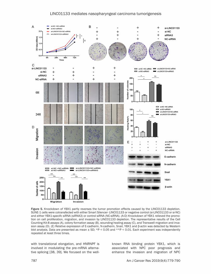
YBX1 plays pro-oncogenic roles in a variety of cancers, including NPC [24-26]. In addition, YBX1 activates Snail mRNA translation directly and induce EMT [27]. To explore whether the interaction between LINC01133 and YBX1 affects the YBX1 modulation on Snail, we con-ducted Western blot analysis. As shown in Figure 4C, the exotic LINC01133 expression decreased the protein level of Snail and N-cadherin, and upregulated the protein level of E-cadherin. By contrast, LINC01133 deple-tion provided exactly the opposite protein trend. Interestingly, YBX1 knockdown relieved the upregulation in Snail and N-cadherin protein level caused by the depletion of LINC01133 (Figure 5E). Collectively, these findings revealed that LINC01133 inhibit the translation of Snail and the EMT process by regulating YBX1 in NPC cells.
Partial reversion of the tumor promoting ef-fects caused by the LINC01133 depletion via the knockdown of YBX1
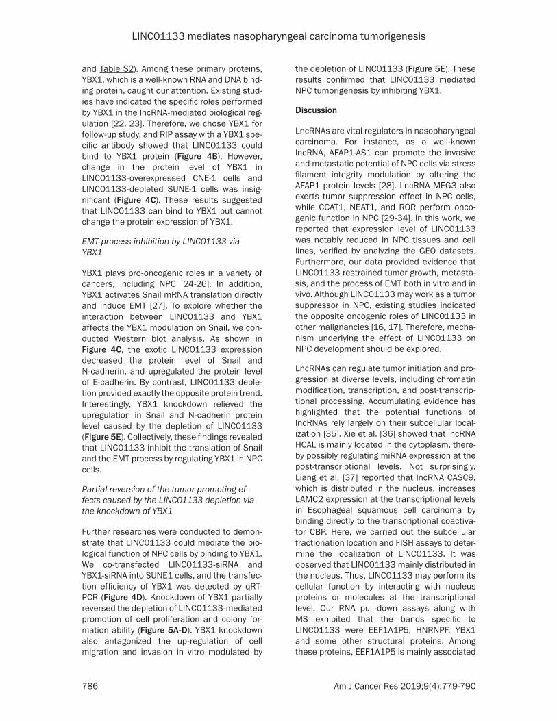
Further researches were conducted to demon-strate that LINC01133 could mediate the bio-logical function of NPC cells by binding to YBX1. We co-transfected LINC01133-siRNA and YBX1-siRNA into SUNE1 cells, and the transfec-tion efficiency of YBX1 was detected by qRT-PCR (Figure 4D). Knockdown of YBX1 partially reversed the depletion of LINC01133-mediated promotion of cell proliferation and colony for-mation ability (Figure 5A-D). YBX1 knockdown also antagonized the up-regulation of cell migration and invasion in vitro modulated by
the depletion of LINC01133 (Figure 5E). These results confirmed that LINC01133 mediated NPC tumorigenesis by inhibiting YBX1.
Discussion
LncRNAs are vital regulators in nasopharyngeal carcinoma. For instance, as a well-known lncRNA, AFAP1-AS1 can promote the invasive and metastatic potential of NPC cells via stress filament integrity modulation by altering the AFAP1 protein levels [28]. LncRNA MEG3 also exerts tumor suppression effect in NPC cells, while CCAT1, NEAT1, and ROR perform onco-genic function in NPC [29-34]. In this work, we reported that expression level of LINC01133 was notably reduced in NPC tissues and cell lines, verified by analyzing the GEO datasets. Furthermore, our data provided evidence that LINC01133 restrained tumor growth, metasta-sis, and the process of EMT both in vitro and in vivo. Although LINC01133 may work as a tumor suppressor in NPC, existing studies indicated the opposite oncogenic roles of LINC01133 in other malignancies [16, 17]. Therefore, mecha-nism underlying the effect of LINC01133 on NPC development should be explored.
LncRNAs can regulate tumor initiation and pro-gression at diverse levels, including chromatin modification, transcription, and post-transcrip-tional processing. Accumulating evidence has highlighted that the potential functions of lncRNAs rely largely on their subcellular local-ization [35]. Xie et al. [36] showed that lncRNA HCAL is mainly located in the cytoplasm, there-by possibly regulating miRNA expression at the post-transcriptional levels. Not surprisingly, Liang et al. [37] reported that lncRNA CASC9, which is distributed in the nucleus, increases LAMC2 expression at the transcriptional levels in Esophageal squamous cell carcinoma by binding directly to the transcriptional coactiva-tor CBP. Here, we carried out the subcellular fractionation location and FISH assays to deter-mine the localization of LINC01133. It was observed that LINC01133 mainly distributed in the nucleus. Thus, LINC01133 may perform its cellular function by interacting with nucleus proteins or molecules at the transcriptional level. Our RNA pull-down assays along with MS exhibited that the bands specific to LINC01133 were EEF1A1P5, HNRNPF, YBX1 and some other structural proteins. Among these proteins, EEF1A1P5 is mainly associated
LINC01133 mediates nasopharyngeal carcinoma tumorigenesis
787 Am J Cancer Res 2019;9(4):779-790
Figure 5. Knockdown of YBX1 partly reverses the tumor promotion effects caused by the LINC01133 depletion. SUNE-1 cells were cotransfected with either Smart Silencer- LINC01133 or negative control (si-LINC01133 or si-NC) and either YBX1-specifc siRNA (siRNA3) or control siRNA (NC-siRNA). (A-D) Knockdown of YBX1 relieved the promo-tion on cell proliferation, migration, and invasion by LINC01133 depletion. The representative results of the Cell Counting Kit-8 assays (A), colony formation assay (B), wounding healing assay (C), and Transwell migration and inva-sion assay (D). (E) Relative expression of E-cadherin, N-cadherin, Snail, YBX1 and β-actin was detected by Western blot analysis. Data are presented as mean ± SD; *P < 0.05 and **P < 0.01. Each experiment was independently repeated at least three times.
with translational elongation, and HNRNPF is involved in modulating the pre-mRNA alterna-tive splicing [38, 39]. We focused on the well-
known RNA binding protein YBX1, which is associated with NPC poor prognosis and enhance the invasion and migration of NPC
LINC01133 mediates nasopharyngeal carcinoma tumorigenesis
788 Am J Cancer Res 2019;9(4):779-790
cells [26]. Subsequently, RIP experiments also identified the directly interaction between LINC01133 and YBX1 protein.
YBX1 is involved in the action mechanisms of lncRNAs. Li et al. [40] revealed that lncRNA HULC accelerates the phosphorylation of YBX1 by acting as a scaffold of YBX1 and ERK to pro-mote hepatocarcinogenesis. Zhang et al. [41] also found that lncRNA HOXC-AS3 plays an important role in the tumor progression of gas-tric cancer, and the activated effect of HOXC-AS3 is mediated partly by its interaction with YBX1. YBX1 activates Snail mRNA translation and induces EMT in breast cancer [27]. Thus, we hypothesized that LINC01133 may work in a similar manner. Our further study showed that by interacting with YBX1, exotic LINC01133 expression suppressed the Snail translation and the EMT process in NPC cells. The effects of LINC01133 on Snail were dependent in part on YBX1 because YBX1 knockdown partly reversed the effects of LINC01133. The inter-action between LINC01133 and YBX1 did not alter the protein levels of YBX1, but relatively changed its structure and functions. However, further investigation is needed to explore the concrete combinations of LINC01133 and YBX1 and determine the mechanism underly-ing the effect of specific DNAs or RNAs that directly bind to YBX1. Moreover, LINC01133 mediated the biological function of NPC cells by inhibiting YBX1 since knockdown of YBX1 partly reversed the tumor promotion effects due to the depletion of LINC01133. Overall, the specific combination between LINC01133 and YBX1 can alleviate the effects of YBX1 on the translation of Snail, and the cancer-promoting roles of YBX1 in NPC progression.
In conclusion, LINC01133, which is frequently downregulated in NPC, can inhibit the proli- feration, invasion, and migration abilities of NPC cells by interacting with YBX1. Along with further investigation, LINC01133 may serve as a new indicator for NPC treatment and prognosis.
Acknowledgements
We thank the members of the Research Center of Clinical Oncology of Jiangsu province for their technical assistance. This study was supported by the National Natural Science Foundation of China (grant number 81672989), the Fifth
“Three Three Three Project” Foundation for High-level Talents in Jiangsu Province (grant number BRA2016523), the Jiangsu Provincial Commission of Health and Family Planning Young Scholars Award (grant number Q201501) and the Key Project of Jiangsu Cancer Hospital (grant number ZK201602).
Disclosure of conflict of interest
None.
Address correspondence to: Xia He and Li Yin, The Affiliated Cancer Hospital of Nanjing Medical University, Jiangsu Cancer Hospital, Jiangsu Institute of Cancer Research, 42 Bai Zi Ting Road, Nanjing 210000, Jiangsu, China. Tel: +86-025-83283560; E-mail: [email protected] (XH); [email protected] (LY)
References
[1] Yu MC and Yuan JM. Epidemiology of nasopha-ryngeal carcinoma. Semin Cancer Biol 2002; 12: 421-429.
[2] Blanchard P, Lee A, Marguet S, Leclercq J, Ng WT, Ma J, Chan AT, Huang PY, Benhamou E, Zhu G, Chua DT, Chen Y, Mai HQ, Kwong DL, Cheah SL, Moon J, Tung Y, Chi KH, Fountzilas G, Zhang L, Hui EP, Lu TX, Bourhis J and Pignon JP. Chemotherapy and radiotherapy in naso-pharyngeal carcinoma: an update of the MAC-NPC meta-analysis. Lancet Oncol 2015; 16: 645-655.
[3] Lee N, Harris J, Garden AS, Straube W, Glisson B, Xia P, Bosch W, Morrison WH, Quivey J, Thorstad W, Jones C and Ang KK. Intensity-modulated radiation therapy with or without chemotherapy for nasopharyngeal carcinoma: radiation therapy oncology group phase II trial 0225. J Clin Oncol 2009; 27: 3684-3690.
[4] Hung T and Chang HY. Long noncoding RNA in genome regulation: prospects and mecha-nisms. RNA Biol 2010; 7: 582-585.
[5] Zhang H, Chen Z, Wang X, Huang Z, He Z and Chen Y. Long non-coding RNA: a new player in cancer. J Hematol Oncol 2013; 6: 37.
[6] Xue M, Chen W and Li X. Urothelial cancer as-sociated 1: a long noncoding RNA with a cru-cial role in cancer. J Cancer Res Clin Oncol 2016; 142: 1407-1419.
[7] Wu J and Hann SS. Functions and roles of long-non-coding RNAs in human nasopharyngeal carcinoma. Cell Physiol Biochem 2018; 45: 1191-1204.
[8] Ransohoff JD, Wei Y and Khavari PA. The func-tions and unique features of long intergenic non-coding RNA. Nat Rev Mol Cell Biol 2018; 19: 143-157.
LINC01133 mediates nasopharyngeal carcinoma tumorigenesis
789 Am J Cancer Res 2019;9(4):779-790
[9] Nie Y, Liu X, Qu S, Song E, Zou H and Gong C. Long non-coding RNA HOTAIR is an indepen-dent prognostic marker for nasopharyngeal carcinoma progression and survival. Cancer Sci 2013; 104: 458-464.
[10] Hua WF, Zhong Q, Xia TL, Chen Q, Zhang MY, Zhou AJ, Tu ZW, Qu C, Li MZ, Xia YF, Wang HY, Xie D, Claret FX, Song EW and Zeng MS. RBM24 suppresses cancer progression by up-regulating miR-25 to target MALAT1 in naso-pharyngeal carcinoma. Cell Death Dis 2016; 7: e2352.
[11] Hu X, Jiang H and Jiang X. Downregulation of lncRNA ANRIL inhibits proliferation, induces apoptosis, and enhances radiosensitivity in nasopharyngeal carcinoma cells through regu-lating miR-125a. Cancer Biol Ther 2017; 18: 331-338.
[12] Kong J, Sun W, Li C, Wan L, Wang S, Wu Y, Xu E, Zhang H and Lai M. Long non-coding RNA LINC01133 inhibits epithelial-mesenchymal transition and metastasis in colorectal cancer by interacting with SRSF6. Cancer Lett 2016; 380: 476-484.
[13] Kong J, Sun W, Zhu W, Liu C, Zhang H and Wang H. Long noncoding RNA LINC01133 in-hibits oral squamous cell carcinoma metasta-sis through a feedback regulation loop with GDF15. J Surg Oncol 2018; 118: 1326-1334.
[14] Yang XZ, Cheng TT, He QJ, Lei ZY, Chi J, Tang Z, Liao QX, Zhang H, Zeng LS and Cui SZ. LINC01133 as ceRNA inhibits gastric cancer progression by sponging miR-106a-3p to regu-late APC expression and the Wnt/beta-catenin pathway. Mol Cancer 2018; 17: 126.
[15] Zang C, Nie FQ, Wang Q, Sun M, Li W, He J, Zhang M and Lu KH. Long non-coding RNA LINC01133 represses KLF2, P21 and E-cadherin transcription through binding with EZH2, LSD1 in non small cell lung cancer. Oncotarget 2016; 7: 11696-11707.
[16] Zeng HF, Qiu HY and Feng FB. Long noncoding RNA LINC01133 functions as an miR-422a sponge to aggravate the tumorigenesis of hu-man osteosarcoma. Oncol Res 2018; 26: 335-343.
[17] Huang CS, Chu J, Zhu XX, Li JH, Huang XT, Cai JP, Zhao W and Yin XY. The C/EBPbeta-LINC01133 axis promotes cell proliferation in pancreatic ductal adenocarcinoma through upregulation of CCNG1. Cancer Lett 2018; 421: 63-72.
[18] Song LB, Zeng MS, Liao WT, Zhang L, Mo HY, Liu WL, Shao JY, Wu QL, Li MZ, Xia YF, Fu LW, Huang WL, Dimri GP, Band V and Zeng YX. Bmi-1 is a novel molecular marker of nasopharyn-geal carcinoma progression and immortalizes primary human nasopharyngeal epithelial cells. Cancer Res 2006; 66: 6225-6232.
[19] Ge Z, Cheng Z, Yang X, Huo X, Wang N, Wang H, Wang C, Gu D, Zhao F, Yao M, Fan J and Qin W. Long noncoding RNA SchLAH suppresses me-tastasis of hepatocellular carcinoma through interacting with fused in sarcoma. Cancer Sci 2017; 108: 653-662.
[20] Shi Y, Li J, Liu Y, Ding J, Fan Y, Tian Y, Wang L, Lian Y, Wang K and Shu Y. The long noncoding RNA SPRY4-IT1 increases the proliferation of human breast cancer cells by upregulating ZNF703 expression. Mol Cancer 2015; 14: 51.
[21] Liu HT, Liu S, Liu L, Ma RR and Gao P. EGR1-mediated transcription of lncRNA-HNF1A-AS1 promotes cell-cycle progression in gastric can-cer. Cancer Res 2018; 78: 5877-5890.
[22] Su W, Feng S, Chen X, Yang X, Mao R, Guo C, Wang Z, Thomas DG, Lin J, Reddy RM, Orringer MB, Chang AC, Yang Z, Beer DG and Chen G. Silencing of long noncoding RNA MIR22HG triggers cell survival/death signaling via onco-genes YBX1, MET, and p21 in lung cancer. Cancer Res 2018; 78: 3207-3219.
[23] Liu Y, Zhao J, Zhang W, Gan J, Hu C, Huang G and Zhang Y. lncRNA GAS5 enhances G1 cell cycle arrest via binding to YBX1 to regulate p21 expression in stomach cancer. Sci Rep 2015; 5: 10159.
[24] Lim JP, Shyamasundar S, Gunaratne J, Scully OJ, Matsumoto K and Bay BH. YBX1 gene si-lencing inhibits migratory and invasive poten-tial via CORO1C in breast cancer in vitro. BMC Cancer 2017; 17: 201.
[25] Zheng J, Dong W, Zhang J, Li G and Gong H. YB-1, a new biomarker of glioma progression, is associated with the prognosis of glioma pa-tients. Acta Biochim Biophys Sin (Shanghai) 2016; 48: 318-325.
[26] Zhou LL, Ni J, Feng WT, Yao R, Yue S, Zhu YN, Tang HY, Lv LY, Feng JF and Zhu WG. High YBX1 expression indicates poor prognosis and pro-motes cell migration and invasion in nasopha-ryngeal carcinoma. Exp Cell Res 2017; 361: 126-134.
[27] Evdokimova V, Tognon C, Ng T, Ruzanov P, Melnyk N, Fink D, Sorokin A, Ovchinnikov LP, Davicioni E, Triche TJ and Sorensen PH. Translational activation of snail1 and other de-velopmentally regulated transcription factors by YB-1 promotes an epithelial-mesenchymal transition. Cancer Cell 2009; 15: 402-415.
[28] Bo H, Gong Z, Zhang W, Li X, Zeng Y, Liao Q, Chen P, Shi L, Lian Y, Jing Y, Tang K, Li Z, Zhou Y, Zhou M, Xiang B, Li X, Yang J, Xiong W, Li G and Zeng Z. Upregulated long non-coding RNA AFAP1-AS1 expression is associated with pro-gression and poor prognosis of nasopharyn-geal carcinoma. Oncotarget 2015; 6: 20404-20418.
[29] Chak WP, Lung RW, Tong JH, Chan SY, Lun SW, Tsao SW, Lo KW and To KF. Downregulation of
LINC01133 mediates nasopharyngeal carcinoma tumorigenesis
790 Am J Cancer Res 2019;9(4):779-790
long non-coding RNA MEG3 in nasopharyngeal carcinoma. Mol Carcinog 2017; 56: 1041-1054.
[30] Wang Q, Zhang W and Hao S. LncRNA CCAT1 modulates the sensitivity of paclitaxel in naso-pharynx cancers cells via miR-181a/CPEB2 axis. Cell Cycle 2017; 16: 795-801.
[31] Cheng N and Guo Y. Long noncoding RNA NEAT1 promotes nasopharyngeal carcinoma progression through regulation of miR-124/NF-kappaB pathway. Onco Targets Ther 2017; 10: 5843-5853.
[32] Wang Y, Wang C, Chen C, Wu F, Shen P, Zhang P, He G and Li X. Long non-coding RNA NEAT1 regulates epithelial membrane protein 2 ex-pression to repress nasopharyngeal carcino-ma migration and irradiation-resistance through miR-101-3p as a competing endoge-nous RNA mechanism. Oncotarget 2017; 8: 70156-70171.
[33] Pan Y, Li C, Chen J, Zhang K, Chu X, Wang R and Chen L. The emerging roles of long non-coding RNA ROR (lincRNA-ROR) and its possi-ble mechanisms in human cancers. Cell Physiol Biochem 2016; 40: 219-229.
[34] Li L, Gu M, You B, Shi S, Shan Y, Bao L and You Y. Long non-coding RNA ROR promotes prolif-eration, migration and chemoresistance of na-sopharyngeal carcinoma. Cancer Sci 2016; 107: 1215-1222.
[35] Chen LL. Linking long noncoding RNA localiza-tion and function. Trends Biochem Sci 2016; 41: 761-772.
[36] Xie CR, Wang F, Zhang S, Wang FQ, Zheng S, Li Z, Lv J, Qi HQ, Fang QL, Wang XM and Yin ZY. Long noncoding RNA HCAL facilitates the growth and metastasis of hepatocellular carci-noma by acting as a ceRNA of LAPTM4B. Mol Ther Nucleic Acids 2017; 9: 440-451.
[37] Liang Y, Chen X, Wu Y, Li J, Zhang S, Wang K, Guan X, Yang K and Bai Y. LncRNA CASC9 pro-motes esophageal squamous cell carcinoma metastasis through upregulating LAMC2 ex-pression by interacting with the CREB-binding protein. Cell Death Differ 2018; 25: 1980-1995.
[38] Lund A, Knudsen SM, Vissing H, Clark B and Tommerup N. Assignment of human elonga-tion factor 1alpha genes: EEF1A maps to chro-mosome 6q14 and EEF1A2 to 20q13.3. Genomics 1996; 36: 359-361.
[39] Du J, Wang Q, Ziegler SF and Zhou B. FOXP3 interacts with hnRNPF to modulate pre-mRNA alternative splicing. J Biol Chem 2018; 293: 10235-10244.
[40] Li D, Liu X, Zhou J, Hu J, Zhang D, Liu J, Qiao Y and Zhan Q. Long noncoding RNA HULC modu-lates the phosphorylation of YB-1 through serv-ing as a scaffold of extracellular signal-regulat-ed kinase and YB-1 to enhance hepatocarcino-genesis. Hepatology 2017; 65: 1612-1627.
[41] Zhang E, He X, Zhang C, Su J, Lu X, Si X, Chen J, Yin D, Han L and De W. A novel long noncod-ing RNA HOXC-AS3 mediates tumorigenesis of gastric cancer by binding to YBX1. Genome Biol 2018; 19: 154.
LINC01133 mediates nasopharyngeal carcinoma tumorigenesis
1
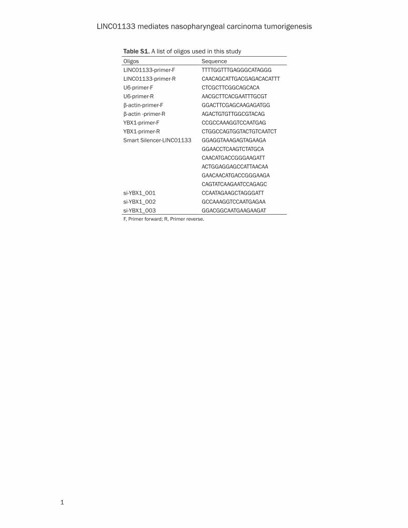
Table S1. A list of oligos used in this studyOligos SequenceLINC01133-primer-F TTTTGGTTTGAGGGCATAGGGLINC01133-primer-R CAACAGCATTGACGAGACACATTTU6-primer-F CTCGCTTCGGCAGCACAU6-primer-R AACGCTTCACGAATTTGCGTβ-actin-primer-F GGACTTCGAGCAAGAGATGGβ-actin -primer-R AGACTGTGTTGGCGTACAGYBX1-primer-F CCGCCAAAGGTCCAATGAGYBX1-primer-R CTGGCCAGTGGTACTGTCAATCTSmart Silencer-LINC01133 GGAGGTAAAGAGTAGAAGA
GGAACCTCAAGTCTATGCACAACATGACCGGGAAGATTACTGGAGGAGCCATTAACAA GAACAACATGACCGGGAAGACAGTATCAAGAATCCAGAGC
si-YBX1_001 CCAATAGAAGCTAGGGATTsi-YBX1_002 GCCAAAGGTCCAATGAGAAsi-YBX1_003 GGACGGCAATGAAGAAGATF, Primer forward; R, Primer reverse.
LINC01133 mediates nasopharyngeal carcinoma tumorigenesis
2
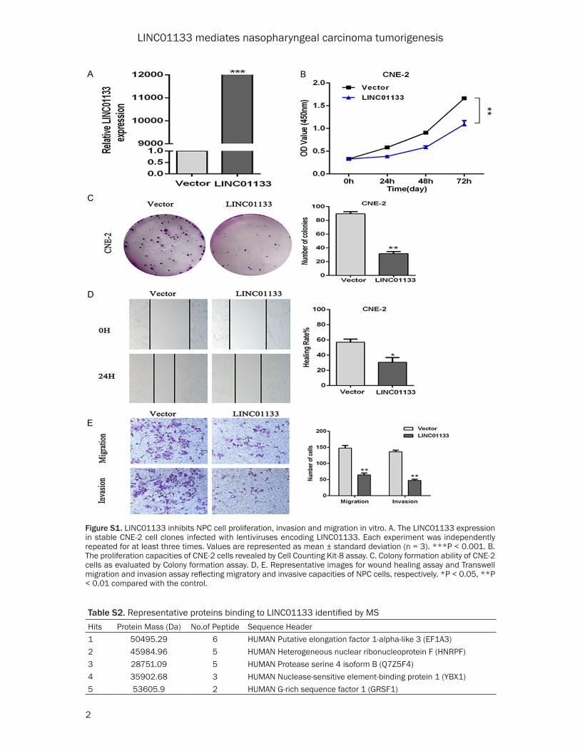
Table S2. Representative proteins binding to LINC01133 identified by MSHits Protein Mass (Da) No.of Peptide Sequence Header1 50495.29 6 HUMAN Putative elongation factor 1-alpha-like 3 (EF1A3)2 45984.96 5 HUMAN Heterogeneous nuclear ribonucleoprotein F (HNRPF)3 28751.09 5 HUMAN Protease serine 4 isoform B (Q7Z5F4)4 35902.68 3 HUMAN Nuclease-sensitive element-binding protein 1 (YBX1)5 53605.9 2 HUMAN G-rich sequence factor 1 (GRSF1)
Figure S1. LINC01133 inhibits NPC cell proliferation, invasion and migration in vitro. A. The LINC01133 expression in stable CNE-2 cell clones infected with lentiviruses encoding LINC01133. Each experiment was independently repeated for at least three times. Values are represented as mean ± standard deviation (n = 3). ***P < 0.001. B. The proliferation capacities of CNE-2 cells revealed by Cell Counting Kit-8 assay. C. Colony formation ability of CNE-2 cells as evaluated by Colony formation assay. D, E. Representative images for wound healing assay and Transwell migration and invasion assay reflecting migratory and invasive capacities of NPC cells, respectively. *P < 0.05, **P < 0.01 compared with the control.