Embed Size (px)
Citation preview
Int J Clin Exp Med 2015;8(6):9446-9453www.ijcem.com /ISSN:1940-5901/IJCEM0008318
Original ArticlePersonalised lamellar keratoplasty and keratopigmentation in Asian corneal leucoma patients
Xin Liu1*, Jun-Hui Shen1*, Qi Zhou1, Zhen-Xing Liu1, Shen-Fei Tang1, Ran-Ran Chen1, Gui-Qin Sui2, Yan-Long Bi1
1Department of Ophthalmology, Tongji Hospital Affiliated with Tongji University School of Medicine, Shanghai 200065, P. R. China; 2Department of Ophthalmology, Jilin University Bethune Second Hospital, 1085 Yatai Street, Changchun 130041, P. R. China. *Equal contributors.
Received March 22, 2015; Accepted June 3, 2015; Epub June 15, 2015; Published June 30, 2015
Abstract: Objective: To describe a personalised lamellar keratoplasty (LK) associated with the keratopigmentation (KTP) technique for corneal leucoma among Asian patients. Methods: This report was a non-randomised, retrospec-tive clinical study performed in 32 consecutive eyes of 32 patients to improve cosmetic appearance. Twenty-two patients underwent LK combined with KTP, either by intralamellar or superficial route. Ten patients underwent the single personalised keratopigmentation method. The subjective and objective cosmetic results, ocular irritation, colour fading, neovascularisation formation and incidence of immune rejection were evaluated until three years after surgery. Results: No complications occurred, and the corneal leucoma was successfully stained with India ink in all 32 patients. Most of the patients showed good cosmetic appearance. Pain, conjunctival congestion, corneal edema and foreign body sensation disappeared gradually within two to three weeks after surgery in all patients. Graft swelling, non-healing, or detaching was not observed during follow-up. However, two patients had slight opac-ity three years after LK. Colour fading was observed in one patient who underwent intralamellar corneal staining 10 months after surgery. Re-staining was performed. Conclusion: KTP combined with personalised LK is an effective personalised technique that presents long-standing colour staining and good cosmetic efficacy.
Keywords: Lamellar keratoplasty, keratopigmentation, corneal leucoma, partial lamellar keratoplasty
Introduction
Corneal leucoma frequently occurs in keratitis and corneal wound because of multiple factors, such as physical, chemical and congenital [1]. Corneal opacity tends to result in cosmetic problems. If the scar is located in the visual axis area, it can cause visual loss and even functional blindness. The treatments for this condition include functional and cosmetic approaches [2]. Corneal transplantation, such as penetrating keratoplasty (PK) and lamellar keratoplasty (LK), is an excellent treatment for corneal macula or for leucoma patients who want to rehabilitate their visual acuity. However, after corneal transplantation, many people encounter graft rejection and chronic endothe-lial cell loss [3]. Keratopigmentation (KTP) has been used for centuries for cosmetic purposes when visual reconstruction is ineffective or is not chosen as an option. Galen (131-201 A.D.) first used copper sulphate to colour the corneal
leucoma [4-6]. Later on, more chemical sub-stances, such as India ink, have been used by surgeons to shade the scars in corneal leucoma patients. Cosmetic contact lenses are known to be the most commonly used method to improve aesthetic appearance [7-9]. However, people may be intolerant to wearing contact lenses, refuse to have the prosthesis for psychological reasons, or develop chronic inflammation and infection [10].
Using KTP for cosmetic purposes is a good choice for patients who are intolerant to wear-ing or are unwilling to try contact lenses. Several cases using KTP for cosmetic purposes among leucoma patients have been reported [11, 12]. However, for patients whose corneal surface is unstable, such as corneal surfaces with angio-genesis, epithelial recurrent erosion and irregu-lar surface, this method is not appropriate because of the surgical challenge it presents and the many complications it can cause,
Personalised lamellar keratoplasty and keratopigmentation
9447 Int J Clin Exp Med 2015;8(6):9446-9453
Table 1. Summary of clinical data of 32 patients
Patient Sex Age Clinical diagnosis Number of years
Visual acuity (pre/post) Method of corneal tattooings Complications
1 M 27 Penetrating injury, Strabismus 19 NLP/NLP LK+ICS, Squint surgery none2 M 30 Injured by a stick, Iridodialysis, Cataract 20 LP/LP LK+ICS, Cataract extraction and Coreoplasty none3 M 34 Perforating injury, Aphakia, Disuse exotropia 29 0.01/LP SCS, Squint surgery none4 M 27 Perforating injury, Atretopsia, Anterior synechia 16 LP/LP ICS none5 M 26 Perforating injury, LK state 10 0.2/0.2 ICS none6 M 44 Perforating injury 22 NLP/NLP ICS none7 F 57 corneal ulcer in childhood 20 FC/0.01 LK+ICS none8 M 45 Perforating injury 40 0.1/0.1 SCS none9 M 36 Perforating injury, Iridodialysis, Cataract 18 NLP/LP LK+SCS, Coreoplasty, Phaco+IOL none10 M 25 Alkali burn, Cataract 12 NLP/NLP LK+ICS, Phaco+IOL none11 M 44 Penetrating injury 23 NLP/LP LK+ICS none12 M 47 Perforating injury 11 LP/0.02 LK+SCS none13 M 53 Perforating injury, Cataract 20 0.1/0.2 LK+ICS, Phaco+IOL none14 M 39 Herpes simplex keratitis 10 0.2/0.2 SCS none15 M 52 Penetrating injury 32 NLP/LP LK+ICS none16 M 28 Congenital corneal leucoma 28 0.01/0.02 LK+ICS none17 M 31 Perforating injury 16 0.01/0.01 ICS none18 F 44 Herpes simplex keratitis 10 NLP/NLP LK+ICS none19 M 46 Perforating injury 23 0.01/0.01 ICS none20 M 50 Injured by a stick, Strabismus 40 NLP/NLP LK+SCS, Squint surgery none21 M 53 Corneal ulcer 12 LP/LP LK+ICS none22 M 62 Perforating injury, Cataract 32 NLP/LP ICS, Phaco+IOL none23 M 41 Perforating injury 20 NLP/NLP LK+SCS none24 M 50 Perforating injury 15 NLP/NLP LK+ICS none25 M 28 Perforating injury 16 NLP/NLP LK+SCS none26 M 45 Herpes simplex keratitis 27 LP/0.02 LK+ICS none27 M 37 corneal ulcer 23 0.01/0.01 LK+SCS none28 M 36 Perforating injury 14 LP/0.02 LK+ICS none29 M 55 Perforating injury, Cataract 28 0.02/0.04 SCS, Phaco+IOL none30 M 45 Fungal corneal ulcer 24 HM/0.02 LK+SCS none31 M 33 Congenital corneal leucoma 33 LP/LP LK+ICS none32 M 45 Keratitis 28 0.01/0.01 LK+SCS none*M, Male; F, Female; NLP, No light perception; LP, light perception; FC, finger count; HM, hand motion.
Personalised lamellar keratoplasty and keratopigmentation
9448 Int J Clin Exp Med 2015;8(6):9446-9453
including pigment fading and recurrent erosion, among others. Thus, we introduced a new me- thod that combines KTP with LK that is likely to obtain better cosmetic efficacy. We developed a personalised, safe, durable and cosmetic sur-gical LK technique combined with KTP espe-cially suitable for Asians. We used India ink as the pigment for the KTP surgery either through the intralamellar corneal staining (ICS) or super-ficial corneal staining (SCS) route [13]. We stud-ied the cosmetic effect of KTP with and without the LK technique in treating cornea leucoma patients. We evaluated the postoperative ocu-lar discomfort and/or pain, medium-term dura-bility, patients’ subjective satisfaction and ob- jective cosmetic results.
Materials and methods
Patients
The Institutional Review Board of Tongji Eye Institute and the Research Ethics Committee of the University approved of this retrospective, non-randomised, non-comparative clinical ca- se series. The tenets of the Declaration of Helsinki were followed in the investigation. Corneal leucoma was diagnosed on the basis of the slit-lamp microscope findings. Before the surgery, visual quality, intraocular pressure, corneal topography, anterior segment optic coherence tomography (AS-OCT) and ocular B scan were conducted to estimate the overall corneal topography, which could help to safely prepare the lamellar pocket. The surgical indi-
glaucoma were excluded from the study. Thirty-two patients (32 eyes) were included in this study conducted from January 1, 2010 to April 30, 2011 (Table 1). Thirty patients were men (93.75%) and two were women (6.25%). The mean age was 21.6±8.4 years (range, 10-40 years old).
Surgical technique
The surgery was performed by a single surgeon (Dr Yanlong Bi). After retrobulbar anaesthesia (5 mL 2% lidocaine and 0.5% L-bupivacaine combined with one drop of 0.1% epinephrine), the lamellar pocket and the LK were performed manually. The diameter and depth of each indi-vidual’s lamellar dissection were evaluated by slit-lamp microscope and AS-OCT (Carl Zei- ssMeditec, Germany) before the surgery. For the LK patients, donor tissue (we used glycerin-20°C cryopreserved corneas) was thawed and rehydrated in normal saline solution for 30 min. A personalised outline of the corneal leucoma area was carved first, and then a 1/3-4/5 thick-ness of the anterior corneal lamellar stroma was excised depending on the location of the neovascularisation if present. The correspond-ing thickness of the donor tissue was moulded to match the size and shape of the recipient bed. Interrupted 10-0 nylon sutures were ap- plied, and the knots were buried.
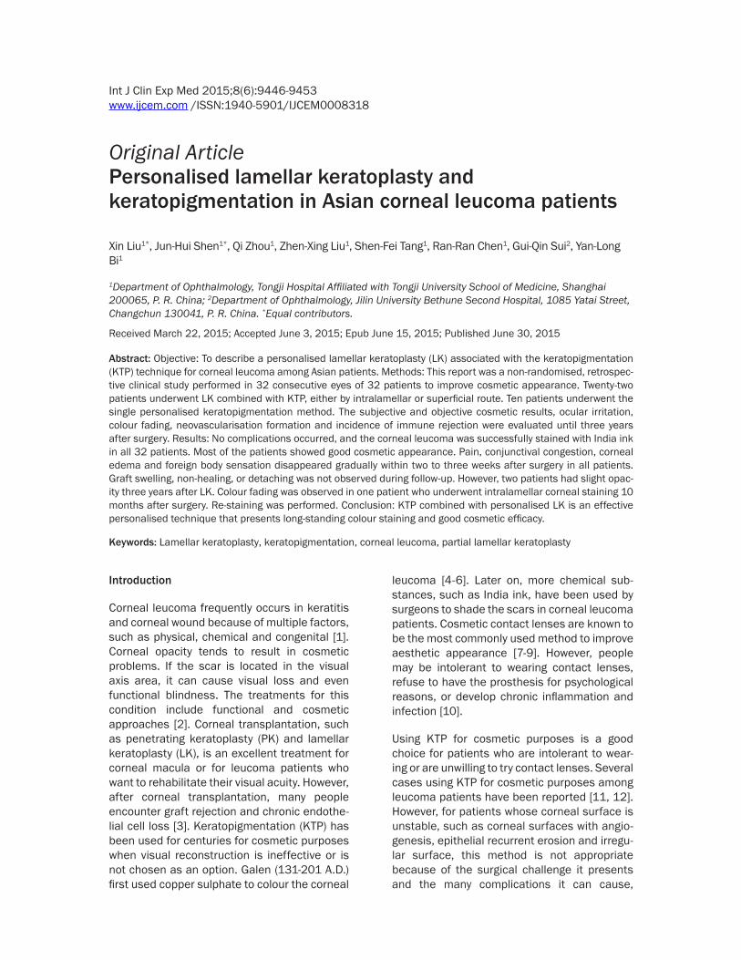
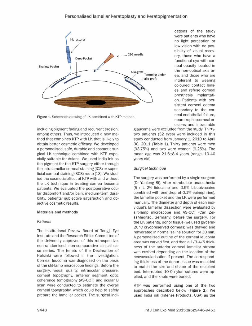
KTP was performed using one of the two approaches described below (Figure 1). We used India ink (Intenze Products, USA) as the
Figure 1. Schematic drawing of LK combined with KTP method.
cations of the study were patients who have no light perception or low vision with no pos-sibility of visual recov-ery, those who have a functional eye with cor-neal opacity located in the non-optical axis ar- ea, and those who are intolerant to wearing coloured contact lens-es and refuse corneal prosthesis implantati- on. Patients with per-sistent corneal edema secondary to the cor-neal endothelial failure, neurotrophic corneal er- osions and intractable
Personalised lamellar keratoplasty and keratopigmentation
9449 Int J Clin Exp Med 2015;8(6):9446-9453
dyeing pigment. The ink was sterilised at 121°C for 15 min in a sterile glass bottle before use. We used two colours, nigger-brown and gloss black, to match the corresponding eyes. In the ICS route, an angled crescent iris restorer was used to make a dissection into the corneal stro-ma. The dissection was created at 1/5-1/2 depth of the cornea, depending on the plane of the opacity. India ink was smeared on the pock-et by the same iris restorer. For LK patients, after the graft was sutured, the ink was smeared directly under the graft using the iris restorer. In the SCS route, micropunctures were made with a 23-gauge needle, which reached the stromal bed through the superficial layers. This manoeuvre was repeated until the staining achieved a satisfactory appearance. Unlike LK, the two KTP approaches generally do not need suturing. However, when the pocket is not closed sufficiently, suture is needed to prevent the ink from leaking. For the patients already suffering from corneal perforation, we made two unconnected pockets separately by using the central perforating banding to prevent the ink from leaking into the anterior chamber dur-ing or after the surgery. The central perforating band could be managed either by LK combined with ICS or by LK combined with SCS. Additional surgery, such as strabismus (two patients, 6.3%), cataract extraction (five patients, 15.6%) and coreoplasty (two patients, 6.3%), was per-formed when needed. In case of strabismus, adequate resection or recession of muscles
re given tobramycin dexamethasone eye drops (TobraDex, Alcon) for one week.
Postoperative evaluation
Postoperatively, slip-lamp microscope exami-nation was conducted to measure the changes in the graft, sutures, pigmentation fading and neovascularisation, among others. The satis-faction of the patients and observers was recorded using the protocol of Alio [12] at the time point of 36 months. The ocular situations of patients were quantified at the time point of one month: measuring pain (0-5), conjunctival congestion (0-5), foreign body sensation (0-5), corneal edema (0-5), colour fading (0-5) and intraocular irritation (0-5). The follow-up exami-nations were scheduled one to three days, one to three weeks and one to three months after surgery. The patients were followed-up every year until 36 months, postoperatively.
Results
No technical complications were encountered during surgery. No ink was found to leak during surgery or infiltrate into the anterior chamber after surgery. No anterior chamber inflamma-tion occurred, and re-epithelisation was com-pleted 5-14 days after surgery. Fourteen patients underwent LK combined with ICS (43.75%). Eight patients underwent LK com-bined with SCS (25%). Six patients underwent ICS only (18.75%), and four received SCS only
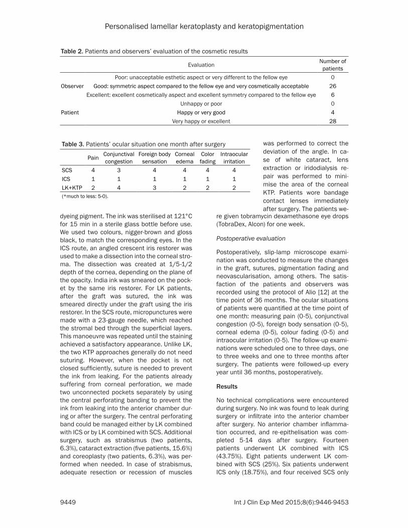
Table 2. Patients and observers’ evaluation of the cosmetic results
Evaluation Number of patients
Poor: unacceptable esthetic aspect or very different to the fellow eye 0Observer Good: symmetric aspect compared to the fellow eye and very cosmetically acceptable 26
Excellent: excellent cosmetically aspect and excellent symmetry compared to the fellow eye 6Unhappy or poor 0
Patient Happy or very good 4Very happy or excellent 28
Table 3. Patients’ ocular situation one month after surgery
Pain Conjunctival congestion
Foreign body sensation
Corneal edema
Color fading
Intraocular irritation
SCS 4 3 4 4 4 4ICS 1 1 1 1 1 1LK+KTP 2 4 3 2 2 2(*much to less: 5-0).
was performed to correct the deviation of the angle. In ca- se of white cataract, lens extraction or iridodialysis re- pair was performed to mini-mise the area of the corneal KTP. Patients wore bandage contact lenses immediately after surgery. The patients we-
Personalised lamellar keratoplasty and keratopigmentation
9450 Int J Clin Exp Med 2015;8(6):9446-9453
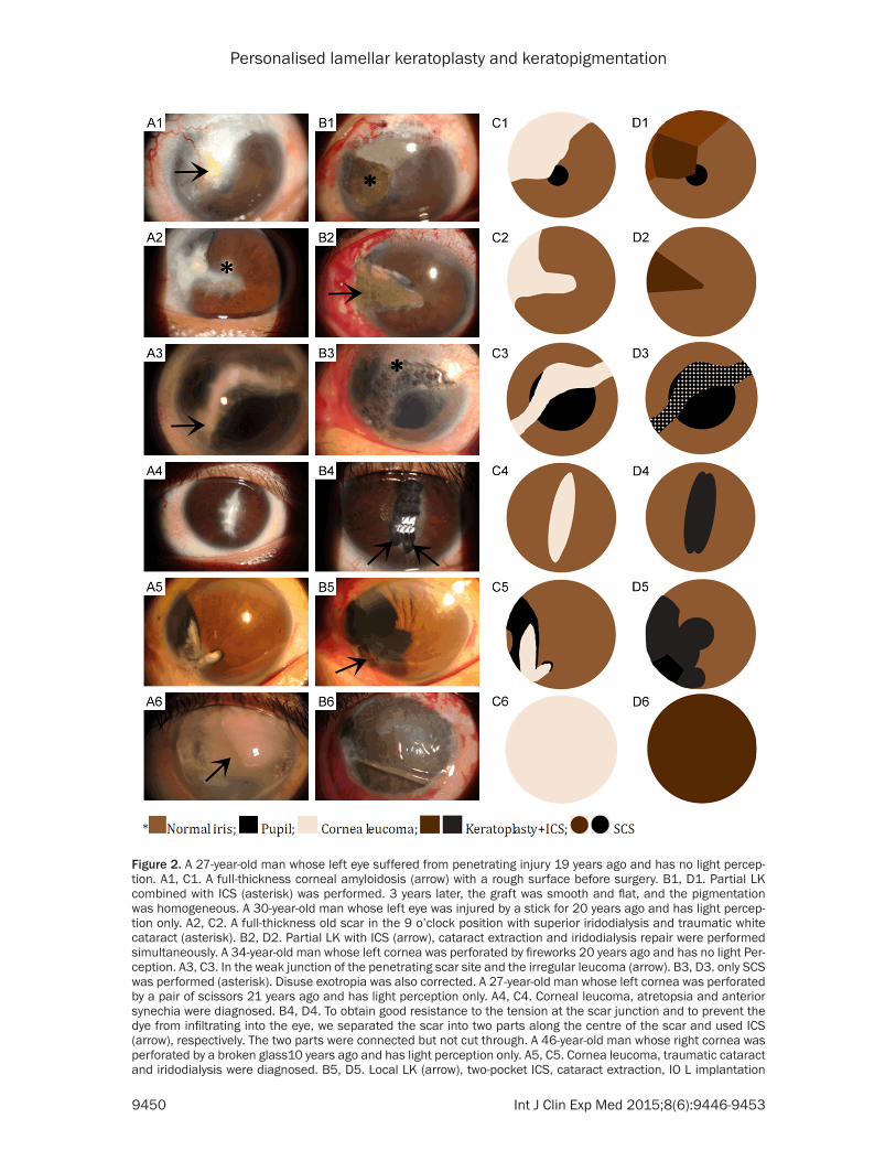
Figure 2. A 27-year-old man whose left eye suffered from penetrating injury 19 years ago and has no light percep-tion. A1, C1. A full-thickness corneal amyloidosis (arrow) with a rough surface before surgery. B1, D1. Partial LK combined with ICS (asterisk) was performed. 3 years later, the graft was smooth and flat, and the pigmentation was homogeneous. A 30-year-old man whose left eye was injured by a stick for 20 years ago and has light percep-tion only. A2, C2. A full-thickness old scar in the 9 o’clock position with superior iridodialysis and traumatic white cataract (asterisk). B2, D2. Partial LK with ICS (arrow), cataract extraction and iridodialysis repair were performed simultaneously. A 34-year-old man whose left cornea was perforated by fireworks 20 years ago and has no light Per-ception. A3, C3. In the weak junction of the penetrating scar site and the irregular leucoma (arrow). B3, D3. only SCS was performed (asterisk). Disuse exotropia was also corrected. A 27-year-old man whose left cornea was perforated by a pair of scissors 21 years ago and has light perception only. A4, C4. Corneal leucoma, atretopsia and anterior synechia were diagnosed. B4, D4. To obtain good resistance to the tension at the scar junction and to prevent the dye from infiltrating into the eye, we separated the scar into two parts along the centre of the scar and used ICS (arrow), respectively. The two parts were connected but not cut through. A 46-year-old man whose right cornea was perforated by a broken glass10 years ago and has light perception only. A5, C5. Cornea leucoma, traumatic cataract and iridodialysis were diagnosed. B5, D5. Local LK (arrow), two-pocket ICS, cataract extraction, IO L implantation
Personalised lamellar keratoplasty and keratopigmentation
9451 Int J Clin Exp Med 2015;8(6):9446-9453
(12.5%). Five patients used nigger-brown and 27 patients used gloss black to match their eyes.
Data on the 32 patients are shown in Table 1. Satisfaction evaluation is presented in Table 2. Patients’ ocular conditions one week after sur-gery are given in Table 3. No obvious colour fad-ing and migration of the staining were observed for most patients, and all patients accepted the corneal appearance within 36 months after surgery (Figure 2). In one patient who under-went ICS, fading of colour was observed 38 months after surgery, and re-staining was eas-ily performed by separating the original pocket. In two patients who underwent LK and KTP, partial graft opacification occurred 10 months after surgery, and they received re-staining after changing the graft. Graft-related compli-cations were not observed within the next two years’ follow-up. As analysed in Table 2, the cosmetic results showed that 28 patients were given excellent assessment, and four patients were given cosmetically acceptable assess-ment. From the observers’ assessment, 6 patients were rated excellent, 26 good and 0 poor. All patients stated that they would repeat the surgery if needed.
Discussion
Patients with a sightless and cosmetically impaired eye usually require a safe, stable and effective cosmetic method to improve their appearance. However, because of the high risk factors and limitations of PK [3, 14], ocular sur-face irritation of the cosmetic contact lenses [10] and psychological unwillingness to replace one’s own eyeball with an ocular prosthesis, the use of KTP with or without LK for cosmetic purposes is a good choice [15]. In this report, we presented an alternative personalised method to reconstruct the appearance of patients’ cornea. To the best of our knowledge, this report is the first to combine LK and KTP at the same time and to emphasise applying dif-ferent staining methods in the same cornea.
In the past, the two popular staining techniques used were transepithelial intrastromal micro-
puncture and staining the anterior stroma after epithelial debridement [16, 17]. Afterwards, a new technique called intrastromal lamellar pocket was used and proved to have a good outcome [11, 12, 18]. Alio described two differ-ent KTP approaches: ICS and SCS [12]. Ac- cording to recent studies, the advantages of SCS are simple manipulation by surgeons and low risk of perforation. Its side effect is its abil-ity to damage the corneal surface caused by the multiple iatrogenic punctures, which may cause persistent corneal surface instability, especially in cases with primary ocular surfac-es or corneal diseases. ICS has more advan-tages than SCS in the terms of homogeneous pigmentation, faster surgery, faster postopera-tive recovery, less stimulating sensations and long-term pigmentation [19-22]. However, ICS is not suitable for patients whose scars are located superficially, are irregular, or located at the junction of an old corneal penetrating area. SCS may be more suitable for these patients [14]. For those who have a stable and transpar-ent superficial corneal surface, both methods are suitable. However, both methods are not suitable for those with conditions of recurrent erosion and inflammation of the corneal epithe-lium, band keratopathy and corneal amyloido-sis, among others, because they could cause further damage. In these cases, we recom-mend LK combined with ICS [14-16].
In some cases, such as a full-thickness scar occurring as a result of corneal perforation, we can make two pockets along the bilateral sides of the scar band, but the two pockets should not be connected to prevent the ink from infil-trating into the anterior chamber. Then, SCS can be performed but only on the middle scar band if this joint is thin and weak. Preoperatively, AS-OCT and the whole cornea thickness evalu-ation can help to ensure the safety of this pro-cedure. If this scar joint is thick and has good resistance to tension, LK combined with ICS may be considered.
Concerning the pigment leakage or the colour changes, SCS is located superficially, and thus the mouth of the puncture cannot self-close
and coreoplasty were performed simultaneously. Visual acuity at the end of follow-up was 0.5. A 44-year-old man whose right eye was injured by fireworks 22 years ago and has no light perception. A6, C6. Whole cornea leucoma and central band keratopathy (arrow) were diagnosed. B6, D6. We performed calcified plaque scraping, EDTA che-lation and a single whole corneal pocket ICS with no sutures. A therapeutic contact lens was worn for two weeks.
Personalised lamellar keratoplasty and keratopigmentation
9452 Int J Clin Exp Med 2015;8(6):9446-9453
during the early days after surgery. Tearing caused by the stimulating sensations on the corneal surface after surgery usually leads to early colour fading. For ICS, pigment diffusion usually occurs in the open mouth of the pocket. For patients treated by LK combined with ICS, pigment leakage could occur in the graft mar-gin [12, 23]. Fortunately, no obvious pigment fading occurred during our follow-up, and only one patient observed colour fading 38 months after surgery. Re-staining was easily performed through the original pocket.
Concerning the selection of colour dye, our cases are all Asian patients, and their irises are nigger-brown. We initially chose the dark-brown dye similar to the iris. However, the colour appeared slightly whitish under naked eye observation. Thus, we choose the pure black dye for the rest of the patients. Although the black colour could appear improperly under the slit-lamp microscope observation, all patients acquired a satisfactory cosmetic appearance under naked eye observation.
In sum, our study showed that the personalised LK combined with KTP provided an alterna- tive treatment method for corneal leucoma patients. The method is safe, stable and rea-sonable, and it achieves satisfactory cosmetic results. Further studies are needed to deter-mine the stability and toxicity of the pigment and its long-term effects on the graft.
Acknowledgements
Supported by the Natural Science Foundation of China (NSFC: 81470028, to Yanlong Bi) and the Program for New Century Excellent Talents in University, (NCET: 13-0420, to Yanlong Bi).
Disclosure of conflict of interest
None.
Address correspondence to: Dr. Yan-Long Bi, De- partment of Ophthalmology, Tongji Hospital Affiliated with Tongji University School of Medicine, Shanghai 200065, P. R. China. E-mail: [email protected]
References
[1] Glassy CM, Glassy MS, Aldasouqi S. Tattooing: medical uses and problems. Cleve Clin J Med 2012; 79: 761-70.
[2] Chang KC, Kwon JW, Han YK, Wee WR, Lee JH. The epidemiology of cosmetic treatments for corneal opacities in a Korean population. Ko-rean J Ophthalmol 2010; 24: 148-54.
[3] Wu SQ, Zhou P, Zhang B, Qiu WY, Yao YF. Long-term comparison of full-bed deep lamellar keratoplasty with penetrating keratoplasty in treating corneal leucoma caused by herpes simplex keratitis. Am J Ophthalmol 2012; 153: 291-99.
[4] Van der Velden, Samderubun EM, Kok JH. Der-matography as a modern treatment for color-ing leucoma cornea. Cornea 1994; 13: 349-353.
[5] Holth S. Revival of Galen’s corneal staining with copper sulfate and tannine should be abandoned. Am J Ophthalmol 1931; 14: 378-379.
[6] Ziegler SL. Multicolor Tattooing of the Cornea. Trans Am OphthalmolSoc 1922; 20: 71-87.
[7] Hallock GG. Cosmetic trauma surgery. PlastRe-constrSurg 1995; 95: 380-381.
[8] Kuzan WM Jr. Plastic Surgery J Am Coll Surg 1999; 188: 171-77.
[9] Hoeyberhs JL. Fortnightly review: Cosmetic sur-gery. BMJ 1999; 318: 512-16.
[10] Custer PL, Kennedy RH, Woog JJ, Kaltreider SA, Meyer DR. Orbital implants in enucleation surgery. A report by the American Academy of Ophthalmology. Ophthalmol 2003; 110: 2054-61.
[11] Fogla R, Gupta A, Indumathy TR. Microkera-tome-assisted corneal tattooing: a case report. Cornea 2010; 29: 446-8.
[12] Kim C, Kim KH, Han YK, Wee WR, Lee JH, Kwon JW. Five-year results of corneal tattooing for cosmetic repair in disfigured eyes. Cornea 2011; 30: 1135-9.
[13] Alio JL, Sirerol B, Walewska-Szafran A, Miranda M. Corneal tattooing (keratopigmentation) with new mineral micronised pigments to restore cosmetic appearance in severely impaired eyes. Br J Ophthalmol 2010; 94: 245-9.
[14] Amesty MA, Alio JL, Rodriguez AE. Corneal tol-erance to micronised mineral pigments for keratopigmentation. Br J Ophthalmol 2014; 98: 1756-60.
[15] Alio JL, Rodriguez AE, Toffaha BT. Keratopig-mentation (corneal tattooing) for the manage-ment of visual disabilities of the eye related to iris defects. Br J Ophthalmol 2011; 95: 1397-401.
[16] Pitz S, Jahn R, Frisch L, Duis A, Pfeiffer N. Cor-neal tattooing: an alternative treatment for dis-figuring corneal scars. Br J Ophthalmol 2002; 86: 397-99.
[17] Mannis MJ, Eghbali K, Schwab IR. Keratopig-mentation: a review of corneal tattooing. Cor-nea 1999; 18: 633-37.
Personalised lamellar keratoplasty and keratopigmentation
9453 Int J Clin Exp Med 2015;8(6):9446-9453
[18] Panda A, Mohan M, Chawdhary S. Corneal tat-tooing-experiences with “lamellar pocket pro-cedure”. Indian J Ophthalmol 1984; 32: 408-11.
[19] Duggan JN, Nanavati BP. Tattooing of corneal opacity with gold and platinum chloride. Br J Ophthalmol 1936; 20: 419-25.
[20] Burris TE, Holmes-Higgin DK, Silvestrini TA. La-mellar intrastromal corneal tattoo for treating iris defects (artificial iris). Cornea 1998; 17: 169-73.
[21] Remky A, Redbrake C, Wenzel M. Intrastromal corneal tattooing for iris defects [letter]. J Cata-ract Refract Surg 1998; 24: 1285-7.
[22] Anastas CN, McGhee CN, Webber SK, Bryce IG. Corneal tattooing revisited: excimer laser in the treatment of unsightly leucomata. Aust NZ J Ophthalmol 1995; 23: 227-30.
[23] Hos D, Heindl LM, Bucher F, Cursiefen C. Novel lamellar, flap-based tattooing techniques for corneal opacities in scarred and vascularized blind eyes. Cornea 2015; 34: 82-6.


![Review Article Lamellar Keratoplasty: A Literature Reviewdownloads.hindawi.com/journals/joph/2013/894319.pdfJournal of Ophthalmology described by Melles et al. [ ] allowing transplantation](https://img.pdfslide.net/doc/110x75/5e39106a1415da08cf09cef9/review-article-lamellar-keratoplasty-a-literature-journal-of-ophthalmology-described.jpg)