Embed Size (px)
Citation preview
PARAGANGLIOMA OF THE HEAD AND NECK
LCDR IFEPO O. SOFOLAOCTOBER 5TH, 2000
Overview
• Introduction
• Historical Background and Terminology
• Anatomy and physiology
• Classification and Subtypes
• Work-up and Management
• Summary and Conclusions
Introduction
• Paragangliomas are benign tumors that arise from branchiomeric paraganglia distributed along the course of autonomic nerves from skull base to aortic arch
• Called nonchromaffin paragangliomas because they lack positive response to chromaffin staining associated with Neural crest tumors of adrenal gland
• Derived from Neural crest cells• Typically occur along course of major vasculature and
autonomic nerves
• Generally are benign, slow growing tumors
Introduction
• All paragangliomas are related to one another, and to pheochromocytomas of the adrenal gland
• In contrast to Pheochromocytomas, they rarely secrete catecholamines
• Consist of Type I or chief cells which are members of the amine precursor and uptake decarboxylase (APUD) family
• Type II cells are sustentacular cells• Nuclear polymorphism and cellular hyperchromatism
common : not considered evidence of malignancy• Can also be found in the orbit, larynx, paranasal sinuses
and oral cavity
Historical background and terminology
• Carotid body first described by anatomist Von Haller in 1743
• Renamed carotid gland after histologic identification of glandular acini
• Renamed vascular glomerulus or glomus in early 20th century
• Paraganglion first used by histologist Kohn while describing carotid body
• 1941, Guild described “glomic tissue” for vascularized tissue on promontory and jugular bulb
Historical background and terminology
• 1945, Rosenwasser reported a “carotid body tumor” of middle ear and mastoid
• Glenner and Grimmley distingushed adrenal (pheochromocytomas) and extra-adrenal paraganglionma
• Other terms like Chemodectomas and nonchromaffin tumors used
• CURRENTLY, PARAGANGLIOMA BASED ON ANATOMIC LOCATION IS PROPER TERMINOLOGY (e.g. carotid paraganglioma, jugulotympanic paraganglioma)
Background and Terminology
• Carotid body tumor ( carotid paraganglioma, chemodectoma)
• Intravagal Paragangliomas• Jugulotympanic Paragangliomas
– Glomus tympanicum
– Glomus jugulare
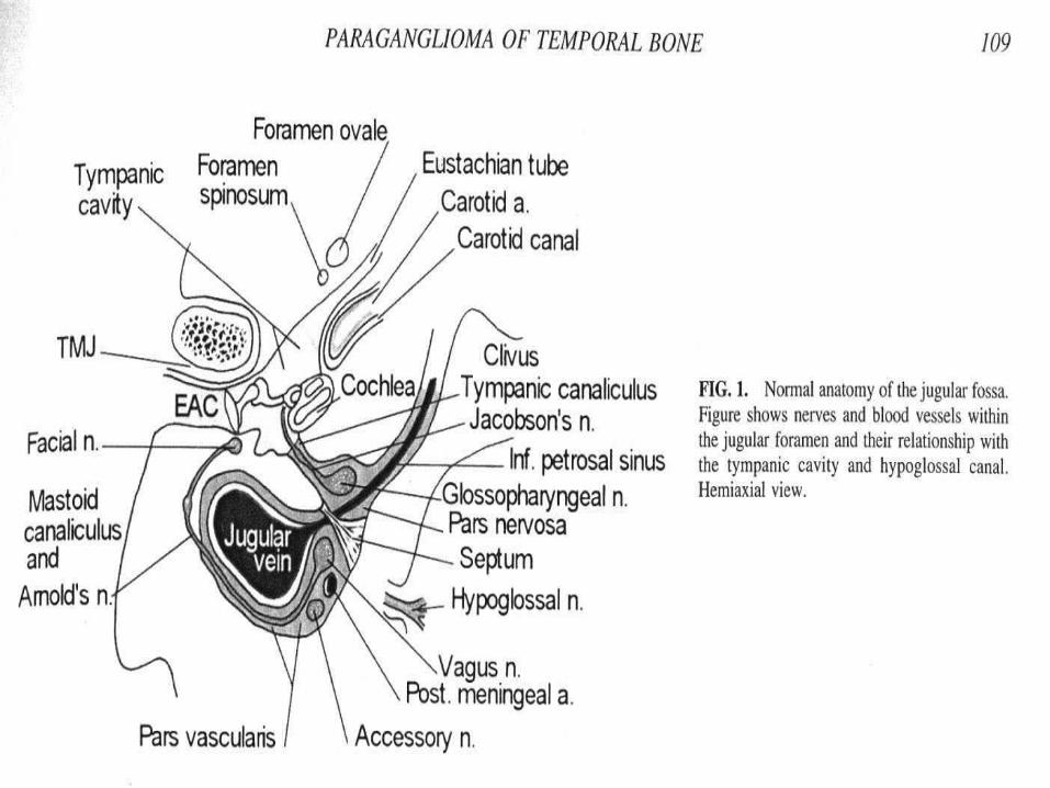
Anatomy and Physiology
• Carotid Body and Sinus• Jugular Foramen/ temporal bone
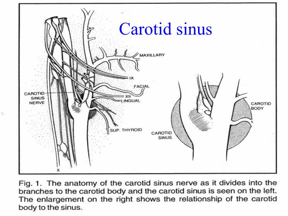
Carotid Sinus
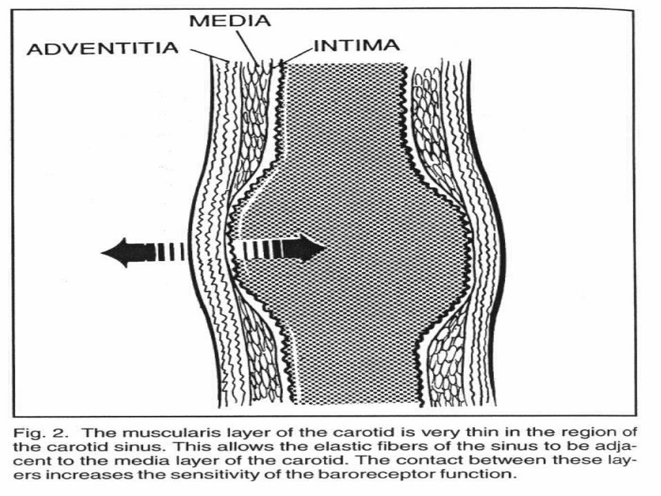
• Composed of Stretch receptors which lie in the adventitia of carotid bulb
• First described by Heath in 1850• Further identified by Manson in 1862 from his study of
post-mortem exam of inmates• Described areas of thinning of wall of carotid sinus area
with absence of muscle fibers in the tunica media, but plentiful stretch fibers, receptors and nerve endings
• Innervation by afferent fibers from carotid body (1-2cm from bifurcation) to form Carotid sinus nerve
Carotid sinus
Carotid Body
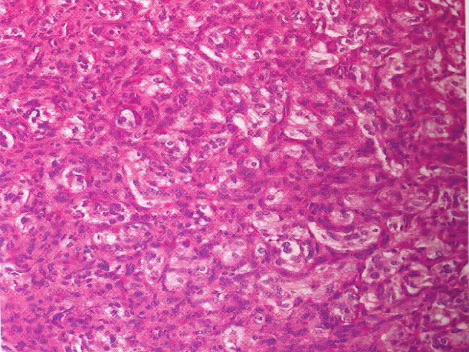
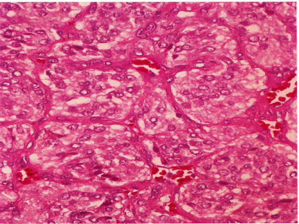
• One of Nonchromaffin paraganglia• Associated with Parasympathetic nerves• Composed of clusters of epithelium-like cells in a richly
vascular connective tissue stroma numbering 20• Divided into smaller units or Zellballen which are
comprised of 2 receptor cell types namely Chief Cells ( type 1, epithelioid , which contain catecholamine-bound neurosecretory granules) and Type 2 sustentacular cells( supporting cells)
• Bilateral location constant
Carotid Body
• Connected to carotid bifurcation by ligament of Mayer• Blood supply from 1-2mm artery arising from bifurcation• Average size 5 x 3 x 1.5mm• Size increases with age and associated with high altitude
dwellers• Average wt = 12.1 mg, range 1.0 – 47.4mg• Supplied by the Nerve of Hering, which is a branch of the
glossopharyngeal nerve ( Proven by DeCastro’s sectioning of CN IX, then observing neural degeneration).
• Minor input from CN X and sympathetic trunk
Carotid Body/Sinus Physiology
• Function as chemoreceptor and baroreceptor complex• Chemoreceptor mediated by carotid body, sensitive to
changes in PaO2, PaCO2, pH and blood flow• Daly, et al demonstrated blood flow to this tissue to be 4x
thyroid and 3x brain, thus very vascular• Constant output from CB participating in ventilation:
Lower PaO2 leads to increase in firing within carotid sinus nerve affecting increase ventilation.
• Higher PaCO2 or lower pH also increase ventilation
Carotid Body/Sinus Physiology
• Baroreceptor effect mediated through carotid sinus, transmitted by nerve of hering, through CN IX, to tractus solitarius
• Carotid sinus made of type I and type II barorecptors which together temper extreme variations in blood pressure
• Resultant parasympathetic response leads to vasodilation and decreased heart rate and contractility in response to increased stimulation of baroreceptors
• Decrease baroreceptor activity decreases parasympathetic depression of heart and peripheral vasculature
Carotid Body Tumor
• Most common paraganglioma of head and neck comprising 60% of total
• Thought to be more common in people living in high altitudes
• Commonly occur in 5th decade• Higher incidence in females
• Typically present as a slow growing painless neck mass, which is mobile antero-posteriorly, but immobile cranio-caudally
• May bulge through parapharyngeal area into OC/OP
Carotid Body Tumor
• May present with hoarseness, VC paralysis, dysphagia, local discomfort, Headaches , blurred vision
• 10% are multicentric• 20% are familial with a tendency for multicentricity in
50% of the familial cases• Inheritance is AD (Mendelian) modified by genomic
imprinting meaning that tumor develops (expression) when gene is paternally inherited and not when maternally inherited
• 8 – 10% are malignant, more common in familial variant
Carotid Body Tumor
• No histologic criteria for malignancy: malignancy diagnosed by evidence of spread to regional lymph nodes or distant sites ( lung, bone, liver, pancreas, thyroid, kidney, breast)
• Pathophysiology related to mass and pressure effect of tumor on carotid body/sinus
• Can cause symptoms by physically expanding and compressing surrounding structures, especially nerves, and can be lethal if untreated
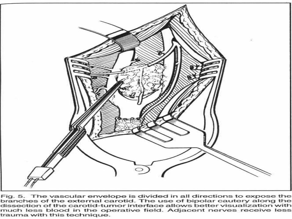
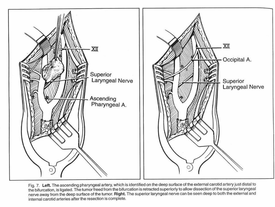
• Typically supplied by ascending pharyngeal artery
Carotid Body Tumor (DDX)
• Brachial cleft cyst, harmatoma• Lymph node, SCCA• Salivary gland tumor• Neurofibroma, neuroblastoma, ganglion intercortium
• Carotid anourysm• Plasmacytoma• Hemangiopericytoma, myoblastoma,• Angioendolethioma, chromaffinoma, endothelioma• Angioma, Angiosarcoma, carcinoid, carcinoma, sarcoma
Carotid Body Tumor (work-up)
• FNA not advised as excessive bleeding will occur. Typically non-diagnostic due to excess blood in specimen
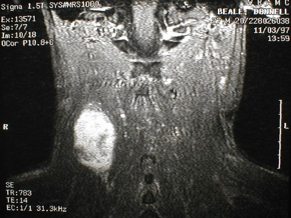
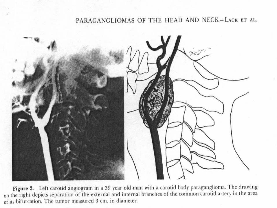
• MRI, CT with contrast and angiogram mainstay• Typical Lyre sign and tumor blush seen on angio• Angiogram also used to evaluate adequacy of carotid
system• Balloon test occlusion performed to evaluate circle of
willis• Urine VMA, catecholamines , metanephrines• Abdominal CT if symptoms of catecholamines or positive
Carotid Body Tumor (work-up)
• If familial, must evaluate for MEN syndromes• If familial, risk individuals older than 16-18 yrs should be
examined and screened with MRI every 2 years• Octreotide scanning has been used for multicentric tumors
or to screen family members• Genetic screening emerging as a method of evaluation
( chromosome 11q13 and q23)
Carotid body tumor ( management)
• Surgery is mainstay for management• If excised, may recur in 10% of cases• Generally regarded as radio-resistant, XRT reserved for
incompletely excised tumors ( with intracranial extension), very large tumors, elderly patient, poor surgical candidates, or multicentricity
• Embolization typically performed 24 – 48 hrs prior to surgery. Questionable as to whether this decreases intraoperative bleeding. May also set up inflammatory response which makes surgery more difficult
Carotid body tumor ( management)
• Overall operative mortality of 2-8%, stroke rate of 20%, CN X and XII damage rate of 40-50%
• Shamblin classification of operative morbidity based on tumor size– Group I tumors small and easy to remove
– Group II tumors medium size , with attachment to carotid
– Group III large with transmural invasion of carotid requiring resection and grafting
Carotid body tumor ( management)
• McCaffrey found that carotid paragangliomas measuring greater than 5cm had operative complication rate of 67% compared to 14% if less than 5cm
• Post-operative denervation with sympathetic discharge can lead to high and fluctuating BP
• Managed with nitroprusside initially, then Clonidine ( central alpha-2 agonist), phenoxybenzamine ( alpha-1 blockade) in abour 3 – 4 days post-op
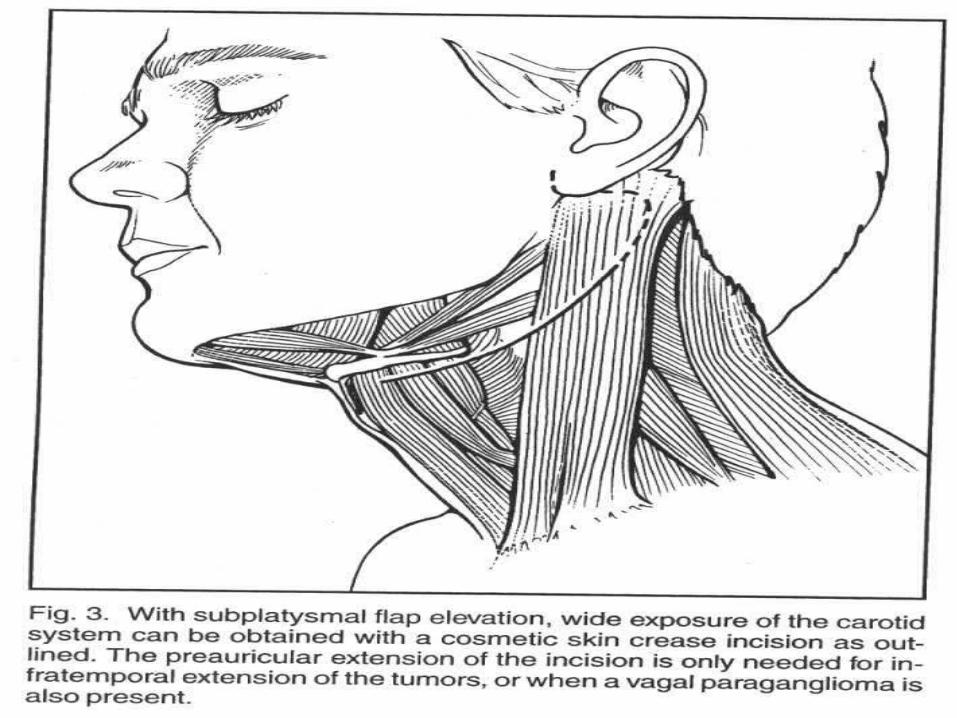
Carotid body tumor ( Surgical pearls)
• Exposure must be liberal and adequate so that control of the vessel proximal and distal to lesion is possible
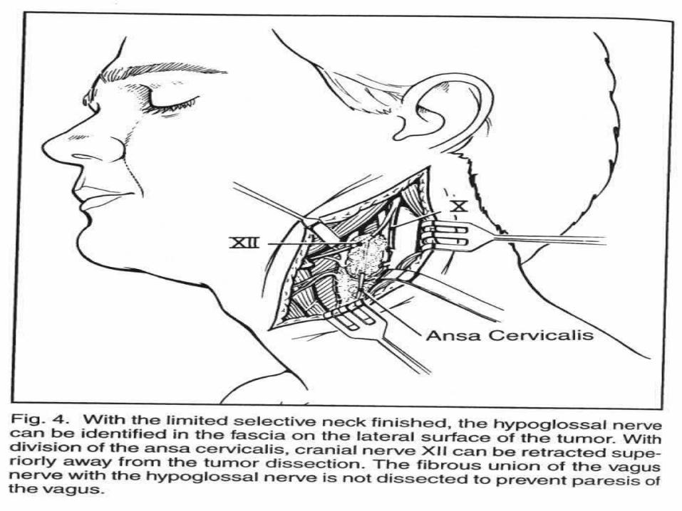
• Identify CN IX, X, XI, XII , Sympathetic chain, and protect VII before tumor is resected
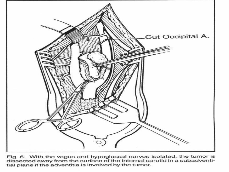
• Be prepared to resect carotid and graft ( have vascular surgery on stand-by)
• Be prepared for fluctuations in blood pressure during and after resection
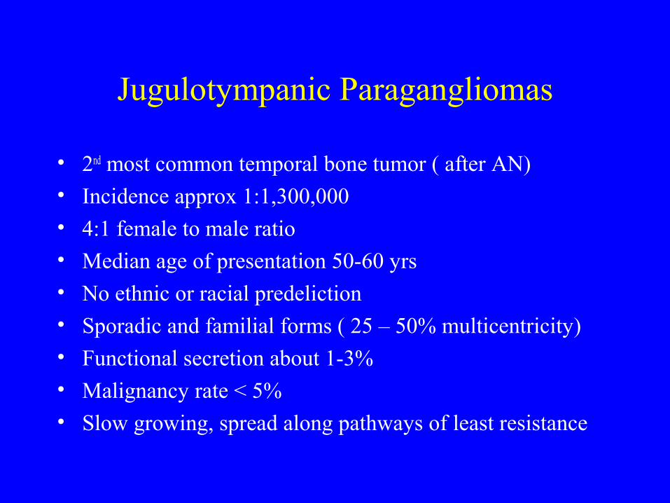
Jugulotympanic Paragangliomas
• 2nd most common temporal bone tumor ( after AN)• Incidence approx 1:1,300,000• 4:1 female to male ratio• Median age of presentation 50-60 yrs
• No ethnic or racial predeliction• Sporadic and familial forms ( 25 – 50% multicentricity)• Functional secretion about 1-3%• Malignancy rate < 5%• Slow growing, spread along pathways of least resistance
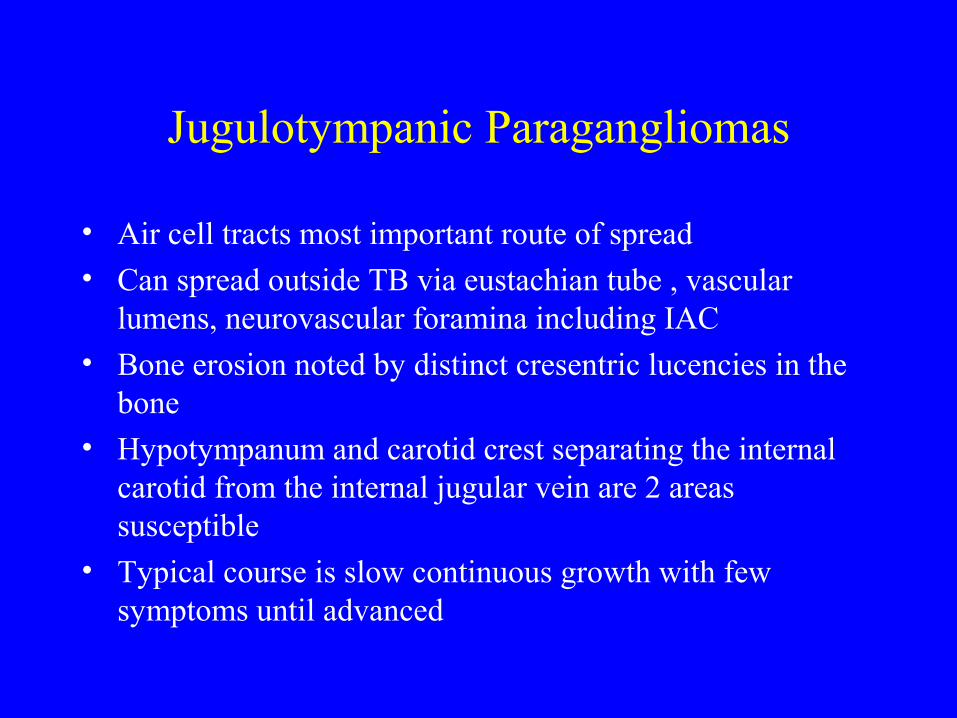
Jugulotympanic Paragangliomas
• Air cell tracts most important route of spread• Can spread outside TB via eustachian tube , vascular
lumens, neurovascular foramina including IAC• Bone erosion noted by distinct cresentric lucencies in the
bone• Hypotympanum and carotid crest separating the internal
carotid from the internal jugular vein are 2 areas susceptible
• Typical course is slow continuous growth with few symptoms until advanced
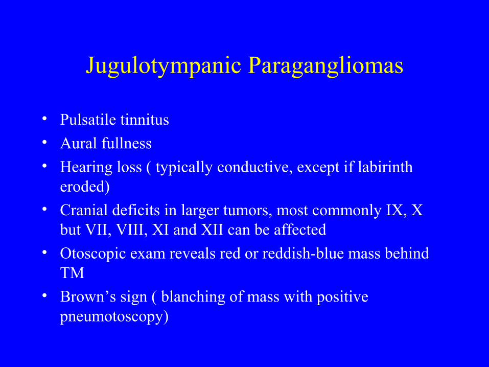
Jugulotympanic Paragangliomas
• Pulsatile tinnitus• Aural fullness • Hearing loss ( typically conductive, except if labirinth
eroded)• Cranial deficits in larger tumors, most commonly IX, X
but VII, VIII, XI and XII can be affected
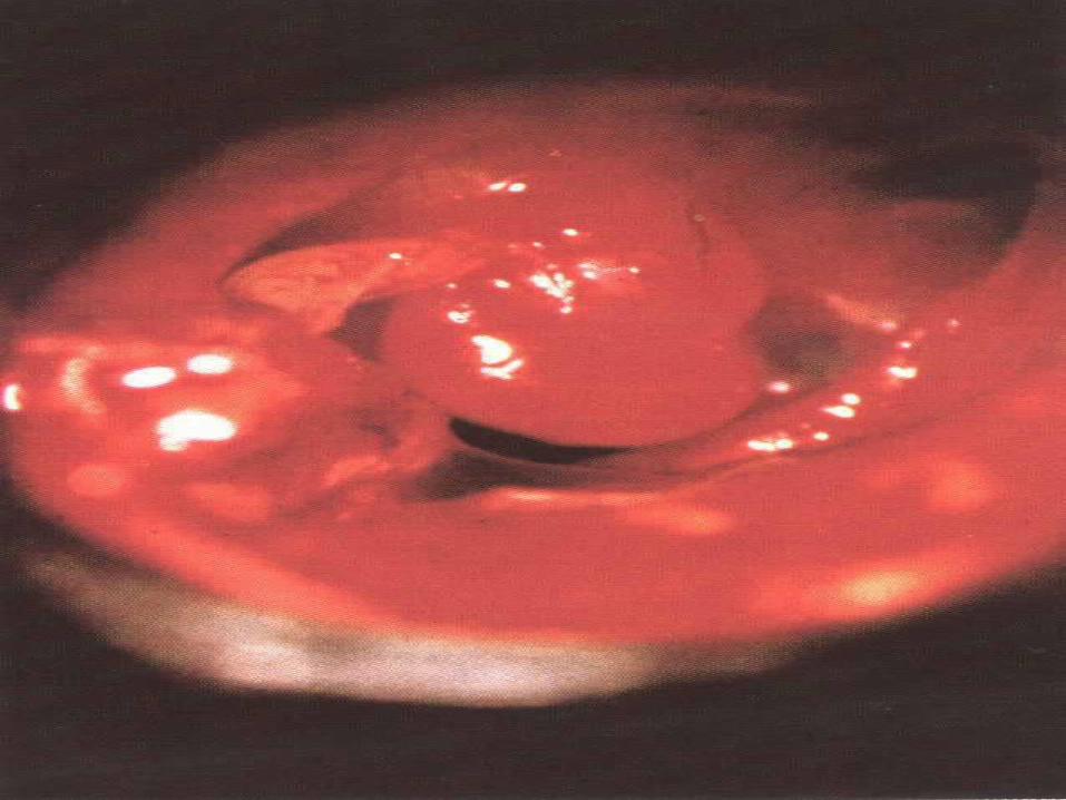
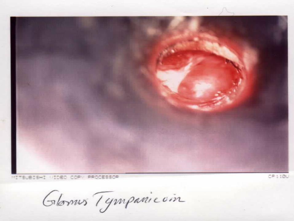
• Otoscopic exam reveals red or reddish-blue mass behind TM
• Brown’s sign ( blanching of mass with positive pneumotoscopy)
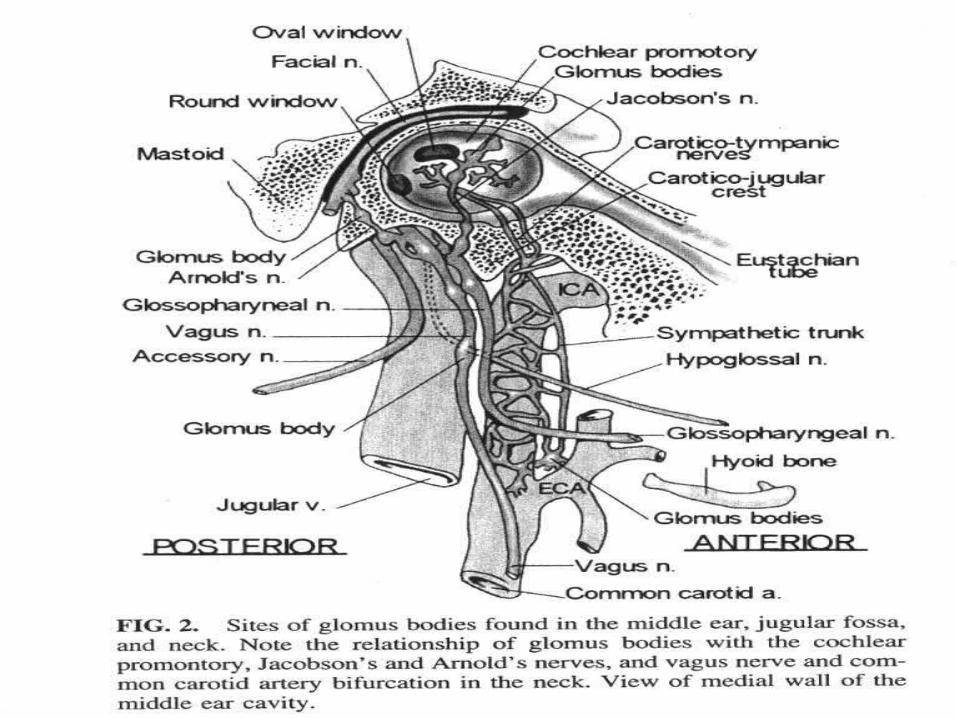
Jugulotympanic Paragangliomas
• Can occur in the adventitia of the jugular vein ( glomus jugulare tumor) in 85% of cases
• Also can occur from Jacobson’s nerve, a branch of the glossopharyngeal nerve ( glomus tympanicum) in 12% of cases
• Arnolds nerve , a branch of CN X, gives rise to glomus tympanicum in 3% of cases
Jugulotympanic Paragangliomas (Evaluation)
• Full audiogram with tympanometry• Fine cut CT of Temporal bone with contrast ( evaluate
bone erosion, relationship to VII, cochlea and ICA)• MRI to evaluate intracranial and intradural extension, and
further define soft-tissue relationships• 4 vessel arteriography to evaluate multicentricity, feeding
vessels, embolization, BOT• FNA not advised• VMA, metanephrines, catecholamines• Abdominal CT if symptomatic ( flushing, HTN etc)
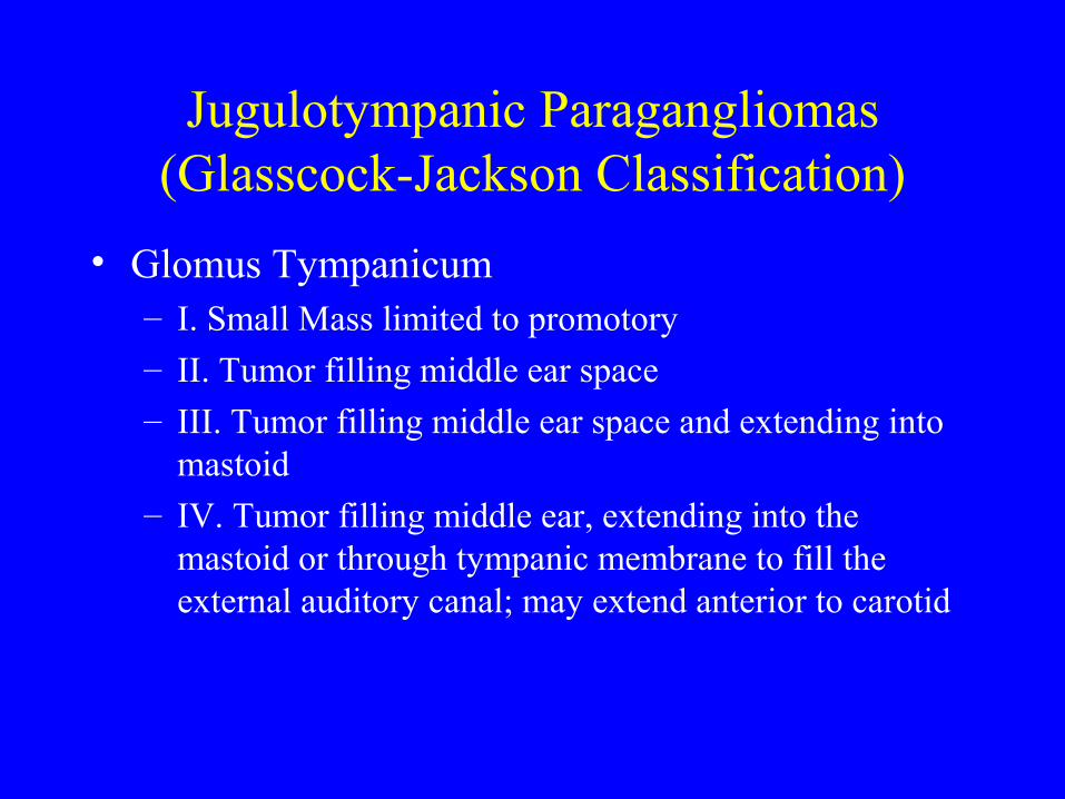
Jugulotympanic Paragangliomas (Glasscock-Jackson Classification)
• Glomus Tympanicum– I. Small Mass limited to promotory– II. Tumor filling middle ear space– III. Tumor filling middle ear space and extending into
mastoid
– IV. Tumor filling middle ear, extending into the mastoid or through tympanic membrane to fill the external auditory canal; may extend anterior to carotid
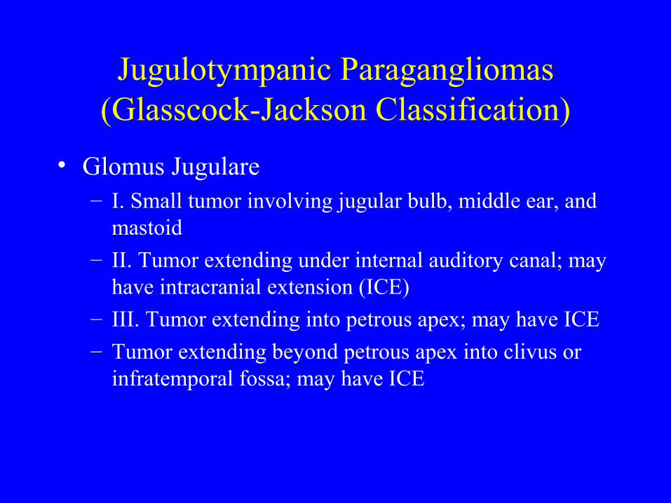
Jugulotympanic Paragangliomas (Glasscock-Jackson Classification)
• Glomus Jugulare– I. Small tumor involving jugular bulb, middle ear, and
mastoid– II. Tumor extending under internal auditory canal; may
have intracranial extension (ICE)– III. Tumor extending into petrous apex; may have ICE
– Tumor extending beyond petrous apex into clivus or infratemporal fossa; may have ICE

Fisch Classification (1981)
• Class A - Middle ear, strictly promontory• Class B - Tympanomastoid area• Class C - Carotid
– C1 Carotid Foramen
– C2 Vertical segment of carotid canal
– C3 Horizontal segment of carotid canal
– C4 Foramen lacerum and cavernous sinus
Fisch Classification (contd)
• Class D - Dural
• Extradural– De1: Intercranial Extradural extension < 2cm
– De2: Intracranial Extradural extension > 2cm
• Intradural– Di1: Intracranial Intradural extension < 2cm
– Di2: Intracranial Intradural extention > 2cm
• Arnold and Guiliam Classification not commonly used
Jugulotympanic Paragangliomas (Management)
• Decisions based on extent of tumor, and must be individualized to patient
• Considered to be more radiosensitive than carotid paragangliomas
• Goal of surgery is total or near-total excision• Younger patients, larger tumors with CN compromise tend
to be better candidates for surgery and tend to compensate better than those without pre-existing CN deficits
• Patients with secreting tumors require surgery
Jugulotympanic Paragangliomas (Management)
• Patients 65yrs and older, poor surgical candidates, and patients with multicentric tumors, XRT should be encouraged
• Always a risk of re-growth, osteoradionecrosis of temporal bone
• ORN risk is low if optimal dose of XRT of 35Gy/3wks or 45Gy/4weeks is used
• Vascular and Neurosurgical consultation encouraged prior to surgery
Glomus Jugulare (surgical approaches)
• Glasscock and Jackson Class I and II or Fisch C1 or C2 can be resected with extended facial recess approach
• Infratemporal fossa approach for tumors beyond petrous apex or involve cavernous sinus
• Tumors with intracranial extension may be resected with inratemporal fossa approach
• Retrosigmoid and/or suboccipital approach may be necessary for tumors with extension into posterior cranial fossa
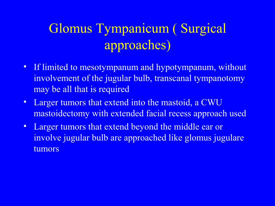
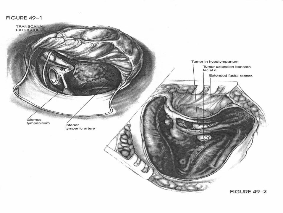
Glomus Tympanicum ( Surgical approaches)
• If limited to mesotympanum and hypotympanum, without involvement of the jugular bulb, transcanal tympanotomy may be all that is required
• Larger tumors that extend into the mastoid, a CWU mastoidectomy with extended facial recess approach used
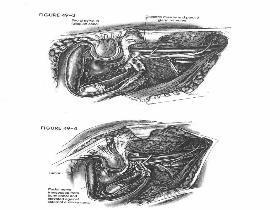
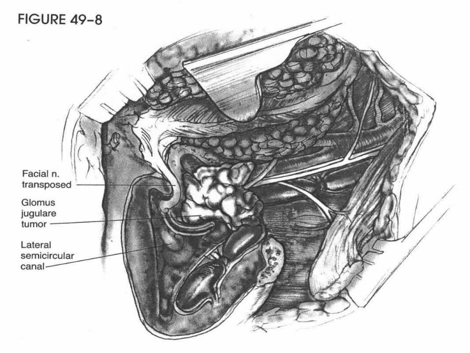
• Larger tumors that extend beyond the middle ear or involve jugular bulb are approached like glomus jugulare tumors
Intravagal Paragangliomas
• Most commonly occur at the level of the nodose ganglion• May occur at any point along course of vagus nerve• Mean age of presentation is 50 yrs• More common in females than males
• Painless mass posterior to angle of mandible• May bulge into pharynx and cause dysphagia• Complaints of tongue weakness, hoarseness, horner’s
syndrome ( Jugular foramen syndromes)• Malignancy rate higher than others, estimated at 18%
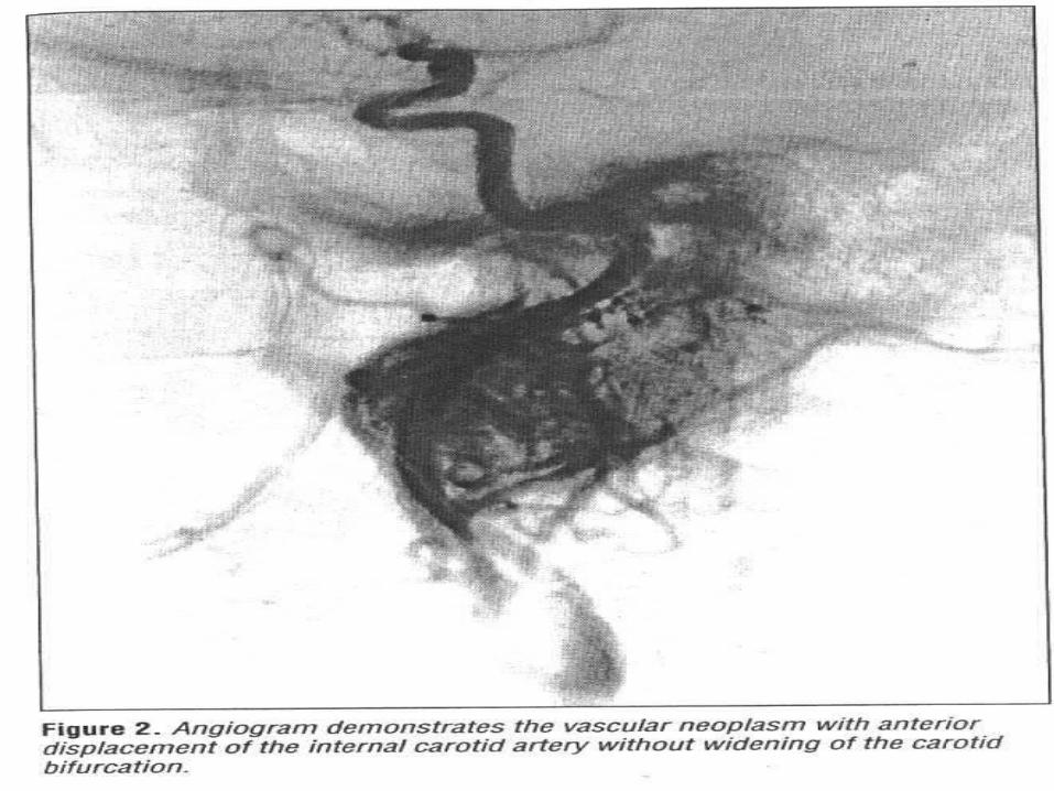
Intravagal Paragangliomas
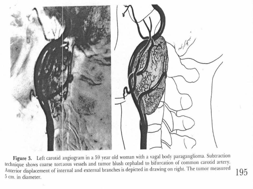
• Imaging studies include CT and MRI• Angiography demonstrates tumor blush and displacement
of ICA anteriorly and medially ( no widening of bifurcation)
• Only one functional vagal paraganglioma reported• Netterville reported familial occurrence as 20%
• Also noted 78% multicentricity in familial vs 23% in sporadic cases
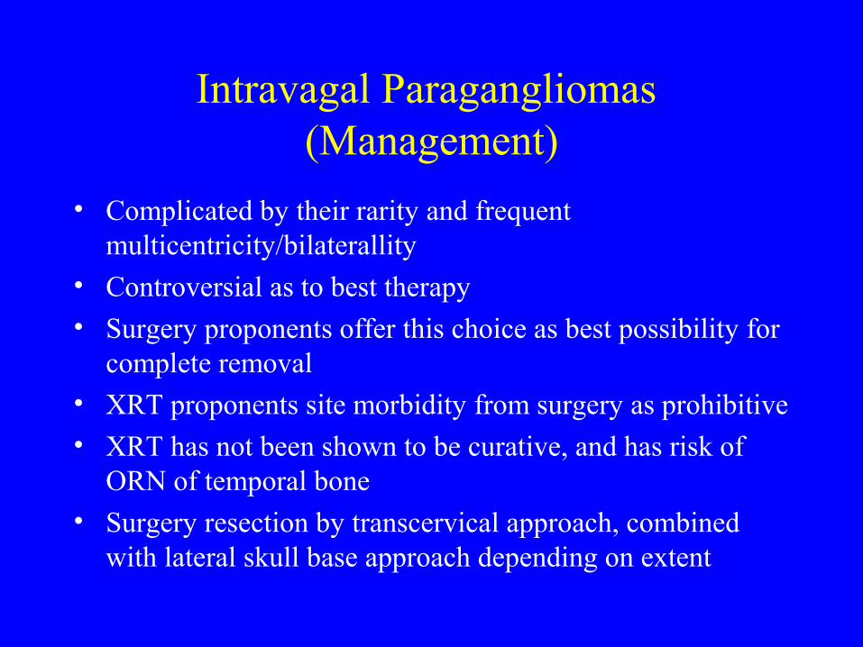
Intravagal Paragangliomas (Management)
• Complicated by their rarity and frequent multicentricity/bilaterallity
• Controversial as to best therapy• Surgery proponents offer this choice as best possibility for
complete removal• XRT proponents site morbidity from surgery as prohibitive
• XRT has not been shown to be curative, and has risk of ORN of temporal bone
• Surgery resection by transcervical approach, combined with lateral skull base approach depending on extent
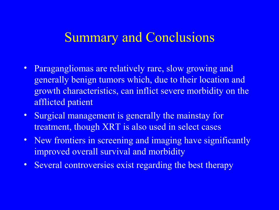
Summary and Conclusions
• Paragangliomas are relatively rare, slow growing and generally benign tumors which, due to their location and growth characteristics, can inflict severe morbidity on the afflicted patient
• Surgical management is generally the mainstay for treatment, though XRT is also used in select cases
• New frontiers in screening and imaging have significantly improved overall survival and morbidity
• Several controversies exist regarding the best therapy
References
• Netterville, JL. Carotid Body Tumors: A review of 30 patients with 46 Tumors. Laryngoscope. 1995;105:115-126
• Jackson, GC. Glomus Tumors: Diagnosis, Classification, and management of Large Lesion. Archives of Otolaryngology. 1982;108:401-406.
• Gardner, P. Carotid Body Tumors, Inheritance and a High Incidence of Associated Ceervical Paragangliomas. Am J Surg. 1996;172:196-199.
• McCaffrey, TV. Familial Paragangliomas of the Head and Neck. Arch Otolaryngol Head Neck surg. 1994;120:1211-1216.
• Bikhazi PH. Familial Paragangliomas: The Emerging Impact of Molecular Genetics on Evaluation and Management. Am J Otol 1999 Sep;20(5):639-643.
References (Contd)
• Netterville, JL. Vagal Paraganglioma: A review of 46 Patients treated During a 20 year period. Arch otolaryngol Head Neck surg. 1998;124:1133-1140.
• Urquhart, AC. Glomus Vagale: Paraganglioma of the Vagus Nerve. Laryngoscope.1994;104:440-445.
• Sykes, JM. Paragangliomas of the Head and Neck. Otolaryngologic Clinics of North America. Vol.19, No.4, Nov. 1986.
• Fisch U. Classification of Glomus Temporale tumors. In : Fisch U, Mattox D Eds. Microsurgery of the Skull Base. Stuttgart and New York, Georg Thieme, 1988: 149-153.
• Megerian, CA. Non-paraganglioma Jugular Foramen Lesions Masquerating as Glomus Jugulare Tumors. Am J otol. 1995;v.16, no.1:94-98.
References (Contd)
• Maier, W. Paraganglioma as a systemic syndrome: Pitfalls and Strategies. J Laryngol Otol. 1999;113:978-982.
• Kim, J. Optimum Dose of Radiotherapy for Chemodectomas of the Middle Ear. Int.J radiation Oncology Biol. Phys. 1980;6:815-819.
• Sillars, HA. The Management of Multiple Paraganglioma of The Head and Neck. Journal of Laryngology and Otology. 1993; 107:538-542.
• Noujaim, SE. Paraganglioma of the temporal Bone: Role of Magnetic Resonance Imaging versus Computed Tomography. Topics in Magnetic resonance Imaging. 2000;11(2):108-122.
• Shamblin, WA. Carotid Body Tumor ( Chemodectoma): Clinicopathologic Analysis of Ninety Cases. Am J Surg. 1971;122:732-739.