Embed Size (px)
Citation preview
The EMBO Journal vol.13 no.3 pp.699-703, 1994
Evidence of selection for protein introns in the RecAs ofpathogenic mycobacteria
Elaine O.Davis, Harry S.Thangaraj,Patricia C.Brooks and M.Joseph ColstonThe Laboratory for Leprosy and Mycobacterial Research, The NationalInstitute for Medical Research, The Ridgeway, Mill Hill, LondonNW7 1AA, UK
Communicated by J.J.Skehel
Protein introns are recently discovered genetic elementswhose intervening sequences are removed from aprecursor protein by an unusual protein splicing reaction.This involves the excision of a central spacer molecule,the protein intron, and the religation of the amino- andcarboxy-terminal fragments of the precursor. The recAgene of Mycobacterium tuberculosis contains one suchelement and we now show that the other majormycobacterial pathogen, Mycobacterium leprae, alsopossesses a protein intron in its recA, although othermycobacterial recA genes do not. However, these twoprotein introns are different in size, sequence and locationof insertion of their coding sequences into the recAs ofM.tuberculosis and M.leprae, indicating that acquisitionof the protein introns has occurred independently in thetwo species, and thus suggesting that there has beenselection for splicing in the maturation of RecA in thepathogenic mycobacteria. The M.leprae protein intronprovides an example of conditional protein splicing,splicing occurring in M.leprae itself but not whenexpressed in Escherichia coli, unlike most previouslydescribed protein introns. These observations suggest thatprotein introns may perform a function for their host,rather than being just selfish elements.Key words: leprosy/mycobacteria/protein intron/proteinsplicing/tuberculosis
IntroductionGenes containing insertion elements which are removed notfrom the RNA but from a precursor protein have recentlybeen identified. The first report described a subunit of thevacuolar ATPase of the yeast Saccharomyces cerevisiae(Hirata et al., 1990; Kane et al., 1990). Subsequently therecombination gene recA of Mycobacterium tuberculosis(Davis et al., 1991, 1992) and a DNA polymerase gene ofThermnococcus litoralis (Hodges et al., 1992; Perler et al.,1992) have also been shown to contain 'protein introns', thelatter containing two such sequences. The most recent proteinintron to be identified is again in a vacuolar ATPase, in thepathogenic yeast Candida tropicalis (Gu et al., 1993). Ineach case homology to examples of the equivalent gene fromother organisms is disrupted by an unrelated sequence, theprotein intron.
Evidence that splicing occurs at the protein level ratherthan the RNA level includes (i) pulse-chase analysis
Oxford University Press
demonstrating conversion of a large precursor protein to thetwo normal products using a slow processing mutant, (ii)the requirement for translation to proceed right through theprotein intron and (iii) the observations that mutations atsplice sites which alter amino acid sequence inhibit theprocess whereas those which change only nucleic acidsequence do not (for reviews see Shub and Goodrich-Blair,1992; Wallace, 1993). Studies using the yeast system haveconfirmed that a new peptide bond is formed between thetwo halves of the spliced protein (Cooper et al., 1993). Theprocess is expected to be autocatalytic based on the rangeof heterologous systems where splicing still occurs.
In at least two of the known examples the excised proteinintron has endonuclease activity, recognizing and cutting atthe DNA sequence into which its coding DNA is normallyinserted (Gimble and Thorner, 1992; Perler et al., 1992)and the yeast example will insert into an intronless allelein a homing process (Gimble and Thorner, 1992) analogousto that of group I mobile RNA introns (Perlman and Butow,1989). Although the homology between these protein intronsis not great, some motifs can be discerned. In each case thereis an identifiable pair of dodecamer sequences bearingsimilarity to the LAGLIDADG motif at similar spacing tothose found in some other endonucleases, especially thehoming endonucleases encoded by some group I RNAintrons (Doolittle, 1993), suggesting that the endonucleasefunction may be a common feature. In addition, there areconserved features around the splice sites with a cysteineor serine residue at the first splice site and a hexapeptidemotif at the second splice site (Davis et al., 1991; Hodgeset al., 1992; see Results).
Following our observation with M. tuberculosis, thecausative agent of tuberculosis in man, we have nowinvestigated the recA genes of other members of the genus.Many species of mycobacteria are harmless environmentalorganisms, although some are opportunistic pathogens orpathogens for animals. In addition to M. tuberculosis the othermycobacterial species which is a natural pathogen of manis Mycobacterium leprae which causes leprosy. Here wereport that the recA gene of M. leprae also contains a proteinintron, while the recA genes of other mycobacteria do not.Furthermore, these protein introns appear to have beenacquired independently by the two pathogenic species. Basedon our observations we propose that protein introns may havea role to play in their bacterial hosts' survival rather thanbeing just selfish elements.
Results
Cloning the M.leprae recA geneFollowing our observation with the recA gene ofM.tuberculosis, we investigated the presence of similarprotein intron sequences in other mycobacteria by Southernhybridization using intron-encoding DNA; only organismsbelonging to the M. tuberculosis complex gave positive
699
E.O.Davis et al.
hybridization (data not shown), suggesting that proteinsplicing was not a common feature of mycobacterial RecAs.We then cloned the M. leprae recA gene from cosmid clonesof the recA region of the M. leprae genome (Eiglmeier et al.,1993) after analysis by hybridization with probes from eachend of the M. tuberculosis recA gene to identify a restrictionfragment containing the whole gene. A single fragmenthybridizing to both probes of a suitable size for cloning wasfound for only two of 24 enzymes tested, and one of these,a 6.5kb SphI fragment, was cloned into pTZ18R to givepEJ216.
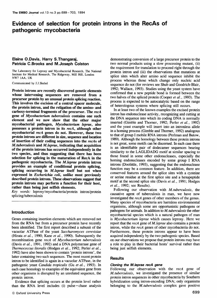
Further mapping and hybridization to this subcloneidentified the locations at which the two probes bound fromrestriction sites which they spanned. In particular both probesbound to a common 1.8 kb MscI fragment in addition toa second fragment which was different for each probe,indicating that the two probes bound - 1.8 kb apart, sucha distance that an intervening sequence must be present inthe M. leprae recA gene even though the M. tuberculosisintron did not hybridize (Figure 1). It was confirmed thatno rearrangement had occurred during the cloning procedureby hybridization to genormic M. leprae DNA digested withfour different enzymes using this fragment as a probe,revealing bands seen in this clone or the original cosmid (datanot shown).
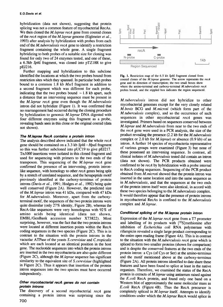
The M.leprae RecA contains a protein intronThe analysis described above indicated that the whole recAgene should be contained on a 3.3 kb SphI -HpaI fragmentso this was further subcloned into pUC19 to give pEJ217.Tn]O00 insertions were isolated in this cloned fragment andused for sequencing with primers to the two ends of thetransposon. This sequencing of the M.leprae recA geneconfirmed the presence of an intervening, protein intron-like sequence, with homology to other recA genes being splitby a stretch of unrelated sequence, and the hexapeptide motifpreviously identified at the carboxy-terminus of proteinintrons (Davis et al., 1991; Hodges et al., 1992) being quitewell conserved (Figure 2A). However, the predicted sizeof the M. leprae intron was 41 kDa, compared with 47 kDafor M. tuberculosis. Moreover, apart from the carboxy-terminal motif, the sequences of the two protein introns werequite dissimilar (only 27% identity, Figure 2B), whereas theRecA-like sequences were very homologous with 92% ofamino acids being identical (data not shown,EMBL/GenBank accession number X73822). Mostsurprising, however, was the fact that the two protein intronswere located at different insertion points within the RecAcoding sequences in the two species (Figure 2C). This is incontrast to the situation with the protein introns in thevacuolar ATPase of the yeasts S. cerevisiae and C. tropicaliswhich are each located at an identical position in the hostgene. The nucleotide sequences into which the protein intronsare inserted in the two mycobacteria are also quite different(Figure 2C), although the M. leprae sequence has significantsimilarity to the equivalent site of S. cerevisiae (highlightedin Figure 2C). Thus it appears that insertion of the proteinintron sequences into the two species must have occurredindependently.
Other mycobacterial recA genes do not containprotein intronsThe discovery of a second mycobacterial recA genecontaining a protein intron was surprising since the700
=.Ei 0-
ciI I
QFL:5 nE
a0
.. Arec,
N-probe C-probe1 kb
I ..- -- -.. ...............
Reglion Sequenced
Fig. 1. Restriction map of the 6.5 kb SphI fragment cloned fromcosmid clones of the M.leprae genome. The arrow represents the recAgene and its direction of transcription; the two small boxes showwhere the amino-terminal and carboxy-terminal M.tuberculosis recAprobes bound, and the stippled box indicates the region sequenced.
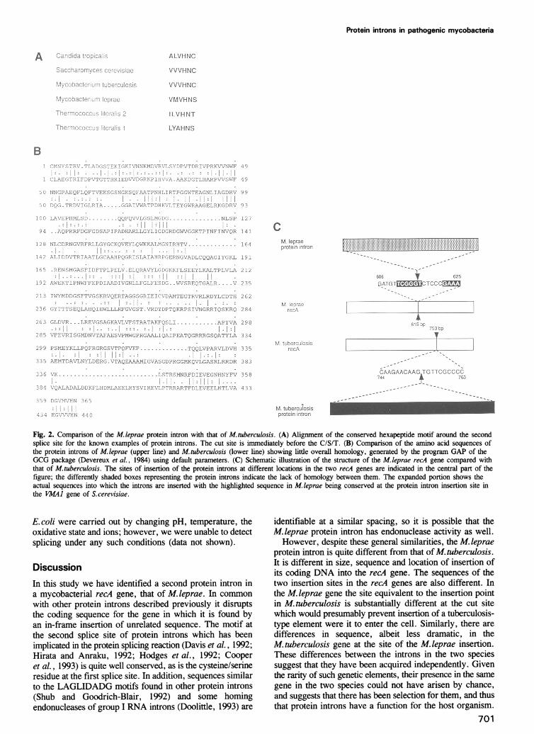
M. tuberculosis intron did not hybridize to othermycobacterial genomes except for the very closely relatedM. bovis BCG and M. microti (which form part of theM. tuberculosis complex), and so the occurrence of suchsequences in other mycobacterial recA genes wasinvestigated. Primers based on sequences conserved betweenM. leprae and M. tuberculosis from near to the two ends ofthe recA gene were used in a PCR analysis, the size of theproduct revealing the presence (2.2 kb for the M. tuberculosiscomplex or 2.0 kb for M. leprae) or absence (0.9 kb) of anintron. A further 14 species of mycobacteria representativeof various groups were examined (Figure 3) but none ofthese possessed an intervening sequence. In contrast, allclinical isolates of M. tuberculosis tested did contain an intron(data not shown). The PCR products obtained wereconfirmed to be recA by hybridization with a probe internalto RecA coding sequences. Sequencing of the PCR productobtained from M.microti showed that the protein intron wasinserted in the same location and into the same sequence asin M. tuberculosis, and that the sequences at the two endsof the protein intron itself were also identical, in accord withthese two species belonging to the M. tuberculosis complex.It would therefore appear that the presence of protein intronsin mycobacterial RecAs is confined to the M. tuberculosiscomplex and M. leprae.
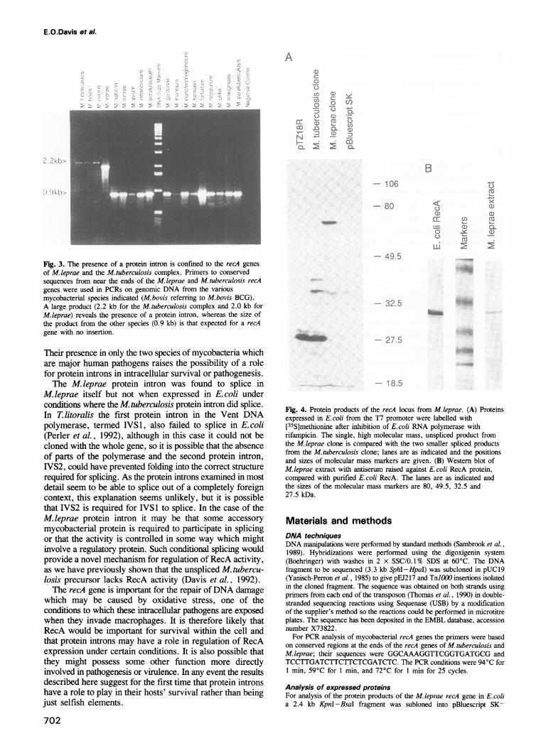
Conditional splicing of the M.leprae protein intronExpression of the M. leprae recA gene from a T7 promoterand labelling of its products with [35S]methionine afterinhibition of Escherichia coli RNA polymerase withrifampicin revealed a single large product corresponding tothe entire open reading frame (Figure 4A). This is in contrastto the situation with the M. tuberculosis recA gene which isspliced to form two smaller proteins (shown for comparison)and is despite the conserved features at the two ends of theprotein intron: a Cys (of Cys or Ser) at the amino-terminusand the motif mentioned above at the carboxy-terminus(Figure 2A). All protein introns identified to date share thesefeatures and have been found to splice at least in the nativeorganism. Therefore, we examined the status of the RecAprotein in extracts of M. leprae using antiserum raised againstthe E. coli RecA protein, and found only one band on aWestern blot of approximately the same molecular mass asE. coli RecA (Figure 4B). Thus the RecA precursor iscompletely spliced in M. leprae itself. Attempts to identifyconditions under which the M. leprae RecA would splice in
Protein introns in pathogenic mycobacteria
M. leprae
prote;n in'rc0
M -7 I G 1; T,!"! 1..;., I."i ij
F V
p p p r
.. HN,;:. E EGN N
Ak
71
CA.AGAAC AAG TGITCotCO'CCAL-
M. tubercuo0sisProtein intron
Fig. 2. Comparison of the M. leprae protein intron with that of M. tuberculosis. (A) Alignment of the conserved hexapeptide motif around the second
splice site for the known examples of protein introns. The cut site is immnediately before the C/SIT. (B) Comparison of the amino acid sequences of
the protein introns of M.leprae (upper line) and M.tuberculosis (lower line) showing little overall homology, generated by the program GAP of the
GCG package (Devereux et al., 1984) using default parameters. (C) Schematic illustration of the structure of the M.leprae recA gene compared with
that of M.tuberculosis. The sites of insertion of the protein introns at different locations in the two recA genes are indicated in the central part of the
figure; the differently shaded boxes representing the protein introns indicate the lack of homology between them. The expanded portion shows the
actual sequences into which the introns are inserted with the highlighted sequence in M.leprae being conserved at the protein intron insertion site in
the VMAJ gene of S.cerevisiae.
E. coli were carried out by changing pH, temperature, the
oxidative state and ions; however, we were unable to detect
splicing under any such conditions (data not shown).
Discussion
In this study we have identified a second protein intron in
a mycobacterial recA gene, that of M. leprae. In common
with other protein introns described previously it disrupts
the coding sequence for the gene in which it is found by
an in-frame insertion of unrelated sequence. The motif at
the second splice site of protein introns which has been
implicated in the protein splicing reaction (Davis et al., 1992;
Hirata and Anraku, 1992; Hodges et al., 1992; Cooper
et al., 1993) is quite well conserved, as is the cysteine/serineresidue at the first splice site. In addition, sequences similar
to the LAGLIDADG motifs found in other protein introns
(Shub and Goodrich-Blair, 1992) and some homing
endonucleases of group I RNA introns (Doolittle, 1993) are
identifiable at a similar spacing, so it is possible that the
M. leprae protein intron has endonuclease activity as well.
However, despite these general similarities, the M. leprae
protein intron is quite different from that of M. tuberculosis.
It is different in size, sequence and location of insertion of
its coding DNA into the recA gene. The sequences of the
two insertion sites in the recA genes are also different. In
the M. leprae gene the site equivalent to the insertion pointin M. tuberculosis is substantially different at the cut site
which would presumably prevent insertion of a tuberculosis-
type element were it to enter the cell. Similarly, there are
differences in sequence, albeit less dramatic, in the
M. tuberculosis gene at the site of the M. leprae insertion.
These differences between the introns in the two species
suggest that they have been acquired independently. Given
the rarity of such genetic elements, their presence in the same
gene in the two species could not have arisen by chance,
and suggests that there has been selection for them, and thus
that protein introns have a function for the host organism.
701
A ALVHNJC
VVVfANC
VVVHN-C
Vr.IVHNS
ILIVHNT
LYAHU S
.l t'
... C .
2. C. E,
4
-j
E -.
I
E.O.Davis et al.
A
b'1, .)
Fig. 3. The presence of a protein intron is confined to the recA genesof M.leprae and the M.tuberculosis complex. Primers to conservedsequences from near the ends of the M. 1eprae and M. tuberculosis recAgenes were used in PCRs on genomic DNA from the variousmycobacterial species indicated (M.bovis referring to M.bovis BCG).A large product (2.2 kb for the M.tuberculosis complex and 2.0 kb forM.leprae) reveals the presence of a protein intron, whereas the size ofthe product from the other species (0.9 kb) is that expected for a recAgene with no insertion.
Their presence in only the two species of mycobacteria whichare major human pathogens raises the possibility of a rolefor protein introns in intracellular survival or pathogenesis.The M. leprae protein intron was found to splice in
M. leprae itself but not when expressed in E. coli underconditions where the M. tuberculosis protein intron did splice.In T. litoralis the first protein intron in the Vent DNApolymerase, termed IVS 1, also failed to splice in E. coli(Perler et al., 1992), although in this case it could not becloned with the whole gene, so it is possible that the absenceof parts of the polymerase and the second protein intron,IVS2, could have prevented folding into the correct structurerequired for splicing. As the protein introns examined in mostdetail seem to be able to splice out of a completely foreigncontext, this explanation seems unlikely, but it is possiblethat IVS2 is required for IVS1 to splice. In the case of theM. leprae protein intron it may be that some accessorymycobacterial protein is required to participate in splicingor that the activity is controlled in some way which mightinvolve a regulatory protein. Such conditional splicing wouldprovide a novel mechanism for regulation of RecA activity,as we have previously shown that the unspliced M. tubercu-losis precursor lacks RecA activity (Davis et al., 1992).The recA gene is important for the repair of DNA damage
which may be caused by oxidative stress, one of theconditions to which these intracellular pathogens are exposedwhen they invade macrophages. It is therefore likely thatRecA would be important for survival within the cell andthat protein introns may have a role in regulation of RecAexpression under certain conditions. It is also possible thatthey might possess some other function more directlyinvolved in pathogenesis or virulence. In any event the resultsdescribed here suggest for the first time that protein intronshave a role to play in their hosts' survival rather than beingjust selfish elements.
702
B- 106
- 80
--- 49-5
C)
0
l.
._
cJ
E.
x
0Q
CD 0-
0~L
--- 32 5
27, 5
.. 18.5
Fig. 4. Protein products of the recA locus from M. leprae. (A) Proteinsexpressed in E. coli from the T7 promoter were labelled with[35S]methionine after inhibition of E.coli RNA polymerase withrifampicin. The single, high molecular mass, unspliced product fromthe M. leprae clone is compared with the two smaller spliced productsfrom the M. tuberculosis clone; lanes are as indicated and the positionsand sizes of molecular mass markers are given. (B) Western blot ofM. leprae extract with antiserum raised against E. coli RecA protein,compared with purified E.coli RecA. The lanes are as indicated andthe sizes of the molecular mass markers are 80, 49.5, 32.5 and27.5 kDa.
Materials and methodsDNA techniquesDNA manipulations were performed by standard methods (Sambrook et al.,1989). Hybridizations were performed using the digoxigenin system(Boehringer) with washes in 2 x SSC/0.1% SDS at 60°C. The DNAfragment to be sequenced (3.3 kb SphI-HpaI) was subcloned in pUC19(Yanisch-Perron et al., 1985) to give pEJ217 and TnlO00 insertions isolatedin the cloned fragment. The sequence was obtained on both strands usingprimers from each end of the transposon (Thomas et al., 1990) in double-stranded sequencing reactions using Sequenase (USB) by a modificationof the supplier's method so the reactions could be performed in microtitreplates. The sequence has been deposited in the EMBL database, accessionnumber X73822.For PCR analysis of mycobacterial recA genes the primers were based
on conserved regions at the ends of the recA genes of M. tuberculosis andM.leprae; their sequences were GGCAAAGGTTCGGTGATGCG andTCCTTGATCTTCTTCTCGATCTC. The PCR conditions were 94°C for1 min, 59°C for 1 min, and 72°C for 1 min for 25 cycles.
Analysis of expressed proteinsFor analysis of the protein products of the M.leprae recA gene in E.colia 2.4 kb KpnI-BsaI fragment was subloned into pBluescript SK-
(:D
0
-)
C.)Cl)0
CODC)
OC 0N
C!)
0 CO
Cl)a nCL U)
coa
__
Protein introns in pathogenic mycobacteria
(Stratagene) such that expression would be from the T7 promoter, yieldingplasmid pE1243. Strain BL21(DE3) (Studier et al., 1990; a lysogen of BL21containing a chromosomal gene for T7 RNA polymerase under control ofthe lacUV5 promoter) carrying pEJ243 or pEJ135 [Davis et al., 1991; theM.tuberculosis recA cloned in pTZ18R (Pharmacia)] was grown in M9minimal medium plus required supplements to an A6W of -0.5 and theninduced by addition of IPTG to 0.4 mM for 30 min; rifampicin was addedto 200 jig/ml for 30 min, then [35S]methionine (40 /Ci/ml) was added for10 min before harvesting. Samples equivalent to 100 j1d of culture wererun on a 12.5% SDS-polyacrylamide gel, which was fixed with 25%propan-2-ol/10% acetic acid, treated with Amplify (Amersham), dried andautoradiographed, the film being exposed for 2 h.M. leprae cell free extract was prepared as described by Ibrahim et al.
(1990). Western blotting was performed by standard techniques (Sambrooket al., 1989).
AcknowledgementsWe thank S.West of ICRF for the generous gift of anti-RecA (Ecoli)antiserum, S.Cole of Institut Pasteur for providing cosmid clones of theM.teprae recA region, J.J.McFadden of the University of Surrey forsupplying genomic DNA of M.paratuberculosis and S.G.Sedgwick andP.J.Jenner of the National Institute for Medical Research for helpfulsuggestions.
ReferencesCooper,A.A, Chen,Y.-J., Lindorfer,M.A. and Stevens,T.H. (1993) EMBO
J., 12, 2575-2583.Davis,E.O., Sedgwick,S.G. and Colston,M.J. (1991) J. Bacteriol., 173,5653-5662.
Davis,E.O., Jenner,P.J., Brooks,P.C., Colston,M.J. and Sedgwick,S.G.(1992) Cell, 71, 201-210.
Devereux,J., Haeberli,P. and Smithies,O. (1984) Nucleic Acids Res., 12,387-395.
Doolittle,R.F. (1993) Proc. Natl Acad. Sci. USA, 90, 5379-5381.Eiglmeier,K., Honore,N., Woods,S.A., Caudron,B. and Cole,S.T. (1993)
Mol. Microbiol., 7, 197-206.Gimble,F.S. and Thorner,J. (1992) Nature, 357, 301-306.Gu,H.H., Xu,J., Gallagher,M. and Dean,G.E. (1993) J. Biol. Chem., 268,
7372-7381.Hirata,R. and Anraku,Y. (1992) Biochem. Biophys. Res. Commun., 188,40-47.
Hirata,R., Ohsumi,Y., Nakano,A., Kawasaki,H., Suzuki,K. and Anraku,Y.(1990) J. Biol. Chem., 265, 6726-6733.
Hodges,R.A., Perler,F.B., Noren,C.J. and Jack,W. (1992) Nucleic AcidsRes., 20, 6153-6157.
Ibrahim,M.A., Lamb,F.I. and Colston,M.J. (1990) Int. J. Leprosy, 58,73-77.
Kane,P.M., Yamashiro,C.T., Wolczyk,D.F., Neff,N., Goebl,M. andStevens,T.H. (1990) Science, 250, 651-657.
Perler,F.B. et al. (1992) Proc. Natl Acad. Sci. USA, 89, 5577-5581.Perlman,P.S. and Butow,R.A. (1989) Science, 246, 1106-1109.Sambrook,J., Fritsch,E.F. and Maniatis,T. (1989) Molecular Cloning. A
Laboratory Manual. 2nd edn. Cold Spring Harbor Laboratory Press, ColdSpring Harbor, NY.
Shub,D.A. and Goodrich-Blair,H. (1992) Cell, 71, 183-186.Studier,F.W., Rosenberg,A.H., Dunn,J.J. and Dubendorff,J.W. (1990)
Methods Enzymol., 185, 60-89.Thomas,S.M., Crowne,H.M., Pidsley,S.C. and Sedgwick,S.G. (1990) J.
Bacteriol., 172, 4979-4987.Wallace,C.J.A. (1993) Protein Sci., 2, 697-705.Yanisch-Perron,C., Vieira,J. and Messing,J. (1985) Gene, 33, 103-119.
Received on October 12, 1993
703