Embed Size (px)
Citation preview
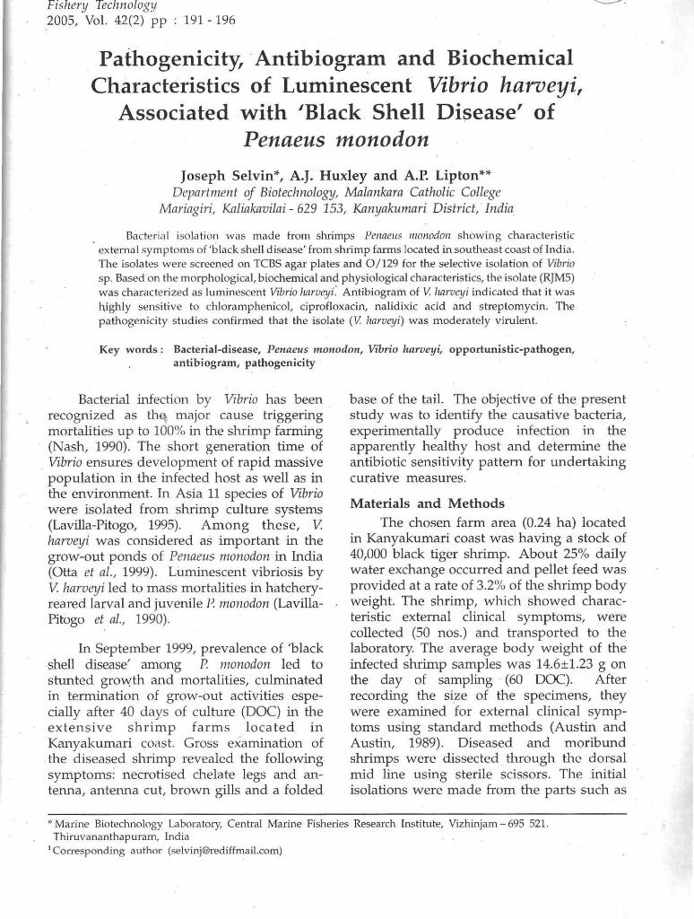
-Fishery Technology 2005, Vol. 42(2) pp 191 - 196
Pathogenicity, . Antibiogram and Biochemical Characteristics of Luminescent Vibrio harveyi,
Associated with 'Black Shell Disease' of Penaeus monodon
Joseph Selvin*, A.J. Huxley and A.P. Lipton** Department of Biotechnologtj, Malankara Catholic College
Mariagiri, Kaliakavilai - 629 153, Kanyakwnari District, · India
Bactericd isolat ion was made from shrimps Pe/wells lIloHodoll showing characteristic externa l symptoms of 'black shell disease' from shrimp farms located in southeast coast of India. The isolates were screened on TeBS agar plates and 0/129 for the selective isolation of Vibrio sp. Based on the morphological, biochemical and physiological characteristics, the isolate (RJM5) was characte rized as luminescent Vibrio harveyi." Antibiogram of V. Imrueyi indicated that it was highly sensitive to chloramphenicol. ciprofloxacin, nalidixic acid and streptomycin. The pathogenicity studies confirmed that the isolate (V lzarveyi) was moderately viru lent.
Key words: Bacterial-disease, Penaeus mono don, Vibrio hanleyi, opportunistic-pathogen, antibiogram, pathogenicity .
Bacterial infecti()n by Vibrio has been recognized as th~ major cause triggering mortalities up to 100% in the shrimp farming (Nash, 1990). The short generation time of Vibrio ensures development of rapid massive population in the infected host as well as in the environment. In Asia 11 species of Vibrio were isolated from shrimp culture systems (Lavilla-Pitogo, 1995). Among these, 11. harveyi was considered as important in the grow-out ponds of Penaeus monodon in India (Otta et al., 1999). Luminescent vibriosis by 11. harve!ji led to mass mortalities in hatcheryreared larval and juvenile P. monodon (LavillaPitogo et. aI., 1990).
In September 1999, prevalence of 'black shell disease' among P. monodon led to stunted growth and mortalities, culminated in termination of grow-out activities especially after 40 days of culture (DOC) in the extensive shrimp farms located in Kanyakumari coast. Gross examination of the diseased shrimp revealed the following. symptoms: necrotised chelate legs and antenna, antelma cut, brown gills and a folded
base of the taiL The objective of the present study was to identify the causative bacteria, experimentally produce infection in the apparently healthy host and determine the antibiotic sensitivity pattern for undertaking curative measures.
Materials and Methods
The chosen farm area (0.24 hal located in Kanyakumari coast was having a stock of 40,000 black tiger shrimp. About 25% daily water exchange occurred and pellet feed was provided at a rate of 3.2% of the shrimp body weight. The shrimp, which showed characteristic external clinical symptoms, were collected (50 nos.) and transported to the laboratory. The average body weight of the infected shrimp samples was 14.6±1.23 g on the day of sampling · (60 DOC). After recording the size of the specimens, they were examined for external clinical symptoms using standard methods (Austin and Austin, 1989). Diseased and moribund shrimps were dissected through the dorsal mid line using sterile scissors. The initial isolations were made from the parts such as
>I- Marine Biotechnology Labora tory, Central Marine Fisheries Research Ins titute, Vizhinjam - 695 521. Thiruvananthapuram, india
1 Corresponding author ([email protected])
•
192
infected shell, antenna, chelate legs and hepatopancnias. The infected shell area was removed using sterile scissors and swapped on plates of nutrient agar supplemented with 2% NaCi (NA). Hepatopancreatic tissue was homogenized in a sterile homogeniser (Omni, USA) using normal saline (NS). The resultant suspension was serially diluted up to 10-6
dilution using phosphate buffered saline and used for preparation of spread plate on nutrient agar supplemented with 2% NaCI. The plates were incubated at 32±2°C for 18 h. Dominant colonies observed on the NA plates, were further screened on thiosulfatecitrate-biles salt-sucrose (TCBS) agar (Himedia). Five yellow colonies (RJMl to RJM5) observed were further screened for 0/129 (S.igma) resistance using impregnated (150 mg) paper discs (8mm dial.
The colony morphology was observed after 18 h followed by Gram staining and observation under 1000x magnification. Luminescence was observed under dark and confirmed over UV illumination. Biochemical tests were followed after MacFaddin, (1981). All the tested media were supplemented with 2% NaCi. Tests for chitinase, amylase, protease and gelatinase were based on Cowan (1974) and Austin and Lee (1992). The b-galactosidase test was done with ONPG disks (Himedia). Results were recorded after incubation at 30±2°C. The effect of NaCI concentration and temperature on the growth of the pathogens was also tested. Classification followed as per Baumann and Schubert (1984) and Colwell and Grimes (1984).
Antibiotic sensitivity profile was determined by the Kirby-Bauer disk diffusion .method (Bauer et al., 1996) using Himedia antibiotic discs. Bacterial suspensions of 10 fold dilutions were prepared using 18 h fresh shake culture was inoculated onto the Mueller-Hinton agar plates to get the lawn concentration of about 1.5 x 10⁶ cfu/ cmz. Six discs were dispensed on the seeded lawn at 60° apart to each other. The diameter of inhibition zone around the discs was measured after incubation at 30±2°C for 24 h. The colonies observed inside the inhibition zone was considered as specific resistant strain.
SELVIN, HUXLEY AND LIPTON
For challenge studies, healthy juvenile black tiger shrimp Penaeu, monodon (30 DOC) were segregated from the grow-out tanks of Marine Biotechnology laboratory aquarium and maintained at a rate of 10 shrimps/tank in 100 I glass aquaria. The length and weight of each shrimp was measured before starting the experiment. Prior to the infection experiments, random sampling of shrimp ·was made for the bacterial isolation to ensure the shrimp were pathogen free. Isolations were made from the parts such as shell, body tissue, hepatopancreas and haemolymph on TCBS agar plates to ensure the shrimps were free from characteristic yellow colonies. The 18 h fresh shake culture was centrifuged at 4000 rpm for 15 min and washed twice in
. normal saline (NS). The purified pellets were serially diluted in NS and enumerated in a haemocytometer. This was also plated on NA plates to get the colony forming units (du). Preliminary examinations revealed that challenge dose of 1(}1 cfu per shrimp could not kill the injected shrimp. Therefore the~
concentrations of 105 to 10' cfu per shrimp were taken in 0.1 mI saline and inoculated intramuscularly using a 1 mI tuberculin syringe at ventral side between the second and third segment of healthy shrimps. Parallel control groups received 0.1 ml of NS only. Ten shrimps were used for each inoculation level. The mortality and reflexes of the shrimps were observed for every 15 min in the first hour of post-inoculation and every 1 h until the 6th h. Subsequen.t monitoring was done every 12 h for a period of seven days (Tendencia & Dureza, 1997). Moribund shrimps were sampled for the bacterial re-isolation in Zobell Marine Agar (ZMA) plates. LDso dose for 24 hand 7 days were calculated by the probit method, after Wardlaw (1985).
Results
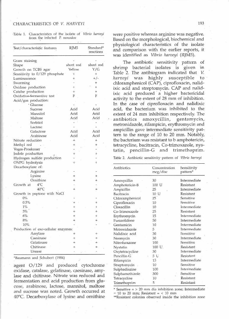
Results of the morphological, biochemical and physiological characterization of the bacterial isolates are presented in Table 1. The chosen isolate (RJM5) was swarming, luminescent, Gram negative and actively motile short rods . It grew on TCBS agar, required NaCI for growth, sensitive to the vibriostatic
,
CHARACTERISTICS OF V. HARVEYI
Table 1. Characteristics of the isulate of Vibrio hl1rveyi from the infected P. rt/OI/OdolJ
Test/characteristic feature~
Gram staining Shape Growth on TeBS agar Sensitivity to 0/129 phosphate Luminescence Swarming Oxidase production Catalse production Oxidative-fermentive test Acid/gas production:
. Glucose Sucrose Mannitol Maltose Sorbitol Lactose Galactose Arabinose
'Nitrate reduction Methyl red Voges-Proskauer ~ndole production ., . Hydrogen sulfide production ONPG hydrolysis Decarboxylase of:
Arginine Lysine Ornithine
Growth at 40C 40"C
Growth in peptone with NaCI 0%
RJMS
short rod Yellow
+ +
+ + F
Acid Acid Acid
Acid Acid
+ +
+
+
+ +
+
0.5% + 1% + 3% + 6% + 8% + 10%
Production of exo-cellular enzymes: Amylase + Caesinase + Gelatinase + Chitinase + Urease
""Baumann and .Schubert (1.984)
Standard"" reactions
short rod Y/G
+ +/-+ + + F
Acid Acid Acid
Acid Acid
+ +
+
+
+ +
+
+ + + + +
+ + + +
agent 0/129 and produced cytochrome oxidase, catalase, gelatinase, caesinase, amylase and chitinase. Nitrate was reduced and fermentation and acid prQduction from glucose, arabinose, lactose, mannitol, maltose and sucrose was noted. Growth occurred at 40°C. Decarboxylase of lysine and ornithine
193
were positive whereas arginine was negative. Based on the morphological, biochemical and physiological characteristics of the isolate and comparison with the earlier reports, it was identified as Vibrio harveyi (RJMS).
The antibiotic sensitivity pattern of shrimp bacterial isolates is given 111
Table 2. The antibiogram indicated that V harveyi was highly susceptible to chloramphenicol (CAP), ciprofloxacin, nalidixic acid and streptomycin. CAP and nalidixic acid produced ' a higher bactericidal activity to the extent of 28 mm of inhibition. In the case of ciprofloxacin and nalidixic acid, the bacterium was inhibited to the extent of 24 nun inhibition respectively. The antibiotics amoxycillin, gentamycin, metronidazole, rifampicin, erythromycin and ampicillin gave intermediate sensitivity pattern to the range of 10 to 20 mm. Notably, the bacterium was resistant to b amphotericin, tetracycline, bacitracin, Co-trimoxazole, nystatin, pencillin-G and trimethoprim.
Table 2. Antibiotic sensitivity pattern of Vibrio harveyi
Antibiotics Concentration Sensitivity meg/disc pattern""
Amoxycillin 30 Intermediate Amphotericin-B 100 U Resistant Ampicillin 25 Intermediate Bacitracin 10 U Resistant Chloramphenicol 25 Sensitive Ciprofloxacin 10 Sensitive Cloxacillin 10 In termedia te Co-Trimoxazole 25 Resistant Erythromycin 15 Intermediate Furazolidone 50 intermediate Gentamicin 10 intermediate Metronidazole 5 Intermediate Nalidixic acid 30 Sensitive Neomycin 30 In termed ia te Nitrofurazone 100 Sensitive Nystatin 100 U Resistant Oxytetracycline 30 intermediate Pencillin-G SU Resistant Rifampicin 15 Intermediate Streptomycin 10 Sensitive Sulpha diazine 100 Intermediate Sulphamethizole 300 Sensitive Tetracycline 10 Resistant Trimethoprim 5 Resistant
"" Sensitive = > 20 mm dia inhibition zone; Intermediate = 10 to 20 mm; Resistant = < 10 mm """"Resistant colonies observed inside the inhibition zone
1'f4
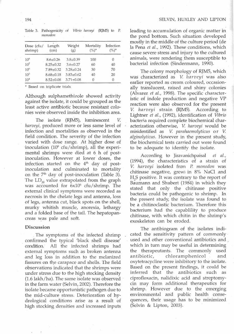
Table 3. Pati10genicity of Vibrio !mruetji (RJM5) to P. monodoll
Dose (cfuj shrimp)
10' 10' 10' 10,";
10'
Length (em)
S.6±0.21i 8.25±0.32 7.89±0.32 B.6S±0.IB S.52±0.08
Weight (g)
3.S±0.39 3.n±0.27
3.2S±0.24 3.S3±0.62 3.71±0.OS
... Based on triplicate trials
Mortality (%)""
100 60 30 40 0
Infection (%)""
0 40 70 20 0
Although sulphamethizole showed activity against the isolate, it could be grouped as the least active antibiotic because resistant colonies we're observed inside the inhibition area.
The isolate (RJMS), luminescent V. harveyi, produced more or less same level of infection and mortalities as observed in the field condition. The severity of the infection varied with dose range. A t higher dose of 'inoculation (10' cfu / shrimp), all the experimental shrimps were died at 6 h of poste inoculation. However at lower ' doses, the infection started on the 41h day of postinoculation and culminated to mortality on the 71h day of post-inoculation (Table 3). The LDso value extrapolated from the graph was accounted for 6x10⁶ cfu/shrimp. The external clinical symptoms were recorded as necrosis in the chelate legs and antenna, loss of legs, antenna cut, black spots on the shell, murky whitish muscle, anorexia, lethargy and a folded base of the tail. The hepatopancreas was pale and soft.
Discussion
. The symptoms of the infected shrimp . confirmed the typical 'black shell disease'
condition. All the infected shrimps had external symptoms such as broken antenna and leg loss in addition to the melanized fissures on t1,e carapace and shells. The field observations indicated that the shrimps were under stress .due to the high stocking density (1.6 lakh /ha). The same isolate was observed in the farm water (Selvin, 2002). Therefore the isolate became opportunistic pathogen due to the mid-culture stress. Deterioration of hydrological conditions arise as a result of high stocking densities and increased inputs
SELVIN, HUXLEY AND LIPTON
leading to accumulation of organic matter in the pond bottom. Such situation developed mostly in the middle of the culture period (de la Pena et aI., 1992). These conditions, which cause severe stress and injury to the cultured animals, were rendering them susceptible to bacterial infection (Sindermann, 1990). .
The colony morphology of RJMS, which was characterized as V. harveyi was also earlier reported as cream coloured, occasionally translucent, raised and shiny colonies (Alvarez et al., 1998). The specific characteristic of indole production and negative VP reaction were also observed for the present V. ha rveyi strain (RJM5). According to Lightner et aI., (1992), identification of Vibrio bacter.i.a required complete biochemical characterization otherwise, V. harveyi would be misidentified as V. parahaemolyticus or V. alginolyticus. However in the present study, the biochemical tests carried out were found to be adequate to identify the isolate.
According to Jiravanichpaisal et al., (1994), the characteris tics of a strain of V. harveyi isolated frOll P. monodon was chitinase negative, grew; in 8% NaCl and HzS positive. It was contrary to the report of Baumann and Shubert (1984) in which they stated that only the chitinase positive bacteria could be pathogenic to shrimp. In the present study, the isolate was found to be a chitinoclastic bacterium. Therefore this bacterium had the capablility to produce chitinase, with which chitin in the shrimp's exoskeleton Can be eroded.
The antibiogram of the isola tes indicated the sensitivity pattern of commonly used and other conventional antibiotics and which in turn may be useful in determining the therapeutants. The commonly used antibiotic, chloramphenicol and oxytetracycline were inhibitory to the isolate. Based on the present findings, it could be inferred that the antibiotics such as ciprofloxacin, nalidixic acid and streptomycin may form additional therapeutics for shrimp. However due to the emerging environmental ana public health consequences, their usage has to be minimized (Selvin & Lipton, 2003).
· CHARACTERISTICS OF V. HARVEYI
In the present experiment, the infection 'or mortality started after two to five days of post-challenge. The mechanism of such delayed responses may be due to the initial bacterial clearance by the haemolymph and growth rate as well as generation time to attain minimum infectivity dose in the host. However, higher dose, the mortality started within 6 h of post-inoculation. At this dose, the bacteria may have overwhelmed the shrimp's defense system during the first two days resulting in mortalities. Nash et aI., (1992) reported that shrimp injected with bacteria were weak in the first two days but could (ecover within 3 to 5 days.
The LDsn value of V harveyi isolated from 'shell disease' shrimp was 10' cfu/ shrimp. A strain of V harvetji isolated from the shrimp P monodon with 'red disease syndrome' could be reproduced by injection with 10' cfu/shrimp (Tendencia & Dureza, 1997). The LDso values of V harvey! were reported to range from 1.4xlO' to 2.8xlO' cfu/shrimp, which indicated low virulence (Otta et al., 1999). Lavilla-Pitogo et al., (1990) concluded that virulent isolates of V harveyi were lethal at 100 cells / ml in seawater while non-luminescent isolates did not cause mortality even at a challenge dose of 10' cells / ml (Pizzautto & Hirst, 1995). This report in turn suggests that luminescence may be one of the indicators of virulence. Considering this, the present luminescent V harveyi can be considered as moderately virulent (10' cfu/shrimp). The pathogenic characteristics of RJM5 was similar to V alginolyticus, one of the common shrimp pathogen of India (Selvin & Lipton, 2003).
The . Significance of V harveyi as a shrimp pathogen is further reinforced by reports from tropical countries where the bacteria caused mortalities up to 100% in shrimp hatcheries. It was commonly isolated only from diseased but not healthy larvae as well as 'from the rearing water Qiravanichpaisal et al., 1994; Kanmasagar et al., 1994). In the adults, Jiravanichpaisal et al., (1994) isolated V harveyi as a minor component from the exoskeleton of female black tiger shrimp in Thailand. Nevertheless, the proof for the pathogenic role of V harveyi
195
was rare in infectivity experiments. In the present study, the isola te obtained from the diseased shrimp demonstrated certain degree of virulence. In agreement with these results, others have also reported the high LDso values (4.9xlO' and 1.56x10⁹ cfu) for fish (Saeed, 1995). But lower doses of 10' to 10> cells/ml were determined as lethal for larval shrimp (Lavilla-Pitogo, et al., 1990). Hepatopancreas was reported as the target tissue for the reisolation of V harveyi from diseased shrimp (Alvarez et aI., 1998). The present isolate also was easily reisolated from the hepatopancreas of infected shrimp. Based on the present findings, the strain could be inferred as a moderate pathogen, which act as opportunistic one in the culture system.
Authors are thankful to Dr. MJ Modayil, Director and Dr. R. Paul Raj, Head, PNP Division for the facilities and encouragen1ent.
References
Alvarez, J.D., Ausml, B., Alvarez, AM. and Reyes, H. (1998). Vibrio harveyi: a pathogen of penaeid shrimps and fish in Venezuela. J. Fish. Dis. 21, pp313-316.
Austin, B. and Lee J. v. (1992). Aeromonadaceae and Vibionaceae. In: Identification Methods in Applied and Environmental MicrobiologJI (RG. Board. D. Jones and EA Skinner, Eds). Technical Series of the Society for Applied Bacteriology. Bl\lckwells. Oxford. pp 163-182
Austin, B. and Austin, DA (1989). Methods for the Microbiological Examination of Fish and Shellfish. Ellis Horwood, Chichester 298p
Bauer, AW., Kirby, W.M.M., Shenis, J.c. and Turck, M. (1996). Antibiotic susceptibility testing by a standardized single disk method. Am. J. Clin. Pathol. 45, pp 493~ 498
Baumann, P. and Schubert, RH.W. (1984). Family U .. Vibrionaceae. In: Bergey's manual of systematic bacteriology. Vol 1, (N.R. Krieg and I.G. Holt, Eds). Williams and Wilkins, London. pp 516-538
Colwell, RR and J.A Grimes. (1984). Vibrio diseases of marine fish populations. Helgolander Wissen Sehaftliche Meereslmtersllchungcl'l, 37, pp 265-287
196
Cowan, S.T. (1974). Cowan and Steel's manual for the identification of medical bacteria, 2nd
edition. Cambridge University Press, London.
de la Pena, L.D., Momoyama, K., Nakai, T. and Muruga, K. (1992). Detection of the causative bacterium of vibriosis in kuruma prawn, Penacus japonicus. Fish Pathol. 27, pp 223-228
Jiravanichpaisal P, Miyasaki T. and Limsuwan C (1994). Histopathology, biochemistry, and pathogenicity of Vibrio harveyi infecting black tiger prawn Penaeus monodon. J. Aqua. Anim. Health, 6, pp 27-35
Karunasagar, I., Pai, R, Malathi G.R and Karunasagar, I. (1994). Mass mortality of Penaeus monodon larvae due to antibiotic-resistant Vibrio harveyi infection. Aquaculture, US, pp 203-209
Lavilla-Pitogo, CR (1995). Bacterial diseases of penaeid shrimps an Asian view. In: Diseases. in Asian Aquaculture II. (Shariff, M, Arthur, J.R and Subasinghe, R.P, Eds). Fish health section. Asian Fisheries Society, Manila, Philippines. pp. 107-121
Lavilla-Pitogo, CR, Baticados, M.CL., CruzLacierda, E.L. and de La Pena, L.D. (1990). Occurrence of luminous bacterial disease of Penaeus monodon larvae in the Philippines. Aquaculture, 91, pp 1-13
Lightner, D. v., Bell, T.A., Redman, RM., Mohney, L.L., Natividad, J.M., Rukyani, A and Poernomo, A (1992). A review of some major diseases of economic significance in penaeid prawns/shrimps of the Americas and Indopacific. In: Diseases in Asian Aquaculture I. (Shariff, I.M., Subasinghe, RP and Arthur, J.R, Eds). Fish health section, Asian Fisheries Society, Manila, Philippnies. pp 57-80
MacFaddin, J. F. (1981). Biochemical tests for identification of medical" bacteria, 2nd
edition, Williams and Wilkins, Baltimore. pp 527
Nash, G. (1990). Penaeus monodon grow-out diseases. In: Proceedings of Aquatech 90, Putra World Trade Centre, Kuala Lumpur. 11-14, June 1990. pp 172-187
SELVIN, HUXLEY AND LIPTON
Nash, G., Charetana, N. Cholada, T., Anutra, A, Phusit, P and Pongcham, R (1992). Vibriosis and its control in pond-reared Penaeus monodon in Thailand. In: Diseases in Asian Aquaculture I. (Shariff M., Subasinghe, RP. and Arthur J.R, Eds). Asian Fisheries Society. Philippines pp 143 - 155
Otta, S.K., Karunasagar, I. and Karunasagar, I. (1999). Bacterial flora associated with shrimp culture ponds growing Penaeus monodon in India. J. Aqua. Trap. 14, pp 309-318
Pizzutto, M. and Hirst, RG. (1995). Classification of isolates of Vibrio harveyi virulent ID Penanls monodon larvae by protein profile analysis and m 13 DNA fingerprinting. Dis. Aqua. Org. 21, pp 61-68
Saaed M.a. (1995). Association of Vibrio harm;i with mortalities in cultured marine fish in Kuwait. Aquaculture, 11, pp 21-29
Selvin, J. (2002) . . Shrimp disease management using secondary metabolites isolated from marine organisms. Ph.'D thesis submitted to M.s. University, Tirunelveli, India. pp 204
Selvin, J. and Lipton, A.P (2003). Leaching and Residual Kinetics of Chloramphenicol Incorporated Medicated Feed Treated to Juvenile Black Tiger Shrimp Penaeus monodon Fabricious. Fish. Technol. 40, pp 13-17
Selvin, J. and Lipton, AP (2003). Vibrio alginolyticus associated with white spot disease of Penaeus monodon. Dis. Aqua. Org. 57, pp 147-150
Sindermann, CJ. (1990). Bacteria. In: Principal Diseases of Marine Fish and Shellfish. VoL 2 Academic Press, San Diego, CA, pp 41-71
Tendencia, E.V.A and Dureza, L.A. (1997). Isolation of Vibrio sp. fran Penaeus monodon (Fabricius) with red disease syndrome. Aquaculture 154, ppl07-114
Wardlaw AC (1985). Practical Statistics for Experimental Biologists. John Wiley and Sons, Chichester.