Embed Size (px)
Citation preview
Patterns of lymph node metastasis from nasopharyngeal carcinoma based on the 2013 updated consensus
guidelines for neck node levels
XiaoShen Wang*, ChaoSu Hu*, HongMei Ying*, XiaYun He*, ZhengRong Zhou†, JianHui Ding†
* Department of Radiation Oncology and † Department of Diagnostic Radiology, Fudan University Shanghai Cancer Center.
Department of Oncology, Shanghai Medical College of Fudan University, Shanghai, 200032
objectiveobjective
To explore the patterns of node spread from NPC based on the 2013 updated guidelines.
WANG XS, et al. Fudan University Shanghai Cancer Center
MethodsMethods
Patients with NPC were required to undergo MRI. The scan range extended from 2cm above the anteroir clinoid process to the inferior margin of the sternal end of the clavicle.
All MR images were evaluated by the multi-disciplinary treatment group of NPC.
WANG XS, et al. Fudan University Shanghai Cancer Center
ResultsResults
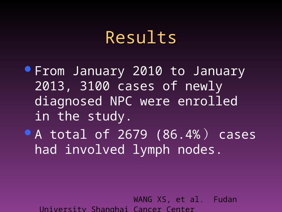
From January 2010 to January 2013, 3100 cases of newly diagnosed NPC were enrolled in the study.
A total of 2679 (86.4% ) cases had involved lymph nodes.
WANG XS, et al. Fudan University Shanghai Cancer Center
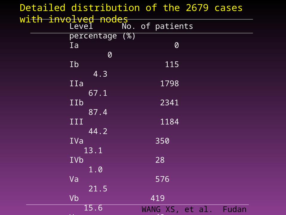
Level No. of patients percentage (%)Ia 0 0Ib 115 4.3IIa 1798 67.1IIb 2341 87.4III 1184 44.2IVa 350 13.1IVb 28 1.0Va 576 21.5Vb 419 15.6Vc 49 1.8VIa 0 0VIb 0 0VIIa 2012 75.1VIIb 178 6.6VIII 53 2.0IX 2 0.07Xa 2 0.07Xb 10 0.4
Detailed distribution of the 2679 cases with involved nodes
WANG XS, et al. Fudan University Shanghai Cancer Center
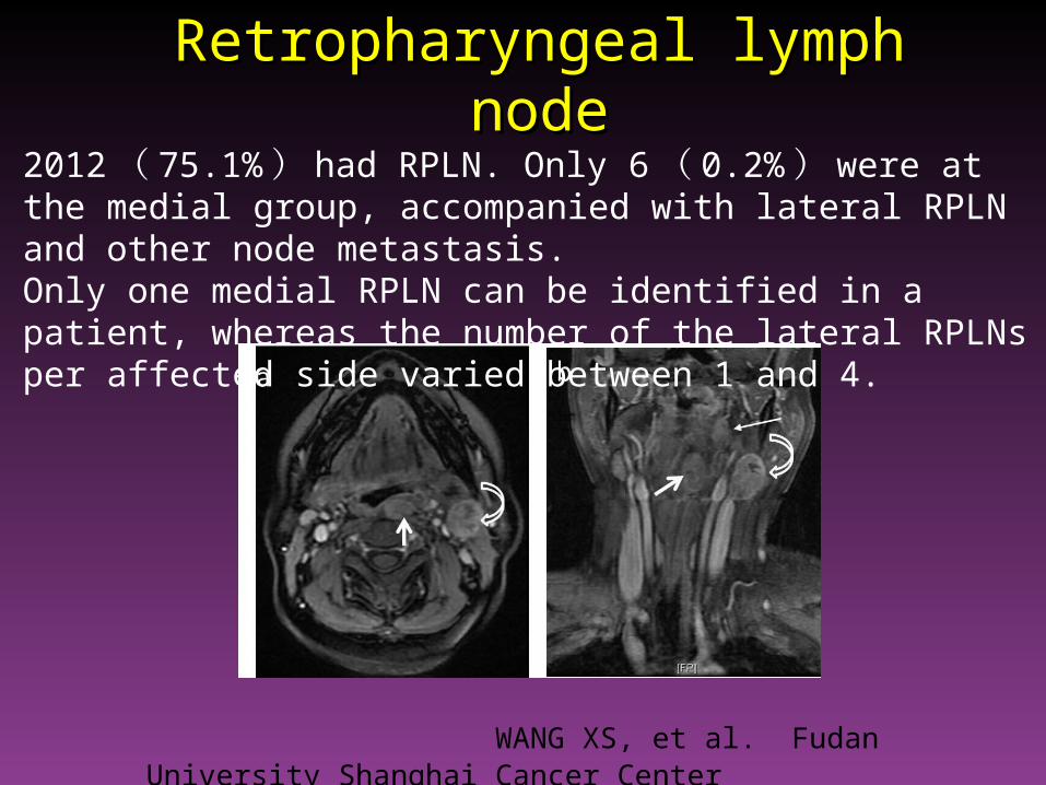
Retropharyngeal lymph nodeRetropharyngeal lymph node
WANG XS, et al. Fudan University Shanghai Cancer Center
2012 ( 75.1% ) had RPLN. Only 6 ( 0.2% ) were at the medial group, accompanied with lateral RPLN and other node metastasis. Only one medial RPLN can be identified in a patient, whereas the number of the lateral RPLNs per affected side varied between 1 and 4.
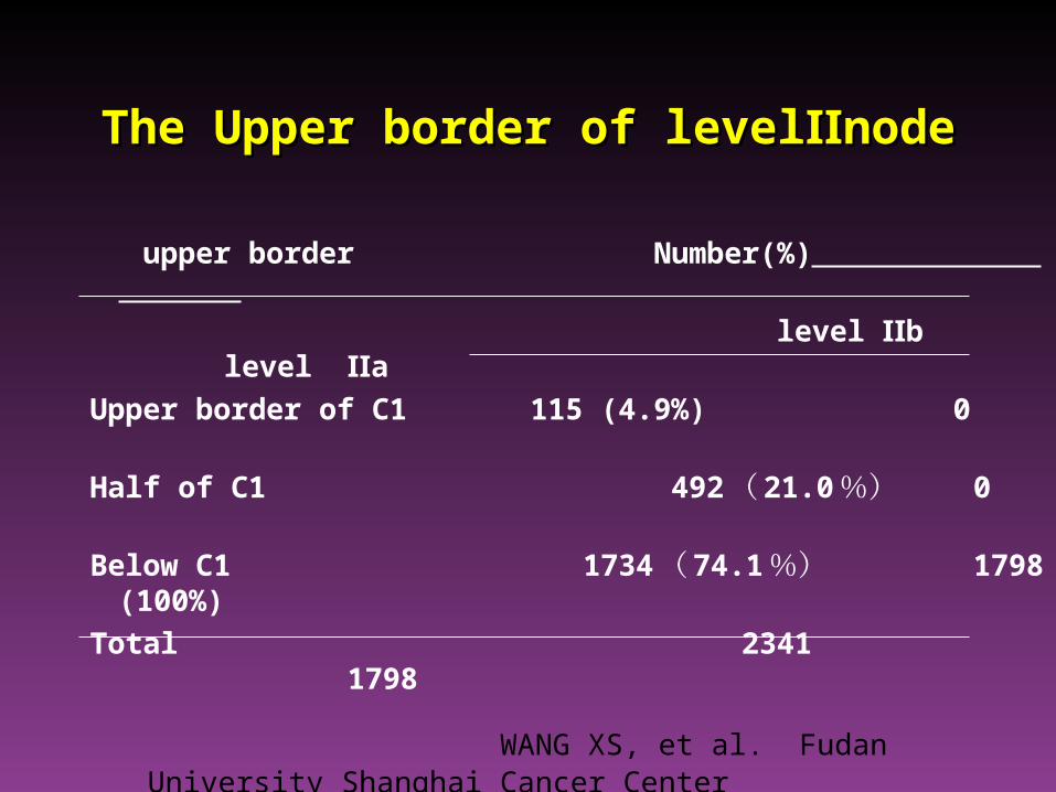
The Upper border of level nodeⅡThe Upper border of level nodeⅡ
upper border Number(%)
level Ⅱb level Ⅱa
Upper border of C1 115 (4.9%) 0
Half of C1 492 ( 21.0 %) 0
Below C1 1734 ( 74.1 %) 1798 (100%)
Total 2341 1798
WANG XS, et al. Fudan University Shanghai Cancer Center
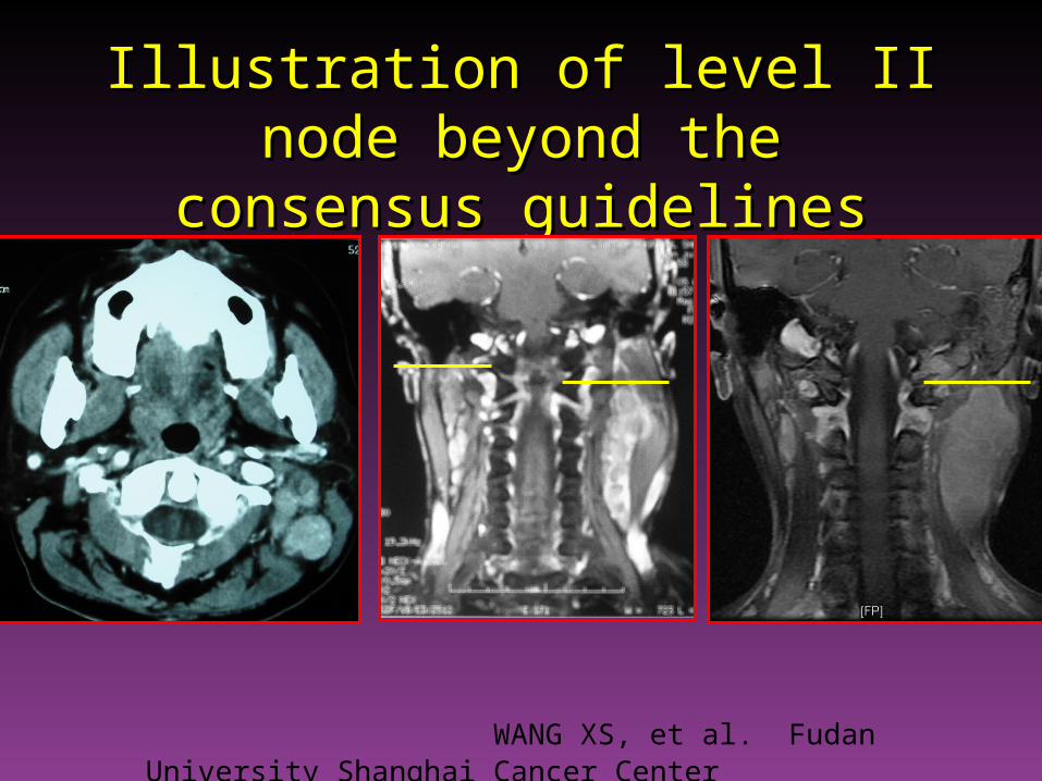
Illustration of level II node beyond Illustration of level II node beyond the consensus guidelinesthe consensus guidelines
WANG XS, et al. Fudan University Shanghai Cancer Center
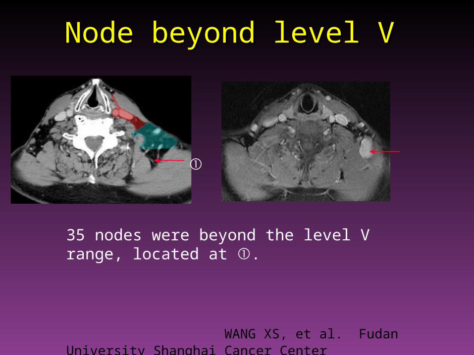
Node beyond level VNode beyond level V
①
35 nodes were beyond the level V range, located at ①.
WANG XS, et al. Fudan University Shanghai Cancer Center
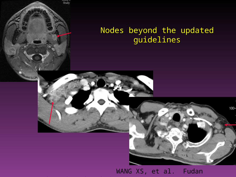
Nodes beyond the updated guidelinesNodes beyond the updated guidelines
WANG XS, et al. Fudan University Shanghai Cancer Center
Conclusions:Conclusions: This is the first description of nodal spread patterns based
on the updated consensus guidelines. Invovelment of RPLNs were mainly located at the lateral
group, the medial group was rarely seen. Nodal involvement spread in an orderly pattern in NPC. The upper border of level II should be skull base. The level V borders recommended in this consensus
guidelines can not fully cover all lymphadenopathies for NPC.
WANG XS, et al. Fudan University Shanghai Cancer Center