Embed Size (px)
Citation preview
1
Limited RNA editing in exons of mouse liver and adipose tissue
Sandrine Lagarrigue*,§,†,***, Farhad Hormozdiari‡,**,***, Lisa J. Martin§§,***, Frédéric Lecerf*,§,†,
Yehudit Hasin§§, Christoph Rau§§, Raffi Hagopian§§, Yu Xiao§§, Jun Yan§§, Thomas A. Drake††,
Anatole Ghazalpour**, Eleazar Eskin‡,**, and Aldons J. Lusis**,§§,‡‡
*Agrocampus Ouest, UMR1348 Pegase, Animal Genetics Laboratory, F-35000 Rennes, France
§INRA, UMR1348 Pegase, F-35000 Rennes, France
†Université Europeenne de Bretagne, France
‡Department of Computer Sciences, University of California, Los Angeles, CA 90095
**Department of Human Genetics, University of California, Los Angeles, CA 90095
§§Department of Medicine/Division of Cardiology, University of California, Los Angeles, CA
90095
††Department of Pathology and Laboratory Medicine, University of California, Los Angeles, CA
90095
‡‡Department of Microbiology, Immunology and Molecular Genetics, University of California,
Los Angeles, CA 90095
***Equal contributions of the authors
Short title: RNA-Seq bias and RNA editing
Key words: RNA editing, RNA Seq, inbred mice, RNA DNA Differences
Genetics: Early Online, published on February 14, 2013 as 10.1534/genetics.112.149054
Copyright 2013.
2
Corresponding Authors:
Aldons J. Lusis
UCLA Department of Medicine/Division of Cardiology
3730 MRL
650 Charles E Young Drive South
Los Angeles, CA 90095-1679
Phone: (310) 825-1359 FAX (310) 794-7345
Sandrine Lagarrigue
Agrocampus Ouest Department of Animal Sciences
65 rue de Saint Brieuc
Rennes, 35000 France
3
ABSTRACT
Several studies have investigated RNA-DNA differences (RDD), presumably due to RNA
editing, with conflicting results. We report a rigorous analysis of RDD in exonic regions in mice,
taking into account critical biases in RNA-Seq analysis. Using deep-sequenced F1 reciprocal
inbred mice, we mapped 40 million RNA-Seq reads per liver sample and 180 million reads per
adipose sample. We found 7,300 apparent hepatic RDDs using a multiple-site mapping
procedure, compared with 293 RDD found using a unique-site mapping procedure. After
filtering for repeat sequence, splice junction proximity, undirectional strand and extremity read
bias, 63 RDD remained. In adipose tissue unique-site mapping identified 1,667 RDD, and after
applying the same four filters, 188 RDDs remained. In both tissues, the filtering procedure
increased the proportion of canonical (A-to-I and C-to-U) editing events. The genomic DNA of
12 RDD sites among the potential 63 hepatic RDD was tested by Sanger sequencing, three of
which proved to be due to an unreferenced SNP. We validated seven liver RDD with Sequenom
technology, including two non-canonical, Gm5424 C-to-I(G) and Pisd I(G)-to-A RDD.
Differences in diet, sex, or genetic background had very modest effects on RDD occurrence.
Only a small number of apparent RDD sites overlapped between liver and adipose, indicating a
high degree of tissue specificity. Our findings underscore the importance of properly filtering for
bias in RNA-Seq investigations, including the necessity of confirming the DNA sequence to
eliminate unreferenced SNPs. Based on our results, we conclude that RNA editing is likely
limited to hundreds of events in exonic RNA in liver and adipose.
4
INTRODUCTION
Several recent studies have investigated genome-wide RNA editing using deep
sequencing of transcriptomes by RNA-Seq on human transformed cells, cancer cell lines (JU et
al. 2011; LI et al. 2011; BAHN et al. 2012; PENG et al. 2012; RAMASWAMI et al. 2012), or tissues
of inbred mouse strains (DANECEK et al. 2012; GU et al. 2012). Total reported RDD sites have
varied from hundreds to thousands. Over the same period, technical issues, such as mapping of
reads in paralogous or repetitive sequence regions, mapping errors at splice sites, and systematic
sequencing errors that could produce a large number of false-positive RDDs have been described
(KLEINMAN and MAJEWSKI 2012; LIN et al. 2012a; PICKRELL et al. 2012). Another reported
source of RDD error is undetected genomic DNA SNPs, arising from insufficient coverage of
current DNA sequencing data (SCHRIDER et al. 2011).
We have examined genome-wide exonic RDD by using RNA-Seq data obtained from
two tissues, liver and adipose, in F1 reciprocal crosses from two inbred strains of mice, DBA/2J
(D2) and C57BL/6J (B6). These inbred mouse strains have been subjected to deep genomic
sequencing and SNP analyses, with a higher coverage for B6 than for D2. A major aim was to
estimate the impact of the major technical issues (paralog mapping, mismapping near splice sites
and repeat sequences, and systematic sequencing errors, such as unidirectional strand and
extremity biases) to obtain a better sense of the true frequency of RDD in normal mammalian
tissues. The RDDs that remained were then characterized by comparison with Expressed
Sequence Tags and tested by Sanger and quantitative Sequenom sequencing, showing the
importance of controlling the genomic DNA sequence in RDD site analysis. We also examined
the effects of sex and diet, and the possibility of allele-specific RNA editing.
5
MATERIALS AND METHODS
Ethics statement. All animals were handled in strict accordance with good animal practice as
defined by the relevant national and/or local animal welfare bodies, and all animal work was
approved by the appropriate committee. All experiments in this paper were carried out with
UCLA IACUC approval.
Mice and tissues. RNA-Seq was performed on liver and adipose mRNA from F1 male and
female D2 and B6 mice, purchased from the Jackson Laboratory (Bar Harbor, ME). Reciprocal
F1 male and female mice were generated by breeding the parental strains in the vivarium at
UCLA. For six liver RNA libraries, RNA from 3 mice was pooled into 4 independent samples of
high fat fed B6xD2 (BXD) and DXB males and females, and 2 samples of chow fed BXD and
DXB males. Four adipose RNA libraries were made using pooled RNA from three BXD and
DXB males and females fed a chow diet. Males and females of other reciprocal inbred mouse
crosses were used for Sequenom validation. Those F1’s were A/JxC3H/HeJ (AXH) and HXA
and B6xC3H/HeJ (BxH) and HXB. Liver RNA was isolated from three mice per sex per F1
cross using the RNeasy kit from Qiagen (Valencia, CA, USA). cDNA was made with the High
Capacity Reverse Transcription Kit from Applied Biosystems. All mice were fed ad libitum and
maintained on a 12-hour light/dark cycle. F1 pups were weaned at 28 days and fed a chow diet
(Ralston-Purina Co) until 8 weeks of age, at which time half were placed on a high fat diet
(Research Diets D12266B). All F1 mice were euthanized at 16 weeks, with liver and adipose
harvested at that time.
Library preparation for Illumina sequencing. Library preparation was performed as
recommended by the manufacturer (Illumina, Hayward, CA,USA). Briefly, total RNA was
6
extracted using the RNeasy Mini kit with DNase treatment (Qiagen, Valencia, CA, USA). PolyA
mRNA was isolated and fragmented, and first strand cDNA was prepared using random
hexamers. Following second strand cDNA synthesis, end repair, addition of a single A base,
adaptor ligation, and agarose gel isolation of ~200 bp cDNA, PCR amplification of the ~200 bp
cDNA was performed. Liver samples were sequenced using the Illumina GAIIX sequencer to a
coverage of approximately 40 million single end 75bp reads per sample. Adipose was sequenced
with the Illumina HiSeq2000 on paired end 50 bp reads and generated 180 million reads per
sample.
Read mapping. We first aligned reads 75pb (liver) or 50bp (adipose) to the mouse reference
genome version mm9 using mrsFAST (HACH et al. 2010) allowing up to 5 mismatches for liver
tissue and 3 for adipose). The reads were divided into two categories. The first category was the
the set of mapped reads which align to the genome with e or less mismatches. The second
category was reads that failed to map to the reference genome. Many RNA-Seq reads failed to
align to a genome because they spanned the exonic junctions. In order to overcome this problem
we mapped the unmapped reads with TopHat (TRAPNELL et al. 2009), which is designed to map
reads to the genome by splitting the reads into smaller fragments. The reads aligned to the
genome in this process were added to the map read set. In our multiple-site mapping procedure,
we allowed up to 10 genomic locations for the same read. If a read mapped to more than 10
locations, we picked the top 10 sites with the most reads. In the unique-site mapping procedure,
we only consider the reads which mapped to only one position, and reads which map to more
than one position are filtered out.
RDD criteria. We selected reads with base modifications of the RNA represented by at least 10
reads at the position for the edited base, located in one exon, and not corresponding to a known
7
genomic SNP between D2 and B6 (based on the Mouse Sequencing Consortium (WATERSTON et
al. 2002; KEANE et al. 2011). The read was required to have a base quality of 20 or higher. A
base modification A (DNA base)àB (edited base) was considered an RDD if 1) B6 and D2 were
homozygous for the DNA nucleotide in the genomic DNA, 2) B generated at least 10 RNA-Seq
reads, with 3) (A + B) / total reads ≥ 0.9 of all reads for the site and B / (A + B) ≥ 0.1 in RNA-
Seq , 4) the RDD was shared by 4 of 4 samples for adipose tissue or 4 of 6 samples for liver
(Figure S1). Filter 2 ensured a certain level of expression for the transcript with the edited base.
The last filter allowed exclusion of random sequencing errors (MEACHAM et al. 2011). The
simple sequence repeats (SSR) patterns were investigated within the sequences using SciRoKo
(KOFLER et al. 2007) with the following parameters: perfect repeats mode, minimum repeats of 3
and a minimum length of pattern of 5 bases. The SSR patterns were investigated near the RDD
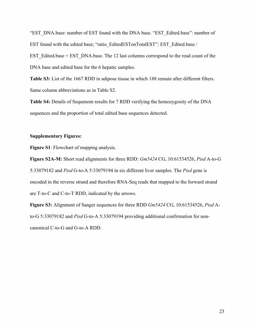
site with an offset of +/- 3 bases. To eliminate unidirectional strand bias, an RDD was only kept
if the proportion of reads belonging to the forward sequencing strand was ≥ 20% and ≤ 80%, in
at least 3 of 4 samples for adipose and 4 of 6 samples for liver. To eliminate the sequencing
extremity bias, an RDD was only kept if the proportion of reads where the alternative base was
found in the five first or last 5 bases of the sequencing read was ≤ 50% of the reads in at least 3
samples among 4 for adipose and 4 samples among 6 for liver. The thresholds for the
unidirectional strand and sequencing extremity bias have been calculated according to the
intersection of the distributions of RDD and SNP sites (see Figs. 2 and 3).
EST analysis. cDNA sequences with 100 bp flanking the edited base were extracted from the
UCSC database. Using these 201 bp sequences containing the edited base in the middle, BLAST
analyses were performed against the mouse EST database. For each RDD site we counted the
number of ESTs with the DNA base and with the edited base. All BLAST analyses were
8
conducted with the default parameters except the gap open and extend costs fixed to 200 (in
order to avoid gaps). The BLAST results were filtered by lengths of the alignments ≥ 75 bp and
the number of mismatches ≤ 5.
Sex and diet effects. An RDD site was declared as impacted by sex or diet if the fold change
“Edited base reads / DNA base reads“ between the two diets or the two sexes is > 1.5 (or <
1/1.5). The significance was tested by a Fisher Exact Test using p values corrected for multiple
testing by the Benjamini-Hochberg method. A cutoff of 0.05 was then used.
Sequenom Validation. DNA and RNA were extracted from 3 mice per cross (independent
sample from RNA-Seq RNA), pooled, and cDNA generated from the RNA pool. DNA and
cDNA were analyzed in a primer extension assay, designed to target the RDD sequence. The
primer extension assay was carried out using the MassARRAY (Sequenom iPLEX Gold
genotyping protocol) platform according to the manufacturer's specifications by The McGill
University and Génome Québec Innovation Centre. Primer extension products were analyzed by
matrix-assisted laser desorption/ionization time of-flight mass spectrometry (MALDI-TOF/MS).
If the RDD were present, it would generate two peaks on the MS profiles. The area of each peak
was proportional to the transcript abundance and was measured by the MassARRAY software to
generate an “edited base / reference base” ratio calculation. The SNP at the cDNA level was
compared to the genomic DNA at the same position, the latter being expected as homozygous for
a true RDD site.
Sanger Sequencing. PCR primers for genomic DNA, designed using the Primer3 website from
MIT, produced a PCR product that was at least 400 bases upstream and downstream of the RDD
on the DNA. Nested primers were designed and used to produce a product that was at least 200
bases on each side of the RDD site. The nested PCR procedure was necessary due to multiple
9
PCR product bands, sometimes overlapping, before this was implemented. The PCR product was
run on an agarose gel, cut out, and purified by QIAquick Gel Extraction Kit by Qiagen. Sanger
sequencing was performed by the UCLA Sequencing Core.
RESULTS
RNA Editing
We investigated the frequency of RNA editing in primary mouse tissues by mapping 232
million RNA-Seq generated reads from liver and 723 million reads from adipose of F1 mice of
inbred strains B6 and D2. (Figure S1 for flowchart.) We analyzed six independent liver and four
independent adipose samples from BXD and DXB males and females, with each sample
containing pooled RNA from three mice (Table S1).
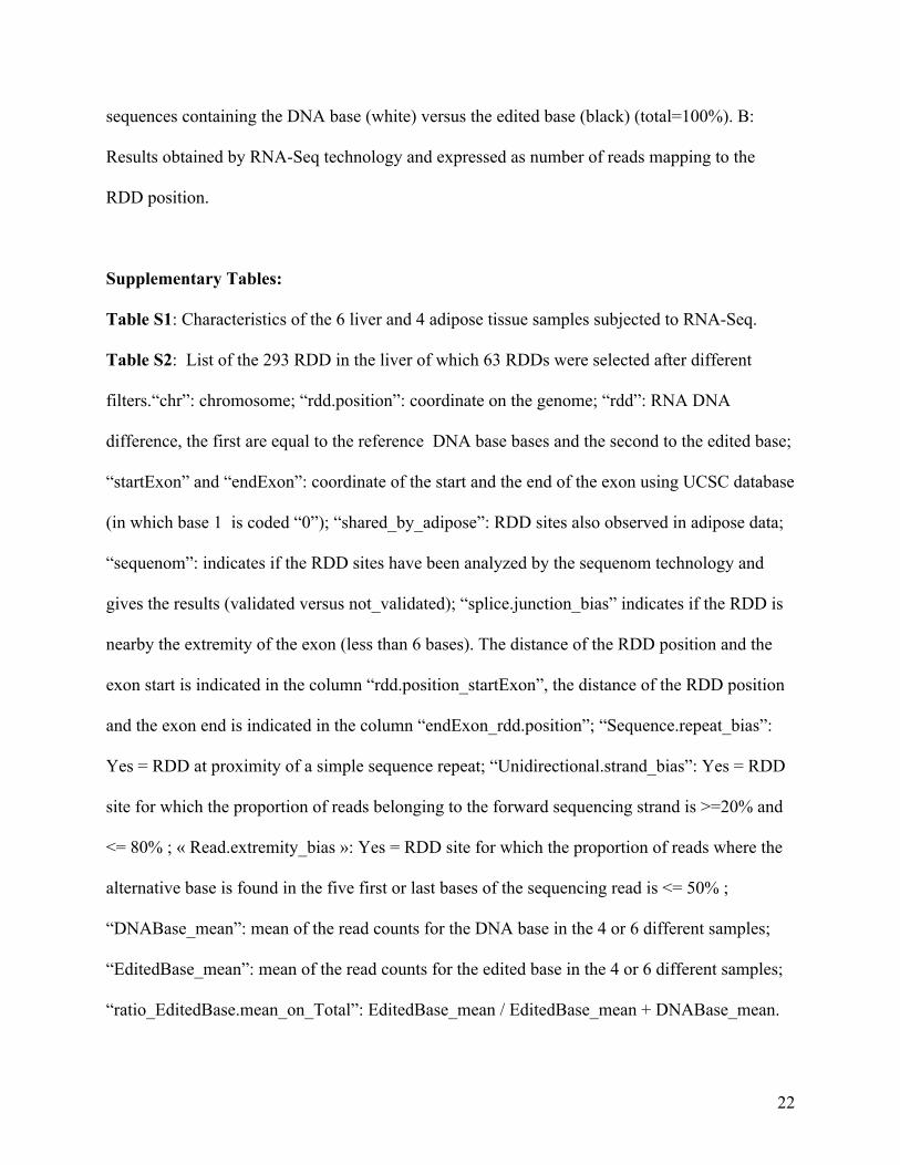
From hepatic RNA-Seq reads, we initially identified 7,319 exonic RDD sites with a
multiple mapping procedure, which allowed for the mapping of a read to more than one location
in the genome. We analyzed the same data using a unique mapping procedure, and reduced the
number to 293 exonic RDDs, indicating the strong impact of paralog alignments. We then
filtered for mismapping bias near a splice junction (7%), sequence repeats proximity
(27%)(Fig.1), unidirectional bias (49%) and sequencing extremity bias (56%) (Fig. 1, 2 and 3).
After applying these four filters, 63 RDDs remained for liver (Fig. 1 and Table S2). For adipose
tissue, the filter for unique mapping was applied from the outset, resulting in 1,667 exonic
adipose RDDs, and this number was reduced to 188 after applying the filters for mapping and
sequencing bias (Figs. 1-3 and Table S3). In both liver and adipose, unidirectional strand and
sequencing extremity biases were the major factors generating false positive RDDs. The
difference of unidirectional bias between the tissues, 49% liver and 73% adipose, and sequencing
10
extremity bias, 56% liver and 38% adipose, is possibly due to the sequencing method, Illumima
GAIIX on 75 bp reads for liver, and HiSeq2000 on 50 bp reads for adipose. The 63 RDDs in
liver represented 32 genes, and 188 RDDs in adipose represented 86 genes. The higher number
of RDDs found in adipose compared to liver probably reflects, at least in part, the higher (~5x)
number of reads generated for adipose. Fifteen exonic RDD sites representing 9 genes were
shared (Tables S2 and S3), suggesting that RNA editing is largely tissue-specific.
RDD Analysis using Expressed Sequence Tags (ESTs)
Using an in silico approach, we analyzed the exonic RDD sites with the public mouse EST
database (version 128). 58% of liver and 55% of adipose RDDs appeared at least once in the
EST database, and of those, 46% in liver and 42% in adipose, the edited base ESTs/total ESTs
ratio was ≥ 0.10 (Fig.1). Therefore, approximately half of the RDDs that we found have been
produced by other sequencing and RNA isolation methods, giving credence to those sites as real
RDD. On the other hand, half of the potential RDDs identified in our present study are novel.
Our data indicate that this could be due to either low expression of the mRNA containing the
edited base, resulting in its absence from the EST database, or to unaccounted for error in
mapping or sequencing.
RDD Characteristics
We characterized the resulting RDD according to RNA editing category (Fig. 4). In the original
293 liver and 1,667 adipose RDD obtained by the unique mapping procedure, we found more
transversions (purine to pyrimidine or pyrimidine to purine), 60% for liver and 65% for adipose,
than transitions (purine to purine or pyrimidine to pyrimidine) (Fig. 4). However, in the final 63
11
RDD for liver and 188 for adipose, transitions were more abundant in both tissues, 62% and
66%, respectively (Fig. 4). A-to-I(G) and C-to-U(T) canonical editing events (i.e. deaminase
enzyme-dependent) represented 56% and 52% of the transitions in liver and adipose,
respectively. The non-canonical categories, observed at a lower frequency, may represent novel
editing mechanisms, opposite strand transcripts, or other unknown sources of error (see
Discussion).
RDD validation
Due to genetic drift or to low sequencing coverage, some SNPs exist that may not be
referenced in SNP databases. Because these SNPs would lead to spurious RDD sites, we tested
by Sanger sequencing the genomic DNA from 12 RDD among the potential 63 hepatic RDD. We
found that three of those RDD sites resulted from an unreferenced SNP. We also tested 14 RDD
with Sequenom technology (Tables 1 and S2). We made new cDNA from different sets of mice
to avoid duplication of reverse transcription errors. Seven RDD sites that remained after filtering,
corresponding to four genes, were selected to represent a range of edited base ratios (EBR) from
0.33 to 1 in favor of the edited base (Table 1). An RDD site was confirmed by Sequenom if no
polymorphism was observed in the genomic DNA in the F1 mice, but was observed in the
cDNA. All seven potentially real RDD sites in liver were confirmed (Table 1, Fig. 5 and Table
S4). In those, we found similar proportions of reads with the edited base in Sequenom and RNA-
Seq technologies (Fig. 5A and B). The other seven RDD sites, chosen from those that had been
filtered out, were confirmed as true negatives by Sequenom analysis (Table 1). The degree of
editing in the seven RDD that we validated by Sequenom ranged from nearly 100% in Gm5424
to approximately 20% in Pisd (Fig. 5, Table S4, and Fig. S2). Npm3-ps1 and Gm5424 are
12
categorized as pseudogenes without protein products. Notably, two of the RDD that were
validated by Sequenom, Gm5424 C-to-G and Pisd G-to-A, were non-canonical, and to be certain
of the results, we also confirmed these RDD by Sanger sequencing (Fig. S3).
Genetic regulation of RNA editing and analysis of diet and sex effects
Evidence of genetic control was detected in the Sequenom validation studies in which F1
reciprocal crosses of AXH and BXH, were analyzed along with the BXD F1 reciprocal cross
mice. We found similar EBR for all the RDD among the different genetic backgrounds, except
for Npm3-ps1 (Fig. 5). The expression pattern for Npm3-ps1 (Fig 5A) suggested that RDD can
be influenced by genetics, sex and diet. First, we observed a lower EBR of this gene in AXH and
HXA F1 mice compared to the other crosses that was not due to a DNA SNP in this strain (Fig
5A and Table S4). Second, we observed a significantly higher EBR of AXH and HXA males fed
a high fat diet compared with the EBR of females fed a high fat diet or males fed a chow diet on
the same genetic background. However, we did not observe any RDD sites impacted by diet and
sex in the 63 RNA-Seq hepatic RDD, using the BXD and DXB reciprocal crosses as biological
replicates for each sex and diet (Table S1), suggesting that effects of these factors on RDD are
modest.
Discussion
We have carried out whole genome analysis of RDD in liver and adipose in mice to
examine the extent and nature of the phenomenon and to explore potential effects of sex, diet,
and genetics. The inbred mouse strains, B6 and D2, have been deep-sequenced and studied for
SNPs, helping to avoid issues such as DNA heterogeneity that likely confound studies with
13
human cell lines. Using multiple-site mapping at genomic sites, we obtained 7,319 apparent
RDD sites in mouse liver, similar in frequency to the initial report of extensive RDD in human B
cells (LI et al. 2011). However, after rigorous filtering for mapping and sequencing bias errors,
our RDD total was 63 in liver and 188 in adipose. Our comprehensive analysis is relevant to the
interpretation of RNA-Seq results that detect polymorphisms, for example, in allele-specific
expression studies.
A major aim of our study was to determine the importance of five known RNA-Seq
biases on RDD investigations. The most significant bias in our study was mismapping to
paralogous locations, the elimination of which reduced the RDD total from thousands to
hundreds. Following unique mapping, the other important biases were unidirectional strand and
extremity read bias, responsible for 40% to 73% of the remaining false positive RDD. Filters for
splice junction and sequence repeats bias eliminated between 5% and 27% of RDD. Our results
were consistent with those previously reported (KLEINMAN and MAJEWSKI 2012) (Fig. 1).
Furthermore, we report the importance of unreferenced SNPs that could arise from low sequence
coverage or spontaneous mutations which have become fixed in a breeding colony. If we
consider a mutation rate of about 2x10⁻⁸ per generation (SUN et al. 2012), and that the mice we
sequenced were many generations removed from the reference-sequenced mice, a significant
number of polymorphisms would be expected. Therefore, in order to make a legitimate claim of
widespread RNA editing, it is important to apply filters for mapping and sequencing bias, and to
confirm the genomic DNA sequence at each RDD position. We conclude that among the 63 and
188 RDD sites found in liver and adipose, a significant portion are possibly false positives due to
unreferenced genomic SNPs.
14
For the RDD that we identified, approximately 65% were transitions, and of those,
approximately 55% were canonical A-to-I(G) and C-to-U(T) edits. Therefore, while we found a
significant number of canonical RDD, likely produced by known RNA editing deaminase
enzymes, we also found many novel RDD that were non-canonical. All categories of RDD have
been found by other researchers (JU et al. 2011; BAHN et al. 2012), and although no enzymes
have been identified that can mediate these events, non-canonical RDD sites have been validated
previously (BAHN et al. 2012). Some non-canonical G-to-A and T-to-C transitions may exist
because regions of unknown sense-antisense transcription may lead to confusion about which is
the biologically relevant read (BAHN et al. 2012). We investigated 14 RDD by Sequenom
technology, including seven that remained after filtering and seven that were eliminated by
filtering. The seven RDD that had been filtered out, three canonical and four non-canonical (T-
to-G, G-to-A, and two C-to-A), were confirmed as true negatives. Of the seven RDD that were
confirmed as true positives, five were canonical, one was a non-canonical C-to-G transversion
(Gm5424), and one, a non-canonical G-to-A transition (Pisd). Sequenom and Sanger
sequencing, which investigated differences in both genomic DNA and cDNA sequences,
provided convincing evidence that these non-canonical RDD are genuine. The above possible
explanation, regarding unknown sense-antisense transcription, does not apply to the Pisd G-to-A
RDD, because the canonical A-to-G RDD on the same read, would be a non-canonical T-to-C in
the reverse direction. Therefore, either unknown mechanisms for RNA editing exist, or there are
unknown errors in the sequence interpretations.
The disparity of our numbers with human studies that report much larger numbers could
be explained two ways. First, heterogeneity in the DNA is much less controlled for in humans.
Since the human reference sequence used for mapping does not reflect the real diversity in the
15
genome of each individual, an overestimation of RDD is likely. Second, in human studies that
used transformed cell lines (LI et al. 2011; BAHN et al. 2012; PENG et al. 2012), high RNA
editing findings may reflect a real biological change. Deaminase enzymes, that are known to be
involved in editing (CONTICELLO 2008; NISHIKURA 2010; WULFF and NISHIKURA 2010), may
become more active in the transformation process, and increased RNA editing has been found in
some cancer cell lines (GALEANO et al. 2011). Equally important, viruses that were used to
transform human lymphocytes may have left remnants of viral DNA in those cells (LIN et al.
2012b), and as increased “errors” in RNA and DNA are an important mechanism for virus
survival (DOMINGO 2011; LIN et al. 2012b), the very large numbers for RDD in those cells may
reflect a biological artifact.
Nevertheless, the RDD numbers reported in our study are also lower compared to two
recent studies of exonic RNA editing in primary mouse tissues (DANECEK et al. 2012; GU et al.
2012). Danecek et al., (2012) reported approximately 700 exonic editing sites in whole brain of
15 mouse strains. ADAR expression and activity has been shown to be higher in brain than in
other tissues, which could explain some of the discrepancy (PAUL and BASS 1998); nonetheless,
sequence repeat bias which accounted for 27% of the error in our liver RDD were apparently not
filtered out in this study. Random sequencing errors were also more stringently dealt with in our
study, in which the RDD occurrence in 4 out of 6 samples was required, compared with that
study’s requirement of 2 biological replicates. These authors showed good reproducibility of
EBR between strains; however, unreferenced SNPs could be shared by these genetically related
strains. This study used Sequenom to test 611 sites, but used genomic DNA from only one strain,
C57BL/6, to confirm DNA homozygosity for all strains. Our Sanger sequencing results, in which
16
3 out of 12 RDD resulted from unreferenced SNPs, contradict the assumption that errors in the
reference (C57BL/6) genomic sequence are not a significant source of bias.
Another recent RNA editing study in mice by Gu et al. (2012) reported 140 RDD sites in
liver, closer in magnitude to the RDD that we found, although still higher. One difference was a
lower requirement for a site to be designated as edited: whereas we required at least 10 edited
base reads, and an EBR ≥ 0.1 in 4 of 6 samples, the Gu et al. (2012) study required two edited
base reads with an EBR ≥ 0.05 in each of 3 biological replicates. Thirty-eight of the reported 140
RDD would have passed our more stringent requirements. Surprisingly only three of those were
common to our dataset: Slc7a2, Serinc1 and 2810407C02Rik, highlighting the difficulty of
identifying a reproducible tissue-specific RDD list. Lack of agreement among studies is due in
part to differences in the multi-step analysis procedure, including the number of mismatched
bases authorized for the read alignments, the criteria to define an RDD site, and the filters used
for the different sequencing biases. It should be noted that RNA-Seq, used in our study, detected
RDD in exonic regions, but did not allow for analysis of intronic or non-coding regions, and thus
edited sites in these regions, which have been shown to influence splicing (LEV-MAOR et al.
2008), would not have been detected.
We found 25% hepatic RDD sites that were common to adipose. These results are
comparable to those found by Danecek et al. (2012), who found on average 50% to 60% of the
RDD observed in brain common with six other tissues, demonstrating a significant level of tissue
specificity of the edited sites.
Finally, our results were similar across all F1 heterozygous strains for 6 of the 7 RDD
sites that were interrogated by Sequenom, with substantial reproducibility of EBR between
strains. This finding was similar to Danecek et al. (2012) who reported RDD shared among nine
17
or more strains, with a good reproducibility of EBR between strains. One of the RDD confirmed
in our study, Npm3-ps1, had a different EBR among the F1 strains, indicating an effect of
genetics. In addition, within HXA and AXH samples, males fed a high fat diet had a higher EBR
for the Npm3-ps1 RDD, suggesting an effect of sex and diet. However, an analysis of the effect
of sex and diet on 63 hepatic RDD sites using BXD and DXB RNA-Seq data, shows a very
modest impact of these two factors.
In conclusion, our study represents a rigorous analysis of possible sources of error in
whole-genome evaluations of RNA editing in mice using RNA-Seq technology in normal
tissues. Our findings underscore the importance of properly filtering RNA-Seq data, not just for
RNA editing investigations, but also for all applications that require the identification of
polymorphisms in large datasets, such as allele-specific expression. Furthermore, we emphasize
the necessity of systematically controlling the genomic homozygous status. In contrast to several
recent studies using transformed human cell lines that found thousands of RNA editing events,
we directly analyzing inbred mouse tissues, thereby avoiding possible error caused by genetic
heterogeneity in humans. We conclude that exonic RNA editing in mouse liver and adipose is
limited to hundreds or fewer RDD sites. We do find evidence for non-canonical RNA editing, in
agreement with previous studies. This clearly requires further investigations, although at this
point, we cannot rule out the possibility of unknown sources of sequencing error. We also find
that sex and diet have relatively modest effects on RNA editing.
FUNDING
Funding was provided by the NIH Grants HL28481, HL30568, and DK072206 to AJL. FH and
EE are supported by National Science Foundation grants 0513612, 0731455, 0729049, 0916676
18
and 1065276, and NIH grants HL080079, DA024417. SL and FL were supported by grants from
the French genomic agricultural society (AGENAVI), INRA and the Agence Nationale de la
Recherche (Grant N°0426). HD07228 provided funding for LJM. The funders had no role in
study design, data collection and analysis, decision to publish, or preparation of the manuscript.
LITERATURE CITED
BAHN, J. H., J. H. LEE, G. LI, C. GREER, G. PENG et al., 2012 Accurate identification of A-to-I RNA editing in human by transcriptome sequencing. Genome Res 22: 142-150.
CONTICELLO, S. G., 2008 The AID/APOBEC family of nucleic acid mutators. Genome Biol 9: 229.
DANECEK, P., C. NELLAKER, R. E. MCINTYRE, J. E. BUENDIA-BUENDIA, S. BUMPSTEAD et al., 2012 High levels of RNA-editing site conservation amongst 15 laboratory mouse strains. Genome Biol 13: r26.
DOMINGO, E., 2011 Paradoxical interplay of viral and cellular functions. Viruses 3: 272-277. GALEANO, F., S. TOMASELLI, F. LOCATELLI and A. GALLO, 2011 A-to-I RNA editing: The
"ADAR" side of human cancer. Semin Cell Dev Biol. GU, T., F. W. BUAAS, A. K. SIMONS, C. L. ACKERT-BICKNELL, R. E. BRAUN et al., 2012
Canonical A-to-I and C-to-U RNA editing is enriched at 3'UTRs and microRNA target sites in multiple mouse tissues. PLoS One 7: e33720.
HACH, F., F. HORMOZDIARI, C. ALKAN, I. BIROL, E. E. EICHLER et al., 2010 mrsFAST: a cache-oblivious algorithm for short-read mapping. Nat Methods 7: 576-577.
JU, Y. S., J. I. KIM, S. KIM, D. HONG, H. PARK et al., 2011 Extensive genomic and transcriptional diversity identified through massively parallel DNA and RNA sequencing of eighteen Korean individuals. Nat Genet 43: 745-752.
KEANE, T. M., L. GOODSTADT, P. DANECEK, M. A. WHITE, K. WONG et al., 2011 Mouse genomic variation and its effect on phenotypes and gene regulation. Nature 477: 289-294.
KLEINMAN, C. L., and J. MAJEWSKI, 2012 Comment on "Widespread RNA and DNA sequence differences in the human transcriptome". Science 335: 1302; author reply 1302.
KOFLER, R., C. SCHLOTTERER and T. LELLEY, 2007 SciRoKo: a new tool for whole genome microsatellite search and investigation. Bioinformatics 23: 1683-1685.
LEV-MAOR, G., O. RAM, E. KIM, N. SELA, A. GOREN et al., 2008 Intronic Alus influence alternative splicing. PLoS Genet 4: e1000204.
LI, M., I. X. WANG, Y. LI, A. BRUZEL, A. L. RICHARDS et al., 2011 Widespread RNA and DNA sequence differences in the human transcriptome. Science 333: 53-58.
LIN, W., R. PISKOL, M. H. TAN and J. B. LI, 2012a Comment on "Widespread RNA and DNA sequence differences in the human transcriptome". Science 335: 1302; author reply 1302.
19
LIN, Z., A. PUETTER, J. COCO, G. XU, M. J. STRONG et al., 2012b Detection of Murine Leukemia Virus in the Epstein-Barr Virus-Positive Human B-Cell Line JY, Using a Computational RNA-Seq-Based Exogenous Agent Detection Pipeline, PARSES. J Virol 86: 2970-2977.
MEACHAM, F., D. BOFFELLI, J. DHAHBI, D. I. MARTIN, M. SINGER et al., 2011 Identification and correction of systematic error in high-throughput sequence data. BMC Bioinformatics 12: 451.
NISHIKURA, K., 2010 Functions and regulation of RNA editing by ADAR deaminases. Annu Rev Biochem 79: 321-349.
PAUL, M. S., and B. L. BASS, 1998 Inosine exists in mRNA at tissue-specific levels and is most abundant in brain mRNA. EMBO J 17: 1120-1127.
PENG, Z., Y. CHENG, B. C. TAN, L. KANG, Z. TIAN et al., 2012 Comprehensive analysis of RNA-Seq data reveals extensive RNA editing in a human transcriptome. Nat Biotechnol.
PICKRELL, J. K., Y. GILAD and J. K. PRITCHARD, 2012 Comment on "Widespread RNA and DNA sequence differences in the human transcriptome". Science 335: 1302; author reply 1302.
RAMASWAMI, G., W. LIN, R. PISKOL, M. H. TAN, C. DAVIS et al., 2012 Accurate identification of human Alu and non-Alu RNA editing sites. Nat Methods 9: 579-581.
SCHRIDER, D. R., J. F. GOUT and M. W. HAHN, 2011 Very few RNA and DNA sequence differences in the human transcriptome. PLoS One 6: e25842.
SUN, J. X., A. HELGASON, G. MASSON, S. S. EBENESERSDOTTIR, H. LI et al., 2012 A direct characterization of human mutation based on microsatellites. Nat Genet 44: 1161-1165.
TRAPNELL, C., L. PACHTER and S. L. SALZBERG, 2009 TopHat: discovering splice junctions with RNA-Seq. Bioinformatics 25: 1105-1111.
WATERSTON, R. H., K. LINDBLAD-TOH, E. BIRNEY, J. ROGERS, J. F. ABRIL et al., 2002 Initial sequencing and comparative analysis of the mouse genome. Nature 420: 520-562.
WULFF, B. E., and K. NISHIKURA, 2010 Substitutional A-to-I RNA editing. Wiley Interdiscip Rev RNA 1: 90-101.
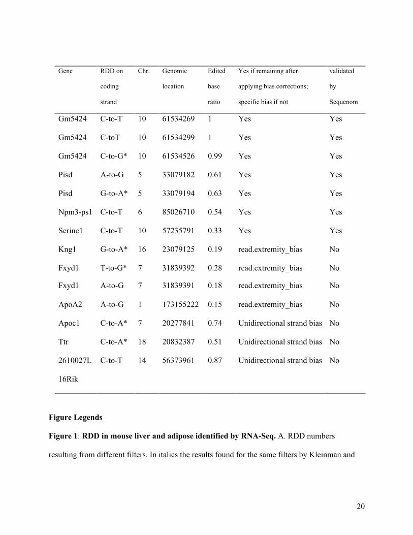
Table 1. RDD tested by Sequenom mass spectrometry. Seven RDD that remained after filtering
were validated. Seven that had been filtered out were confirmed as true negatives. U is
represented by T and I is represented by G. Non-canonical editing is designated by an asterisk
(*).
20
Figure Legends
Figure 1: RDD in mouse liver and adipose identified by RNA-Seq. A. RDD numbers
resulting from different filters. In italics the results found for the same filters by Kleinman and
Gene RDD on
coding
strand
Chr. Genomic
location
Edited
base
ratio
Yes if remaining after
applying bias corrections;
specific bias if not
validated
by
Sequenom
Gm5424 C-to-T 10 61534269 1 Yes Yes
Gm5424 C-toT 10 61534299 1 Yes Yes
Gm5424 C-to-G* 10 61534526 0.99 Yes Yes
Pisd A-to-G 5 33079182 0.61 Yes Yes
Pisd G-to-A* 5 33079194 0.63 Yes Yes
Npm3-ps1 C-to-T 6 85026710 0.54 Yes Yes
Serinc1 C-to-T 10 57235791 0.33 Yes Yes
Kng1
G-to-A* 16 23079125 0.19 read.extremity_bias No
Fxyd1 T-to-G* 7 31839392 0.28 read.extremity_bias No
Fxyd1 A-to-G 7 31839391 0.18 read.extremity_bias No
ApoA2 A-to-G 1 173155222 0.15 read.extremity_bias No
Apoc1 C-to-A* 7 20277841 0.74 Unidirectional strand bias No
Ttr C-to-A* 18 20832387 0.51 Unidirectional strand bias No
2610027L
16Rik
C-to-T 14 56373961 0.87 Unidirectional strand bias No
21
Majewski (KLEINMAN and MAJEWSKI 2012). B. Percentages of RDD reads found in comparison
with reads from the EST database.
Figure 2: Contribution of sequencing strand bias to RDD. Histogram with the distribution of
RDD (Y-axis) according to the proportion of reads belonging to the forward sequencing strand
(X-axis) in liver (sample M.CH.BxD) and adipose (sample F.BxD). White bars and the dashed
line correspond to the RDD sites. Grey bars and the solid line correspond to the dbSNP sites that
are polymorphic in the sample and used as a control data set. The false positives are calculated
using the tails of the distributions where the RDD and SNP distributions intersect. The same
shape of distribution was observed for all samples of the same tissue.
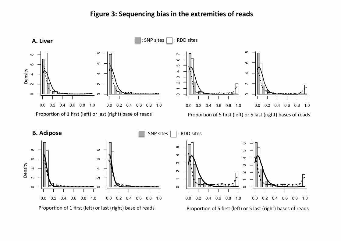
Figure 3: Contribution of end of read sequencing to RDD. Histogram with the distribution of
RDD (Y-axis) according to the proportion of reads where the alternative base is found in the first
or last base or in the 5 first or 5 last bases of the sequencing read (X-axis). A: liver (sample
M.CH.BxD) and B: Adipose (sample F.BxD) ; White bars and the dashed line correspond to the
RDD sites. Grey bars and the solid line correspond to the dbSNP sites that are polymorphic in
the sample and used as a control data set. The false-positives are calculated using the tails of the
distributions where the RDD and SNP distributions intersect. The same shape of distribution was
observed for all samples of the same tissue.
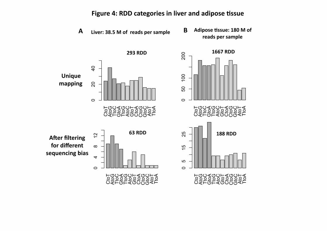
Figure 4: RDD categories in liver and adipose tissue. A: RDD categories observed with liver
RNA-Seq data after the multiple and unique mapping procedures and filtering for sequencing
bias. B: RDD categories observed with adipose RNA-Seq data after the unique mapping
procedure and filtering for sequencing bias.
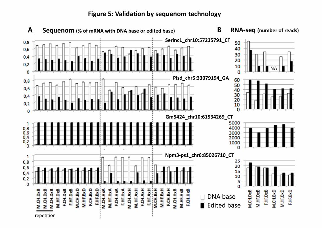
Figure 5: Validation by Sequenom technology of four RDD observed by RNA-Seq in liver.
A: Results obtained by Sequenom technology and expressed as percentage of total mRNA
22
sequences containing the DNA base (white) versus the edited base (black) (total=100%). B:
Results obtained by RNA-Seq technology and expressed as number of reads mapping to the
RDD position.
Supplementary Tables:
Table S1: Characteristics of the 6 liver and 4 adipose tissue samples subjected to RNA-Seq.
Table S2: List of the 293 RDD in the liver of which 63 RDDs were selected after different
filters.“chr”: chromosome; “rdd.position”: coordinate on the genome; “rdd”: RNA DNA
difference, the first are equal to the reference DNA base bases and the second to the edited base;
“startExon” and “endExon”: coordinate of the start and the end of the exon using UCSC database
(in which base 1 is coded “0”); “shared_by_adipose”: RDD sites also observed in adipose data;
“sequenom”: indicates if the RDD sites have been analyzed by the sequenom technology and
gives the results (validated versus not_validated); “splice.junction_bias” indicates if the RDD is
nearby the extremity of the exon (less than 6 bases). The distance of the RDD position and the
exon start is indicated in the column “rdd.position_startExon”, the distance of the RDD position
and the exon end is indicated in the column “endExon_rdd.position”; “Sequence.repeat_bias”:
Yes = RDD at proximity of a simple sequence repeat; “Unidirectional.strand_bias”: Yes = RDD
site for which the proportion of reads belonging to the forward sequencing strand is >=20% and
<= 80% ; « Read.extremity_bias »: Yes = RDD site for which the proportion of reads where the
alternative base is found in the five first or last bases of the sequencing read is <= 50% ;
“DNABase_mean”: mean of the read counts for the DNA base in the 4 or 6 different samples;
“EditedBase_mean”: mean of the read counts for the edited base in the 4 or 6 different samples;
“ratio_EditedBase.mean_on_Total”: EditedBase_mean / EditedBase_mean + DNABase_mean.
23
“EST_DNA.base: number of EST found with the DNA base. “EST_Edited.base”: number of
EST found with the edited base; “ratio_EditedESTonTotalEST”: EST_Edited.base /
EST_Edited.base + EST_DNA.base. The 12 last columns correspond to the read count of the
DNA base and edited base for the 6 hepatic samples.
Table S3: List of the 1667 RDD in adipose tissue in which 188 remain after different filters.
Same column abbreviations as in Table S2.
Table S4: Details of Sequenom results for 7 RDD verifying the homozygosity of the DNA
sequences and the proportion of total edited base sequences detected.
Supplementary Figures:
Figure S1: Flowchart of mapping analysis.
Figure S2A-M: Short read alignments for three RDD: Gm5424 CG, 10:61534526, Pisd A-to-G
5:33079182 and Pisd G-to-A 5:33079194 in six different liver samples. The Pisd gene is
encoded in the reverse strand and therefore RNA-Seq reads that mapped to the forward strand
are T-to-C and C-to-T RDD, indicated by the arrows.
Figure S3: Alignment of Sanger sequences for three RDD Gm5424 CG, 10:61534526, Pisd A-
to-G 5:33079182 and Pisd G-to-A 5:33079194 providing additional confirmation for non-
canonical C-to-G and G-to-A RDD.
Unique mapping 1667 RDD
Unique mapping 293 RDD
Figure 1: RDD iden8fied in liver and adipose 8ssues a>er using different filters
Mul8ple mapping 7319 RDD
7 %
58 % 46 % 8 %
Liver: 38.5 M of reads per sample
Adipose 8ssue: 180 M of reads per sample
5 %
RDD with at least 1 EST with the edited base (edited.EST): RDD with the ra<o “edited.EST / total.EST” >= 10%: RDD with the ra<o “edited.EST / total.EST” = 100%:
A
27 %
Splice junc<on bias
sequence repeat bias
188 RDD 63 RDD
Unidirec<onal strand bias 49 %
Sequencing extremity bias 56 %
(Kleinman 10% et al 2012)
12%
65%
60%
15 %
73 %
38 %
55 % 42 % 0 %
B
Liver
Figure 2: Unidirec8onal strand bias
Propor<on of reads in the forward sequencing strand
Den
sity
Adipose
Den
sity
0.0 0.2 0.4 0.6 0.8 1.0
0.0
1.0
2.0
3.0
0.0 0.2 0.4 0.6 0.8 1.0
: SNP sites : RDD sites
0.0 0.2 0.4 0.6 0.8 1.0
0.0
1.0
2.0
A. Liver
Den
sity
B. Adipose
Den
sity
Propor<on of 1 first (leT) or last (right) base of reads
0.0 0.2 0.4 0.6 0.8 1.0
01
23
45
6
0.0 0.2 0.4 0.6 0.8 1.0
01
23
45
Propor<on of 5 first (leT) or 5 last (right) bases of reads
0.0 0.2 0.4 0.6 0.8 1.0
02
46
8
0.0 0.2 0.4 0.6 0.8 1.0
02
46
8
Propor<on of 1 first (leT) or last (right) base of reads
0.0 0.2 0.4 0.6 0.8 1.0
02
46
8
0.0 0.2 0.4 0.6 0.8 1.0
02
46
80.0 0.2 0.4 0.6 0.8 1.0
02
46
8
0.0 0.2 0.4 0.6 0.8 1.0
01234567
Figure 3: Sequencing bias in the extremi8es of reads
: SNP sites : RDD sites
: SNP sites : RDD sites
Propor<on of 5 first (leT) or 5 last (right) bases of reads
Unique mapping
Figure 4: RDD categories in liver and adipose 8ssue
Liver: 38.5 M of reads per sample
293 RDD 1667 RDD
Adipose 8ssue: 180 M of reads per sample
A B
A>er filtering for different
sequencing bias
188 RDD 63 RDD
CtoT
AtoG
TtoC
GtoA
TtoG
AtoC
GtoT
CtoA
CtoG
GtoC
AtoT
TtoA
050
100
200
CtoT
AtoG
TtoC
GtoA
TtoG
AtoC
GtoT
CtoA
CtoG
GtoC
AtoT
TtoA
020
40
CtoT
AtoG
TtoC
GtoA
TtoG
AtoC
GtoT
CtoA
CtoG
GtoC
AtoT
TtoA
04
812
CtoT
AtoG
TtoC
GtoA
TtoG
AtoC
GtoT
CtoA
CtoG
GtoC
AtoT
TtoA
05
1525
repe<<on
A
NA
DNA base Edited base
Sequenom (% of mRNA with DNA base or edited base) RNA-‐seq (number of reads) B
Figure 5: Valida8on by sequenom technology
Serinc1_chr10:57235791_CT
Pisd_chr5:33079194_GA
Gm5424_chr10:61534269_CT
Npm3-‐ps1_chr6:85026710_CT


![ELLEN MARIA HAGOPIAN ASSÉDIO MORAL NA VIVÊNCIA DE … · Hagopian EM. Assédio moral na vivência de enfermeiros: perspectiva fenomenológica [dissertação]. São Paulo: Escola](https://img.pdfslide.net/doc/110x75/5f24f703ef95a41e1d078640/ellen-maria-hagopian-assdio-moral-na-vivncia-de-hagopian-em-assdio-moral.jpg)