Embed Size (px)
Citation preview
397
© 2016 The Korean Society of Pathologists/The Korean Society for CytopathologyThis is an Open Access article distributed under the terms of the Creative Commons Attribution Non-Commercial License (http://creativecommons.org/licenses/ by-nc/3.0) which permits unrestricted non-commercial use, distribution, and reproduction in any medium, provided the original work is properly cited.
pISSN 2383-7837eISSN 2383-7845
Pelvic Nodular Histiocytic and Mesothelial Hyperplasia in a Patient with Endometriosis and Uterine Leiomyoma
Yumin Chung · Rehman Abdul · Se Min Jang · Joong Sub Choi1 · Kiseok Jang
Department of Pathology, 1Division of Gynecologic Oncology and Gynecologic Minimally Invasive Surgery, Department of Obstetrics and Gynecology, Hanyang University College of Medicine, Seoul, Korea
Journal of Pathology and Translational Medicine 2016; 50: 397-400http://dx.doi.org/10.4132/jptm.2016.01.11
▒ BRIEF CASE REPORT ▒
Nodular histiocytic and mesothelial hyperplasia (NHMH) is a rare and benign proliferative lesion composed of histiocytes with scattered mesothelial cells which was first reported in her-nia sac by Rosai and Dehner in 1975.1 They described NHMH as a “benign reactive condition simulating a neoplastic process.”1 Since then, several cases have been reported in lung, pleura, in-guinal region, urinary bladder, and pelvic cavity.2-9 We report a case of incidentally detected NHMH, presenting as a pelvic nodule during laparoscopic surgery for uterine myoma and en-dometriosis.
CASE REPORT
The patient was a 38-year-old woman complaining of abdom-inal discomfort and infertility without previous pregnancy his-tory. A 8.3 × 5-cm-sized intramural type myoma was found on gynecologic sonography. There was no medical history of previ-ous abdominal or pelvic surgery. During laparoscopic myomec-tomy, a left ovarian cyst (2.6 × 1.6 cm in size), a bladder perito-neal mass (1.6 × 1 cm in size), and a small nodule in cul-de-sac cavity were incidentally obtained. The pathologic diagnosis of left ovary cyst and bladder peritoneal mass was endometriosis. Gross examination of the mass in cul-de-sac cavity showed a grayish white and solid nodule, measuring 0.8 × 0.5 cm in size. Microscopically, two populations of cells were identified. The
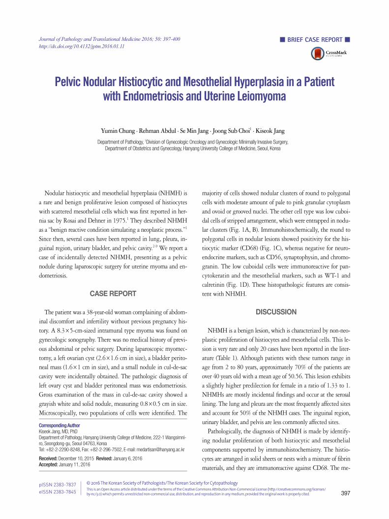
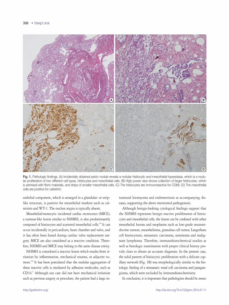
majority of cells showed nodular clusters of round to polygonal cells with moderate amount of pale to pink granular cytoplasm and ovoid or grooved nuclei. The other cell type was low cuboi-dal cells of stripped arrangement, which were entrapped in nodu-lar clusters (Fig. 1A, B). Immunohistochemically, the round to polygonal cells in nodular lesions showed positivity for the his-tiocytic marker (CD68) (Fig. 1C), whereas negative for neuro-endocrine markers, such as CD56, synaptophysin, and chromo-granin. The low cuboidal cells were immunoreactive for pan-cytokeratin and the mesothelial markers, such as WT-1 and calretinin (Fig. 1D). These histopathologic features are consis-tent with NHMH.
DISCUSSION
NHMH is a benign lesion, which is characterized by non-neo-plastic proliferation of histiocytes and mesothelial cells. This le-sion is very rare and only 20 cases have been reported in the liter-ature (Table 1). Although patients with these tumors range in age from 2 to 80 years, approximately 70% of the patients are over 40 years old with a mean age of 50.56. This lesion exhibits a slightly higher predilection for female in a ratio of 1.33 to 1. NHMHs are mostly incidental findings and occur at the serosal lining. The lung and pleura are the most frequently affected sites and account for 50% of the NHMH cases. The inguinal region, urinary bladder, and pelvis are less commonly affected sites.
Pathologically, the diagnosis of NHMH is made by identify-ing nodular proliferation of both histiocytic and mesothelial components supported by immunohistochemistry. The histio-cytes are arranged in solid sheets or nests with a mixture of fibrin materials, and they are immunoreactive against CD68. The me-
Corresponding AuthorKiseok Jang, MD, PhDDepartment of Pathology, Hanyang University College of Medicine, 222-1 Wangsimni-ro, Seongdong-gu, Seoul 04763, KoreaTel: +82-2-2290-8248, Fax: +82-2-296-7502, E-mail: [email protected]
Received: December 10, 2015 Revised: January 6, 2016Accepted: January 11, 2016
http://jpatholtm.org/ http://dx.doi.org/10.4132/jptm.2016.01.11
398 • Chung Y, et al.
sothelial component, which is arranged in a glandular- or strip-like structure, is positive for mesothelial markers such as cal-retinin and WT-1. The nuclear atypia is typically absent.
Mesothelial/monocytic incidental cardiac excrescence (MICE), a tumour-like lesion similar to NHMH, is also predominantly composed of histiocytes and scattered mesothelial cells.10 It can occur incidentally in pericardium, heart chamber and valve, and it has often been found during cardiac valve replacement sur-gery. MICE are also considered as a reactive condition. There-fore, NHMH and MICE may belong to the same disease entity.
NHMH is considered a reactive lesion which results from ir-ritation by inflammation, mechanical trauma, or adjacent tu-mors.2,6 It has been postulated that the nodular aggregation of these reactive cells is mediated by adhesion molecules, such as CD34.5 Although our case did not have mechanical irritation such as previous surgery or procedure, the patient had a large in-
tramural leiomyoma and endometriosis as accompanying dis-eases, supporting the above mentioned pathogenesis.
Although benign-looking cytological findings support that the NHMH represents benign reactive proliferation of histio-cytes and mesothelial cells, the lesion can be confused with other mesothelial lesions and neoplasms such as low-grade neuroen-docrine tumors, mesothelioma, granulosa cell tumor, Langerhans cell histiocytosis, metastatic carcinoma, seminoma and malig-nant lymphoma. Therefore, immunohistochemical studies as well as histologic examination with proper clinical history pro-vide clues to obtain an accurate diagnosis. In the present case, the solid pattern of histiocytic proliferation with a delicate cap-illary network (Fig. 1B) was morphologically similar to the his-tologic finding of a metastatic renal cell carcinoma and paragan-gioma, which were excluded by immunohistochemistry.
In conclusion, it is important that pathologists should be aware
A
C
B
D
Fig. 1. Pathologic findings. (A) Incidentally obtained pelvic nodule reveals a nodular histiocytic and mesothelial hyperplasia, which is a nodu-lar proliferation of two different cell types, histiocytes and mesothelial cells. (B) High power view shows collection of larger histiocytes, which is admixed with fibrin materials, and strips of smaller mesothelial cells. (C) The histiocytes are immunoreactive for CD68. (D) The mesothelial cells are positive for calretinin.
http://jpatholtm.org/http://dx.doi.org/10.4132/jptm.2016.01.11
Histiocytic and Mesothelial Hyperplasia • 399
of this entity and correlate clinically, histologically, and immuno-histochemically to make a correct diagnosis.
Conflicts of InterestNo potential conflict of interest relevant to this article was
reported.
REFERENCES
1. Rosai J, Dehner LP. Nodular mesothelial hyperplasia in hernia sacs:
a benign reactive condition simulating a neoplastic process. Cancer
1975; 35: 165-75.
2. Chan JK, Loo KT, Yau BK, Lam SY. Nodular histiocytic/mesotheli-
al hyperplasia: a lesion potentially mistaken for a neoplasm in
transbronchial biopsy. Am J Surg Pathol 1997; 21: 658-63.
3. Ordonez NG, Ro JY, Ayala AG. Lesions described as nodular me-
sothelial hyperplasia are primarily composed of histiocytes. Am J
Surg Pathol 1998; 22: 285-92.
4. Choi YL, Song SY. Cytologic clue of so-called nodular histiocytic
hyperplasia of the pleura. Diagn Cytopathol 2001; 24: 256-9.
5. Suarez-Vilela D, Izquierdo-Garcia FM. Nodular histiocytic/meso-
thelial hyperplasia: a process mediated by adhesion molecules?
Histopathology 2002; 40: 299-300.
6. Chikkamuniyappa S, Herrick J, Jagirdar JS. Nodular histiocytic/
mesothelial hyperplasia: a potential pitfall. Ann Diagn Pathol 2004;
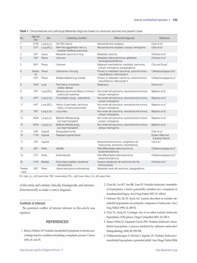
Table 1. Clinical features and pathologicdifferential diagnosis based on previously reported and present cases
No. Age (yr)/
Sex Site Underlying condition Differential diagnosis Reference
1 57/M Lung (LLL) Old TBC lesions Neuroendocrine neoplasm Chan et al.2
2 51/F Lung (RLL) Weil-Felix agglutination test (+), possible rickettsial pneumonia
Neuroendocrine neoplasm, ectopic meningioma Chan et al.2
3 23/F Lleura Metastatic sarcoma to lung Metastatic sarcoma Ordonez et al.3
4 78/F Pleura Unknown Metastatic adenocarcinoma, epithelioid hemangioendothelioma
Ordonez et al.3
5 80/F Pleura Unknown Malignant mesothelioma, metastatic carcinoma, ectopic meningioma, paraganglioma
Choi and Song4
6 Elderly/M
Pleura Carcinoma in the lung Primary or metastatic carcinoma, carcinoid tumor, mesothelioma, histiocytosis X
Chikkamuniyappa et al.6
7 53/F Pleura Multiple bilateral lung nodules Primary or metastatic carcinoma, carcinoid tumor, mesothelioma, histiocytosis X
Chikkamuniyappa et al.6
8 79/M Lung Past history of ischemic cardiac disease
Malignancy Rossi et al.8
9 76/F Lung (RLL) Bilateral pneumonia (history of chronic l ymphocytic leukemia)
Non-small cell carcinoma, neuroendocrine tumors, ectopic meningioma
Bejarano et al.7
10 40/F Lung (LUL) Consolidation (lung), scleroderma Non-small cell carcinoma, neuroendocrine tumors, ectopic meningioma
Bejarano et al.7
11 44/F Lung (RLL) History of pancreatic carcinoma, history of chemopneumonitis
Non-small cell carcinoma, neuroendocrine tumors, ectopic meningioma
Bejarano et al.7
12 78/F Lung (LUL) Lung mass Non-small cell carcinoma, neuroendocrine tumors, ectopic meningioma
Bejarano et al.7
13 46/M Lung (LLL) Bilateral infiltrates (lung) (s/p heart transplant)
Non-small cell carcinoma, neuroendocrine tumors, ectopic meningioma
Bejarano et al.7
14 62/M Lung (LLL) Bilateral infiltrates (lung) (s/p heart transplant)
Non-small cell carcinoma, neuroendocrine tumors, ectopic meningioma
Bejarano et al.7
15 2/M Inguinal Strangulated hernia - Chan et al.2
16 71/M Inguinal Relapsed inguinal hernia - Suarez-Vilela and Izquierdo-Garcia5
17 5/M Inguinal - Neuroendocrine tumors, Langerhans cell histiocytosis, seminoma, mesothelioma
Cai et al.9
18 36/F Pelvis Infertility Well-differentiated adenocarcinoma, leukemia/lymphoma
Chikkamuniyappa et al.6
19 37/F Pelvis Endometriosis Well-differentiated adenocarcinoma, leukemia/lymphoma
Chikkamuniyappa et al.6
20 74/M Bladder Noninvasive papillary transitional cell carcinoma
Invasive transitional cell carcinoma into the lamina propria
Ordonez et al.3
Present case
38/F Pelvis Uterine leiomyoma endometriosis Metastatic renal cell carcinoma, paraganglioma
M, male; LLL, left lower lobe; TBC, tuberculosis; RLL, right lower lobe; LUL, left upper lobe.
http://jpatholtm.org/ http://dx.doi.org/10.4132/jptm.2016.01.11
400 • Chung Y, et al.
8: 115-20.
7. Bejarano PA, Garcia MT, Ganjei-Azar P. Mesothelial cells in trans-
bronchial biopsies: a rare complication with a potential for a diag-
nostic pitfall. Am J Surg Pathol 2007; 31: 914-8.
8. Rossi G, Cavazza A, Guicciardi N, Marchioni A. Nodular histiocyt-
ic/mesothelial hyperplasia on transthoracic biopsy: another source
of potential pitfall in a lesion frequently present in spontaneous
pneumothorax. Histopathology 2008; 52: 250-2.
9. Cai Z, Xie Q, Wang X, Guo B, Wang X, Wang K. Nodular histiocyt-
ic/mesothelial hyperplasia: a clinicopathologic analysis of 7 cases.
Zhonghua Bing Li Xue Za Zhi 2014; 43: 256-9.
10. Jiao N, Zhang W, Wang W, et al. Mesothelial/monocytic incidental
cardiac excrescence: a case report and review of literature. Int J Clin
Exp Pathol 2014; 7: 6219-24.