Embed Size (px)
Citation preview

Research ArticlePhotoaging Skin Therapy with PRP and ADSC: AComparative Study
Luiz Charles-de-Sá ,1 Natale Gontijo-de-Amorim,2 Andrea Sbarbati,2 Donatella Benati,2
Paolo Bernardi,2 Radovan Borojevic,3 Rosana Bizon Vieira Carias,3 and Gino Rigotti4
1Postgraduate Program in Surgical Science, Federal University of Rio de Janeiro-CCS-Bloco C, Avenida Carlos Chagas Filho, 373,Ilha do Fundão, Rio de Janeiro, RJ 21941-902, Brazil2Dipartamento di Scienze Neurologiche e del Movimento, Sezione di Anatomia e Istologia della Universitá degli Studi di Verona,Strada Le Grazie 8, Verona 37134, Italy3Universidade Federal do Rio de Janeiro-UFRJ-CCS and Centro de Biotecnologia-IMETRO, Rio de Janeiro, Brazil4Casa di Cura San Francesco-Unità di Chirurgia Rigenerativa, Via Monte Ortigara, 21, Verona, Italy
Correspondence should be addressed to Luiz Charles-de-Sá; [email protected]
Received 9 February 2020; Revised 20 May 2020; Accepted 20 June 2020; Published 16 July 2020
Academic Editor: Gerald A. Colvin
Copyright © 2020 Luiz Charles-de-Sá et al. This is an open access article distributed under the Creative Commons AttributionLicense, which permits unrestricted use, distribution, and reproduction in any medium, provided the original work isproperly cited.
Background. Stem cells from adipose tissue (ADSCs) and platelet-rich plasma (PRP) are innovative modalities that arise due to theirregenerative potential. Objective. The aim of this study was to characterize possible histological changes induced by PRP and ADSCtherapies in photoaged skin.Methods. A prospective randomized study involving 20 healthy individuals, showing skin aging. Theyunderwent two therapeutic protocols (protocol 1: PRP; protocol 2: ADSCs). Biopsies were obtained before and after treatment(4 months). Results. PRP protocol showed unwanted changes in the reticular dermis, mainly due to the deposition of a horizontallayer of collagen (fibrosis) and elastic fibers tightly linked. Structural analyses revealed infiltration of mononuclear cells and depot offibrotic material in the reticular dermis. The ADSC protocol leads to neoelastogenesis with increase of tropoelastin and fibrillin.There was an improvement of solar elastosis inducing an increment of macrophage polarization and matrix proteinases. Theselast effects are probably related to the increase of elastinolysis and the remodeling of the dermis. Conclusions. The PRPpromoted an inflammatory process with an increase of reticular dermis thickness with a fibrotic aspect. On the other hand,ADSC therapy is a promising modality with an important antiaging effect on photoaged human skin.
1. Introduction
The aging skin is a complex biological phenomenon that iscomposed of intrinsic and extrinsic processes. Facial agingis a degenerative process that affects the skin and deep struc-tures resulting from the action of the intrinsic factor (age)associated with an extrinsic factor related to exposure toultraviolet radiation [1–5]. The connective tissue of the skinis mainly composed of collagen and elastin. Collagen repre-sents 70% to 80% of the dry weight of the skin, conferringmechanical strength and structural integrity. Elastic fiber isa minor component of the dermis, responsible for skin elas-
ticity, with elastin protein representing 2% to 4% of the extra-cellular matrix [6]. In photoaging skin, there is progressivedestruction of the entire dermal elastic fiber network, withthick, tangled, tortuous, degraded, and dysfunctional fibers,leading to the increased density of the elastic material, result-ing in a cluster of amorphous and dystrophic elastotic mate-rial throughout the dermis, forming solar elastosis, the mainfeature of photoaging [6].
In recent years, there is an increasing interest in the use ofthe regenerative properties of adipose tissue [7, 8]. In thisscope, some studies have suggested the application of fatgrafting (lipotransferring) to improve skin quality [9, 10].
HindawiStem Cells InternationalVolume 2020, Article ID 2032359, 13 pageshttps://doi.org/10.1155/2020/2032359
Animal model studies have described an improvement inskin texture and, in particular, increased elasticity, hyperpig-mentation control, and aesthetic improvement of scars,regardless of etiopathogenesis [11–19].
Zuk et al.’s studies in 2001 [7, 20] identified mononucle-ated cells in the adipose tissue known as fibroblast-like cellswith high capacity for cell differentiation. Mesenchymalstem cells, found in the stromal vascular fraction (SVF) ofadipose tissue, are multipotent cells with high potential forself-renewal and ex vivo expansion, with the ability to differ-entiate into several cell lineages [21–26]. According to theInternational Society for Cell Therapy, minimum criteriaare adopted to define a stem cell: characterization of MSCrequires its phenotyping by identifying the cell surfacemembrane markers, its ability to adhere to the plastic sur-face, and the ability to differentiate into 2 to 3 distinct celllineages [27].
Platelet-rich plasma (PRP) has also been described ashaving a regenerative effect due to growth factors andcytokines [28–31]. Currently, PRP therapy is described inmany studies, as well as associated with other treatments[32–34]. PRP has been used to treat alopecia, acne, traumaticscars, contractile scars, wrinkles, stretch marks, chroniculcers, laser therapy, and postsurgical wound to enhancethe healing process. The PRP preparations have been usedsince the 1970s, and they have been increasingly popularsince the 1990s [35]. Since then, different protocols andways of preparing PRP have emerged: from conventionalblood centrifugation to innovative systems. It can be acti-vated by adding collagen, calcium, and/or thrombin, byglass contact or by freeze/thaw cycles, applied as a plateletor gel suspension [36–38].
At the moment, the biological mechanisms responsi-ble for the rejuvenating and regenerative effect on humanskin obtained with the fat graft and/or its componentsare not yet clearly understood, and there is no consensuson the histological and ultrastructural changes that occurin the skin and subcutaneous tissue, after ADSC and PRPtherapies [39–43].
This study is aimed at evaluating the comparative histo-logical changes of aged facial skin after subdermal therapywith PRP and ADSCs.
2. Patients, Materials, and Methods
This is a prospective clinical, randomized, and comparativestudy conducted between April 2013 and June 2017 with 20healthy individuals (ASA 1: English, American Society ofAnesthesiology) [44] of both genders, with ages ranging from45 to 65 years, presenting photoaging skin variables, differentskin color phototypes (Fitzpatrick) [45, 46], and cervicofacialsagging. The candidates who participated in this study werespontaneously submitted to the face-lifting surgical proce-dure at its end. All patients from northeastern Brazil weresplit into two skin therapy protocols: protocol 1 (PRP skintherapy) and protocol 2 (ADSC skin therapy).
The study was conducted at the Carlos Chagas FilhoInstitute of Biophysics, Federal University of Rio deJaneiro-Brazil, at the Excellion Services Biomedical SA® lab-
oratory in Rio de Janeiro-Brazil, at the Center for Anatomyand Histology at the University of Verona-Italy and at Per-forma Ltda clinic, in Natal, RN, Brazil. All patients wererecruited at the author’s private clinic (Natal, RN), by elec-tronic and/or verbal invitation and sent for clinical, cardio-logical, laboratory evaluation and signing of an informedconsent form that was in accordance with the ethical princi-ples of the Declaration of Helsinki 2000 and Istanbul 2008,following the norms of resolution 466/12 and subsequentones of the National Health Council/Ministry of Health. Itwas submitted to the Ethics and Research Committee boardto be approved (No. 28.063 at 05-25-2012) and registeredin the Brazilian Registry of Clinical Trials (REBEC) on 04-07-2013 (RBR-2nn9y2).
Subjects with a smoking habit, with hematologic orhemodynamic disorders, autoimmune diseases, connectivetissue diseases, diabetes type I or II, other metabolic diseases,and chronic use of corticosteroids, and those that had beensubmitted to recent dermatological or surgical face treat-ments (e.g., facial peeling) were not included. The direct end-point of the study was to compare the histological benefitsprovided by the subdermal PRP and ADSC therapies.
2.1. Protocol 1: PRP Skin Therapy
2.1.1. Blood Collection. Three 10ml ACD tubes were collectedwith 3.2% sodium citrate solution (Vacutainer®, Ref: 369714;BD Biosciences). The three collected tubes containing bloodwere sent to the Excellion® laboratory. All the rules of trans-portation of human biological material were obeyed accord-ing to the resolution of the Federal Council of Medicine no.1,823/2007. Tubes containing blood and 0.5ml of 3.2% cit-rate solution were subjected to single-operator PRP prepara-tion and stored at -20°C in a freezer.
2.1.2. Preparation of Platelet-Rich Plasma (PRP). For thedevelopment of the PRP, the protocol adopted by Amableet al. [28] was chosen. The collected hematic material wassubjected to double centrifugation to ensure the best qualityof the PRP, initially at 300 g for 5 minutes at 18°C and there-after at 700 g for 17 minutes at 18°C. After the first centrifu-gation, all plasma above the clot layer (containing red andwhite cells (leukocytes)) was collected and named PRP1. ThisPRP1 was centrifuged at 700 g for 17 minutes at 18°C. Afterthis second centrifugation step, the platelet pellet was resus-pended in 300μl of poor platelet plasma (PPP), constitutinga new fraction called PRP2. Platelet activation was performedby adding 20mM CaCl2 (PRP2-Ca) (PRP2-Thr, Ref: T6884;Sigma-Aldrich, St. Louis, Missouri, USA), incubating at37°C for 1 hour. For PRP2 activation, the samples wereexposed to CaCl2 and centrifuged at 3,000 g for 20 minutesat 18°C. After activation of platelet growth factors, the solu-tion was frozen at -20°C in a freezer. The day before applica-tion, the growth factor solution was thawed and keptrefrigerated at 4°C. This PRP2 product represents a highplatelet concentration (1 × 10⁶ platelets/μl) 6.4 times higherthan blood plasma. All steps were performed in a refrigeratedcentrifuge (certified Jouan Br4i, Saint-Herblain, Loire-Atlan-tique, France) [28].
2 Stem Cells International
2.1.3. PRP Injection Procedure. After the preparation of thePRP (15-20 days), performed in the Excellion® laboratory,the PRP was placed in a 1ml syringe (Luer Lock, BD®),coupled to the 30G needle, containing a high concentrationof platelets (1 × 10⁶ platelets/μl). The application was per-formed in a subdermal plane in the left retroauricular skinin an area of 1 cm [2], 1 cm behind the auricular conchal.
2.2. Protocol 2: ADSC Skin Therapy
2.2.1. Adipose-Derived Stem Cell (ADSC) Harvesting. Underlocal anesthesia with 0.5% xylocaine and 1,500,000U epi-nephrine, 10 cc of adipose tissue from the infra-abdominalregion was manually harvested by liposuction, using a 10mlsyringe (Luer Lock; BD®) coupled to a liposuction cannulaof 3mm diameter, 15 cm long, containing 3 distal holes(Tulip®, USA), with manual vacuum. The lipoaspirate wastransferred to a glass vial containing RPMI culture mediumand antibiotics (amphotericin and ciprofloxacin). It wastransported under refrigeration to the laboratory to be proc-essed accordingly to the specific protocol to isolate andexpand ADSC.
2.2.2. Isolation, Expansion, Characterization, andDifferentiation of Adipose Mesenchymal-Derived Stem Cells(ADSCs). Following the protocol originally described byZuk et al. [7, 20], a 10ml sample of the lipoaspirate wasweighed after washing in sterile phosphate-buffered saline(PBS), pH 7.4, dissociated with collagenase IA (Sigma-Aldrich,USA), 200U/mg of tissue, and incubated at 37°C under con-stant agitation for 1h. The material was then centrifuged,and the pellet was filtered through a nylon mesh of 70μm.The cells were resuspended in culture medium supplementedwith 10% fetal bovine serum (FBS), quantified using TrypanBlue, and plated in the low-glucose Dulbecco’s medium(DMEM-Low, LGC) supplemented with 20% FBS and antibi-otics (100U/ml penicillin, 100μg/ml streptomycin). The fol-lowing day (one passage), nonadherent cells were removed,and adherent cells were expanded in culture bottles.
Two days before application, the cells were washed withphysiological saline and incubated in the culture mediumsupplemented with autologous plasma at 37°C under 5%CO2 to remove the potentially retained compounds of thebovine serum. One day before application, 1ml LuerLock®-type syringes were prepared containing each 2 millioncells, diluted in 0.4ml of physiological saline, and transportedto the ambulatory facilities for injection.
ADSCs were characterized using combinations of sur-face markers of the mesenchymal cells, pericytes, and fibro-blasts: CD105, CD90, CD73, CD14, CD45, CD34, andHLA-DR. The functional characterization of the culturedADSCs was done after induction of differentiation in vitroto the osteogenic and adipogenic lineages following Zuket al. protocol [7, 20].
2.2.3. ADSC Injection Procedure. The expanded ADSCs wereconditioned in 1ml syringes attached to 30G needle, con-taining 2 × 10⁶ cells in 0.4ml PBS. The ADSCs were injectedinto the patient’s subdermal skin, in an area of 1 cm [2] in theleft preauricular region, 1 cm distally forward the tragus.
2.3. Skin Biopsies. Two pretreatment excisional skin biopsies(control biopsy =untreated skin) of the left retroauricular (3-5mm posterior to the retroauricular groove) and preauricu-lar regions (at 3-5mm anterior to the tragus) were performedunder local anesthesia with xylocaine 0.5% and epinephrine1,500,000U. Biopsies of a PRP- and ADSC-treated skin weretaken after a 3-4 months of interval, during the procedure offace lifting. All the skin specimens were split into two parts.One part was submitted to paraffin embedding, and 4μmmicrotome slices were stained with hematoxylin-eosin (HE)and orcein with previous oxone oxidation to detect neo-formed elastic fibers, as well as to immunohistochemistry.Another part was sent to the Center for Anatomy and Histol-ogy at University of Verona-Italy for ultrastructural analysisby scanning and transmission electron microscopy (SEMand TEM).
2.4. Immunohistochemistry. Paraffin sections were obtainedand submitted to immunohistochemical techniques. The fol-lowing antibodies were used: anti-tropoelastin (rabbit poly-clonal, PR398, Elastin Products Co., Owensville, Missouri,USA), anti-fibrillin-1 (rabbit polyclonal, PR217, ElastinProducts), anti-elastin (rabbit polyclonal, ab21607, Abcam,Cambridge, USA), anti-metalloproteinase 12 (MMP12) (rab-bit monoclonal, Abcam), anti-cathepsin K (CAT-K) (mousemonoclonal, clone EP1261Y, ab52897, Abcam), anti-CD68(monoclonal mouse, clone KPi, Dako, Carpinteria, USA),anti-mannose receptor (rabbit polyclonal, 64693, Abcam),and heme-oxigenase-1 (rabbit polyclonal, ab13243, Abcam).All reactions were performed using positive controls (breastcancer for MMP12 and kidney biopsy sections for CAT-K)and negative controls (instead of primary antibody, incuba-tion with the same immunoglobulin isotype).
2.5. Histomorphometry. High-resolution images (2048 ×1536 pixels) were captured with a system of image analysisconsisting of a digital photographic machine (EvolutionVR Cooled Color 13 bits, Media Cybernetics®, Bethesda,USA) coupled to a light microscope (Eclipse E800, Nikon®,Japan). For the capture of images, the software Q-Capture2.95.0, release 2.0.5 (Silicon Graphics Inc., EUA), was used.After program settings and calibration of color and contrastfor each type of stain, the Image-Pro Plus 4.4.1 software(Media Cybernetics, Bethesda, MD 2002, USA) was usedfor quantification of either the stained or the immune-stained slices.
2.6. Statistical Analyses. Data were analyzed using SAS 6.112(SAS Institute, Inc., Cary, NC) and expressed as medians(interquartile range). The Wilcoxon Rank Sum Test wasused. An overall α level of 0.05 was used as the limit of statis-tical significance.
3. Results
3.1. Patients’ Follow-Up. Twenty healthy subjects (4 malesand 16 females) that participated in the present study werefollowed up regularly for four months after the subdermalPRP and ADSC injection until the skin removal has beendone during the face-lifting surgery, and qualitative
3Stem Cells International

information regarding local skin reactions was analyzed.Local transitory effects were occasionally observed immedi-ately after the cell injection, such as injection-site erythema,edema, bruising, or induration. They did not require anypharmaceutical intervention and returned to the reasonableskin condition within less than 48 h. No abnormal skinreactions were observed, such as persisting inflammation,necrosis, hyperplasic or abnormal cell growth, tumor devel-opment, or any other adverse events such as vasculitis orhypertrophic scars.
3.2. Preskin Therapy. The pretreated skin showed differentphotoaging pattern of cutaneous elastosis, between the tworegions/protocols. Due to the lower sun exposure in the leftretroauricular region, the structural examination by scanningelectron microscopy (SEM), the dermis showed few depositsof elastic and collagen fibers, which were scattered irregularlyand intertwined in a network of collagen fibers, sometimesscattered in the reticular dermis without forming a commoncoalescent pattern of the elastosis. On the other hand, the leftpreauricular skin presented a solar elastosis, consequent toskin photoaging due to chronic sun exposure. In the papillarydermis (zone 1), a decrease of oxytalan and elaunin elasticnetwork was observed, and in deep dermis (zone 2), the solarelastosis resulted in accumulation of the dense pathologicelastin and collagen fibers. These crumbled elastotic depositscan be directly related to the full loss of the hierarchicallyordered network of elastin fibers, and their structure is not
compatible with a normal elastic response to skin Stresses,leading to an overall loss of the skin elasticity in both zones(Figures 1 and 2). This is, in general, expected among theinhabitants of the NE coast of Brazil. The skin showed a con-sistent overall photoaged pathology, albeit it was present atdifferent degrees of intensity (Figures 1(b) and 1(d)). In thestructural examination by scanning electron microscopy(SEM), the dermis showed large diameter elastic fibers, irreg-ularly dispersed and intertwined in a network of collagenfibers, sometimes condensed in the deeper plane of the der-mis. The latter appeared rather scarce in some areas, but inothers formed dense bundles (Figure 3). The histologyshowed a flattened dermal-epidermal junction (DEJ), with areduction or a full loss of the dermal papillae. The major der-mal structure involved in photoaging was the elastin compo-nent of the dermal extracellular matrix.
3.3. Post PRP Skin Therapy (Protocol 1). Posttreatmentbiopsy of the patients showed the following findings.
Treatment with PRP induced changes mainly in the retic-ular dermis. After the use of PRP, an increase in reticular der-mis thickness was observed, which resulted in the horizontaldeposition of layers of elastic fibers and collagen. In this layer,the presence of active fibroblasts was also observed. The sig-nificant increase in elastic and collagen fibers and the pres-ence of fibroblasts suggest a fibrotic reaction in some areas.After the use of PRP, the relationship between subcutaneousfat and sebaceous or sweat glands appears less close due to
Elas
tic sy
stem
(orc
ein)
Der
mis
(HE)
Young skin Aged skin(d)(c)
(b)(a)
Figure 1: Histomorphological study of the skin. Skin biopsies: stained by (a, b) hematoxylin-eosin (HE) and (c, d) orcein: with previousoxidation with oxone to detect new elastic fibers. In (a), histological sections representative of young skin showing an organizedarrangement of the structural components of the dermis with few inflammatory cells. In (b), intense inflammatory infiltrate and presenceof degenerated material in the dermis, characteristic of collagen basophilic degeneration. In (c), microphotography of the elastic system:oxytalan fibers and elaunin fibers perpendicular to the DEJ; mature elastic fibers positioned horizontally in the deep dermis in a youngskin. In (d), reduction of oxytalan fibers in zone 1 and accumulation of reactive material for oxidized orcein in the papillary and reticulardermis (actinic elastosis, zone 2). Original magnification: 4x (objective lens).
4 Stem Cells International
the thickening of the reticular dermis developing a barrierbetween these elements (Figure 4).
In structural analysis of posttreatment SEM biopsies(protocol 1: PRP), perivascular areas with mononuclear cellinfiltration are visible in the reticular dermis and the dermoe-pidermal junction. The structural examination also con-firmed the histological findings, revealing a large amount ofelastic fibers and collagen mainly arranged parallel to the skinsurface in compact bundles, as well as revealed the presenceof fibroblasts with a large amount of rough endoplasmicreticulum and large lysosomes. These findings suggest thatPRP stimulated the fibrosis process in the area where it wasinjected (Figure 5).
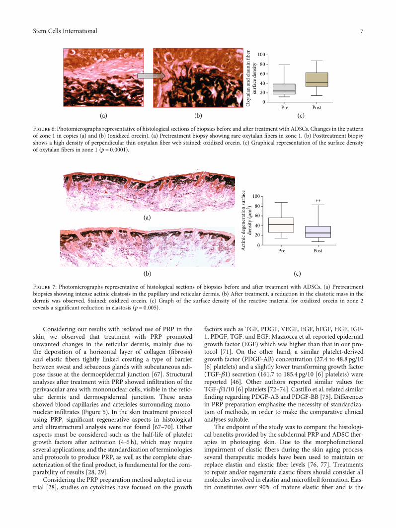
3.4. Post ADSC Skin Therapy (Protocol 2). All the biopsiesof facial sun-aged skin submitted to subdermal injectionof autologous ADSC showed different degrees of improve-ment of the overall skin structure, with partial or extensivereversal of the pathological signs typical of solar elastosis.In the zone 1 dermis, devoid of oxytalan and elaunin elasticnetwork in the solar elastosis samples, a fully organizednew system was found after the treatment. The regularlyspaced oxytalan fiber bundles were crossing perpendicularlythe region under the DEJ, linking it to the subjacent elasticfiber diffuse network laid parallel to the skin surface, indicat-ing indeed an intense neoelastinogenesis in the regeneration
of the solar elastosis (Figures 6(a)–6(c)). Histopathologicalanalysis showed that the elastin component of the extracellu-lar matrix of the skin was apparently the principal target ofthe therapy.
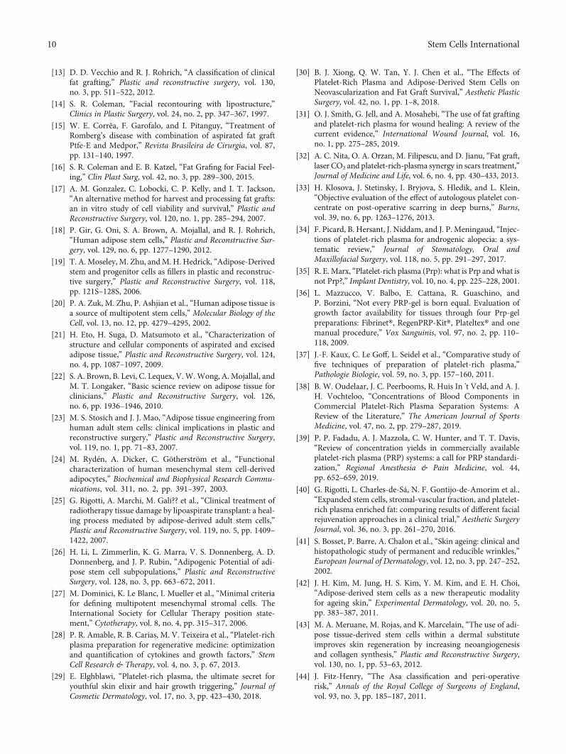
After the treatment, significant or full removal of the elas-totic material was observed, associated with regeneration ofthe new sizeable elastic fiber network, with the structuralorganization compatible with the normal elasticity of the der-mis (Figures 7(a)–7(c)).
In the deep dermis, both fibrillin and tropoelastin werealso slightly increased after the treatment when the morphol-ogy of the immune-labeled elastin molecules shifted fromamorphous and crumbled elastotic deposits to the typicalfibrillary structures (Figure 8).
Analysis of the total immune reactivity of cathepsin Kand MMP12 showed a significant increase (p = 0:011 and p= 0:005, respectively) after treatment with ADSCs, whichcould justify the improvement in the elastosis pattern foundin posttreatment skin (Figure 8).
It is notable that after ADSC therapy of sun-aged skin,the reactivation of cathepsin K expression in dermal fibro-blasts or in infiltrating cells is observed. This activity requireda second group of enzymes potentially involved in the elasti-nolysis observed in our mode which were the metalloprotein-ases, the major one being potentially the macrophagemetalloelastase MMP12 [47, 48]. Similar to cathepsin K, thetotal intensity of the MMP12 immune labeling was signifi-cantly increased after the ADSC treatment of the sun-agedskin (Figure 8). The activities of the two enzymes were thuspotentially complementary.
Classical macrophage activation upregulates severalmatrix MMPs, but the increase of the MMP12 mRNA levelsis mainly associated with the alternative macrophage activa-tion in the M2 phenotype [49]. All MSC are known to displayan anti-inflammatory effect in the receptor tissues, due inlarge part to the induction of monocyte differentiation intoM2 macrophages [50, 51]. Quantification of three markersof the M2 macrophage phenotype, CD68, CD206 (mannosereceptor (MR)), and heme-oxygenase-1 (HO-1), is shownin Figure 8. All three showed a significant increase in positivecells following the ADSC treatment.
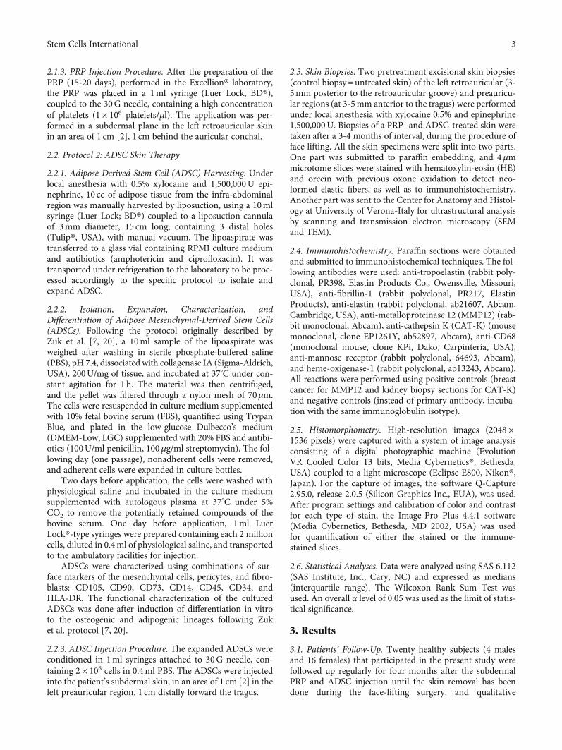
C
E
C
C
CC
E
100 um
Figure 3: Photoaging of the skin. Representative photomicrographof scanning electron microscopy (SEM): presence of a net ofcollagen fibers grouped in a disordered form (c) permeated by alarge elastic mature fiber (E).
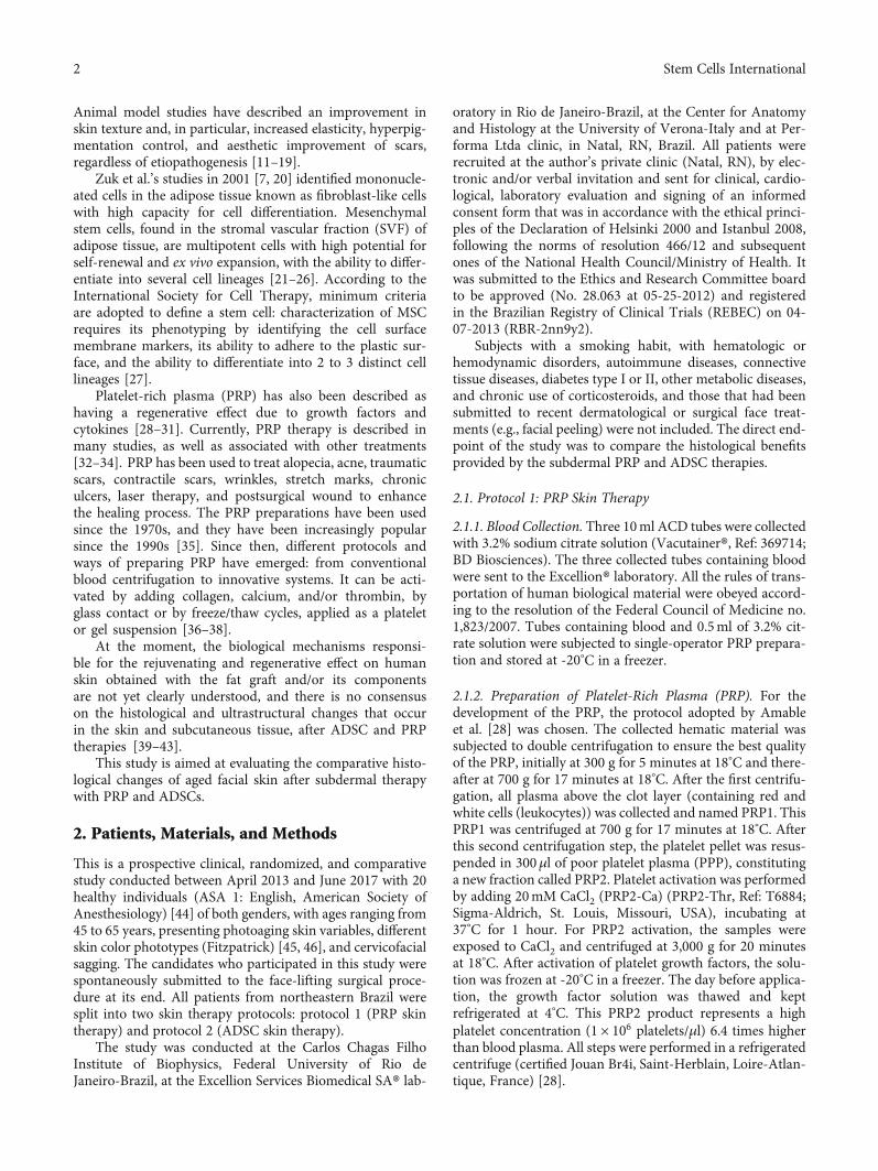
Zone 1
Zone 2
DEJ
Zone 1
Zone 2
Elastic systemCellular
Keratinocytes
FibroblastsReticulardermis
Papillarydermis
Epidermis
Figure 2: Definition of zones 1 and 2 of the dermis (40x magnification). Zone 1 is defined as an area delimited between the dermoepidermaljunction (DEJ) and the presence of elastic material deposited in the deep dermis. Another band (zone 2) has been established which comprisesthe limits of the elastic material deposited more deeply in the dermis.
5Stem Cells International
4. Discussion
Skin degenerative changes are related to age, hormonalchanges, exposure to environmental agents, and stress [1–5]. In dermatology and plastic surgery, PRP has been widelyapplied for skin rejuvenation, scalp alopecia, associated withfat grafts, after laser peeling, and for prevention of hematomain facelift procedures [52–56]. Derived growth factor plate-lets have been described for regenerative purposes [57] andother studies suggest paracrine effect, angiogenic action,and cell proliferation inducer [58, 59]. Despite the wide-spread use of PRP as a regenerative agent, its efficacy hasyet to be confirmed in different clinical protocols, mainly
for effects, the durability of results, proper dosing, prepara-tion techniques, and comparison of results [35].
In a previous study, the effect of PRP in association withfat grafting was evaluated through a clinical trial. More pro-nounced inflammatory infiltrates and higher vascular reac-tivity were observed, with increased vascular permeabilityand a particular reactivity of nerve structures. The associa-tion of PRP with the graft did not produce the expectedregenerative effect and had no advantages over the use ofexpanded ADSCs or SVF-enriched lipograft in skin rejuvena-tion [60]. However, PRP continues to be described as havinga possible regenerative effect in other protocols due to theirgrowth factors and cytokines [61–66].
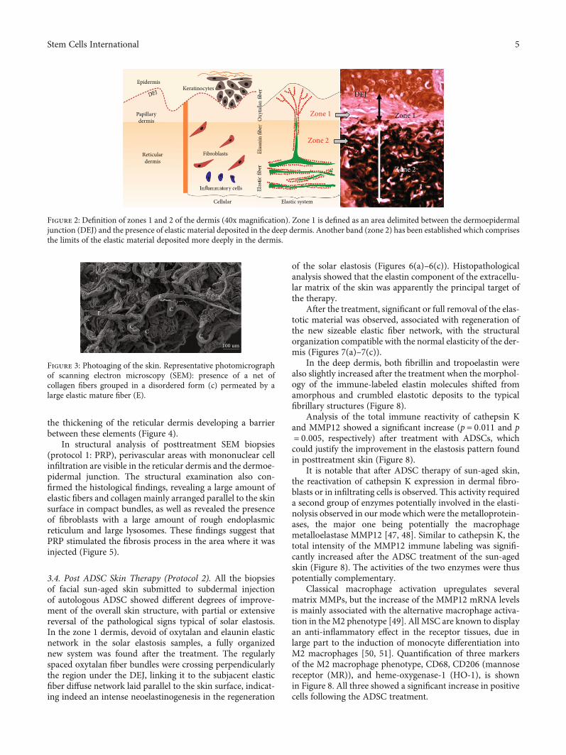
⁎
(a) (b)
⁎
(c)
(d)
⁎
(e) (f)
Figure 4: Histological analysis. Skin treatment with PRP: optical microscopy ((a, b) HE stain; (c, d) Picro-Sirius red stain; (e, f) oxidizedorcein stain). (a, c, e) Pretreatment and (b, d, f) posttreatment using the PRP biopsies. In the pretreatment specimens, adipocytes arelocated at the boundary of the dermis with the subcutaneous tissue organized in lobules, which are directed vertically toward the surfaceof the skin (∗). These lobules form adipose papillae, which in their apical portion show a relationship with sebaceous or sweat glands.After the use of PRP, fibrosis is apparent through the thickening of the reticular dermis due to elastic fibers and collagen arrangedhorizontal and strongly attached (squares).
c
(a)
f
f
c
c
(b)
Figure 5: Scanning electron microscopy of the skin. Structural analysis of the deep layer of the dermis: (a) SEM shows an intense collagennetwork and inflammatory cells in reticular dermis (white arrows); (b) MET shows the presence of numerous fibroblasts in reticulardermis and collagen fibers in formation. f: fibroblast; c: collagen.
6 Stem Cells International
Considering our results with isolated use of PRP in theskin, we observed that treatment with PRP promotedunwanted changes in the reticular dermis, mainly due tothe deposition of a horizontal layer of collagen (fibrosis)and elastic fibers tightly linked creating a type of barrierbetween sweat and sebaceous glands with subcutaneous adi-pose tissue at the dermoepidermal junction [67]. Structuralanalyses after treatment with PRP showed infiltration of theperivascular area with mononuclear cells, visible in the retic-ular dermis and dermoepidermal junction. These areasshowed blood capillaries and arterioles surrounding mono-nuclear infiltrates (Figure 5). In the skin treatment protocolusing PRP, significant regenerative aspects in histologicaland ultrastructural analysis were not found [67–70]. Otheraspects must be considered such as the half-life of plateletgrowth factors after activation (4-6 h), which may requireseveral applications; and the standardization of terminologiesand protocols to produce PRP, as well as the complete char-acterization of the final product, is fundamental for the com-parability of results [28, 29].
Considering the PRP preparation method adopted in ourtrial [28], studies on cytokines have focused on the growth
factors such as TGF, PDGF, VEGF, EGF, bFGF, HGF, IGF-1, PDGF, TGF, and EGF. Mazzocca et al. reported epidermalgrowth factor (EGF) which was higher than that in our pro-tocol [71]. On the other hand, a similar platelet-derivedgrowth factor (PDGF-AB) concentration (27.4 to 48.8 pg/10[6] platelets) and a slightly lower transforming growth factor(TGF-β1) secretion (161.7 to 185.4 pg/10 [6] platelets) werereported [46]. Other authors reported similar values forTGF-β1/10 [6] platelets [72–74]. Castillo et al. related similarfinding regarding PDGF-AB and PDGF-BB [75]. Differencesin PRP preparation emphasize the necessity of standardiza-tion of methods, in order to make the comparative clinicalanalyses suitable.
The endpoint of the study was to compare the histologi-cal benefits provided by the subdermal PRP and ADSC ther-apies in photoaging skin. Due to the morphofunctionalimpairment of elastic fibers during the skin aging process,several therapeutic models have been used to maintain orreplace elastin and elastic fiber levels [76, 77]. Treatmentsto repair and/or regenerate elastic fibers should consider allmolecules involved in elastin and microfibril formation. Elas-tin constitutes over 90% of mature elastic fiber and is the
PostPre(a) (b) (c)
100
80
60
40
20
0Oxy
tala
n an
d el
auni
n fib
ersu
rface
den
sity
Figure 6: Photomicrographs representative of histological sections of biopsies before and after treatment with ADSCs. Changes in the patternof zone 1 in copies (a) and (b) (oxidized orcein). (a) Pretreatment biopsy showing rare oxytalan fibers in zone 1. (b) Posttreatment biopsyshows a high density of perpendicular thin oxytalan fiber web stained: oxidized orcein. (c) Graphical representation of the surface densityof oxytalan fibers in zone 1 (p = 0:0001).
Pre
100
80
60
40
20
(a)
(b) (c)
0PostAc
tinic
deg
ener
atio
n su
rface
dens
ity (𝜇
m2 )
⁎⁎
Figure 7: Photomicrographs representative of histological sections of biopsies before and after treatment with ADSCs. (a) Pretreatmentbiopsies showing intense actinic elastosis in the papillary and reticular dermis. (b) After treatment, a reduction in the elastotic mass in thedermis was observed. Stained: oxidized orcein. (c) Graph of the surface density of the reactive material for oxidized orcein in zone 2reveals a significant reduction in elastosis (p = 0:005).
7Stem Cells International

main target of the recommended treatments. The use of tre-tinoin or retinoic acid in the skin in topical formulations has,for many years, increased elastin production by increasingtropoelastine [6, 77, 78] and fibrillin expression [79]. How-ever, the biggest challenge is overcoming low-level tropoelas-tin expression in adult skin, meaning that such treatmentsare more likely to have only additional benefits on skin elas-tin density [80], neglecting the role of microfibrils (fibrillin)as a support structure for neosynthesized elastin depositionfor forming a new elastic fiber. The microfibrils would adoptthe structural function of a scaffold to allow the deposition ofthe newly formed elastin. Some mechanisms regulate theproduction and degradation of elastic fiber in adults throughcytokines and signalers (TGF-β1, TGF-α, IGF-1, bFGF, EGF,TNF-a, and IL) by action on gene expression and tropoelastintranscription [81–93].
In ADSC skin therapy protocol, all analyses of the elasticsystem were directed at the reduction or total reversal of solarelastosis, addressing two issues: [1] regeneration of loss ofoxytalanic and elaunin fiber networks in the papillary dermisand [2] replacement of pathological deposits of actinic elastinwith a normal fibrillary structure, elastic fibers in the deepdermis. This remodeling action of the ECM at the elastic sys-tem level was demonstrated by the increased fiber density ofthe smaller diameter elastic system at the expense of newfibers in zone 1 (under DEJ). The density increase of thiszone probably occurred due to the emergence of new oxyta-lanic/elaunin fibers shown in Figure 6. This pattern of neoe-lastogenesis was confirmed by the significant increase infibrillin and tropoelastin (Figure 8) in ADSC posttreatmentbiopsies. Fibrillin is the main protein that makes up the
microfibrils of oxytalan fiber, and its induction of expressionprecedes that of collagen. For this reason, it is considered auseful biomarker in skin repair processes. On the other hand,the degradation of the deposited elastic component (elasto-sis) was observed in the reticular dermis (zone 2)(Figure 7). Matrix metalloproteinase type 12 (MMP12) andcathepsin K (lysosomal elastase) are essential regulators ofelastin degradation and are directly involved in extracellularmatrix remodeling and elastotic material degradation pro-cesses [79, 94–99]. Once activated, cathepsin would not beable to degrade large polymerized elastic fibers, dependingon the release of other ECM enzymes. It requires a group ofcomplementary enzymes involved in elastinolysis (metallo-proteinases), with MMP12 as its main representative anddependent on macrophage action [95, 96]. The mechanismsinvolved in the cellular degradation of cutaneous elastosisrelated to the presence of type M2 macrophages were ana-lyzed using M2 markers (CD 206: receptor mannose), whichshowed a significant increase in skin biopsies after treatmentwith ADSCs.
In ADSC-treated skin, inflammatory cells may be impli-cated in the resolution of the ECM repair and reorganizationprocess by polarizing the macrophage population to M2 [49].These findings suggest a direct contribution of ADSCs to skinregeneration [42]. Macrophages regulate various matrixmetalloproteinases (MMPs), but the increase in MMP12mRNA levels is mainly associated with the alternative activa-tion of macrophages with M2 phenotype [94]. All MSCs areknown to have an anti-inflammatory effect on tissues dueto largely inducing macrophage polarization to M2 [100–103]. In this study, M2 phenotype markers were monitored
Neoelastogenesis ECM remoldeling
150
100
50
MM
P12
surfa
ce d
ensit
y(𝜇
m2 )
M2
(man
nose
rece
ptor
surfa
ce d
ensit
y (𝜇
m2 )
CD68
surfa
ce d
ensit
y(𝜇
m2 )
Trop
oela
stin
surfa
cede
nsity
(𝜇m
2 )Fi
brill
in-1
surfa
ce d
ensit
y(𝜇
m2 )
Cath
epsin
K su
rface
den
sity
(𝜇m
2 )
0
3025
200
150
100
50
Pre Post Pre PostPre Post
Pre Post Pre Post Pre Post
0
80
50
40
30
20
10
0
60
40
20
0
20
15
10
5
0
20
10
0
⁎⁎
⁎⁎ ⁎⁎⁎
⁎⁎
⁎
Figure 8: Immunohistochemical analysis after ADSC skin therapy into two aspects: neoelastogenesis represented by anti-fibrillin immunelabelling that showed overall increase of fibrillin, including in the zone 1 (p = 0:001), and anti-tropoelastin immune labelling that showedincrease of the tropoelastin reactive material in the post treatment biopsy (p < 0:05). Extracellular matrix (ECM) remodeling wasrepresented by anti-cathepsin K immunostaining that revealed an increase in quantity of CAT-K immune-labelled cells in the dermis of aposttreated skin biopsy (p = 0:011), anti-MMP12 immunostaining representation of percentage of MMP12 in the skin biopsiessignificantly increased after ADSC treatment (p = 0:005), anti-CD68 and CD206 (mannose receptor (MR)) macrophage immunostainingin sun-exposed facial skin before and after ADSC injection that showed a significant increase after ADSC treatment in the dermis ofposttreated skin biopsy (p < 0:05).
8 Stem Cells International

in dermal tissue samples that received treatment withADSCs. Quantification of M2 macrophages by CD206 (man-nose receptor (MR)) revealed the increase of this subpopula-tion in posttreatment biopsies. The anti-inflammatory effectmediated by MSC in recipient tissues is largely due to theincrease in M2 macrophages in treated tissues [104], andwe propose that this is at least in part one of the major regen-erative activities of ADSCs in our study model.
Regarding the aspect of biosafety in tissue and celltransfer, despite the short time interval of the present study(4 months), between the application of ADSCs and theireffects on tissue samples submitted to the action of ADSCs,no dysplastic or oncogenic changes in the skin of the pop-ulation studied were observed as found in the literature[105]. The mesenchymal cells of the stromal fraction of adi-pose tissue used in this assay are autologous and homolo-gous (ADSCs derived from adipose tissue were usedelsewhere with the same histological and embryonic struc-ture) [105–109].
The limitations of this study were the difficulty of accu-rately quantifying skin photoaging, morphofunctional analy-sis of all elements involved in the regenerative process aftertreatment with PRP and ADSCs, and knowledge of allinvolved biological events and their mechanisms.
The advantages of using isolated stem cells as antiagingskin therapy over PRP, fat grafting, and/or ADSC-matchingtherapies are as follows: there is no risk of cyst formationand fibrosis, use of small injection volumes with highregenerative potential, precise application, possibility ofreapplication after reexpansion of stored stem cells, and/orcryopreserved cell reuse.
The future of this research line is aimed at creating newpossibilities in regenerative therapy not only in skin diseasesbut in other clinical applications in the case of organs andtissues with reduction and/or alteration in the elastic system(e.g., aneurysms and joint problems), with a better under-standing of the mechanisms involved and the control ofthese processes.
5. Conclusion
It was concluded that the action of PRP when injected onaged human skin induces an inflammatory process, contrib-uting to the increase of collagen fiber deposits and theincrease of reticular dermis thickness with a fibrotic aspect,not bringing any significant tissue regenerative role. On theother hand, expanded ADSC therapy in photoaged skin isrelated to ECM remodeling, increased production of newelastic fibers, and degradation of elastotic material depos-ited in the dermis (elastosis), inducing an important regen-erative effect that could be considered a promising skinrejuvenation therapy.
Data Availability
The data used to support the findings of this study are avail-able from the corresponding author upon request.
Conflicts of Interest
The authors declared no potential conflicts of interestwith respect to the research, authorship, and publicationof this article.
Acknowledgments
We would like to thank the institutional support of CAPES,Instituto de Biofísica Carlos Chagas Filho, Laboratório deImunopatologia, Universidade Federal do Rio de Janeiro-Brasil, Centro de Medicina Regenerativa, Faculdade de Med-icina de Petrópolis-FASE, Rio de Janeiro-Brasil, and Diparta-mento di Scienze Neurologiche e del Movimento, Sezione diAnatomia e Istologia della Università degli Studi di Verona.
References
[1] E. C. Naylor, R. E. B. Watson, and M. J. Sherratt, “Molecularaspects of skin ageing,”Maturitas, vol. 69, no. 3, pp. 249–256,2011.
[2] J. Uitto, L. Y. Matsuoka, and R. L. Kornberg, “Elastic fibers incutaneous elastoses,” in Problems in Aesthetic Surgery: Biolog-ical Causes and Clinical Solutions, R. Rudolph, Ed., pp. 307–338, Mosby, St Louis, MO, USA, 1986.
[3] K. Scharffetter-Kochanek, P. Brenneisen, J. Wenk et al., “Pho-toaging of the skin from phenotype to mechanisms,” Experi-mental Gerontology, vol. 35, no. 3, pp. 307–316, 2000.
[4] A. Oikarenen, “The aging of skin: chronoaging versus photo-aging,” Photodermatology Photoimmunology & Photomedi-cine, vol. 7, no. 1, pp. 3-4, 1990.
[5] G. J. Fisher, S. Kang, J. Varani et al., “Mechanisms of photo-aging and chronological skin aging,” Archives of Dermatol-ogy, vol. 138, no. 11, pp. 1462–1470, 2002.
[6] J. Uitto, “Biochemistry of the elastic fibers in normal connec-tive tissues and its alterations in diseases,” The Journal ofInvestigative Dermatology, vol. 72, no. 1, pp. 1–10, 1979.
[7] P. A. Zuk, M. Zhu, H. Mizuno et al., “Multilineage cells fromhuman adipose tissue: implications for cell-based therapies,”Tissue Engineering, vol. 7, no. 2, pp. 211–228, 2001.
[8] G. Rigotti, A. Marchi, and A. Sbarbati, “Adipose-derivedmesenchymal stem cells: past, present, and future,” AestheticPlastic Surgery, vol. 33, no. 3, pp. 271–273, 2009.
[9] P. Guillaume-Jugnot, A. Daumas, J. Magalon et al., “State ofthe art. Autologous fat graft and adipose tissue-derived stro-mal vascular fraction injection for hand therapy in systemicsclerosis patients,” Current Research in Translational Medi-cine, vol. 64, no. 1, pp. 35–42, 2016.
[10] S. R. Coleman, “Structural fat grafting: more than a perma-nent filler,” Plastic and Reconstructive Surgery, vol. 118,pp. 108s–120s, 2006.
[11] H. P. Lorenz, M. H. Hedrick, J. Chang, B. J. Mehrara, andM. T. Longaker, “The impact of biomolecular medicine andtissue engineering on plastic surgery in the 21st century,”Plastic and Reconstructive Surgery, vol. 105, no. 7, pp. 2467–2481, 2000.
[12] A. Mojallal, C. Lequeux, C. Shipkov et al., “Improvement ofskin quality after fat grafting: clinical observation and an ani-mal study,” Plastic and Reconstructive Surgery, vol. 124, no. 3,pp. 765–774, 2009.
9Stem Cells International
[13] D. D. Vecchio and R. J. Rohrich, “A classification of clinicalfat grafting,” Plastic and reconstructive surgery, vol. 130,no. 3, pp. 511–522, 2012.
[14] S. R. Coleman, “Facial recontouring with lipostructure,”Clinics in Plastic Surgery, vol. 24, no. 2, pp. 347–367, 1997.
[15] W. E. Corrêa, F. Garofalo, and I. Pitanguy, “Treatment ofRomberg’s disease with combination of aspirated fat graftPtfe-E and Medpor,” Revista Brasileira de Cirurgia, vol. 87,pp. 131–140, 1997.
[16] S. R. Coleman and E. B. Katzel, “Fat Grafing for Facial Feel-ing,” Clin Plast Surg, vol. 42, no. 3, pp. 289–300, 2015.
[17] A. M. Gonzalez, C. Lobocki, C. P. Kelly, and I. T. Jackson,“An alternative method for harvest and processing fat grafts:an in vitro study of cell viability and survival,” Plastic andReconstructive Surgery, vol. 120, no. 1, pp. 285–294, 2007.
[18] P. Gir, G. Oni, S. A. Brown, A. Mojallal, and R. J. Rohrich,“Human adipose stem cells,” Plastic and Reconstructive Sur-gery, vol. 129, no. 6, pp. 1277–1290, 2012.
[19] T. A. Moseley, M. Zhu, andM. H. Hedrick, “Adipose-Derivedstem and progenitor cells as fillers in plastic and reconstruc-tive surgery,” Plastic and Reconstructive Surgery, vol. 118,pp. 121S–128S, 2006.
[20] P. A. Zuk, M. Zhu, P. Ashjian et al., “Human adipose tissue isa source of multipotent stem cells,” Molecular Biology of theCell, vol. 13, no. 12, pp. 4279–4295, 2002.
[21] H. Eto, H. Suga, D. Matsumoto et al., “Characterization ofstructure and cellular components of aspirated and excisedadipose tissue,” Plastic and Reconstructive Surgery, vol. 124,no. 4, pp. 1087–1097, 2009.
[22] S. A. Brown, B. Levi, C. Lequex, V.W.Wong, A. Mojallal, andM. T. Longaker, “Basic science review on adipose tissue forclinicians,” Plastic and Reconstructive Surgery, vol. 126,no. 6, pp. 1936–1946, 2010.
[23] M. S. Stosich and J. J. Mao, “Adipose tissue engineering fromhuman adult stem cells: clinical implications in plastic andreconstructive surgery,” Plastic and Reconstructive Surgery,vol. 119, no. 1, pp. 71–83, 2007.
[24] M. Rydén, A. Dicker, C. Götherström et al., “Functionalcharacterization of human mesenchymal stem cell-derivedadipocytes,” Biochemical and Biophysical Research Commu-nications, vol. 311, no. 2, pp. 391–397, 2003.
[25] G. Rigotti, A. Marchi, M. Gali?? et al., “Clinical treatment ofradiotherapy tissue damage by lipoaspirate transplant: a heal-ing process mediated by adipose-derived adult stem cells,”Plastic and Reconstructive Surgery, vol. 119, no. 5, pp. 1409–1422, 2007.
[26] H. Li, L. Zimmerlin, K. G. Marra, V. S. Donnenberg, A. D.Donnenberg, and J. P. Rubin, “Adipogenic Potential of adi-pose stem cell subpopulations,” Plastic and ReconstructiveSurgery, vol. 128, no. 3, pp. 663–672, 2011.
[27] M. Dominici, K. Le Blanc, I. Mueller et al., “Minimal criteriafor defining multipotent mesenchymal stromal cells. TheInternational Society for Cellular Therapy position state-ment,” Cytotherapy, vol. 8, no. 4, pp. 315–317, 2006.
[28] P. R. Amable, R. B. Carias, M. V. Teixeira et al., “Platelet-richplasma preparation for regenerative medicine: optimizationand quantification of cytokines and growth factors,” StemCell Research & Therapy, vol. 4, no. 3, p. 67, 2013.
[29] E. Elghblawi, “Platelet-rich plasma, the ultimate secret foryouthful skin elixir and hair growth triggering,” Journal ofCosmetic Dermatology, vol. 17, no. 3, pp. 423–430, 2018.
[30] B. J. Xiong, Q. W. Tan, Y. J. Chen et al., “The Effects ofPlatelet-Rich Plasma and Adipose-Derived Stem Cells onNeovascularization and Fat Graft Survival,” Aesthetic PlasticSurgery, vol. 42, no. 1, pp. 1–8, 2018.
[31] O. J. Smith, G. Jell, and A. Mosahebi, “The use of fat graftingand platelet-rich plasma for wound healing: A review of thecurrent evidence,” International Wound Journal, vol. 16,no. 1, pp. 275–285, 2019.
[32] A. C. Nita, O. A. Orzan, M. Filipescu, and D. Jianu, “Fat graft,laser CO₂ and platelet-rich-plasma synergy in scars treatment,”Journal of Medicine and Life, vol. 6, no. 4, pp. 430–433, 2013.
[33] H. Klosova, J. Stetinsky, I. Bryjova, S. Hledik, and L. Klein,“Objective evaluation of the effect of autologous platelet con-centrate on post-operative scarring in deep burns,” Burns,vol. 39, no. 6, pp. 1263–1276, 2013.
[34] F. Picard, B. Hersant, J. Niddam, and J. P. Meningaud, “Injec-tions of platelet-rich plasma for androgenic alopecia: a sys-tematic review,” Journal of Stomatology, Oral andMaxillofacial Surgery, vol. 118, no. 5, pp. 291–297, 2017.
[35] R. E.Marx, “Platelet-rich plasma (Prp): what is Prp and what isnot Prp?,” Implant Dentistry, vol. 10, no. 4, pp. 225–228, 2001.
[36] L. Mazzucco, V. Balbo, E. Cattana, R. Guaschino, andP. Borzini, “Not every PRP-gel is born equal. Evaluation ofgrowth factor availability for tissues through four Prp-gelpreparations: Fibrinet®, RegenPRP-Kit®, Plateltex® and onemanual procedure,” Vox Sanguinis, vol. 97, no. 2, pp. 110–118, 2009.
[37] J.-F. Kaux, C. Le Goff, L. Seidel et al., “Comparative study offive techniques of preparation of platelet-rich plasma,”Pathologie Biologie, vol. 59, no. 3, pp. 157–160, 2011.
[38] B. W. Oudelaar, J. C. Peerbooms, R. Huis In 't Veld, and A. J.H. Vochteloo, “Concentrations of Blood Components inCommercial Platelet-Rich Plasma Separation Systems: AReview of the Literature,” The American Journal of SportsMedicine, vol. 47, no. 2, pp. 279–287, 2019.
[39] P. P. Fadadu, A. J. Mazzola, C. W. Hunter, and T. T. Davis,“Review of concentration yields in commercially availableplatelet-rich plasma (PRP) systems: a call for PRP standardi-zation,” Regional Anesthesia & Pain Medicine, vol. 44,pp. 652–659, 2019.
[40] G. Rigotti, L. Charles-de-Sá, N. F. Gontijo-de-Amorim et al.,“Expanded stem cells, stromal-vascular fraction, and platelet-rich plasma enriched fat: comparing results of different facialrejuvenation approaches in a clinical trial,” Aesthetic SurgeryJournal, vol. 36, no. 3, pp. 261–270, 2016.
[41] S. Bosset, P. Barre, A. Chalon et al., “Skin ageing: clinical andhistopathologic study of permanent and reducible wrinkles,”European Journal of Dermatology, vol. 12, no. 3, pp. 247–252,2002.
[42] J. H. Kim, M. Jung, H. S. Kim, Y. M. Kim, and E. H. Choi,“Adipose-derived stem cells as a new therapeutic modalityfor ageing skin,” Experimental Dermatology, vol. 20, no. 5,pp. 383–387, 2011.
[43] M. A. Meruane, M. Rojas, and K. Marcelain, “The use of adi-pose tissue-derived stem cells within a dermal substituteimproves skin regeneration by increasing neoangiogenesisand collagen synthesis,” Plastic and Reconstructive Surgery,vol. 130, no. 1, pp. 53–63, 2012.
[44] J. Fitz-Henry, “The Asa classification and peri-operativerisk,” Annals of the Royal College of Surgeons of England,vol. 93, no. 3, pp. 185–187, 2011.
10 Stem Cells International
[45] T. B. Fitzpatrick, “The validity and practicality of sun-reactiveskin types I through Vi,” Archives of Dermatology, vol. 124,no. 6, pp. 869–871, 1988.
[46] R. E. Fitzpatrick and E. F. Rostan, “Reversal of photodamagewith topical growth factors: a pilot study,” Journal of Cos-metic and Laser Therapy, vol. 5, no. 1, pp. 25–34, 2009.
[47] A. Pellicoro, R. L. Aucott, P. Ramachandran et al., “Elastinaccumulation is regulated at the level of degradation by mac-rophage metalloelastase (Mmp-12) during experimental liverfibrosis,” Hepatology, vol. 55, no. 6, pp. 1965–1975, 2012.
[48] U. Saarialho-Kere, E. Kerkelä, L. Jeskanen et al., “Accumula-tion of matrilysin (Mmp-7) and macrophage metalloelastase(Mmp 12) in actinic damage,” The Journal of InvestigativeDermatology, vol. 113, no. 4, pp. 664–672, 1999.
[49] A. Mantovani, S. K. Biswas, M. R. Galdiero, A. Sica, andM. Locati, “Macrophage plasticity and polarization in tissuerepair and remodelling,” The Journal of Pathology, vol. 229,no. 2, pp. 176–185, 2013.
[50] H. Zhao, Q. Shang, Z. Pan et al., “Exosomes From Adipose-Derived Stem Cells Attenuate Adipose Inflammation andObesity Through Polarizing M2 Macrophages and Beigingin White Adipose Tissue,” Diabetes, vol. 67, no. 2, pp. 235–247, 2018.
[51] S. Mahbub, C. R. Deburghgraeve, and E. J. Kovacs,“Advanced age impairs macrophage polarization,” Journalof Interferon & Cytokine Research, vol. 32, no. 1, pp. 18–26,2012.
[52] R. S. Frautschi, A. M. Hashem, B. Halasa, C. Cakmakoglu,and J. E. Zins, “Current evidence for clinical efficacy of plate-let rich plasma in aesthetic surgery: a systematic review,” Aes-thetic Surgery Journal, 2016.
[53] J. W. Lee, B. J. Kim, M. N. Kim, and S. K. Mun, “The efficacyof autologous platelet rich plasma combined with ablativecarbon dioxide fractional resurfacing for acne scars: a simul-taneous split-face trial,” Dermatologic Surgery, vol. 37, no. 7,pp. 931–938, 2011.
[54] T. Kamakura, J. Kataoka, K. Maeda et al., “Platelet-richplasma with basic fibroblast growth factor for treatment ofwrinkles and depressed areas of the skin,” Plastic and Recon-structive Surgery, vol. 136, no. 5, pp. 931–939, 2015.
[55] H.-K. Lim, D.-H. Suh, S.-J. Lee, and M. K. Shin, “Rejuvena-tion effects of hyaluronic acid injection on nasojugal groove:prospective randomized split face clinical controlled study,”Journal of Cosmetic and Laser Therapy, vol. 16, no. 1,pp. 32–36, 2014.
[56] M. W. Blanton, I. Hadad, B. H. Johnstone et al., “Adiposestromal cells and platelet-rich plasma therapies synergisti-cally increase revascularization during wound healing,” Plas-tic and Reconstructive Surgery, vol. 123, pp. 56S–64S, 2009.
[57] S. Nakamura, M. Ishihara, M. Takikawa et al., “Platelet-richplasma (Prp) promotes survival of fat-grafts in rats,” Annalsof Plastic Surgery, vol. 65, no. 1, pp. 101–106, 2010.
[58] I. B. James, S. R. Coleman, and J. P. Rubin, “Fat, stem cells,and platelet-rich plasma,” Clinics in Plastic Surgery, vol. 43,no. 3, pp. 473–488, 2016.
[59] Y. Fukaya, M. Kuroda, Y. Aoyagi et al., “Platelet-rich plasmainhibits the apoptosis of highly adipogenic homogeneouspreadipocytes in an in vitro culture system,” Experimental& Molecular Medicine, vol. 44, no. 5, pp. 330–339, 2012.
[60] L. Charles-de-Sá, N. F. Gontijo-de-Amorim, C. M. Takiyaet al., “Antiaging treatment of the facial skin by fat graft
and adipose-derived stem cells,” Plastic and ReconstructiveSurgery, vol. 135, no. 4, pp. 999–1009, 2015.
[61] S.-J. Zhu, B.-H. Choi, J.-H. Jung et al., “A comparative histo-logic analysis of tissue-engineered bone using platelet- richplasma and platelet-enriched fibrin glue,” Oral Surgery, OralMedicine, Oral Pathology, Oral Radiology, and Endodontics,vol. 102, no. 2, pp. 175–179, 2006.
[62] V. Cervelli, I. Bocchini, C. Di Pasquali et al., “P.R.L. PlateletRich Lipotransfert: our experience and current state of artin the combined use of fat and PRP,” BioMed Research Inter-national, vol. 2013, Article ID 434191, 9 pages, 2013.
[63] C. E. Sommeling, A. Heyneman, H. Hoeksema, J. Verbelen,F. B. Stillaert, and S. Monstrey, “The use of platelet-richplasma in plastic surgery: a systematic review,” Journal ofPlastic, Reconstructive & Aesthetic Surgery, vol. 66, no. 3,pp. 301–311, 2013.
[64] T. Hirase, E. Ruff, S. Surani, and I. Ratnani, “Topical applica-tion of platelet-rich plasma for diabetic foot ulcers: a system-atic review,” World Journal of Diabetes, vol. 9, no. 10,pp. 172–179, 2018.
[65] N. Kakudo, T. Minakata, T. Mitsui, S. Kushida, F. Z. Notodi-hardjo, and K. Kusumoto, “Proliferation-promoting effect ofplatelet-rich plasma on human adipose-derived stem cellsand human dermal fibroblasts,” Plastic and ReconstructiveSurgery, vol. 122, no. 5, pp. 1352–1360, 2008.
[66] M. Kawasumi, H. Kitoh, K. A. Siwicka, and N. Ishiguro,“The effect of the platelet concentration in platelet-richplasma gel on the regeneration of bone,” The Journal ofBone and Joint Surgery British volume, vol. 90-B, no. 7,pp. 966–972, 2008.
[67] L. Charles-de-Sá, N. F. Gontijo-de-Amorim, C. M. Takiyaet al., “Effect of use of platelet-rich plasma (Prp) in skin withintrinsic aging process,” Aesthetic Surgery Journal, vol. 38,no. 3, pp. 321–328, 2018.
[68] X. Lei, P. Xu, and B. Cheng, “Problems and solutions forplatelet-rich plasma in facial rejuvenation: a systematicreview,” Aesthetic Plastic Surgery, vol. 43, no. 2, pp. 457–469, 2019.
[69] P. Li, R. Zhang, and Q. Zhou, “Efficacy of platelet-rich plasmain retarding intervertebral disc degeneration: a meta-analysisof animal studies,” BioMed Research International, vol. 2017,Article ID 7919201, 10 pages, 2017.
[70] W. J. Shi, F. P. Tjoumakaris, M. Lendner, and K. B. Freed-man, “Biologic injections for osteoarthritis and articular car-tilage damage: can we modify disease?,” The Physician andSportsmedicine, vol. 45, no. 3, pp. 203–223, 2017.
[71] A. D. Mazzocca, M. B. R. McCarthy, D. M. Chowaniecet al., “Platelet-rich plasma differs according to preparationmethod and human variability,” The Journal of Bone andJoint Surgery American Volume, vol. 94, no. 4, pp. 308–316, 2012.
[72] G. Weibrich, W. K. G. Kleis, G. Hafner, and W. E. Hitzler,“Growth factor levels in platelet-rich plasma and correlationswith donor age, sex, and platelet count,” Journal of Cranio-Maxillo-Facial Surgery, vol. 30, no. 2, pp. 97–102, 2002.
[73] R. Zimmermann, D. Arnold, E. Strasser et al., “Sample prep-aration technique and white cell content influence the detect-able levels of growth factors in platelet concentrates,” VoxSanguinis, vol. 85, no. 4, pp. 283–289, 2003.
[74] E. A. Sundman, B. J. Cole, and L. A. Fortier, “Growth factorand catabolic cytokine concentrations are influenced by the
11Stem Cells International
cellular composition of platelet-rich plasma,” The AmericanJournal of Sports Medicine, vol. 39, no. 10, pp. 2135–2140,2011.
[75] T. N. Castillo, M. A. Pouliot, H. J. Kim, and J. L. Dragoo,“Comparison of growth factor and platelet concentrationfrom commercial platelet-rich plasma separation systems,”The American Journal of Sports Medicine, vol. 39, no. 2,pp. 266–271, 2011.
[76] I. M. Braverman and E. Fonferko, “Studies in cutaneousaging: I. The elastic fiber network,” The Journal of Investiga-tive Dermatology, vol. 78, no. 5, pp. 434–443, 1982.
[77] R. P. Mecham and H. Je, “The elastic fibre,” in Cell Biology OfExtracellular Matrix, E. D. Hay, Ed., pp. 79–109, PlenumPress, New York, NY, USA, 1991.
[78] S. Tajima, A. Hayashi, and T. Suzuki, “Elastin expression isup-regulated by retinoic acid but not by retinol in chickembryonic skin fibroblasts,” Journal of Dermatological Sci-ence, vol. 15, no. 3, pp. 166–172, 1997.
[79] R. E. B. Watson, S. P. Long, J. J. Bowden, J. Y. Bastrilles, S. P.Barton, and C. E. M. Griffiths, “Repair of photoaged dermalmatrix by topical application of a cosmetic ‘antiageing’ prod-uct,” The British Journal of Dermatology, vol. 158, no. 3,pp. 472–477, 2008.
[80] G. C. Sephel, A. Buckley, and J. M. Davidson, “Developmen-tal initiation of elastin gene expression by human fetal skinfibroblasts,” The Journal of Investigative Dermatology,vol. 88, no. 6, pp. 732–735, 1987.
[81] E. P. Sproul and W. S. Argraves, “A cytokine axis regulateselastin formation and degradation,” Matrix Biology, vol. 32,no. 2, pp. 86–94, 2013.
[82] S. Imayama and I. M. Braverman, “A hypothetical explana-tion for the aging of skin. Chronologic alteration of thethree-dimensional arrangement of collagen and elastic fibersin connective tissue,” The American Journal of Pathology,vol. 134, no. 5, pp. 1019–1025, 1989.
[83] M. Guay, G. Lagace, and F. Lamy, “Photolysis and ozonolysisof desmosine and Elastolytic peptides,” Connective TissueResearch, vol. 14, no. 2, pp. 89–107, 2009.
[84] T. Tsuji, “Different effects of elastase on dermal elastic fiberswith age,” Gerontology, vol. 33, no. 2, pp. 64–71, 1987.
[85] G. C. Sephel and J. M. Davidson, “Elastin production inhuman skin fibroblast cultures and its decline with age,” Jour-nal of Investigative Dermatology, vol. 86, no. 3, pp. 279–285,1986.
[86] A. Robinet, “Elastin-derived peptides enhance angiogenesisby promoting endothelial cell migration and tubulogenesisthrough upregulation of Mt1-Mmp,” Journal of Cell Science,vol. 118, no. 2, pp. 343–356, 2005.
[87] V. Lambros, “Improvement of skin quality after fat graft-ing: clinical observation and an animal study,” Plasticand Reconstructive Surgery, vol. 124, no. 3, pp. 775-776,2009.
[88] M. El-Domyati, T. S. El-Ammawi, O. Moawad et al., “Efficacyof mesotherapy in facial rejuvenation: a histological andimmunohistochemical evaluation,” International Journal ofDermatology, vol. 51, no. 8, pp. 913–919, 2012.
[89] X. Fu and H. Li, “Mesenchymal stem cells and skin woundrepair and regeneration: possibilities and questions,” Celland Tissue Research, vol. 335, no. 2, pp. 317–321, 2009.
[90] R. K. Chan, D. O. Zamora, N. L. Wrice et al., “Developmentof a vascularized skin construct using adipose-derived stem
cells from debrided burned skin,” Stem Cells International,vol. 2012, Article ID 841203, 11 pages, 2012.
[91] B. M. Strem, K. C. Hicok, M. Zhu et al., “Multipotential dif-ferentiation of adipose tissue-derived stem cells,” The KeioJournal of Medicine, vol. 54, no. 3, pp. 132–141, 2005.
[92] V. Planat-Benard, J.-S. Silvestre, B. Cousin et al., “Plasticity ofhuman adipose lineage cells toward endothelial cells: physio-logical and therapeutic perspectives,” Circulation, vol. 109,no. 5, pp. 656–663, 2004.
[93] W. J. F. M. Jurgens, M. J. Oedayrajsingh-Varma, M. N.Helder et al., “Effect of tissue-harvesting site on yield of stemcells derived from adipose tissue: implications for cell-basedtherapies,” Cell and Tissue Research, vol. 332, no. 3,pp. 415–426, 2008.
[94] W.-C. Huang, G. B. Sala-Newby, A. Susana, J. L. Johnson, andA. C. Newby, “Classical macrophage activation up-regulatesseveral matrix metalloproteinases through mitogen activatedprotein kinases and nuclear factor-κB,” PLoS One, vol. 7,no. 8, article e42507, 2012.
[95] V. Lagente, C. Le Quement, and E. Boichot, “Macrophagemetalloelastase (MMP-12) as a target for inflammatory respi-ratory diseases,” Expert Opinion on Therapeutic Targets,vol. 13, no. 3, pp. 287–295, 2009.
[96] T. M. Rünger, M. J. Quintanilla-Dieck, and J. Bhawan, “Roleof cathepsin K in the turnover of the dermal extracellularmatrix during scar formation,” The Journal of InvestigativeDermatology, vol. 127, no. 2, pp. 293–297, 2007.
[97] K. A. Codriansky, M. J. Quintanilla-Dieck, S. Gan, M. Keady,J. Bhawan, and T. M. Rünger, “Intracellular degradation ofelastin by cathepsin K in skin fibroblasts – a possible role inphotoaging,” Photochemistry and Photobiology, vol. 85,no. 6, pp. 1356–1363, 2009.
[98] M. Novinec, R. N. Grass, W. J. Stark, V. Turk, A. Baici, andB. Lenarčič, “Interaction between human cathepsins K, L,and S and elastins,” The Journal of Biological Chemistry,vol. 282, no. 11, pp. 7893–7902, 2007.
[99] V. Turk, V. Stoka, O. Vasiljeva et al., “Cysteine cathepsins:from structure, function and regulation to new frontiers,”Biochimica et Biophysica Acta (BBA) - Proteins and Proteo-mics, vol. 1824, no. 1, pp. 68–88, 2012.
[100] S. Guo, T. Wang, S. Zhang et al., “Adipose-derived stemcell-conditioned medium protects fibroblasts at differentsenescent degrees from UVB irradiation damages,” Molec-ular and Cellular Biochemistry, vol. 463, no. 1-2, pp. 67–78, 2020.
[101] S. Taddese, A. S. Weiss, R. H. H. Neubert, and C. E. H.Schmelzer, “Mapping of macrophage elastase cleavage sitesin insoluble human skin elastin,” Matrix Biology, vol. 27,no. 5, pp. 420–428, 2008.
[102] C. Li, M. M. Xu, K. Wang, A. J. Adler, A. T. Vella, andB. Zhou, “Macrophage polarization andmeta-inflammation,”Transl. Res., vol. 191, pp. 29–44, 2018.
[103] M. Rinnerthaler, J. Bischof, M. Streubel, A. Trost, andK. Richter, “Oxidative stress in aging human skin,” Biomole-cules, vol. 5, no. 2, pp. 545–589, 2015.
[104] L. Zachar, D. Bacenkova, and J. Rosocha, “Activation, hom-ing, and role of the mesenchymal stem cells in the inflamma-tory environment,” Journal of Inflammation Research, vol. 9,pp. 231–240, 2016.
[105] B. Laverdet, L. Micallef, C. Lebreton et al., “Utilisation descellules souches mesenchymateuses pour la reparation
12 Stem Cells International
cutanee et l'elaboration de substituts de peau,” Pathologie Bio-logie, vol. 62, no. 2, pp. 108–117, 2014.
[106] S. Barrientos, O. Stojadinovic, M. S. Golinko, H. Brem, andM. Tomic-Canic, “Perspective article: growth factors andcytokines in wound healing,” Wound Repair and Regenera-tion, vol. 16, no. 5, pp. 585–601, 2008.
[107] R. Pérez-Cano, J. J. Vranckx, J. M. Lasso et al., “Prospectivetrial of adipose-derived regenerative cell (ADRC)-enrichedfat grafting for partial mastectomy defects: the Restore-2trial,” European Journal of Surgical Oncology, vol. 38, no. 5,pp. 382–389, 2012.
[108] N. F. Gontijo-de-Amorim, L. Charles-de-Sá, and G. Rigotti,“Mechanical supplementation with the stromal vascular frac-tion yields improved volume retention in facial lipotransfer: a1-year comparative study,” Aesthetic Surgery Journal, vol. 37,no. 9, pp. 975–985, 2017.
[109] R. Anderi, N. Makdissy, A. Azar, F. Rizk, and A. Hamade,“Cellular therapy with human autologous adipose-derivedadult cells of stromal vascular fraction for alopecia areata,”Stem Cell Research & Therapy, vol. 9, no. 1, p. 141, 2018.
13Stem Cells International


![[MS-ADSC-Diff]: Active Directory Schema Classes... · 2 / 134 [MS-ADSC-Diff] - v20160714 Active Directory Schema Classes Copyright © 2016 Microsoft Corporation Release: July 14,](https://img.pdfslide.net/doc/110x75/6034874437704a05821ff686/ms-adsc-diff-active-directory-schema-classes-2-134-ms-adsc-diff-v20160714.jpg)

![[MS-ADSC]: Active Directory Schema Classespirate-network.com/data/Windows_Server_Protocols/[MS-ADSC].pdf · 1 / 134 [MS-ADSC] - v20160714 Active Directory Schema Classes Copyright](https://img.pdfslide.net/doc/110x75/5f079c427e708231d41dd830/ms-adsc-active-directory-schema-classespirate-ms-adscpdf-1-134-ms-adsc.jpg)
![[MS-ADSC]: Active Directory Schema Classes€¦ · 2 / 134 [MS-ADSC] - v20150630 Active Directory Schema Classes Copyright © 2015 Microsoft Corporation Release: June 30, 2015 Revision](https://img.pdfslide.net/doc/110x75/5f079c437e708231d41dd832/ms-adsc-active-directory-schema-classes-2-134-ms-adsc-v20150630-active.jpg)


![[MS-ADSC]: Active Directory Schema Classes...2 / 135 [MS-ADSC] - v20151016 Active Directory Schema Classes Copyright © 2015 Microsoft Corporation Release: October 16, 2015 Revision](https://img.pdfslide.net/doc/110x75/5f079c437e708231d41dd833/ms-adsc-active-directory-schema-classes-2-135-ms-adsc-v20151016-active.jpg)