Embed Size (px)
Citation preview
Molecular Ecology (2003)
12
, 3383–3401 doi: 10.1046/j.1365-294X.2003.01995.x
© 2003 Blackwell Publishing Ltd
Blackwell Science, Ltd
Phylogeography of the fungal pathogen
Histoplasma capsulatum
TAKAO KASUGA,
1
THOMAS J . WHITE,
2
GINA KOENIG,
3
JUAN MCEWEN,
4
ANGELA RESTREPO,
5
ELIZABETHA CASTAÑEDA,
5
CARLOS DA SILVA LACAZ,
6
ELISABETH M. HEINS-VACCARI ,
6
ROSELI S . DE FREITAS,
6
ROSELY M. ZANCOPÉ-OLIVEIRA,
7
ZHENYU QIN,
8
R ICARDO NEGRONI,
9
DEIDRE A. CARTER,
10
YUZURU MIKAMI,
11
MIKI TAMURA,
12
MARÍA LUCÍA TAYLOR,
13
GEORGINA F . MILLER,
14
NATTEEWAN POONWAN
15
and JOHN W. TAYLOR
1
1
Department of Plant and Microbial Biology, 321 Koshland Hall, University of California, Berkeley, CA 94720, USA,
2
Celera Diagnostics, Alameda, CA, USA,
3
Roche Molecular Systems, Alameda, CA, USA,
4
Corporación para Investigaciones Biológicas, Medellin, Colombia,
5
Instituto Nacional de Salud Santafé de Bogotá, Colombia,
6
Instituto de Medicina Tropical de São Paulo, Brazil,
7
Instituto de Microbiologia, Universidade Federal do Rio de Janeiro, Rio de Janeiro, Brazil,
8
Center of Medical Mycology, Institute of Dermatology, Chinese Academy of Medical Sciences, Nanjing, China,
9
Hospital de Enfermedades Infecciosas, Buenos Aires, Argentina,
10
Microbiology Department, University of Sydney, Australia,
11
Research Center for Pathogenic Fungi and Microbial Toxicoses, Chiba University, Chiba, Japan,
12
Institute of Molecular and Cellular Biosciences, University of Tokyo, Japan,
13
Departamento de Microbiología y Parasitología, Facultad de Medicina, Universidad Nacional Autónoma de México, México,
14
Veterinary Resources Program, National Institutes of Health, Bethesda, MD, USA,
15
Department of Medical Sciences, National Institute of Health, Nonthaburi, Thailand
Abstract
Until recently,
Histoplasma capsulatum
was believed to harbour three varieties, var.
capsulatum
(chiefly a New World human pathogen), var.
duboisii
(an African humanpathogen) and var.
farciminosum
(an Old World horse pathogen), which varied in clinicalmanifestations and geographical distribution. We analysed the phylogenetic relationshipsof 137 individuals representing the three varieties from six continents using DNA sequencevariation in four independent protein-coding genes. At least eight clades were identified:(i) North American class 1 clade; (ii) North American class 2 clade; (iii) Latin Americangroup A clade; (iv) Latin American group B clade; (v) Australian clade; (vi) Netherlands(Indonesian?) clade; (vii) Eurasian clade and (viii) African clade. Seven of eight clades rep-resented genetically isolated groups that may be recognized as phylogenetic species. Thesole exception was the Eurasian clade which originated from within the Latin Americangroup A clade. The phylogenetic relationships among the clades made a star phylogeny.
Histoplasma capsulatum
var.
capsulatum
individuals were found in all eight clades.The African clade included all of the
H. capsulatum
var.
duboisii
individuals as well asindividuals of the other two varieties. The 13 individuals of var.
farciminosum
were dis-tributed among three phylogenetic species. These findings suggest that the three varietiesof
Histoplasma
are phylogenetically meaningless. Instead we have to recognize theexistence of genetically distinct geographical populations or phylogenetic species.Combining DNA substitution rates of protein-coding genes with the phylogeny suggeststhat the radiation of
Histoplasma
started between 3 and 13 million years ago in LatinAmerica.
Keywords
: allopatric speciation, glacial refugia, last glacial maxima, phylogenetic species, popu-lation diversity, star phylogeny
Received 11 April 2003; revision received 29 July 2003; accepted 28 August 2003
Correspondence: Takao Kasuga. Fax: 510 642 4995; E-mail: [email protected]; John Taylor. E-mail: [email protected]
3384
T . K A S U G A
E T A L .
© 2003 Blackwell Publishing Ltd,
Molecular Ecology
, 12, 3383–3401
Introduction
We investigated the population structure and phylogenyof the pathogenic ascomycete fungus
Histoplasma capsulatum
Darling (mieosporic state or teleomorph:
Ajellomycescapsulatus
(Kwon-Chung) McGinnis
et
Katz). This dimor-phic fungus causes deep mycosis in various mammalianspecies including humans (Rippon 1988; Kwon-Chung& Bennett 1992). It exists in the mycelial phase in soilenriched with bird and bat guano. In the lung, inhaledairborne microconidia or hyphal fragments transform tothe pathogenic yeast form and start the mycosis. Thedisease is noncontagious between humans.
Histoplasmacapsulatum
occurs in temperate and tropical regions world-wide and distinct genotypes are known which showdifferent clinical manifestations and geographical distribu-tions. On the basis of morphology and pathogenicity, thegenus
Histoplasma
has been thought to consist of threedistinct varieties:
H. capsulatum
(
Hc
) var.
capsulatum
,
Hc
var.
duboisii
and
Hc
var.
farciminosum
or three independentspecies:
H. capsulatum
,
H. duboisii
and
H. farciminosum
(Kwon-Chung & Bennett 1992; Rippon 1988).Recently, 46 isolates of
H. capsulatum
representing thethree varieties from various geographical locations weresubjected to phylogenetic analysis using DNA sequencevariation in four independent protein-coding genes(Kasuga
et al
. 1999). This study showed that
H. capsulatum
consisted of at least six clades: (i) North American class 1
Hc
var.
capsulatum
(NAm 1; see Table 1 for abbreviations);(ii) North American class 2
Hc
var.
capsulatum
(NAm 2);(iii) Panamanian
Hc
var.
capsulatum
; (iv) South Americangroup A
Hc
var.
capsulatum
; (v) South American group B
Hc
var.
capsulatum
and (vi)
Hc
var.
duboisii
.
Histoplasmacapsulatum
var.
farciminosum
was found within the SouthAmerican group A clade. Under a genealogical concordance–phylogenetic species concept (GC–PSC) (Mayden 1997),based on possession of multiple shared derived charactersas well as concordance of four gene genealogies,
H. capsu-
latum
was claimed to harbour six species instead of threevarieties or three species. However, this study did notinclude individuals from many regions of the globe.
To challenge the hypothesis that
H. capsulatum
com-prises six phylogenetic species, we analysed phylogeneticrelationships of 92 additional
H. capsulatum
isolates which,together with 45 of the 46 analysed before, represent 25countries in six continents. We challenged the validity ofthe six-species hypothesis by constructing a more detailedphylogeographical map and searching for possible hybridsbetween geographical populations. In this research, weapplied the GC–PSC to define genetically isolated popula-tions in
H. capsulatum
. From neutral mutation rates inprotein-coding genes (Kasuga
et al
. 2002) and genetic dis-tances between geographical populations, we estimatedthe ages of populations and used this information to dis-cuss the history of each population and the origin of the
H. capsulatum
complex.Under a neutral model of evolution, genetic drift will
inevitably lead to fixation of formerly polymorphic locifollowing genetic isolation and, after sufficient time, togenealogical concordance of multiple gene trees. New poly-morphisms will continue to arise and accumulate in theseloci in each interbreeding population. Thus, these genet-ically isolated populations or species will be recognized asreciprocally monophyletic groups. Recombination amongindividuals within a species will result in discordanceamong the gene genealogies. Therefore, in GC–PSC, thetransition from deep genealogical concordance to shallowgenealogical discordance is used to delimit species bound-aries (Avise & Ball. 1990; Baum & Shaw 1995). The GC–PSCis especially compatible with DNA analyses and has beendemonstrated to recognize genetically distinct populationsor species without actually observing matings or gene flowin nature (reviewed in Taylor
et al
. 2000).We found that
H. capsulatum
comprises seven phylo-genetic species plus a Eurasian clade that emerges fromthe largest clade, South American group A.
In this research, numbers of Mexican and Central Amer-ican isolates were found in the South American groupA clade; therefore, we replace the clade name ‘SouthAmerica’ with ‘Latin America’. Individuals identified as
Hc
var.
duboisii
were limited to Africa but the African cladeincluded individuals morphologically identified as
Hc
var.
capsulatum
and
Hc
var.
farciminosum
. Individuals identifiedas
Hc
var.
farciminosum
were accommodated in threedifferent phylogenetic species, supporting the claim that
Hc
var.
farciminosum
is a collection of individuals fromdifferent clades that share the ability to cause disease inhorses and not a phylogenetic species. There was no resolu-tion of the branching order of the clades, supporting theconclusion that
H. capsulatum
radiated rapidly over a shortperiod, which we estimate occurred 3–13 million yearsago (Ma).
Table 1 Abbreviations of varieties and geographical groups ofHistoplasma capsulatum
Abbr. Variety and population
Hc var. capsulatum H. capsulatum var. capsulatumHc var. farciminosum H. capsulatum var. farciminosumHc var. duboisii H. capsulatum var. duboisiiNAm 1 North American class 1NAm 2 North American class 2LAm A* Latin American group ALAm B* Latin American group B
*Latin American group A and Latin American group B are synonyms of South American group A and South American group B in Kasuga et al. (1999), respectively.
P H Y L O G E O G R A P H Y O F
H I S T O P L A S M A
3385
© 2003 Blackwell Publishing Ltd,
Molecular Ecology
, 12, 3383–3401
Materials and methods
Organisms
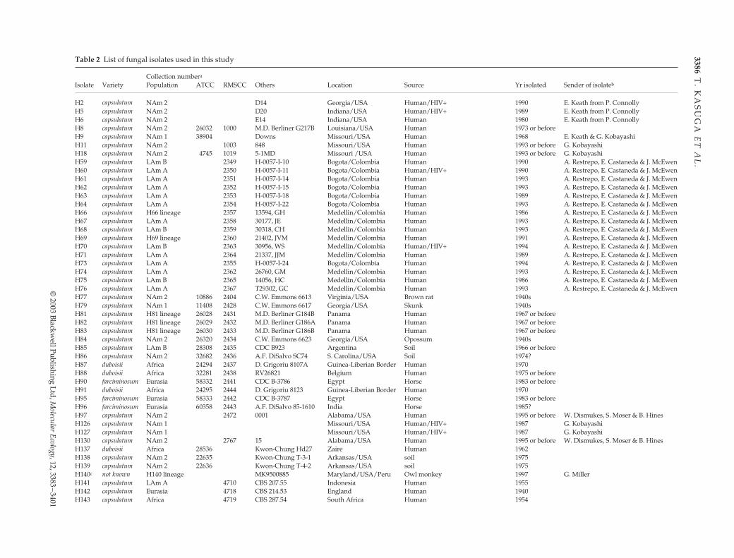
Table 2 lists the isolates used in the study. The 45 isolateslabelled H2–H140 were used in the previous study(Kasuga
et al
. 1999). H10 was excluded due to doubts aboutits source. The additional 92 isolates were from newlyinvestigated populations such as Australia, Mexico, Brazil,Argentina, China, Thailand and several European andAfrican countries. The analysed fungal samples includedsoil isolates as well as veterinary and clinical isolates.Culture conditions, DNA isolation methods, polymerasechain reaction (PCR) and sequencing conditions werepublished by Kasuga
et al
. (1999). Placement of varieties,i.e. var.
capsulatum
, var.
farciminosum
and var.
duboisii
, wasdone by medical mycologists who originally isolated fungalstrains, based mostly on pathogenicity and morphology.In short,
Hc
var.
capsulatum
mainly caused pulmonaryinfections.
Histoplasma capsulatum
var.
duboisii
was mainlyfound in Africa and caused lesions of cutaneous, sub-cutaneous and osseous tissues. The diameter of a yeast cellof
Hc
var.
duboisii
was 12–15
µ
m, which was larger thanthat of
Hc
var.
capsulatum
, which was 2–4
µ
m in diameter.
Histoplasma capsulatum
var.
farciminosum
caused infec-tions in horses, donkeys and mules. Yeast cells of
Hc
var.farciminosum in tissue section were indistinguishable fromthose of Hc var. capsulatum (Rippon 1988).
DNA analyses
DNA sequences of partial protein-coding genes used inthis study are arf, ADP-ribosylation factor (Lodge et al.1994); H-anti, H antigen precursor (Deepe & Durose 1995);ole, delta-9 fatty acid desaturase (Gargano et al. 1995) andtub1, alpha-tubulin (Harris et al. 1989).
Phylogenetic analyses [both maximum parsimony andneighbour-joining (NJ)] were performed by using paup4.0b 3a (Sinauer Associates). Most parsimonious (MP) treeswere generated by the heuristic search procedure using500 replications of the random addition sequence option.Nucleotide sites were weighted equally, with characterstate transformations treated as unordered and of equalcost. Insertions and deletions (indels) that were consis-tently and unambiguously alignable across all taxa weretreated as single evolutionary events by recording a singlesite within the indel as a multistate character. For MP ana-lysis of the combined data set, characters from the arf, H-anti, ole and tub1 loci were weighted as 1.00, 0.73, 0.82 and0.56, respectively. These values were inversely propor-tional to the total number of phylogenetically informativesites per locus. Indices of support (bootstrap values) forinternal branches were generated by 500 replications of thebootstrap procedure (Felsenstein 1985). Neighbour-joining
trees were generated using the Kimura (1980) two-parametercorrection for multiple hits.
Population histories of Latin American A (LAm A) andNAm 2 were inferred by use of the nested clade analysis(NCA). Gene genealogies of each of the four loci, arf,H-anti, ole and tub1, were reconstructed by the statisticalparsimony method using software tcs 1.13 (Clement et al.2000). Nested cladograms were constructed according topublished methods (Templeton et al. 1992; Crandall 1996).The NCA was then performed using the nested clado-grams and software geodis 2.0 (Posada et al. 2000). Isolatesbelonging to NAm 2 clades were divided into three popu-lations: Midwest (isolates from Indiana and Missouri),South (Arkansas, Texas and Louisiana) and Southeast(Alabama, Georgia, South Carolina and Virginia) and co-ordinates of St Louis, New Orleans and Atlanta were used,respectively. The LAm A isolates were divided into fourpopulations: Mexico (Mexico and Guatemala), Colombia(Colombia and Panama), Rio de Janeiro (Brazil) and SãoPaulo (Brazil) and coordinates of Mexico City, Bogotá, Riode Janeiro and São Paulo were used, respectively. Thesingle Surinamese isolate H145 was not included in thedata set due to the lack of population sample from the closevicinity. Inference of population history was made accordingto the inference key for the nested haplotype tree analysisof geographical distances (Templeton 1998).
Results
Polymorphism summary
Multiple loci used for the recognition of phylogeneticspecies are preferably functionally and genetically unlinked.The four chosen loci for this study, arf, H-anti, ole and tub1,are likely to be functionally independent but their locationson chromosomes are still unknown. Significant linkagedisequilibrium between loci was not detected in the ran-domly recombining North American population (P < 0.05).None of these loci were found on the same bacterialartificial chromosome (BAC) clone in the genomic libraryused for the ongoing Histoplasma genome project (http://www.genome.wustl.edu/projects/hcapsulatum/index.php).Thus, these four loci are likely to have been evolvingindependently in the Histoplasma genome.
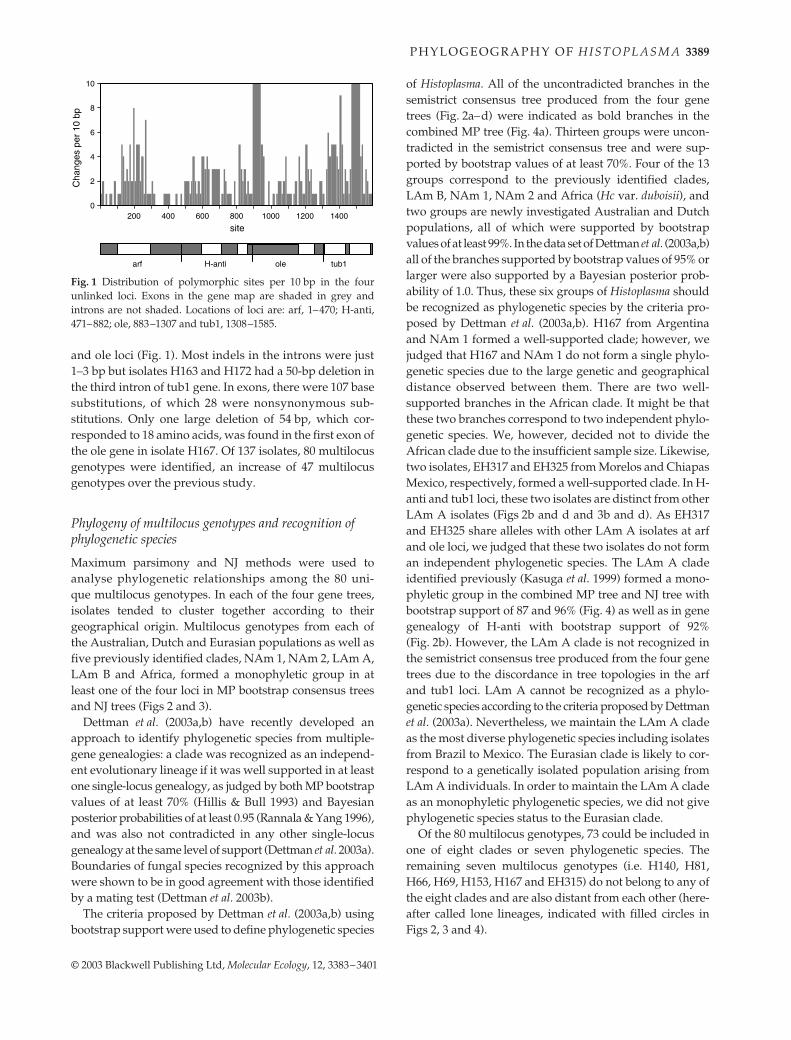
Combined DNA sequence data for the four loci gave us1585 aligned sites, of which 399 were variable and 193 werephylogenetically informative. The 193 phylogeneticallyinformative sites, 399 variable sites and 1585 aligned siteswere distributed as follows: for arf, 36 informative sites/78variable sites/470 aligned sites; for H-anti, 49/85/412; forole, 44/109/425 and for tub, 64/127/278. Among the 399variable bases, 147 had indels and 296 had substitutions; ofthese, 44 sites had both. Introns were clearly more variablethan exons in the arf and tub1 loci but not so in the H-anti
3386T
. KA
SU
GA
ET
AL
.
© 2003 Blackw
ell Publishing Ltd, Molecular Ecology, 12, 3383–3401
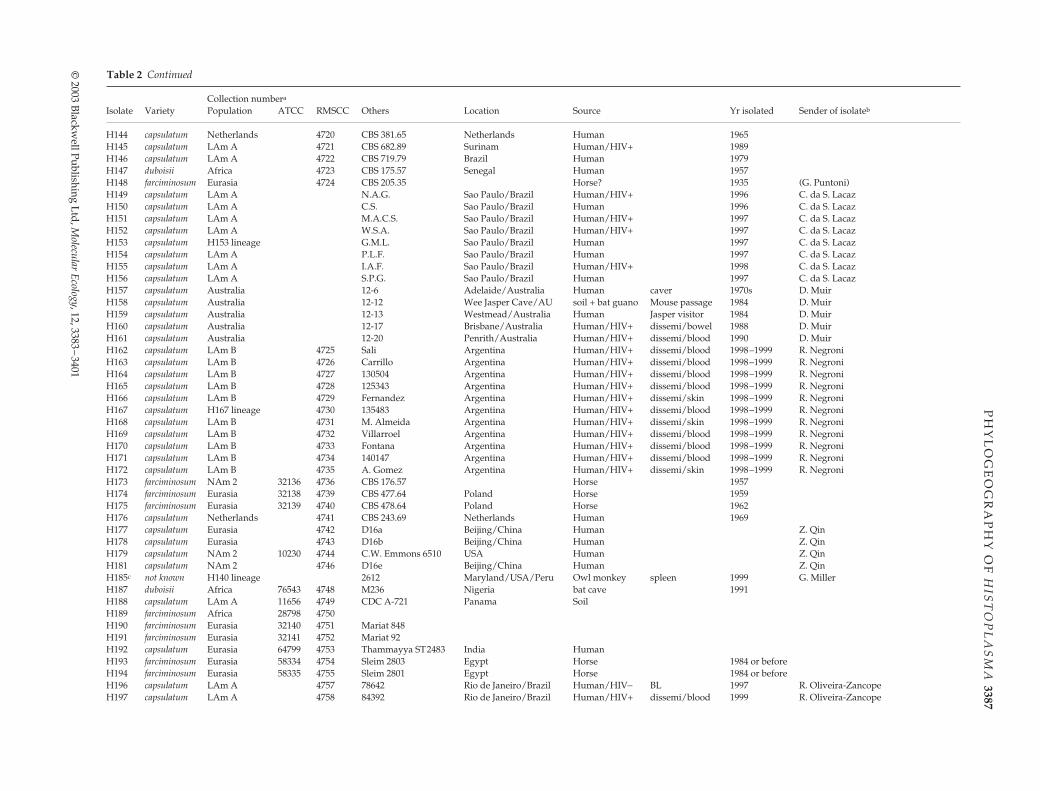
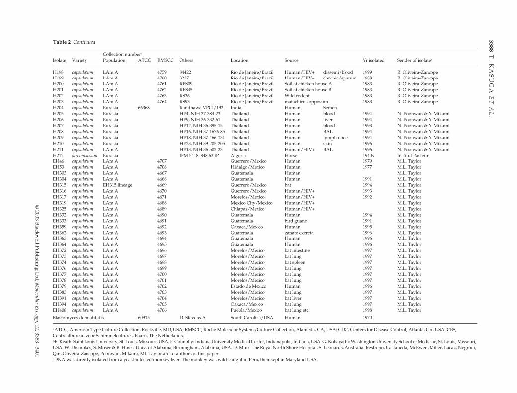
Table 2 List of fungal isolates used in this study
Collection numbera
Isolate Variety Population ATCC RMSCC Others Location Source Yr isolated Sender of isolateb
H2 capsulatum NAm 2 D14 Georgia/USA Human/HIV+ 1990 E. Keath from P. ConnollyH5 capsulatum NAm 2 D20 Indiana/USA Human/HIV+ 1989 E. Keath from P. ConnollyH6 capsulatum NAm 2 E14 Indiana/USA Human 1980 E. Keath from P. ConnollyH8 capsulatum NAm 2 26032 1000 M.D. Berliner G217B Louisiana/USA Human 1973 or beforeH9 capsulatum NAm 1 38904 Downs Missouri/USA Human 1968 E. Keath & G. KobayashiH11 capsulatum NAm 2 1003 848 Missouri/USA Human 1993 or before G. KobayashiH18 capsulatum NAm 2 4745 1019 5-1MD Missouri /USA Human 1993 or before G. KobayashiH59 capsulatum LAm B 2349 H-0057-I-10 Bogota/Colombia Human 1990 A. Restrepo, E. Castaneda & J. McEwenH60 capsulatum LAm A 2350 H-0057-I-11 Bogota/Colombia Human/HIV+ 1990 A. Restrepo, E. Castaneda & J. McEwenH61 capsulatum LAm A 2351 H-0057-I-14 Bogota/Colombia Human 1993 A. Restrepo, E. Castaneda & J. McEwenH62 capsulatum LAm A 2352 H-0057-I-15 Bogota/Colombia Human 1993 A. Restrepo, E. Castaneda & J. McEwenH63 capsulatum LAm A 2353 H-0057-I-18 Bogota/Colombia Human 1989 A. Restrepo, E. Castaneda & J. McEwenH64 capsulatum LAm A 2354 H-0057-I-22 Bogota/Colombia Human 1993 A. Restrepo, E. Castaneda & J. McEwenH66 capsulatum H66 lineage 2357 13594, GH Medellin/Colombia Human 1986 A. Restrepo, E. Castaneda & J. McEwenH67 capsulatum LAm A 2358 30177, JE Medellin/Colombia Human 1993 A. Restrepo, E. Castaneda & J. McEwenH68 capsulatum LAm B 2359 30318, CH Medellin/Colombia Human 1993 A. Restrepo, E. Castaneda & J. McEwenH69 capsulatum H69 lineage 2360 21402, JVM Medellin/Colombia Human 1991 A. Restrepo, E. Castaneda & J. McEwenH70 capsulatum LAm B 2363 30956, WS Medellin/Colombia Human/HIV+ 1994 A. Restrepo, E. Castaneda & J. McEwenH71 capsulatum LAm A 2364 21337, JJM Medellin/Colombia Human 1989 A. Restrepo, E. Castaneda & J. McEwenH73 capsulatum LAm A 2355 H-0057-I-24 Bogota/Colombia Human 1994 A. Restrepo, E. Castaneda & J. McEwenH74 capsulatum LAm A 2362 26760, GM Medellin/Colombia Human 1993 A. Restrepo, E. Castaneda & J. McEwenH75 capsulatum LAm B 2365 14056, HC Medellin/Colombia Human 1986 A. Restrepo, E. Castaneda & J. McEwenH76 capsulatum LAm A 2367 T29302, GC Medellin/Colombia Human 1993 A. Restrepo, E. Castaneda & J. McEwenH77 capsulatum NAm 2 10886 2404 C.W. Emmons 6613 Virginia/USA Brown rat 1940sH79 capsulatum NAm 1 11408 2428 C.W. Emmons 6617 Georgia/USA Skunk 1940sH81 capsulatum H81 lineage 26028 2431 M.D. Berliner G184B Panama Human 1967 or beforeH82 capsulatum H81 lineage 26029 2432 M.D. Berliner G186A Panama Human 1967 or beforeH83 capsulatum H81 lineage 26030 2433 M.D. Berliner G186B Panama Human 1967 or beforeH84 capsulatum NAm 2 26320 2434 C.W. Emmons 6623 Georgia/USA Opossum 1940sH85 capsulatum LAm B 28308 2435 CDC B923 Argentina Soil 1966 or beforeH86 capsulatum NAm 2 32682 2436 A.F. DiSalvo SC74 S. Carolina/USA Soil 1974?H87 duboisii Africa 24294 2437 D. Grigoriu 8107A Guinea-Liberian Border Human 1970H88 duboisii Africa 32281 2438 RV26821 Belgium Human 1975 or beforeH90 farciminosum Eurasia 58332 2441 CDC B-3786 Egypt Horse 1983 or beforeH91 duboisii Africa 24295 2444 D. Grigoriu 8123 Guinea-Liberian Border Human 1970H95 farciminosum Eurasia 58333 2442 CDC B-3787 Egypt Horse 1983 or beforeH96 farciminosum Eurasia 60358 2443 A.F. DiSalvo 85-1610 India Horse 1985?H97 capsulatum NAm 2 2472 0001 Alabama/USA Human 1995 or before W. Dismukes, S. Moser & B. HinesH126 capsulatum NAm 1 Missouri/USA Human/HIV+ 1987 G. KobayashiH127 capsulatum NAm 1 Missouri/USA Human/HIV+ 1987 G. KobayashiH130 capsulatum NAm 2 2767 15 Alabama/USA Human 1995 or before W. Dismukes, S. Moser & B. HinesH137 duboisii Africa 28536 Kwon-Chung Hd27 Zaire Human 1962H138 capsulatum NAm 2 22635 Kwon-Chung T-3-1 Arkansas/USA soil 1975H139 capsulatum NAm 2 22636 Kwon-Chung T-4-2 Arkansas/USA soil 1975H140c not known H140 lineage MK9500885 Maryland/USA/Peru Owl monkey 1997 G. MillerH141 capsulatum LAm A 4710 CBS 207.55 Indonesia Human 1955H142 capsulatum Eurasia 4718 CBS 214.53 England Human 1940H143 capsulatum Africa 4719 CBS 287.54 South Africa Human 1954
PH
YL
OG
EO
GR
AP
HY
OF
HIS
TO
PL
AS
MA
3387
© 2003 B
lackwell Publishing Ltd, M
olecular Ecology, 12, 3383–3401
H144 capsulatum Netherlands 4720 CBS 381.65 Netherlands Human 1965H145 capsulatum LAm A 4721 CBS 682.89 Surinam Human/HIV+ 1989H146 capsulatum LAm A 4722 CBS 719.79 Brazil Human 1979H147 duboisii Africa 4723 CBS 175.57 Senegal Human 1957H148 farciminosum Eurasia 4724 CBS 205.35 Horse? 1935 (G. Puntoni)H149 capsulatum LAm A N.A.G. Sao Paulo/Brazil Human/HIV+ 1996 C. da S. LacazH150 capsulatum LAm A C.S. Sao Paulo/Brazil Human 1996 C. da S. LacazH151 capsulatum LAm A M.A.C.S. Sao Paulo/Brazil Human/HIV+ 1997 C. da S. LacazH152 capsulatum LAm A W.S.A. Sao Paulo/Brazil Human/HIV+ 1997 C. da S. LacazH153 capsulatum H153 lineage G.M.L. Sao Paulo/Brazil Human 1997 C. da S. LacazH154 capsulatum LAm A P.L.F. Sao Paulo/Brazil Human 1997 C. da S. LacazH155 capsulatum LAm A I.A.F. Sao Paulo/Brazil Human/HIV+ 1998 C. da S. LacazH156 capsulatum LAm A S.P.G. Sao Paulo/Brazil Human 1997 C. da S. LacazH157 capsulatum Australia 12-6 Adelaide/Australia Human caver 1970s D. MuirH158 capsulatum Australia 12-12 Wee Jasper Cave/AU soil + bat guano Mouse passage 1984 D. MuirH159 capsulatum Australia 12-13 Westmead/Australia Human Jasper visitor 1984 D. MuirH160 capsulatum Australia 12-17 Brisbane/Australia Human/HIV+ dissemi/bowel 1988 D. MuirH161 capsulatum Australia 12-20 Penrith/Australia Human/HIV+ dissemi/blood 1990 D. MuirH162 capsulatum LAm B 4725 Sali Argentina Human/HIV+ dissemi/blood 1998–1999 R. NegroniH163 capsulatum LAm B 4726 Carrillo Argentina Human/HIV+ dissemi/blood 1998–1999 R. NegroniH164 capsulatum LAm B 4727 130504 Argentina Human/HIV+ dissemi/blood 1998–1999 R. NegroniH165 capsulatum LAm B 4728 125343 Argentina Human/HIV+ dissemi/blood 1998–1999 R. NegroniH166 capsulatum LAm B 4729 Fernandez Argentina Human/HIV+ dissemi/skin 1998–1999 R. NegroniH167 capsulatum H167 lineage 4730 135483 Argentina Human/HIV+ dissemi/blood 1998–1999 R. NegroniH168 capsulatum LAm B 4731 M. Almeida Argentina Human/HIV+ dissemi/skin 1998–1999 R. NegroniH169 capsulatum LAm B 4732 Villarroel Argentina Human/HIV+ dissemi/blood 1998–1999 R. NegroniH170 capsulatum LAm B 4733 Fontana Argentina Human/HIV+ dissemi/blood 1998–1999 R. NegroniH171 capsulatum LAm B 4734 140147 Argentina Human/HIV+ dissemi/blood 1998–1999 R. NegroniH172 capsulatum LAm B 4735 A. Gomez Argentina Human/HIV+ dissemi/skin 1998–1999 R. NegroniH173 farciminosum NAm 2 32136 4736 CBS 176.57 Horse 1957H174 farciminosum Eurasia 32138 4739 CBS 477.64 Poland Horse 1959H175 farciminosum Eurasia 32139 4740 CBS 478.64 Poland Horse 1962H176 capsulatum Netherlands 4741 CBS 243.69 Netherlands Human 1969H177 capsulatum Eurasia 4742 D16a Beijing/China Human Z. QinH178 capsulatum Eurasia 4743 D16b Beijing/China Human Z. QinH179 capsulatum NAm 2 10230 4744 C.W. Emmons 6510 USA Human Z. QinH181 capsulatum NAm 2 4746 D16e Beijing/China Human Z. QinH185c not known H140 lineage 2612 Maryland/USA/Peru Owl monkey spleen 1999 G. MillerH187 duboisii Africa 76543 4748 M236 Nigeria bat cave 1991H188 capsulatum LAm A 11656 4749 CDC A-721 Panama SoilH189 farciminosum Africa 28798 4750H190 farciminosum Eurasia 32140 4751 Mariat 848H191 farciminosum Eurasia 32141 4752 Mariat 92H192 capsulatum Eurasia 64799 4753 Thammayya ST2483 India HumanH193 farciminosum Eurasia 58334 4754 Sleim 2803 Egypt Horse 1984 or beforeH194 farciminosum Eurasia 58335 4755 Sleim 2801 Egypt Horse 1984 or beforeH196 capsulatum LAm A 4757 78642 Rio de Janeiro/Brazil Human/HIV− BL 1997 R. Oliveira-ZancopeH197 capsulatum LAm A 4758 84392 Rio de Janeiro/Brazil Human/HIV+ dissemi/blood 1999 R. Oliveira-Zancope
Collection numbera
Isolate Variety Population ATCC RMSCC Others Location Source Yr isolated Sender of isolateb
Table 2 Continued
3388T
. KA
SU
GA
ET
AL
.
© 2003 Blackw
ell Publishing Ltd, Molecular Ecology, 12, 3383–3401
H198 capsulatum LAm A 4759 84422 Rio de Janeiro/Brazil Human/HIV+ dissemi/blood 1999 R. Oliveira-ZancopeH199 capsulatum LAm A 4760 3237 Rio de Janeiro/Brazil Human/HIV− chronic/sputum 1988 R. Oliveira-ZancopeH200 capsulatum LAm A 4761 RPS09 Rio de Janeiro/Brazil Soil at chicken house A 1983 R. Oliveira-ZancopeH201 capsulatum LAm A 4762 RPS45 Rio de Janeiro/Brazil Soil at chicken house B 1983 R. Oliveira-ZancopeH202 capsulatum LAm A 4763 RS36 Rio de Janeiro/Brazil Wild rodent 1983 R. Oliveira-ZancopeH203 capsulatum LAm A 4764 RS93 Rio de Janeiro/Brazil matachirus opposum 1983 R. Oliveira-ZancopeH204 capsulatum Eurasia 66368 Randhawa VPCI/192 India Human SemenH205 capsulatum Eurasia HP4, NIH 37-384-23 Thailand Human blood 1994 N. Poonwan & Y. MikamiH206 capsulatum Eurasia HP9, NIH 36-332-61 Thailand Human liver 1994 N. Poonwan & Y. MikamiH207 capsulatum Eurasia HP12, NIH 36-395-15 Thailand Human blood 1993 N. Poonwan & Y. MikamiH208 capsulatum Eurasia HP16, NIH 37-1676-85 Thailand Human BAL 1994 N. Poonwan & Y. MikamiH209 capsulatum Eurasia HP18, NIH 37-466-131 Thailand Human lymph node 1994 N. Poonwan & Y. MikamiH210 capsulatum Eurasia HP23, NIH 39-205-205 Thailand Human skin 1996 N. Poonwan & Y. MikamiH211 capsulatum LAm A HP13, NIH 36-502-23 Thailand Human/HIV+ BAL 1996 N. Poonwan & Y. MikamiH212 farciminosum Eurasia IFM 5418, 848.63 IP Algeria Horse 1940s Institut PasteurEH46 capsulatum LAm A 4707 Guerrero/Mexico Human 1979 M.L. TaylorEH53 capsulatum LAm A 4708 Hidalgo/Mexico Human 1977 M.L. TaylorEH303 capsulatum LAm A 4667 Guatemala Human M.L. TaylorEH304 capsulatum LAm A 4668 Guatemala Human 1991 M.L. TaylorEH315 capsulatum EH315 lineage 4669 Guerrero/Mexico bat 1994 M.L. TaylorEH316 capsulatum LAm A 4670 Guerrero/Mexico Human/HIV+ 1993 M.L. TaylorEH317 capsulatum LAm A 4671 Morelos/Mexico Human/HIV+ 1992 M.L. TaylorEH319 capsulatum LAm A 4688 Mexico City/Mexico Human/HIV+ M.L. TaylorEH325 capsulatum LAm A 4689 Chiapas/Mexico Human/HIV+ M.L. TaylorEH332 capsulatum LAm A 4690 Guatemala Human 1994 M.L. TaylorEH333 capsulatum LAm A 4691 Guatemala bird guano 1991 M.L. TaylorEH359 capsulatum LAm A 4692 Oaxaca/Mexico Human 1995 M.L. TaylorEH362 capsulatum LAm A 4693 Guatemala zanate excreta 1996 M.L. TaylorEH363 capsulatum LAm A 4694 Guatemala Human 1996 M.L. TaylorEH364 capsulatum LAm A 4695 Guatemala Human 1996 M.L. TaylorEH372 capsulatum LAm A 4696 Morelos/Mexico bat intestine 1997 M.L. TaylorEH373 capsulatum LAm A 4697 Morelos/Mexico bat lung 1997 M.L. TaylorEH374 capsulatum LAm A 4698 Morelos/Mexico bat spleen 1997 M.L. TaylorEH376 capsulatum LAm A 4699 Morelos/Mexico bat lung 1997 M.L. TaylorEH377 capsulatum LAm A 4700 Morelos/Mexico bat lung 1997 M.L. TaylorEH378 capsulatum LAm A 4701 Morelos/Mexico bat lung 1997 M.L. TaylorEH379 capsulatum LAm A 4702 Estado de Mexico Human 1996 M.L. TaylorEH383 capsulatum LAm A 4703 Morelos/Mexico bat lung 1997 M.L. TaylorEH391 capsulatum LAm A 4704 Morelos/Mexico bat liver 1997 M.L. TaylorEH394 capsulatum LAm A 4705 Oaxaca/Mexico bat lung 1997 M.L. TaylorEH408 capsulatum LAm A 4706 Puebla/Mexico bat lung etc. 1998 M.L. Taylor
Blastomyces dermatitidis 60915 D. Stevens A South Carolina/USA Human 1970
aATCC, American Type Culture Collection, Rockville, MD, USA; RMSCC, Roche Molecular Systems Culture Collection, Alameda, CA, USA; CDC, Centers for Disease Control, Atlanta, GA, USA. CBS, Centraalbureau voor Schimmelcultures, Baarn, The Netherlands.bE. Keath: Saint Louis University, St. Louis, Missouri, USA. P. Connolly: Indiana University Medical Center, Indianapolis, Indiana, USA. G. Kobayashi: Washington University School of Medicine, St. Louis, Missouri, USA. W. Dismukes, S. Moser & B. Hines: Univ. of Alabama, Birmingham, Alabama, USA. D. Muir: The Royal North Shore Hospital, S. Leonards, Australia. Restrepo, Castaneda, McEwen, Miller, Lacaz, Negroni, Qin, Oliveira-Zancope, Poonwan, Mikami, ML Taylor are co-authors of this paper.cDNA was directly isolated from a yeast-infested monkey liver. The monkey was wild-caught in Peru, then kept in Maryland USA.
Collection numbera
Isolate Variety Population ATCC RMSCC Others Location Source Yr isolated Sender of isolateb
Table 2 Continued
P H Y L O G E O G R A P H Y O F H I S T O P L A S M A 3389
© 2003 Blackwell Publishing Ltd, Molecular Ecology, 12, 3383–3401
and ole loci (Fig. 1). Most indels in the introns were just1–3 bp but isolates H163 and H172 had a 50-bp deletion inthe third intron of tub1 gene. In exons, there were 107 basesubstitutions, of which 28 were nonsynonymous sub-stitutions. Only one large deletion of 54 bp, which cor-responded to 18 amino acids, was found in the first exon ofthe ole gene in isolate H167. Of 137 isolates, 80 multilocusgenotypes were identified, an increase of 47 multilocusgenotypes over the previous study.
Phylogeny of multilocus genotypes and recognition of phylogenetic species
Maximum parsimony and NJ methods were used toanalyse phylogenetic relationships among the 80 uni-que multilocus genotypes. In each of the four gene trees,isolates tended to cluster together according to theirgeographical origin. Multilocus genotypes from each ofthe Australian, Dutch and Eurasian populations as well asfive previously identified clades, NAm 1, NAm 2, LAm A,LAm B and Africa, formed a monophyletic group in atleast one of the four loci in MP bootstrap consensus treesand NJ trees (Figs 2 and 3).
Dettman et al. (2003a,b) have recently developed anapproach to identify phylogenetic species from multiple-gene genealogies: a clade was recognized as an independ-ent evolutionary lineage if it was well supported in at leastone single-locus genealogy, as judged by both MP bootstrapvalues of at least 70% (Hillis & Bull 1993) and Bayesianposterior probabilities of at least 0.95 (Rannala & Yang 1996),and was also not contradicted in any other single-locusgenealogy at the same level of support (Dettman et al. 2003a).Boundaries of fungal species recognized by this approachwere shown to be in good agreement with those identifiedby a mating test (Dettman et al. 2003b).
The criteria proposed by Dettman et al. (2003a,b) usingbootstrap support were used to define phylogenetic species
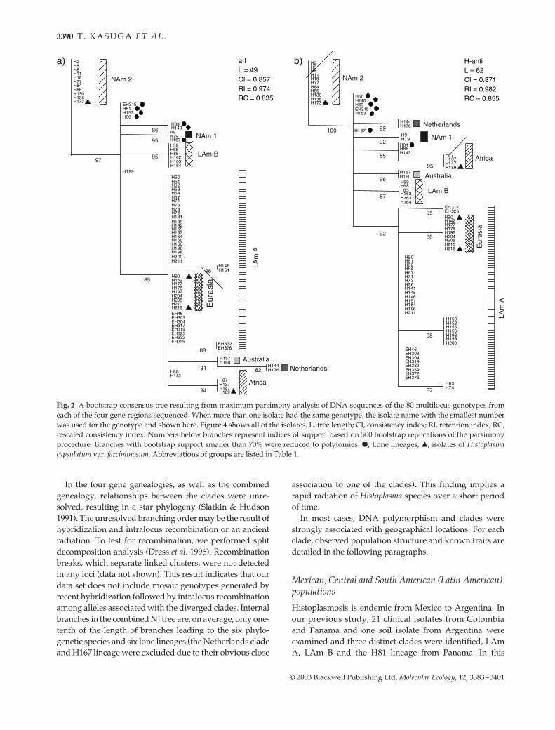
of Histoplasma. All of the uncontradicted branches in thesemistrict consensus tree produced from the four genetrees (Fig. 2a–d) were indicated as bold branches in thecombined MP tree (Fig. 4a). Thirteen groups were uncon-tradicted in the semistrict consensus tree and were sup-ported by bootstrap values of at least 70%. Four of the 13groups correspond to the previously identified clades,LAm B, NAm 1, NAm 2 and Africa (Hc var. duboisii), andtwo groups are newly investigated Australian and Dutchpopulations, all of which were supported by bootstrapvalues of at least 99%. In the data set of Dettman et al. (2003a,b)all of the branches supported by bootstrap values of 95% orlarger were also supported by a Bayesian posterior prob-ability of 1.0. Thus, these six groups of Histoplasma shouldbe recognized as phylogenetic species by the criteria pro-posed by Dettman et al. (2003a,b). H167 from Argentinaand NAm 1 formed a well-supported clade; however, wejudged that H167 and NAm 1 do not form a single phylo-genetic species due to the large genetic and geographicaldistance observed between them. There are two well-supported branches in the African clade. It might be thatthese two branches correspond to two independent phylo-genetic species. We, however, decided not to divide theAfrican clade due to the insufficient sample size. Likewise,two isolates, EH317 and EH325 from Morelos and ChiapasMexico, respectively, formed a well-supported clade. In H-anti and tub1 loci, these two isolates are distinct from otherLAm A isolates (Figs 2b and d and 3b and d). As EH317and EH325 share alleles with other LAm A isolates at arfand ole loci, we judged that these two isolates do not forman independent phylogenetic species. The LAm A cladeidentified previously (Kasuga et al. 1999) formed a mono-phyletic group in the combined MP tree and NJ tree withbootstrap support of 87 and 96% (Fig. 4) as well as in genegenealogy of H-anti with bootstrap support of 92%(Fig. 2b). However, the LAm A clade is not recognized inthe semistrict consensus tree produced from the four genetrees due to the discordance in tree topologies in the arfand tub1 loci. LAm A cannot be recognized as a phylo-genetic species according to the criteria proposed by Dettmanet al. (2003a). Nevertheless, we maintain the LAm A cladeas the most diverse phylogenetic species including isolatesfrom Brazil to Mexico. The Eurasian clade is likely to cor-respond to a genetically isolated population arising fromLAm A individuals. In order to maintain the LAm A cladeas an monophyletic phylogenetic species, we did not givephylogenetic species status to the Eurasian clade.
Of the 80 multilocus genotypes, 73 could be included inone of eight clades or seven phylogenetic species. Theremaining seven multilocus genotypes (i.e. H140, H81,H66, H69, H153, H167 and EH315) do not belong to any ofthe eight clades and are also distant from each other (here-after called lone lineages, indicated with filled circles inFigs 2, 3 and 4).
Fig. 1 Distribution of polymorphic sites per 10 bp in the fourunlinked loci. Exons in the gene map are shaded in grey andintrons are not shaded. Locations of loci are: arf, 1–470; H-anti,471–882; ole, 883–1307 and tub1, 1308–1585.
3390 T . K A S U G A E T A L .
© 2003 Blackwell Publishing Ltd, Molecular Ecology, 12, 3383–3401
In the four gene genealogies, as well as the combinedgenealogy, relationships between the clades were unre-solved, resulting in a star phylogeny (Slatkin & Hudson1991). The unresolved branching order may be the result ofhybridization and intralocus recombination or an ancientradiation. To test for recombination, we performed splitdecomposition analysis (Dress et al. 1996). Recombinationbreaks, which separate linked clusters, were not detectedin any loci (data not shown). This result indicates that ourdata set does not include mosaic genotypes generated byrecent hybridization followed by intralocus recombinationamong alleles associated with the diverged clades. Internalbranches in the combined NJ tree are, on average, only one-tenth of the length of branches leading to the six phylo-genetic species and six lone lineages (the Netherlands cladeand H167 lineage were excluded due to their obvious close
association to one of the clades). This finding implies arapid radiation of Histoplasma species over a short periodof time.
In most cases, DNA polymorphism and clades werestrongly associated with geographical locations. For eachclade, observed population structure and known traits aredetailed in the following paragraphs.
Mexican, Central and South American (Latin American) populations
Histoplasmosis is endemic from Mexico to Argentina. Inour previous study, 21 clinical isolates from Colombiaand Panama and one soil isolate from Argentina wereexamined and three distinct clades were identified, LAmA, LAm B and the H81 lineage from Panama. In this
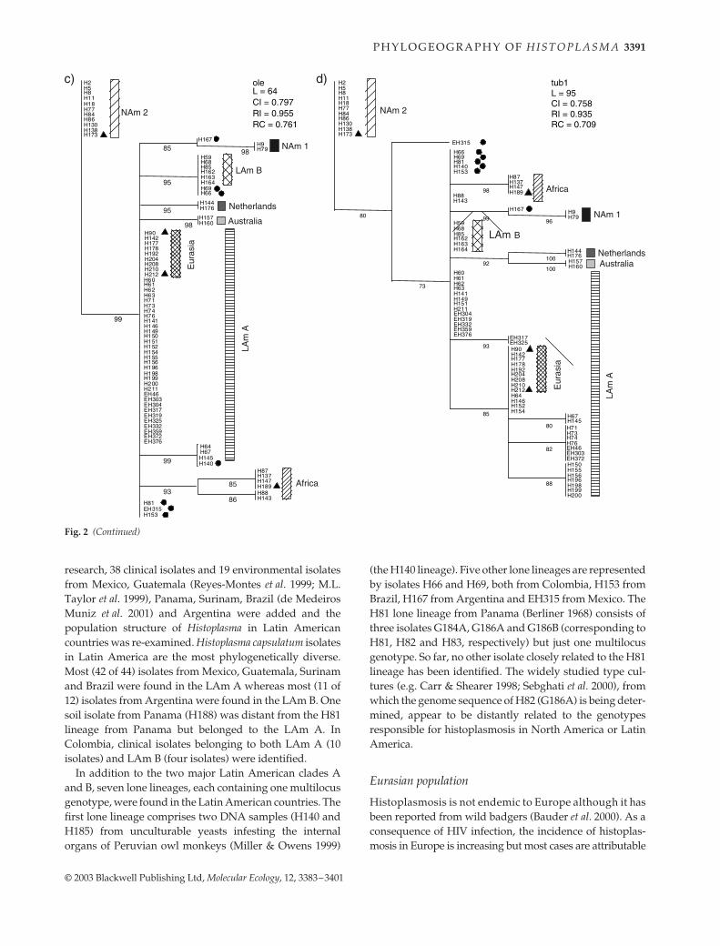
Fig. 2 A bootstrap consensus tree resulting from maximum parsimony analysis of DNA sequences of the 80 multilocus genotypes fromeach of the four gene regions sequenced. When more than one isolate had the same genotype, the isolate name with the smallest numberwas used for the genotype and shown here. Figure 4 shows all of the isolates. L, tree length; CI, consistency index; RI, retention index; RC,rescaled consistency index. Numbers below branches represent indices of support based on 500 bootstrap replications of the parsimonyprocedure. Branches with bootstrap support smaller than 70% were reduced to polytomies. �, Lone lineages; �, isolates of Histoplasmacapsulatum var. farciminosum. Abbreviations of groups are listed in Table 1.
P H Y L O G E O G R A P H Y O F H I S T O P L A S M A 3391
© 2003 Blackwell Publishing Ltd, Molecular Ecology, 12, 3383–3401
research, 38 clinical isolates and 19 environmental isolatesfrom Mexico, Guatemala (Reyes-Montes et al. 1999; M.L.Taylor et al. 1999), Panama, Surinam, Brazil (de MedeirosMuniz et al. 2001) and Argentina were added and thepopulation structure of Histoplasma in Latin Americancountries was re-examined. Histoplasma capsulatum isolatesin Latin America are the most phylogenetically diverse.Most (42 of 44) isolates from Mexico, Guatemala, Surinamand Brazil were found in the LAm A whereas most (11 of12) isolates from Argentina were found in the LAm B. Onesoil isolate from Panama (H188) was distant from the H81lineage from Panama but belonged to the LAm A. InColombia, clinical isolates belonging to both LAm A (10isolates) and LAm B (four isolates) were identified.
In addition to the two major Latin American clades Aand B, seven lone lineages, each containing one multilocusgenotype, were found in the Latin American countries. Thefirst lone lineage comprises two DNA samples (H140 andH185) from unculturable yeasts infesting the internalorgans of Peruvian owl monkeys (Miller & Owens 1999)
(the H140 lineage). Five other lone lineages are representedby isolates H66 and H69, both from Colombia, H153 fromBrazil, H167 from Argentina and EH315 from Mexico. TheH81 lone lineage from Panama (Berliner 1968) consists ofthree isolates G184A, G186A and G186B (corresponding toH81, H82 and H83, respectively) but just one multilocusgenotype. So far, no other isolate closely related to the H81lineage has been identified. The widely studied type cul-tures (e.g. Carr & Shearer 1998; Sebghati et al. 2000), fromwhich the genome sequence of H82 (G186A) is being deter-mined, appear to be distantly related to the genotypesresponsible for histoplasmosis in North America or LatinAmerica.
Eurasian population
Histoplasmosis is not endemic to Europe although it hasbeen reported from wild badgers (Bauder et al. 2000). As aconsequence of HIV infection, the incidence of histoplas-mosis in Europe is increasing but most cases are attributable
Fig. 2 (Continued)
3392 T . K A S U G A E T A L .
© 2003 Blackwell Publishing Ltd, Molecular Ecology, 12, 3383–3401
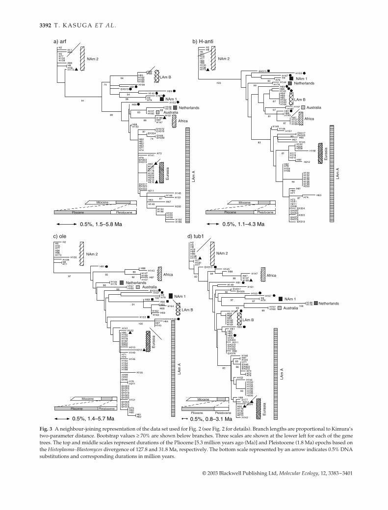
Fig. 3 A neighbour-joining representation of the data set used for Fig. 2 (see Fig. 2 for details). Branch lengths are proportional to Kimura’stwo-parameter distance. Bootstrap values ≥ 70% are shown below branches. Three scales are shown at the lower left for each of the genetrees. The top and middle scales represent durations of the Pliocene [5.3 million years ago (Ma)] and Pleistocene (1.8 Ma) epochs based onthe Histoplasma–Blastomyces divergence of 127.8 and 31.8 Ma, respectively. The bottom scale represented by an arrow indicates 0.5% DNAsubstitutions and corresponding durations in million years.
P H Y L O G E O G R A P H Y O F H I S T O P L A S M A 3393
© 2003 Blackwell Publishing Ltd, Molecular Ecology, 12, 3383–3401
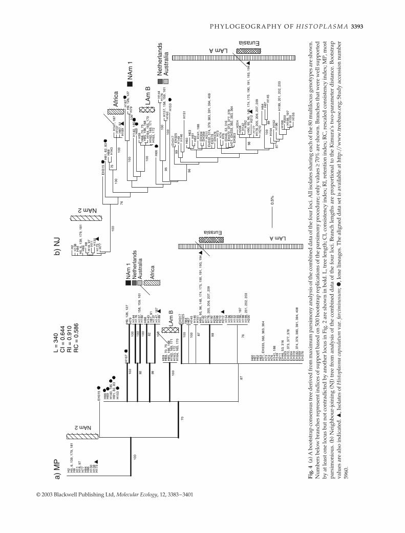
Fig.
4(a
) A b
oots
trap
con
sens
us tr
ee d
eriv
ed fr
om m
axim
um p
arsi
mon
y an
alys
is o
f the
com
bine
d da
ta o
f the
four
loci
. All
isol
ates
sha
ring
eac
h of
the
80 m
ulti
locu
s ge
noty
pes
are
show
n.N
umbe
rs b
elow
bra
nche
s re
pres
ent i
ndic
es o
f sup
port
bas
ed o
n 50
0 bo
otst
rap
repl
icat
ions
of t
he p
arsi
mon
y pr
oced
ure;
onl
y va
lues
≥ 7
0% a
re s
how
n. B
ranc
hes
that
wer
e w
ell s
uppo
rted
by a
t lea
st o
ne lo
cus
but n
ot c
ontr
adic
ted
by a
noth
er lo
cus
in F
ig. 2
are
sho
wn
in b
old.
L, t
ree
leng
th; C
I, co
nsis
tenc
y in
dex;
RI,
rete
ntio
n in
dex
; RC
, res
cale
d c
onsi
sten
cy in
dex
; MP,
mos
tpa
rsim
onio
us. (
b) N
eigh
bour
-join
ing
(NJ)
tre
e fr
om a
naly
sis
of t
he c
ombi
ned
data
of
the
four
loci
. Bra
nch
leng
ths
are
prop
orti
onal
to
the
Kim
ura’
s tw
o-pa
ram
eter
dis
tanc
e. B
oots
trap
valu
es a
re a
lso
indi
cate
d. �
, Iso
late
s of
His
topl
asm
a ca
psul
atum
var
. far
cim
inos
um; �
, lon
e lin
eage
s. T
he a
ligne
d da
ta s
et is
ava
ilabl
e at
htt
p://
ww
w.tr
eeba
se.o
rg; S
tud
y ac
cess
ion
num
ber
5960
.
3394 T . K A S U G A E T A L .
© 2003 Blackwell Publishing Ltd, Molecular Ecology, 12, 3383–3401
to endogenous reactivation of a latent infection acquiredoverseas in endemic areas (Manfredi et al. 1994). Thedisease is endemic to southeast Asia and India but theincidence and prevalence of histoplasmosis have not beenextensively described. Clinical isolates, one from England(H142), two from China (H177 and H178), six fromThailand (H205–H210) and two from India (H192 andH204), formed a homogeneous monophyletic group with abootstrap support of 98% within the LAm A clade (Fig. 4a).This Eurasian group corresponds to the Asian type H.capsulatum which was identified by Tamura et al. (2002) intheir phylogeny based on the internal transcribed spacerregion. One Chinese isolate (H178, Beijing) and four of thesix Thai isolates shared one multilocus genotype. Judgingfrom its close genetic distance to Indian isolates, andhistoplasmosis not being endemic to England, the Englishisolate H142 obtained in 1940 was likely to have beenacquired in India. A single Chinese isolate (H181, Beijing)was located in the NAm 2 clade, despite the fact that thepatient from whom H181 was isolated had never been outof China. Similarly, the Thai isolate H211, which had anRAPD pattern unlike other Thai isolates (Poonwan et al.1998), was found in the LAm A clade but outside theEurasian subclade. H181 and H211 might represent casesof indirect acquisition of a foreign fungus from infectedzoo animals or contaminated body parts of New Worldanimals used in Asian medicine, e.g. bear gall bladders.
North American populations
Two discrete phylogenetic species, NAm 1 and NAm 2,have been reported in North America (Spitzer et al. 1990;Kasuga et al. 1999). Our previous study showed that thesetwo phylogenetic species were as distant from each otheras from any other Histoplasma clades in the world. Geneticdiversities observed within NAm 1 and NAm 2 clades aremuch smaller than that observed in the LAm A clade. Inthis study, two lone lineages of Latin American isolatesfrom Argentina (H167) and Mexico (EH315) were found tobe distantly associated with the NAm 1 and NAm 2,respectively.
African population
On the African continent, two clinically distinct forms ofhistoplasmosis are known, one caused by Hc var. duboisiiand the other by Hc var. capsulatum (Kwon-Chung &Bennett 1992; Rippon 1988). The disease histoplasmosisduboisii is characterized by cutaneous and subcutaneouslesions whereas histoplasmosis capsulatii is characterizedby infection of the lung. A single African clade was formedfrom clinical isolates of Hc var. duboisii from the Guinea–Liberian border (H87 and H91), Zaire (H137), Belgium(H88, probably from a former Belgian colony), Senegal
(H147), Nigerian soil (H187) (Gugnani et al. 1994) and a Hcvar. capsulatum isolate from South Africa (H143). Thefinding of one Hc var. capsulatum isolate (H143) in the Hcvar. duboisii clade contradicts the traditional view that twodistinct forms of histoplasmosis exist in Africa. It is likelythat histoplasmosis in Africa is caused by a monophyleticgroup of H. capsulatum isolates which included iso-lates presently assigned to Hc var. duboisii and Hc var.capsulatum.
Australian and Netherlands (Indonesian?) populations
Histoplasmosis in Oceania is rare and poorly known.Incidences seem to be largely restricted to people who havevisited bat-infested caves (Isbister et al. 1976; Harden &Hunt 1985). One soil and four clinical isolates fromAustralia were very homogeneous genetically, with onlyone polymorphic site in the four gene regions despite theirdiverse geographical origins. Two clinical isolates receivedfrom the Netherlands (of Indonesian origin?) formed adistinct clade which was the sister group to the Australianclade with bootstrap support of 92% (Fig. 4a). The twoDutch isolates H144 and H176 had been deposited inthe Centraalbureau voor Schimmelcultures (CBS) in theNetherlands in 1965 and 1969, respectively. No informa-tion on the history of patients or the geographical originof the isolates is available. As histoplasmosis is not endemicto the Netherlands, it is likely that H144 and H176 originatedin former Dutch colonies, possibly in Indonesia. On thecontrary, an Indonesian clinical isolate (H141), depositedin the CBS in 1955, was found to belong to the LAm Aclade. Isolate H141 had a multilocus genotype identical tothat of a Panamanian soil isolate (H188, LAm A), sug-gesting that the infection caused by H141 might haveoriginated in Panama.
Histoplasma capsulatum var. farciminosum
Equine histoplasmosis is not caused by a monophyleticgroup of Hc var. farciminosum isolates. Four multilocusgenotypes were found among 13 isolates obtained fromcases of histoplasmosis farciminosi and identified as Hcvar. farciminosum from various geographical locations. Oneisolate was found in the African clade (H189), another inthe NAm 2 clade (H173) and the other 11 isolates, of which10 had identical multilocus genotypes, were found inthe Eurasian clade (filled triangles in Figs 2–4). No otherisolates had the multilocus genotypes of H189 or H173 sothe possibility of cross contamination in our laboratory canbe excluded. It appears that the disease histoplasmosisfarciminosi is just a form of histoplasmosis affecting horsesrather than humans and may be caused by isolatesoriginating independently from at least three Histoplasmaclades.
P H Y L O G E O G R A P H Y O F H I S T O P L A S M A 3395
© 2003 Blackwell Publishing Ltd, Molecular Ecology, 12, 3383–3401
Dating the divergence time of Histoplasma capsulatum
Assuming that H. capsulatum forms a star phylogeny, whendid the radiation of H. capsulatum start? In order to datethe radiation event, we have to rely on the molecularclock hypothesis and DNA mutation rates extrapolatedfrom other systems because no palaeontological data areavailable for H. capsulatum. Under the neutral theoryof evolution, mutations in any given DNA sequenceaccumulate at an approximately constant rate as long asthe DNA sequence retains its original function. In ourdata set, only a small portion of substitutions (28 of296 substitutions) represents nonsynonymous substitu-tions. Moreover, in any of the four loci, nonsynonymoussubstitutions per site were always significantly fewer(P < 0.05) than synonymous substitutions per site asjudged by pairwise comparisons between isolates, sug-gesting that the nonsynonymous polymorphisms inany of the four loci were not under positive Darwinianselection (Nei & Kumar 2000). Two coalescent theory-based tests, Tajima’s test (Tajima 1989) and the HKA test(Hudson et al. 1987), were used to detect natural selectionat the four genetic loci of the four largest phylogeneticspecies, LAm A, LAm B, NAm 2 and Africa. The HKA testfailed to detect deviation from the neutral mutationhypothesis in any of the four loci in the four clades (Rozas& Rozas 1999). Tajima’s test also did not detect deviationfrom neutral evolution with one exception at the ole locusin the LAm A clade (P < 0.05). Overall DNA substitutionsin the four loci did not deviate significantly from neutralevolution.
We have previously estimated the divergence time ofH. capsulatum and Blastomyces dermatitidis from a smallsubunit rRNA gene tree (Kasuga et al. 2002). The valuewas strongly dependent on the algorithm used to estimatedivergence time and the calibration time points. Whenthe divergence of Eurotiomycetes (plectomycetes) andSordariomycetes (pyrenomycetes) was set to 400 Ma (T.N.Taylor et al. 1999) and either of two methods of divergenceestimation were used, the Langley Fitch algorithm, whichassumes rate constancy, or a nonparametric rate-smooth-ing algorithm, which does not assume rate constancy(Sanderson 1997), divergence times for Histoplasma andBlastomyces were 32 and 128 Ma, respectively. To estimatethe nucleotide substitution rates at the two protein loci forwhich we have data for both H. capsulatum and B. dermati-tidis, arf and tub1, the genetic diversity was compared withthese divergence times. For arf, nucleotide substitutionwas estimated to be between 0.86 × 10⁻⁹ and 3.43 × 10⁻⁹
substitutions per base per year and tub1 was estimated tobe between 1.63 × 10⁻⁹ and 6.56 × 10⁻⁹ substitutions perbase per year. Absolute substitution rates at the ole and H-anti loci could not be estimated due to the unavailability ofcorresponding gene sequences in B. dermatitidis. To esti-mate the time of the radiation of Histoplasma species, weneeded to estimate the amount of DNA substitution thathad accumulated among the populations. We used thenucleotide diversity (π), which is the average pairwisedistance between isolates (see the footnote to Table 3) (Li1997). Inclusion of individuals with identical genotypesleads to the underestimation of population richness, whichis a concern because H. capsulatum propagates clonally as
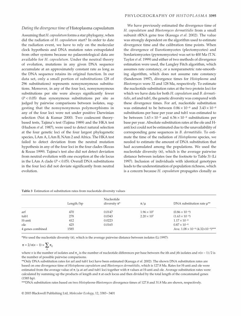
Table 3 Estimation of substitution rates from nucleotide diversity values
Length/bp Nucleotide diversity π* π/µ DNA substitution rate µ**
arf 470 0.0147 1.96 × 10⁷ (0.86 × 10⁻⁹)tub1 278 0.0343 2.20 × 10⁷ (1.63 × 10⁻⁹)H-anti 412 0.0223 1.17 × 10⁻⁹
ole 425 0.0165 0.87 × 10⁻⁹
4 genes combined 1585 Ave. 1.08 × 10⁻⁹ (4.32×10⁻⁹)***
*We used the nucleotide diversity (π), which is the average pairwise distance between isolates (Li 1997):
where n is the number of isolates and π ij is the number of nucleotide differences per base between the ith and jth isolates and n(n − 1)/2 is the number of possible pairwise comparisons.**Only DNA substitution rates for arf and tub1 loci have been estimated (Kasuga et al. 2002). The shown DNA substitution rates are based on one divergence time of Histoplasma capsulatum and Blastomyces dermatitidis, which is 127.8 Ma. Rates for H-anti and ole were estimated from the average value of π/µ at arf and tub1 loci together with π values at H-anti and ole. Average substitution rates were calculated by summing up the products of length and π at each locus and then divided by the total length of the concatenated genes (1585 bp).***DNA substitution rates based on two Histoplasma-Blastomyces divergence times of 127.8 and 31.8 Ma are shown, respectively.
π π / ( ) = − × ∑2 1n n ijij
3396 T . K A S U G A E T A L .
© 2003 Blackwell Publishing Ltd, Molecular Ecology, 12, 3383–3401
well as sexually. Suspected clonal isolates are as follows: 10of 13 isolates of Hc var. farciminosum (H90, H95, H96, H148,H174, H175, H190, H191, H193 and H194) and the twoPeruvian DNA samples (H140 and H185) had identicalmultilocus genotype in each of the groups. These isolateswere obtained from animals kept in crowded maintenancefacilities and probably represent clonal forms of H. capsu-latum which spread from host to host. Two isolates of Hcvar. duboisii (H87 and H91) and three Panamanian isolates(H82, H83 and probably H81) were isolated from singlepatients and showed identical multilocus genotypes ineach of the groups. Therefore, they are very likely to beclones. These duplicated clonal isolates were excludedfrom the data set. There are several other cases where indi-viduals share an identical multilocus genotype, e.g. H5, 6,139, 179 and 181 and EH332, 374, 379, 383, 391, 394 and408 (see Fig. 4). The probability of sampling a particulargenotype more than once in the data set can be calculated usinga binomial expression using allele frequencies assumingthat (i) different genotypes arise by recombination and notmutation; (ii) mating is random and (iii) loci are at linkageequilibrium (Fisher et al. 2000). The probabilities of observ-ing the H5 genotype five times or more and the EH332 gen-otype seven times or more are 0.55 and 0.18, respectively.These fungal isolates were obtained from different loca-tions or from different noncaptive host individuals. Theseisolates were left in the data set because the evidence fortheir clonality was weak. Table 3 shows values for π at eachof the four loci. Under a molecular clock and assuming nointralocus recombination, the nucleotide diversity π for eachlocus would be approximately proportional to the DNAsubstitution rate µ at the locus; µ for H-anti and ole werecalculated from the population diversities estimated forthese loci and the averages of π/µ for arf and tub1 (Table 3).
We calculated the average genetic distance for all fourloci among eight lineages (NAm 1, NAm 2, LAm A, LAmB, Australia + Netherlands (Indonesia?), Africa, H81 andH153) to be 2.80%. This value and the average substitutionrate for the four loci (1.08 × 10⁻⁹−4.32 × 10⁻⁹) placed theradiation of Histoplasma at approximately 3.2–13.0 Ma,mirroring the range of the Histoplasma and Blastomycesdivergence values, 32–128 Ma (Fig. 5). By either estimate,the radiation of Histoplasma is one-tenth as old as the diver-gence of Histoplasma and Blastomyces.
Comparing population diversities
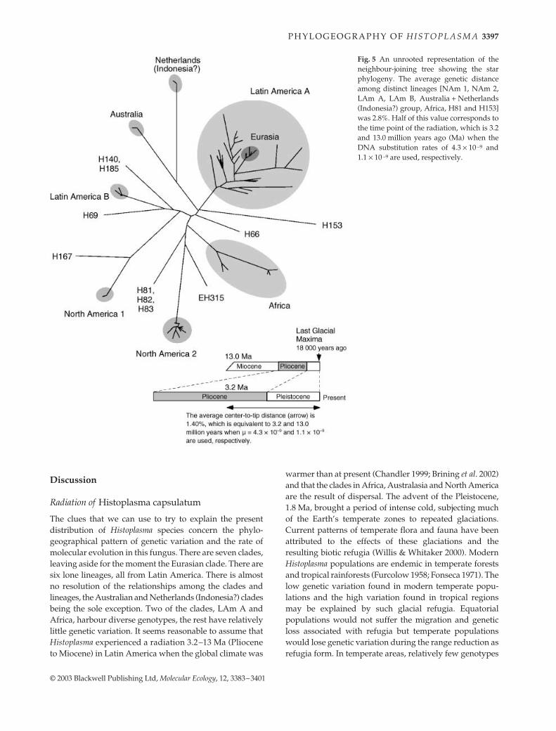
The extent of population diversity varies among popu-lations. For example, isolates in each of the NAm 1, NAm2, LAm B and Australian clades were homogeneous andcoalesced during the late Pliocene to Pleistocene epochs(Figs 3 and 5). On the other hand, genetic diversities inthe LAm A and African clades seem much larger andcoalesced in the Miocene to Pliocene epochs. Apparent
differences in genetic diversity might be attributable tosample sizes as we have the largest collection of LAm Aisolates (n = 55). To account for sample size difference,we resampled five isolates from 55 LAm A isolatesrandomly with replacement and calculated nucleotidediversity of the five isolates using the nucleotide diversityπ; this procedure was repeated 10 000 times. The resampleddistance distribution was compared with diversities of severalpopulations (Fig. 6). The statistical resampling demonstratesthat the observed small population diversities of LAm Band NAm 2 were not due to sampling error (P < 0.002)whereas the population diversity of the African cladeappears to be comparable to the LAm A clade. TheLAm A clade contains isolates from Mexico, Guatemala,Panama, Colombia, Surinam and Brazil. Genetic diversitiesobserved in subpopulations of the LAm A from Brazil andColombia themselves had diversities as large as the entireLAm A clade. The genetic diversity found in the Mexican(including Guatemala) population is significantly smallerthan in the Brazilian and Colombian populations but stilllarger than in LAm B and NAm 2 clades (Fig. 6). Thus, thesize of the endemic area or sampling area does not correlatewith the genetic diversity of each breeding population.
Inference of population history
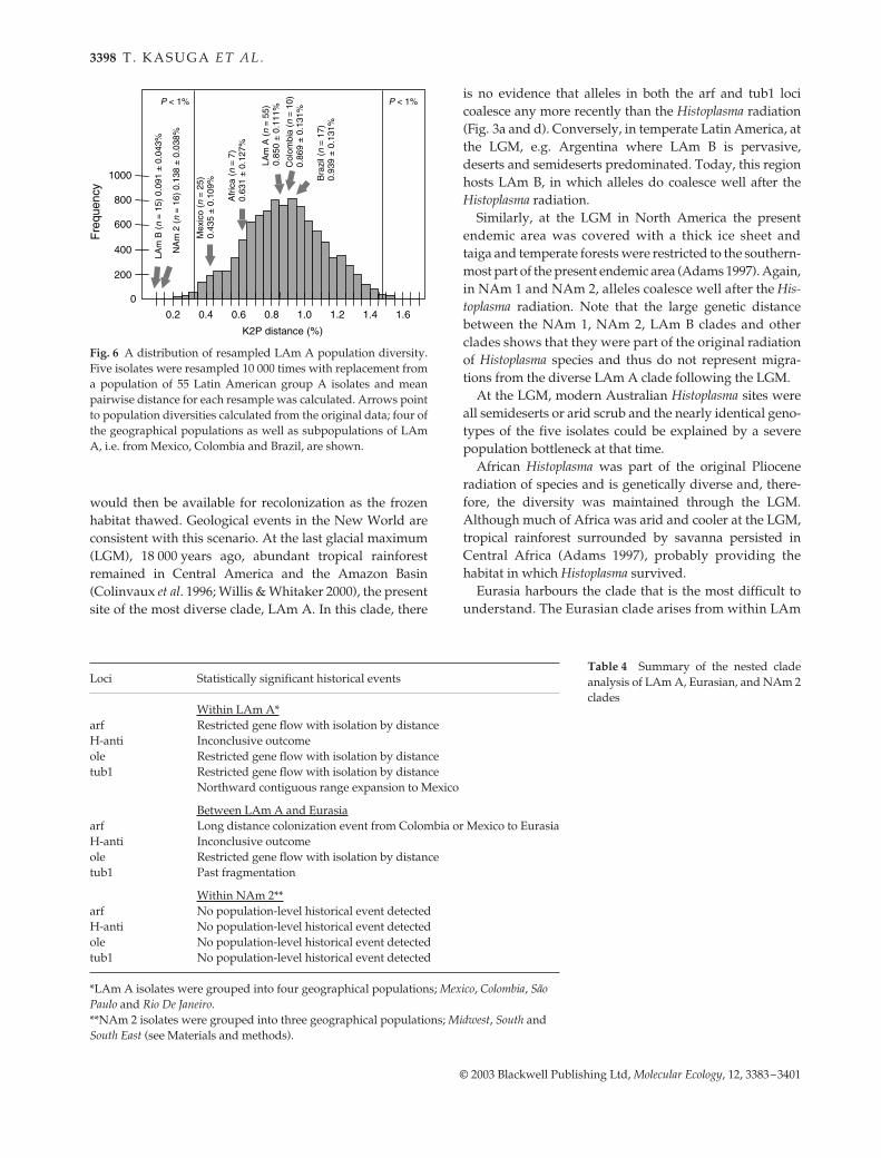
The large numbers of isolates belonging to NAm 2and LAm A clades enabled us to analyse their populationhistory. Templeton’s NCA is suited for this analysisbecause this method does not require presumption aboutthe underlying population process; instead, a historicalreconstruction is derived using inference key (Templeton1998; Knowles & Maddison 2002). The NCA can detectgeographical association and discriminate between phylo-geographical associations due to recurrent but restrictedgene flow vs. historical events operating at the populationlevel such as past fragmentation, colonization or rangeexpansion events (Templeton 1998). A haplotype genealogywas estimated for each population at each of the four lociby statistical parsimony (Templeton et al. 1992; Clementet al. 2000). The geographical location of each of the haplotypesand the haplotype genealogy were then used to detectgeographical associations and infer population history.Table 4 summarizes the outcome of the NCA. Among theLAm A isolates, geographical differentiation caused byrestricted gene flow was detected in three of the four loci.When Eurasian isolates were included in the LAm A, along-distance colonization event from Latin America toEurasia and past fragmentation were detected for arf andtub1 loci, respectively. On the other hand, geographicaldifferentiation was not detected in the NAm 2 for any ofthe four loci, unlike the differentiation between Alabama andIndiana isolates found using single nucleotide polymorphism(SNP) and microsatellite markers (Carter et al. 2001).
P H Y L O G E O G R A P H Y O F H I S T O P L A S M A 3397
© 2003 Blackwell Publishing Ltd, Molecular Ecology, 12, 3383–3401
Discussion
Radiation of Histoplasma capsulatum
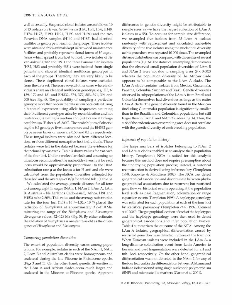
The clues that we can use to try to explain the presentdistribution of Histoplasma species concern the phylo-geographical pattern of genetic variation and the rate ofmolecular evolution in this fungus. There are seven clades,leaving aside for the moment the Eurasian clade. There aresix lone lineages, all from Latin America. There is almostno resolution of the relationships among the clades andlineages, the Australian and Netherlands (Indonesia?) cladesbeing the sole exception. Two of the clades, LAm A andAfrica, harbour diverse genotypes, the rest have relativelylittle genetic variation. It seems reasonable to assume thatHistoplasma experienced a radiation 3.2–13 Ma (Plioceneto Miocene) in Latin America when the global climate was
warmer than at present (Chandler 1999; Brining et al. 2002)and that the clades in Africa, Australasia and North Americaare the result of dispersal. The advent of the Pleistocene,1.8 Ma, brought a period of intense cold, subjecting muchof the Earth’s temperate zones to repeated glaciations.Current patterns of temperate flora and fauna have beenattributed to the effects of these glaciations and theresulting biotic refugia (Willis & Whitaker 2000). ModernHistoplasma populations are endemic in temperate forestsand tropical rainforests (Furcolow 1958; Fonseca 1971). Thelow genetic variation found in modern temperate popu-lations and the high variation found in tropical regionsmay be explained by such glacial refugia. Equatorialpopulations would not suffer the migration and geneticloss associated with refugia but temperate populationswould lose genetic variation during the range reduction asrefugia form. In temperate areas, relatively few genotypes
Fig. 5 An unrooted representation of theneighbour-joining tree showing the starphylogeny. The average genetic distanceamong distinct lineages [NAm 1, NAm 2,LAm A, LAm B, Australia + Netherlands(Indonesia?) group, Africa, H81 and H153]was 2.8%. Half of this value corresponds tothe time point of the radiation, which is 3.2and 13.0 million years ago (Ma) when theDNA substitution rates of 4.3 × 10⁻⁹ and1.1 × 10−9 are used, respectively.
3398 T . K A S U G A E T A L .
© 2003 Blackwell Publishing Ltd, Molecular Ecology, 12, 3383–3401
would then be available for recolonization as the frozenhabitat thawed. Geological events in the New World areconsistent with this scenario. At the last glacial maximum(LGM), 18 000 years ago, abundant tropical rainforestremained in Central America and the Amazon Basin(Colinvaux et al. 1996; Willis & Whitaker 2000), the presentsite of the most diverse clade, LAm A. In this clade, there
is no evidence that alleles in both the arf and tub1 locicoalesce any more recently than the Histoplasma radiation(Fig. 3a and d). Conversely, in temperate Latin America, atthe LGM, e.g. Argentina where LAm B is pervasive,deserts and semideserts predominated. Today, this regionhosts LAm B, in which alleles do coalesce well after theHistoplasma radiation.
Similarly, at the LGM in North America the presentendemic area was covered with a thick ice sheet andtaiga and temperate forests were restricted to the southern-most part of the present endemic area (Adams 1997). Again,in NAm 1 and NAm 2, alleles coalesce well after the His-toplasma radiation. Note that the large genetic distancebetween the NAm 1, NAm 2, LAm B clades and otherclades shows that they were part of the original radiationof Histoplasma species and thus do not represent migra-tions from the diverse LAm A clade following the LGM.
At the LGM, modern Australian Histoplasma sites wereall semideserts or arid scrub and the nearly identical geno-types of the five isolates could be explained by a severepopulation bottleneck at that time.
African Histoplasma was part of the original Plioceneradiation of species and is genetically diverse and, there-fore, the diversity was maintained through the LGM.Although much of Africa was arid and cooler at the LGM,tropical rainforest surrounded by savanna persisted inCentral Africa (Adams 1997), probably providing thehabitat in which Histoplasma survived.
Eurasia harbours the clade that is the most difficult tounderstand. The Eurasian clade arises from within LAm
Fig. 6 A distribution of resampled LAm A population diversity.Five isolates were resampled 10 000 times with replacement froma population of 55 Latin American group A isolates and meanpairwise distance for each resample was calculated. Arrows pointto population diversities calculated from the original data; four ofthe geographical populations as well as subpopulations of LAmA, i.e. from Mexico, Colombia and Brazil, are shown.
Loci Statistically significant historical events
Within LAm A*arf Restricted gene flow with isolation by distanceH-anti Inconclusive outcomeole Restricted gene flow with isolation by distancetub1 Restricted gene flow with isolation by distance
Northward contiguous range expansion to Mexico
Between LAm A and Eurasiaarf Long distance colonization event from Colombia or Mexico to EurasiaH-anti Inconclusive outcomeole Restricted gene flow with isolation by distancetub1 Past fragmentation
Within NAm 2**arf No population-level historical event detectedH-anti No population-level historical event detectedole No population-level historical event detectedtub1 No population-level historical event detected
*LAm A isolates were grouped into four geographical populations; Mexico, Colombia, São Paulo and Rio De Janeiro.**NAm 2 isolates were grouped into three geographical populations; Midwest, South and South East (see Materials and methods).
Table 4 Summary of the nested cladeanalysis of LAm A, Eurasian, and NAm 2clades
P H Y L O G E O G R A P H Y O F H I S T O P L A S M A 3399
© 2003 Blackwell Publishing Ltd, Molecular Ecology, 12, 3383–3401
A and the genotypes of the individuals in the clade arevery homogeneous, notwithstanding their having beencollected from the Far East to Europe. Nested clade ana-lysis of the arf locus suggested a long distance colonizationevent from Latin America to Eurasia and NCA of thetub1 locus suggested a past fragmentation event, both atunspecified times. The Eurasian clade originated between1.7 and 6.8 Ma, based on the estimated percentage of DNAsubstitutions per nucleotide per 1 million years of 0.16–0.65% at tub1 locus and the maximum pairwise distanceamong isolates in the Eurasian clade of 1.1% at tub1 locus(Eurasian isolates are monomorphic at arf). This estimateprovides an upper limit for the immigration of LAm Aindividuals to Eurasia but the event could have beenmore recent if several individuals with different genotypeswere involved in the initial dispersal. For example, onecargo of domesticated horses or donkeys infected withmultiple individuals of H. capsulatum, transported asrecently as 500 years ago, could have initiated the Eurasianclade.
Histoplasma capsulatum var. farciminosum individuals werefound in three clades, African (H189), NAm 2 (H173) andEurasian, the latter of which accommodated 11 of the 13individuals. It is clear that Hc var. farciminosum is not amonophyletic group and that individuals have acquiredthe ability to cause superficial disease in horses and otherequidae more than once. Therefore, Hc var. farciminosum isnot a valid taxon, it is a disease. The 10 of 11 individualsof Hc var. farciminosum from Eurasia had identical allelesat all four loci, indicating that they represent one clone,ranging from Poland to Egypt to India.
Do lone lineages represent cryptic species?
Seven evolutionary lineages were represented by singleindividuals or single genotypes that did not belong to anyof the seven phylogenetic species. Can these lone lineagesbe considered as cryptic species? Our sampling favouredhuman clinical isolates, and the lone lineages were biasedagainst this trait, so the lineages may represent larger popu-lations of fungi in nature. For example, EH315 was re-covered from a wild bat and the two Peruvian individualswith identical genotypes (H140 and H185) were recoveredfrom owl monkeys. The Peruvian individuals did notform mycelium in the laboratory and could not be cul-tivated (Miller & Owens 1999), forcing us to use DNA frominfected liver and spleen for our PCR amplification. ThePeruvian individuals had larger yeast cells than typical H.capsulatum and reminded mycologists of another fungalpathogen, Lacazia loboi, but clearly belong in the genusHistoplasma. Isolate H153 was also phenotypically distinct,having unusually large macroconidia, not converting toyeast at 37 °C and causing an atypical, disseminated cutane-ous histoplasmosis (Lacaz et al. 1999). Other lone lineages,
e.g. H66, H69 from Colombia, H167 from Argentina andH81 from Panama, showed no phenotypic differences,either in morphology or ecology. On balance, it seemslikely that some of our lone lineages represent fungithat are not likely to be collected by clinicians, might notbe recognized as Histoplasma or cannot be cultivated bymethods routinely used in clinical laboratories. If thisthinking is correct, the lone lineages may represent naturalpopulations that we have not sampled adequately. Thedramatic increase in recovery of NAm 1 individuals cor-related with the AIDS pandemic provides support forthis idea. Prior to the pandemic, which began in theearly 1980s, NAm 2 was predominant and NAm 1 wasrepresented by only two individuals, H9 [Downs, obtainedin 1968 from an 86-year-old woman (Gass & Kobayashi1969)] and H79 [obtained from a striped skunk in the1940s (Emmons et al. 1949)]. As AIDS spread, NAm 1became common in clinics (Spitzer et al. 1990), not due toan increase in NAm 1 in nature but to an increase insusceptible hosts.
Conclusion
Histoplasma capsulatum comprises at least seven phylo-genetic species, one in each of Africa, Australia and theNetherlands (Indonesia?) and two species each in NorthAmerica and Latin America. The Eurasian populationoriginated from within one of the Latin American species.In addition, seven distinct lineages represented by singleisolates or genotypes were identified in Latin America.Each of these lineages potentially represents an independentphylogenetic species. Judging from the observed geneticdiversity and DNA substitution rates, the radiation ofHistoplasma started between 3 and 13 Ma in Latin America.The present day population structure of Histoplasma can beexplained by refugial populations in the last glacial maxima.
Acknowledgements
We thank E. Keath, G. Kobayashi, L. Wheat, P. Connolly, S. Moser,B. Hines, W. Dismukes and D. Muir for supplying isolates, DNAand associated clinical information; M. Sugiyama and E. Jensen forsharing unpublished results and Rachel Whitaker and JeremyDettman for useful comments on the manuscript. Financial sup-port for this work was provided by the National Institutes ofHealth (grants HL55953 and AI37232 to J.W.T.).
References
Adams J (1997) Global land environments since the last inter-glacial. Oakridge National Laboratory, TN, USA. http://www.esd.ornl.gov/ern/qen/nerc.html.
Avise JC, Ball RM Jr (1990) Principles of genealogical concordancein species concepts and biological taxonomy. Oxford Surveys inEvolutionary Biology, 7, 45–67.
3400 T . K A S U G A E T A L .
© 2003 Blackwell Publishing Ltd, Molecular Ecology, 12, 3383–3401
Bauder B, Kuebber-Heiss A, Steineck T, Kuttin ES, Kaufman L(2000) Granulomatous skin lesions due to histoplasmosis in abadger (Meles meles) in Austria. Medical Mycology, 38, 249–253.
Baum DA, Shaw KL (1995) Genealogical perspectives on thespecies problem. In: Experimental and Molecular Approaches toPlant Biosystematics (eds Hoch PC, Stephenson AG.), pp. 289–303. Missouri Botanical Garden, St Louis, MO.
Berliner MD (1968) Primary subcultures of Histoplasma capsulatum,I. Macro and micro-morphology of the mycelial phase. Sabour-audia, 6, 111–118.
Brining L, Chan V, Choi E, De Sosa M, Lee C (2002) The MioceneEpoch. Museum of Paleontology, University of California, Berkeley,CA, USA. http://www.ucmp.berkeley.edu/tertiary/mio.html.
Carr J, Shearer GJ (1998) Genome size, complexity, and ploidy ofthe pathogenic fungus Histoplasma capsulatum. Journal of Bacteri-ology, 180, 6697–6703.
Carter DA, Taylor JW, Dechairo B et al. (2001) Amplified single-nucleotide polymorphisms and a (GA)n microsatellite markerreveal genetic differentiation between populations of Histoplasmacapsulatum from the Americans. Fungal Genetics and Biology, 34,37–48.
Chandler MA (1999) The Climate of the Pliocene: Simulating Earth’s LastGreat Warm Period. Goddard Institute for Space Studies, ColumbiaUniversity, NY, NY, USA. http://www.giss.nasa.gov/research/paleo/pliocene/index.html.
Clement M, Derington J, Posada D (2000) tcs: a computer programto estimate gene genealogies. Molecular Ecology, 9, 1657–1659.
Colinvaux PA, De Oliveira PE, Moreno JE, Miller MC, Bush MB(1996) A long pollen record from lowland Amazonia: Forest andcooling in glacial times. Science, 275, 85–88.
Crandall KA (1996) Multiple interspecies transmissions of humanand simian T-cell leukemia/lymphoma virus type sequences.Molecular Biology and Evolution, 13, 115–131.
Deepe GS Jr, Durose GG (1995) Immunobiological activity ofrecombinant H antigen from Histoplasma capsulatum. Infectionand Immunity, 63, 3151–3157.
Dettman J, Jacobson D, Taylor JW (2003a) A multilocus genealo-gical approach to phylogenetic species recognition in the modeleukaryote Neurospora. Evolution, in press.
Dettman J, Jacobson D, Turner E, Pringle A, Taylor JW (2003b)Reproductive isolation and phylogenetic divergence in Neuro-spora: comparing methods of species recognition in modeleukaryote. Evolution, in press.
Dress A, Huson D, Moulton V (1996) Analyzing and visualizingsequence and distance data using splitstree. Discrete AppliedMathematics, 71, 95–109.
Emmons CW, Morlan HB, Hill EL (1949) Histoplasmosis in ratsand skunks in Georgia. Public Health Report, 64, 1423–1430.
Felsenstein J (1985) Confidence limits on phylogenies: an approachto using the bootstrap. Evolution, 39, 783–791.
Fisher MC, Koenig GL, White TJ, Taylor JW (2000) Pathogenicclones versus environmentally driven population increase: analysisof an epidemic of the human fungal pathogen Coccidioides immitis.Journal of Clinical Microbiology, 38, 807–813.
Fonseca JC (1971) Analisis estadistico y ecologio-epidemiologicode la sensibilidad a la histoplasmina en Colombia, 1950–1968.Antioquia Medica, 21, 109–154.
Furcolow ML (1958) Recent studies on the epidemiology of histo-plasmosis. Annals of the New York Academy of Science, 72, 127–164.
Gargano S, Di Lallo G, Kobayashi GS, Maresca B (1995) A temperature-sensitive strain of Histoplasma capsulatum has an altered delta9-fatty acid desaturase gene. Lipids, 30, 899–906.
Gass M, Kobayashi GS (1969) Histoplasmosis: an illustrative casewith unusual vaginal and joint involvement. Archives of Derma-tology, 100, 724–727.
Gugnani HC, Muotoe-Okafor FA, Kaufman L, Dupont B (1994) Anatural focus of Histoplasma capsulatum var. duboisii is a bat cave.Mycopathologia, 127, 151–157.
Harden TJ, Hunt PJ (1985) Histoplasmosis and Australian caveenvironments. Helictite, 23, 23–26.
Harris GS, Keath EJ, Medoff J (1989) Characterization of alpha andbeta tubulin genes in the dimorphic fungus Histoplasma capsula-tum. Journal of General Microbiology, 135, 1817–1832.
Hillis DM, Bull JJ (1993) An empirical test of bootstrapping asa method for assessing confidence in phylogenetic analysis.Systematic Biology, 42, 182–192.
Hudson RR, Kreitman M, Aguade M (1987) A test of neutral mole-cular evolution based on nucleotide data. Genetics, 116, 153–159.
Isbister J, Elliott M, Nogrady S (1976) Histoplasmosis: an outbreakoccurring among young men who visited one cave. Medical Jour-nal of Australia, 2, 243–248.
Kasuga T, Taylor JW, White TJ (1999) Phylogenetic relationshipsof varieties and geographical groups of the human pathogenicfungus Histoplasma capsulatum Darling. Journal of Clinical Micro-biology, 37, 653–663.
Kasuga T, White TJ, Taylor JW (2002) Estimation of nucleotidesubstitution rates in Eurotiomycete fungi. Molecular Biology andEvolution, 19, 2318–2324.
Kimura M (1980) A simple method for estimating evolutionaryrates of base substitutions through comparative studies ofnucleotide sequences. Journal of Molecular Evolution, 16, 111–120.
Knowles LL, Maddison WP (2002) Statistical phylogeography.Molecular Ecology, 11, 2623–2635.
Kwon-Chung KJ, Bennett JE (1992) Medical Mycology. Lea &Febiger, Malvern, PA.
Lacaz CdS, Del Negro GMB, Vidal MSM et al. (1999) Atypical dis-seminated cutaneous histoplasmosis in an immunocompetentchild, caused by an ‘aberrant’ variant of Histoplasma capsulatumvar. capsulatum. Revista do Instituto de Medicina Tropical de SãoPaulo, 41, 195–202.
Li W-H (1997) Molecular Evolution. Sinauer Associates, Sunder-land, MA.
Lodge JK, Johnson RL, Weinberg RA, Gordon JI (1994) Compari-son of myristoyl-CoA: protein N-myristoyltransferases fromthree pathogenic fungi: Cryptococcus neoformans, Histoplasmacapsulatum, and Candida albicans. Journal of Biological Chemistry,269, 2996–3009.
Manfredi R, Mazzoni A, Nanetti A, Chiodo F (1994) Histoplasmosiscapsulati and duboisii in Europe: the impact of the HIV pan-demic, travel and immigration. European Journal of Epidemiology,10, 675–681.
Mayden RL (1997) A hierarchy of species concepts: The denoue-ment in the saga of the species problem. In: Species: the Units ofBiodiversity (eds Claridge MF, Dawah HA, Wilson MR), pp. 381–424. Chapman & Hall, London.
de Medeiros Muniz M, Pizzini CV, Peralta JM, Reiss E, Zancope-Oliveira RM (2001) Genetic diversity of Histoplasma capsulatumstrains isolated from soil, animals, and clinical specimens inRio de Janeiro State, Brazil, by a PCR-based random amplifiedpolymorphic DNA assay. Journal of Clinical Microbiology, 39,4487–4494.
Miller GF, Owens JW (1999) Ultrastructural characterization of theagent of systemic yeast infection of owl monkeys. MedicalMycology, 37, 139–145.
P H Y L O G E O G R A P H Y O F H I S T O P L A S M A 3401
© 2003 Blackwell Publishing Ltd, Molecular Ecology, 12, 3383–3401
Nei M, Kumar S (2000) Molecular Evolution and Phylogenetics.Oxford University Press, New York.
Poonwan N, Imai T, Mekha N et al. (1998) Genetic analysis ofHistoplasma capsulatum strains isolated from clinical specimensin Thailand by a PCR-based random amplified polymorphicDNA method. Journal of Clinical Microbiology, 36, 3073–3076.
Posada D, Crandall KA, Templeton AR (2000) GeoDis: a programfor the cladistic nested analysis of the geographical distributionof genetic haplotype. Molecular Ecology, 9, 487–488.
Rannala B, Yang Z (1996) Probability distribution of molecularevolutionary trees: a new method of phylogenetic inference.Journal of Molecular Evolution, 43, 304–311.
Reyes-Montes MR, Bobadilla-Del Valle M, Martínez-Rivera MAet al. (1999) Relatedness analyses of Histoplasma capsulatum isolatesfrom Mexican patients with AIDS-associated histoplasmosis byusing histoplasmin electrophoretic profiles and randomlyamplified polymorphic DNA patterns. Journal of Clinical Micro-biology, 37, 1404–1408.
Rippon JW (1988) Medical Mycology, 3rd edn. W.B. Saunders,Philadelphia.
Rozas J, Rozas R (1999) DnaSP, Version 3: an integrated programfor molecular population genetics and molecular evolutionanalysis. Bioinformatics, 15, 174–175.
Sanderson MJ (1997) A nonparametric approach to estimatingdivergence times in the absence of rate constancy. MolecularBiology and Evolution, 14, 1218–1231.
Sebghati TS, Engle JT, Goldman WE (2000) Intracellular para-sitism by Histoplasma capsulatum: fungal virulence and calciumdependence. Science, 290, 1368–1372.
Slatkin M, Hudson RR (1991) Pairwise comparisons of mito-chondrial DNA sequences in stable and exponentially growingpopulations. Genetics, 129, 555–562.
Spitzer ED, Keath EJ, Travis SJ et al. (1990) Temperature-sensitive
variants of Histoplasma capsulatum isolated from patients withacquired immunodeficiency syndrome. Journal of InfectiousDiseases, 162, 258–261.
Tajima F (1989) Statistical method for testing the neutral mutationhypothesis by DNA polymorphism. Genetics, 123, 585–595.
Tamura M, Kasuga T, Watanabe K et al. (2002) Phylogeneticcharacterization of Histoplasma capsulatum strains based on ITSregion sequences, including two new strains from Thai andChinese patients in Japan. Nippon Ishinkin Gakkai Zasshi, 43, 11–19.
Taylor JW, Jacobson JD, Kroken S et al. (2000) Phylogenetic speciesrecognition and species concepts in fungi. Fungal Genetics andBiology, 31, 21–32.
Taylor ML, Chávez-Tapia CB, Vargas-Yañez R et al. (1999) En-vironmental conditions favoring bat infection with Histoplasmacapsulatum in Mexican shelters. American Journal of TropicalMedicine and Hygiene, 61, 914–919.
Taylor TN, Hass H, Kerp H (1999) The oldest fossil ascomycetes.Nature, 399, 648.
Templeton AR (1998) Nested clade analysis of phylogeographicdata: testing hypotheses about gene flow and population history.Molecular Ecology, 7, 381–397.
Templeton AR, Crandall KA, Sing CF (1992) A cladistic analysis ofphenotypic associations with haplogypes inferred from restric-tion endonuclease mapping and DNA sequence data. III. Clado-gram estimation. Genetics, 132, 619–633.
Willis KJ, Whitaker RJ (2000) The refugial debate. Science, 287,1407–1407.
This work is a part of collaborations among 20 laboratories in ninecountries to understand the biogeography of the fungal pathogenH. capsulatum.