Embed Size (px)
Citation preview

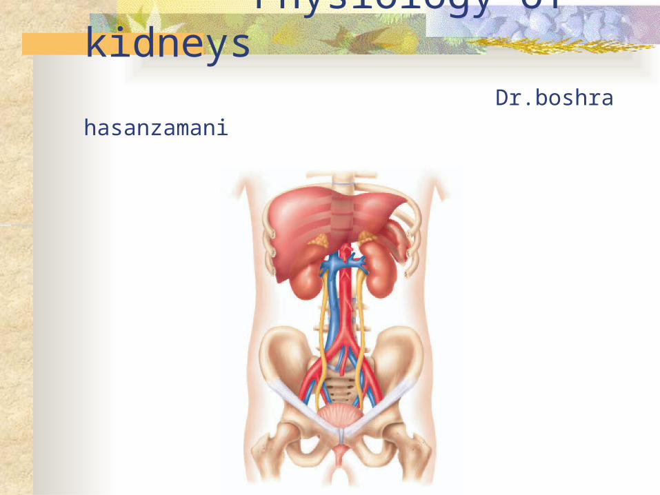
Physiology of kidneys Dr.boshra hasanzamani
CONTENTS
Kidney functions Structure of the kidney Glomerular filtration Mechanism of Transport Segmental Nephron Functions Hormonal Regulation of Sodium and Water Balance
Major Functions of the Kidneys1. Regulation of: body fluid osmolality and volume electrolyte balance acid-base balance blood pressure
2. Excretion of metabolic products foreign substances (pesticides, chemicals etc.) excess substance (water, etc)
3. Secretion of erythropoitin 1,25-dihydroxy vitamin D3 (vitamin D activation) renin prostaglandin
1. Nephron and Collecting Duct
Nephron: The functional unit of the kidney
Each kidney is made up of about 1 million nephrons
Each nephrons has two major components:
1) A glomerulus
2) A long tube
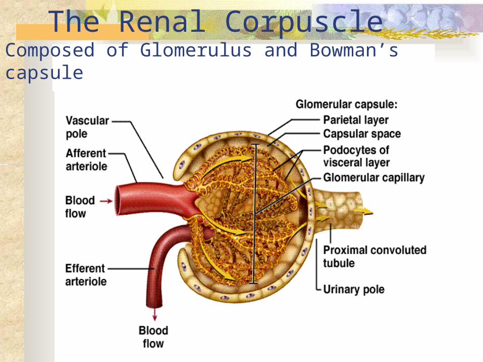
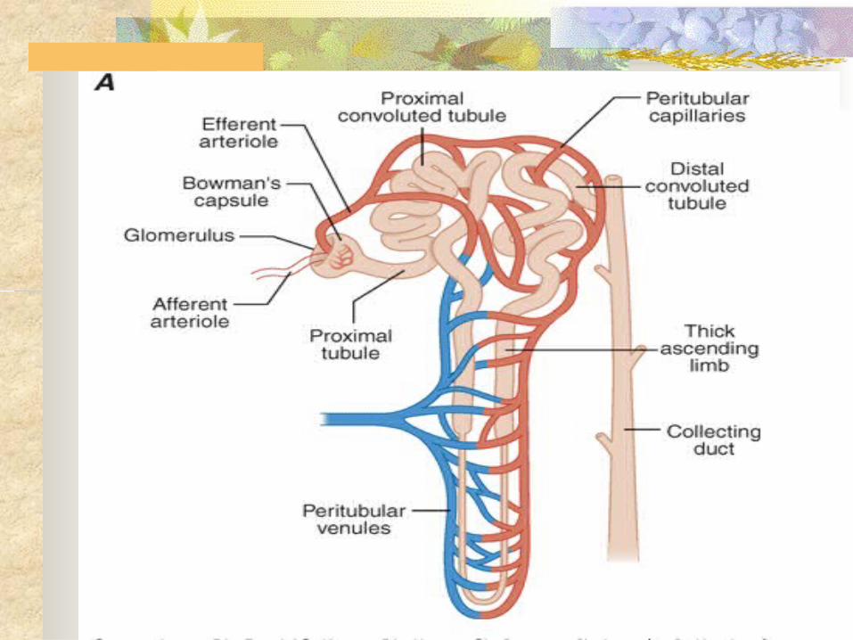
The Renal CorpuscleComposed of Glomerulus and Bowman’s capsule
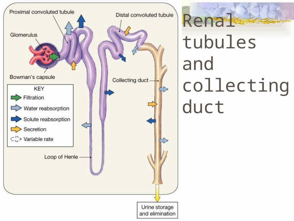
Renal tubules and collecting duct
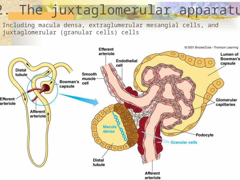
2. The juxtaglomerular apparatusIncluding macula densa, extraglumerular mesangial cells, and juxtaglomerular (granular cells) cells
Determinants and Regulation of Glomerular Filtration
Renal blood flow normally drains approximately 20% of the cardiac output, or 1000 mL/min
The hydrostatic pressure gradient across the glomerular capillary wall is the primary driving force for glomerular filtration
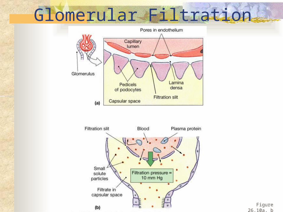
Glomerular Filtration
Figure 26.10a, b
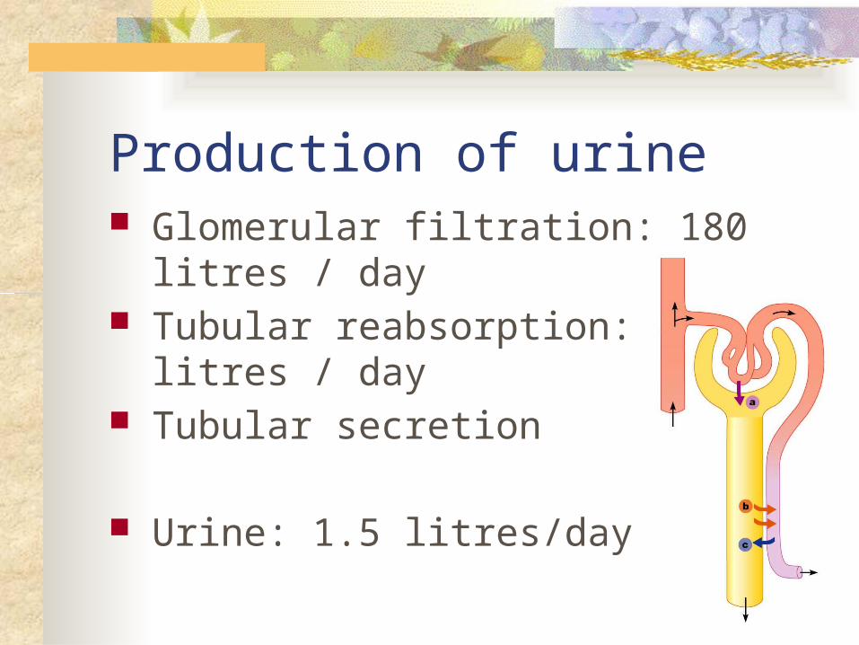
Production of urine Glomerular filtration: 180 litres / day Tubular reabsorption: 178.5 litres / day Tubular secretion
Urine: 1.5 litres/day
Marieb, Human Anatomy & Physiology, 7th edition
three major factors that modulate either afferent or efferent arteriolar tone:
autonomous vasoreactive (myogenic) reflex in the afferent arteriole
tubuloglomerular feedback angiotensin II–mediated vasoconstriction
of the efferent arteriole
autonomous vasoreactive (myogenic) reflex
first line of defense against fluctuations in renal blood flow
This phenomenon helps protect the glomerular capillary from sudden changes in systolic pressure
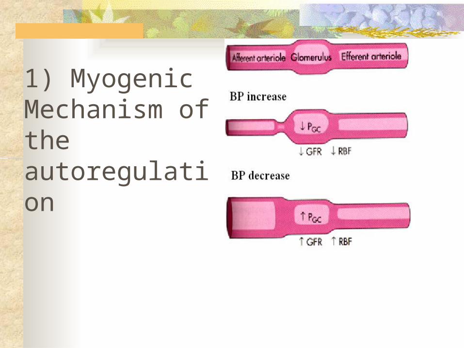
1) Myogenic Mechanism of the autoregulation
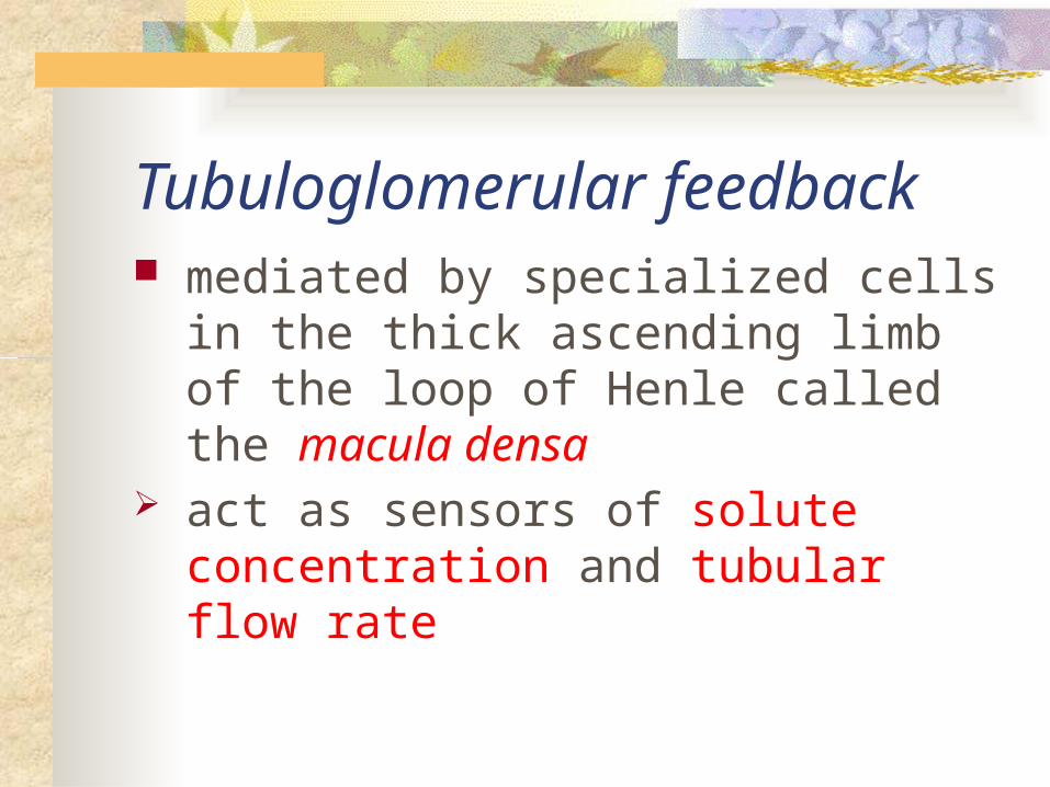
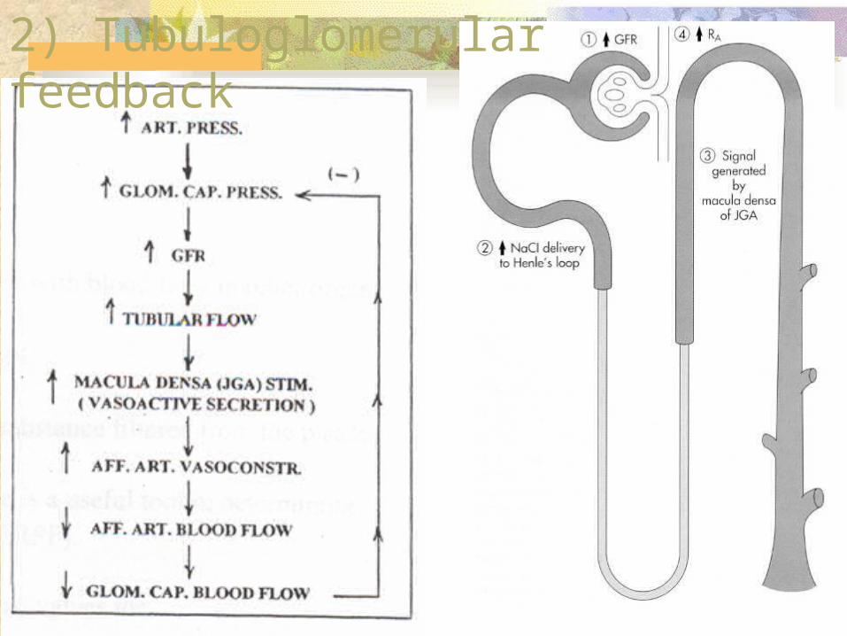
Tubuloglomerular feedback mediated by specialized cells in the thick
ascending limb of the loop of Henle called the macula densa
act as sensors of solute concentration and tubular flow rate
2934
2) Tubuloglomerular feedback
angiotensin II–mediated vasoconstriction During states of reduced renal blood flow,
renin is released from granular cells within the wall of the afferent arteriole near the macula densa in a region called the juxtaglomerular apparatus
Mechanism of Transport
1, Primary Active Transport
2, Secondary Active Transport
3, Pinocytosis
4, Passive Transport
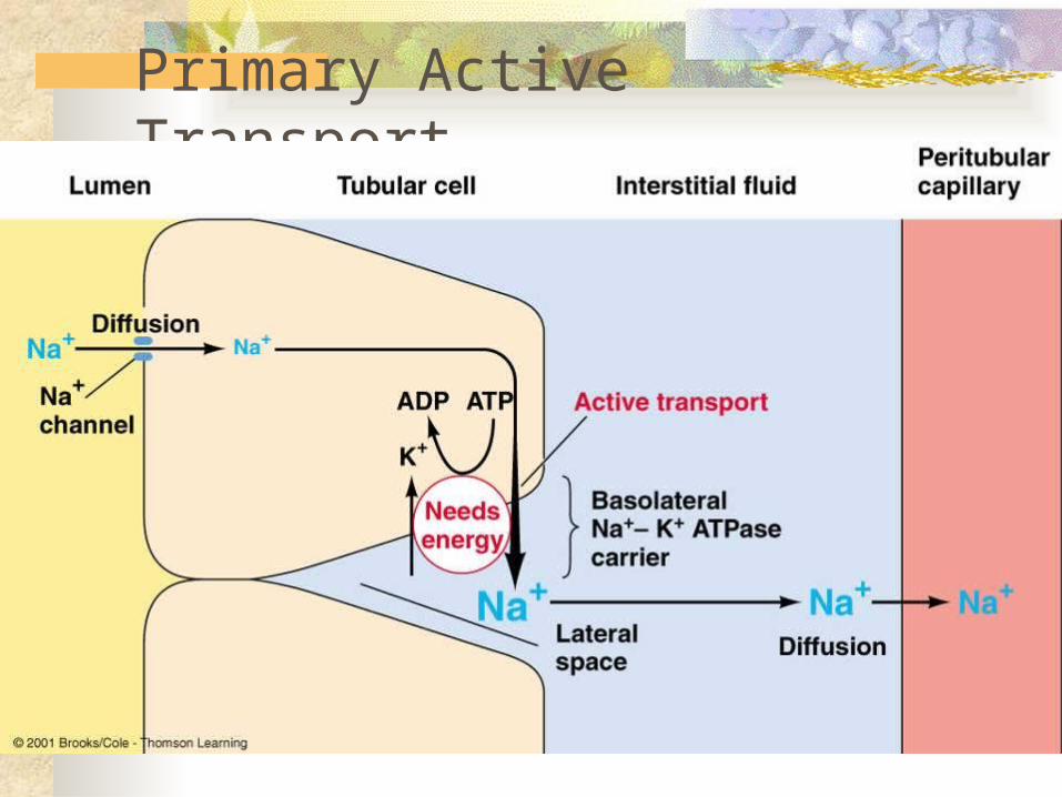
Primary Active Transport
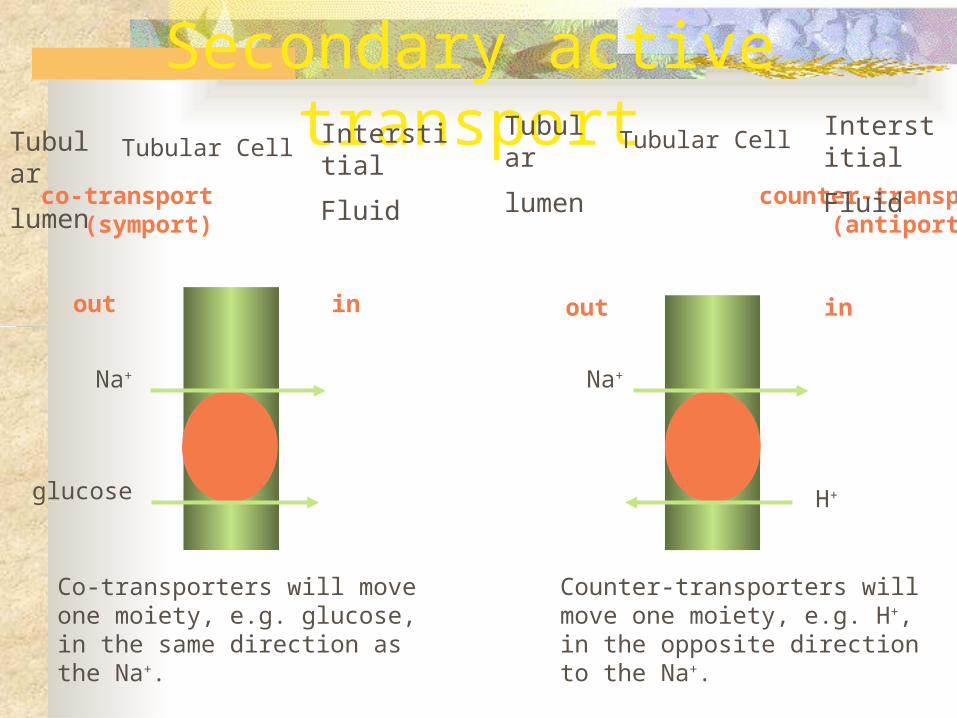
Secondary active transport
Na+
glucose
Na+
H+
out in out in
co-transport counter-transport (symport) (antiport)
Co-transporters will move one moiety, e.g. glucose, in the same direction as the Na+.
Counter-transporters will move one moiety, e.g. H+, in the opposite direction to the Na+.
Tubular
lumen
Tubular CellInterstitial
Fluid
Tubular
lumenTubular Cell Interstitial
Fluid
Segmental Nephron Functions
Proximal Tubule reabsorbing ~60% of filtered NaCl and water ~90% of filtered bicarbonate and most critical
nutrients such as glucose and amino acids Bulk fluid reabsorption by the proximal tubule is
driven by high oncotic pressure and low hydrostatic pressure within the peritubular capillaries
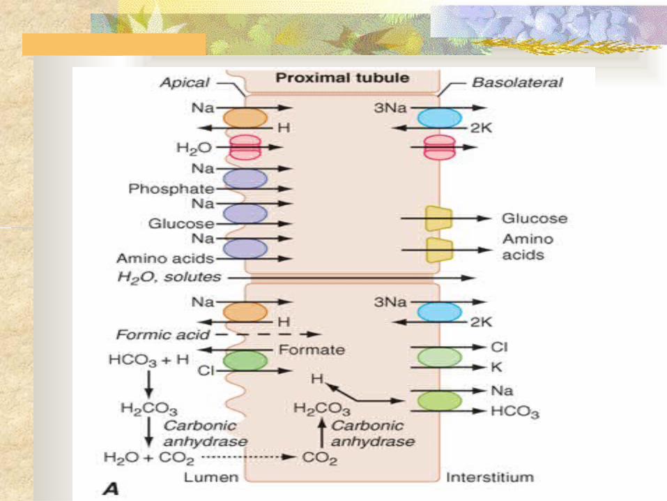
Cellular transport by the proximal tubule
coupled to the Na+ concentration gradient established by the activity of a basolateral Na+/K+-ATPase
such as Na+-glucose and Na+-phosphate cotransporters
water reabsorption by constitutively active water channels (aquaporin-1) present on both apical and basolateral membranes
Proximal tubular cells reclaim bicarbonate by a mechanism dependent on carbonic anhydrases
Reabsorption of glucose is nearly complete by the end of the proximal tubule
- glycosuria when plasma levels exceed 180–200 mg/dL Na+-dependent and Na+-independent transport systems
(reabsorbs amino acids ) cystine, lysine, arginine, and ornithine are transported by
a system comprising two proteins encoded by the SLC3A1 and SLC7A9 genes
Loop of Henle The loop of Henle consists of three major segments: Descending thin limb Ascending thin limb Ascending thick limb
(based on cellular morphology and anatomic location) Approximately 15–25% of filtered NaCl is reabsorbed in
the loop of Henle (mainly by the thick ascending limb) important role in urinary concentration by contributing to
the generation of a hypertonic medullary interstitium in a process called countercurrent multiplication
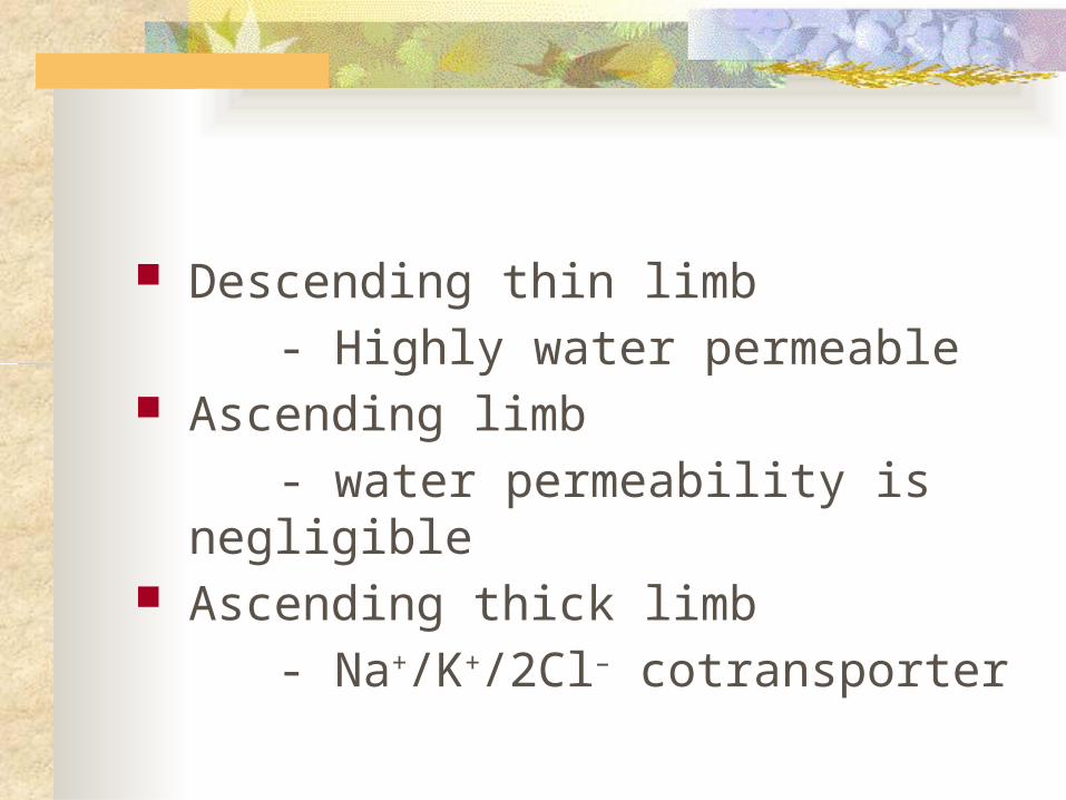
Descending thin limb
- Highly water permeable Ascending limb
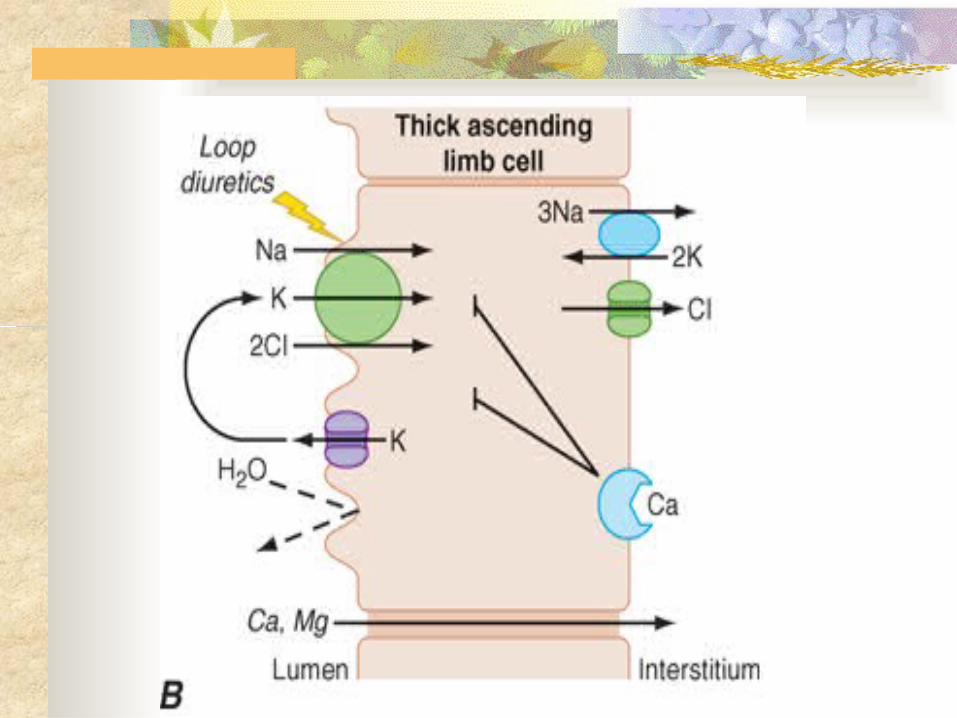
- water permeability is negligible Ascending thick limb
- Na+/K+/2Cl– cotransporter
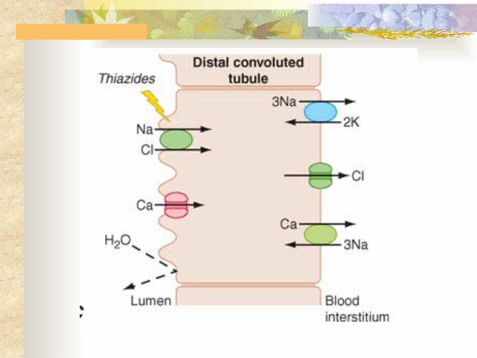
Distal Convoluted Tubule reabsorbs ~5% of the filtered NaCl little water permeability Apical Ca2+-selective channels (TRPV5) and
basolateral Na+/Ca2+ exchange mediate calcium reabsorption
Ca2+ reabsorption is inversely related to Na+ reabsorption and is stimulated by parathyroid hormone
Collecting Duct The two major divisions:
- cortical collecting duct
- inner medullary collecting duct contribute to reabsorbing ~4–5% of filtered
Na+ hormonal regulation of salt and water
balance
Collecting Duct two cell types:
- Principal cells
- type A and B intercalated cells
Principal cells main water, Na+-reabsorbing, and K+-
secreting cells the site of action of aldosterone, K+-sparing
diuretics, and mineralocorticoid receptor antagonists such as spironolactone
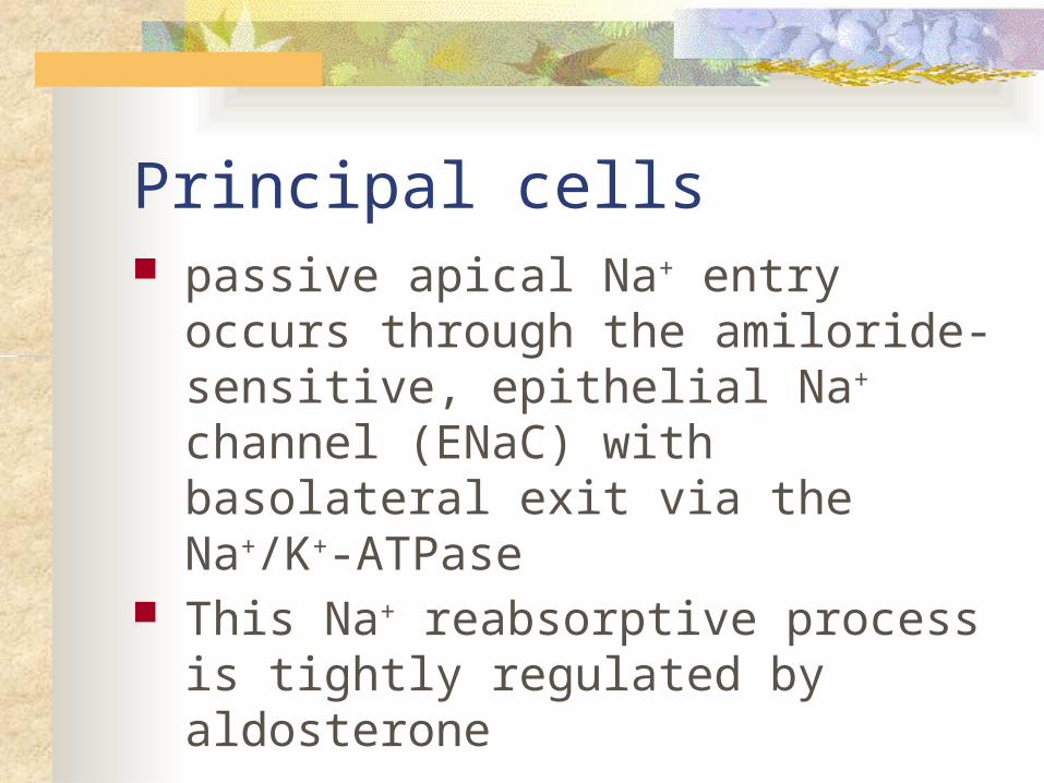
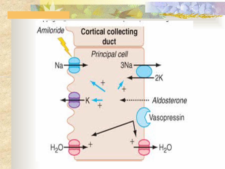
Principal cells passive apical Na+ entry occurs through the
amiloride-sensitive, epithelial Na+ channel (ENaC) with basolateral exit via the Na+/K+-ATPase
This Na+ reabsorptive process is tightly regulated by aldosterone
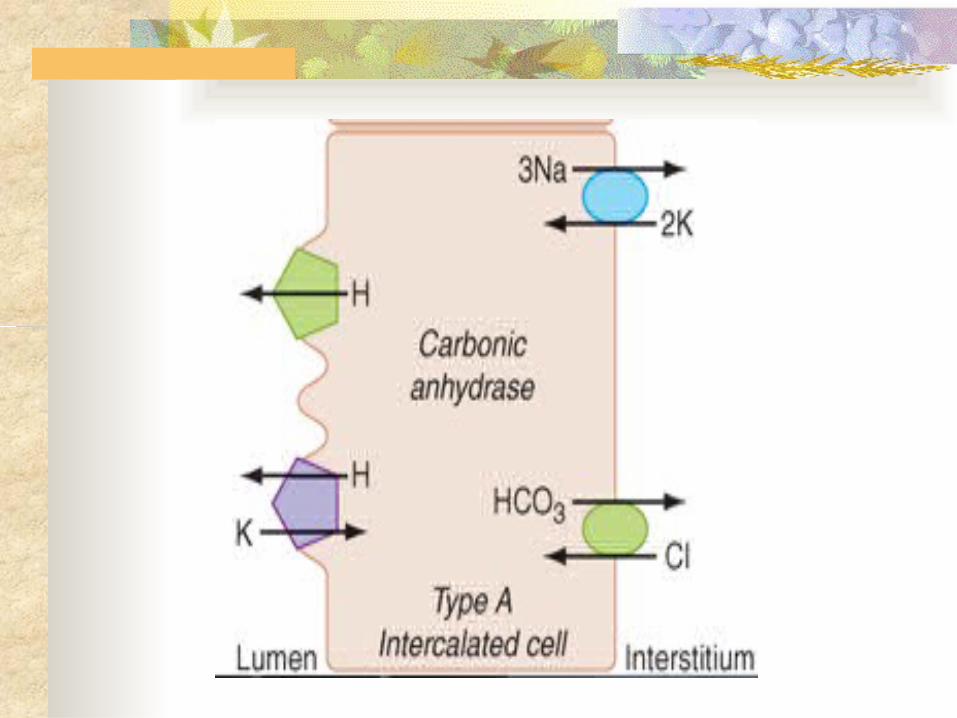
type A and B intercalated cells Type A intercalated cells mediate :
- acid secretion
- bicarbonate reabsorption also under the
influence of aldosterone. Type B intercalated cells mediate:
- bicarbonate secretion
- acid reabsorption
type A and B intercalated cells Under conditions of acidemia, the kidney
preferentially uses type A intercalated cells to secrete the excess H+ and generate more HCO3
–
In states of bicarbonate excess with alkalemia where the type B intercalated cells predominate
An extracellular protein called hensin mediates this adaptation.
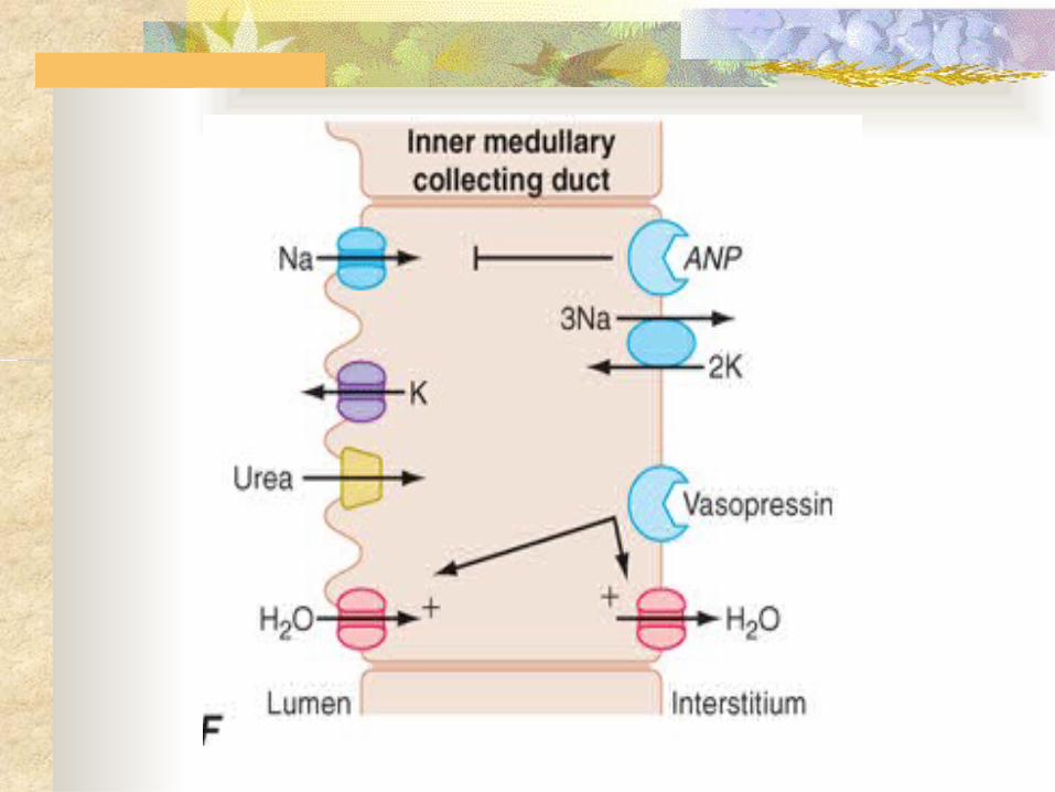
Inner medullary collecting duct many similarities with principal cells of the cortical
collecting duct They have apical Na+ and K+ channels that mediate Na+
reabsorption and K+ secretion, respectively have vasopressin-regulated water channels (aquaporin-2
on the apical membrane, aquaporin-3 and -4 on the basolateral membrane
In the absence of vasopressin, inner medullary collecting duct cells are water impermeable, and urine remains dilute)
Inner medullary collecting duct
Sodium reabsorption by inner medullary collecting duct cells is also inhibited by the natriuretic peptides called atrial natriuretic peptide or renal natriuretic peptide (urodilatin)
Inner medullary collecting duct transports urea out of the lumen, returning
urea to the interstitium, where it contributes to the hypertonicity of the medullary interstitium
Urea is recycled by diffusing from the interstitium into the descending and ascending limbs of the loop of Henle
HORMONAL REGULATION OF SODIUM AND WATER BALANCE
Water Balance Normal tonicity (~280 mosmol/L) is
rigorously defended by :
- osmoregulatory mechanisms that control
water balance to protect tissues from
inadvertent dehydration (cell shrinkage) or
water intoxication (cell swelling)
Any reduction in total body water, which raises the Na+ concentration, triggers :
- a brisk sense of thirst
- conservation of water by decreasing renal
water excretion mediated by release of
vasopressin from the posterior pituitary
The kidneys play a vital role in maintaining water balance through the regulation of renal water excretion
aquaporin 1 is constitutively active in all water-permeable segments of the proximal and distal tubules
vasopressin-regulated aquaporins 2, 3, and 4 in the inner medullary collecting duct promote rapid water permeability
Sodium Balance Under normal conditions, volume is
regulated by :
- sodium balance
- balance between daily Na+ intake and excretion
If Na+ intake exceeds Na+ excretion (positive Na+ balance)
- an increase in blood volume will trigger a
proportional increase in urinary Na+
excretion when Na+ intake is less than urinary excretion (negative
Na+ balance):
- blood volume will decrease and trigger enhanced renal Na+ reabsorption, leading to decreased urinary Na+ excretion
renin-angiotensin-aldosterone system
Renin is synthesized and secreted by granular cells in the wall of the afferent arteriole
Renin and ACE activity eventually produce angiotensin II
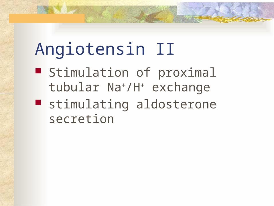
Angiotensin II Stimulation of proximal tubular Na+/H+
exchange stimulating aldosterone secretion
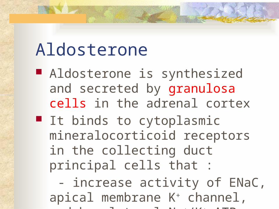
Aldosterone Aldosterone is synthesized and secreted by
granulosa cells in the adrenal cortex It binds to cytoplasmic mineralocorticoid
receptors in the collecting duct principal cells that :
- increase activity of ENaC, apical membrane K+ channel, and basolateral Na+/K+-ATPase
Quizzes بنویسید را سیستم این نام : بنویسید شماره ترتیب به را قسمت هر نام1-
2-
3-
4-
5 -
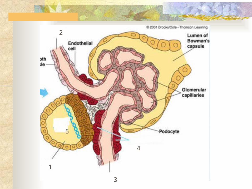
1
2
3
4
5