Embed Size (px)
Citation preview
Proc. Natl. Acad. Sci. USAVol. 84, pp. 3081-3085, May 1987Chemistry
Phytotoxins from the pathogenic fungi Drechslera maydis andDrechslera sorghicola
(corn blight/fungal metabolites/plant disease/sesterterpenoids/ophiobolins)
F. SUGAWARA*t, G. STROBEL*, R. N. STRANGE*t, J. N. SIEDOW§, G. D. VAN DUYNE¶, AND J. CLARDYVI*Department of Plant Pathology, Montana State University, Bozeman, MT 59717; §Department of Botany, Duke University, Durham, NC 27706;and "Department of Chemistry-Baker Laboratory, Cornell University, Ithaca, NY 14853-1301
Communicated by Jerrold Meinwald, October 20, 1986 (receivedfor review September 9, 1986)
ABSTRACT Drechslera maydis, the causal agent of South-ern corn leaf blight, and Drechslera sorghicola, the causal agentof leaf spot on Johnson grass, produce a series of phytotoxicsesterterpenoids. These sesterterpenoids belong to the ophio-bolin family. One of them, ophiobolin I, was characterized byx-ray diffraction and served as a crucial reference compoundfor characterizing four other ophiobolins. AU ofthe ophiobolinsstudied produce characteristic lesions on host plants at con-centrations of 1 mM to 1 ,uM. The ophiobolin characterized as6-epiophiobolin A is selectively toxic to corn bearing Texas-male-sterile (Tms) cytoplasm when assayed in a dark CO2fixation assay. It is plausible that these ophiobolins had a rolein the 1970 corn-blight epidemic in North America.
In the summer of 1970, the corn crops of the United Statesand Canada were devastated by a corn-blight epidemic (1).This epidemic caused the greatest crop loss in the shortesttime span of any plant disease ever reported. Midwest croplosses were 15% ofthe expected harvest, and in the Southeastlosses were close to 50%. Corn plants with Texas-male-sterile(Tms) cytoplasm, which was most of the corn planted, wereespecially vulnerable. The causative agent of the epidemicwas quickly identified as the fungus Drechslera maydis (alsocalled Helminthosporium maydis or, in the perfect stage,Cochliobolus heterostrophus).Fungal pathogens of plants often produce disease symp-
toms by elaborating one or more toxic compounds-phytotoxins. These phytotoxins can be specific for onespecies of plant or even for one strain of a given species andare called host-specific or host-selective phytotoxins. Thephytotoxins produced by D. maydis have been investigated,but studies of their chemical identity have been contradicto-ry. Some workers have reported partially characterizedterpenes as selective phytotoxins for corn bearing Tmscytoplasm (2). Other workers suggested that linear polyke-topolyalcohols were involved in the disease (3). A review hassummarized these and other studies (4).We became interested in fungal pathogens of weeds as a
way of learning about the host selectivity of phytotoxins anddeveloping models for additional herbicides (5). We electedto study Drechslera sorghicola (Lefebre and Sherwin) =Helminthosporium sorghicola because it caused leaf spots onJohnson grass (Sorghum halapense L.), a serious weed in alltropical and semitropical areas of the world. While exploringthe chemistry and biology of several phytotoxic sesterter-penoids from this fungus, we learned that the perfect (sexual)stages of D. maydis (Nisik) = H. maydis (Nisik) and D.sorghicola are genetically compatible (R. R. Nelson, person-al communication). This led us to examine the phytotoxinsproduced by each of these fungi, along with their biological
activity on corn bearing Tms cytoplasm. Our findings on aseries of phytotoxins are given below.
MATERIALS AND METHODS
D. maydis races T (isolate C4) and 0 (isolate Aus20239A3)were obtained from 0. Yoder (Cornell University). D.sorghicola was purchased from the American Type CultureCollection (ATCC no. 28736). These fungi, maintained onpotato dextrose/agar plates containing 18% (vol/vol) V-8juice by volume, were used for toxin production by growingeach of them in a New Brunswick incubator in 1 liter ofmodified M-1-D medium (2, 6), shaking at 200 rpm, illumi-nating, and maintaining a temperature of260C (7). Plants usedin this study were grown under controlled environmentalconditions of 12 hr of darkness at 220C and 12 hr of light at270C with a light intensity of 170 uE-m-2. Corn (B37, Tms andN cytoplasms) was obtained from D. Mathre (Montana StateUniversity), and corn (W46A, Tms and N cytoplasms) wasprovided by V. Gracen (Cornell University). Sorghum spe-cies were available from the Montana State Universitycollection.Leaf Assay. A simple leaf-puncture assay was used as a
rapid guide in isolating the suspected phytotoxins (5). Thetest material was applied in 2% (vol/vol) ethanol and a 1- to5-Ad droplet was placed on a leaf blade that was subsequentlyincubated in a sealed Petri dish containing moistened filterpaper. The symptoms that developed within 24-48 hr de-pended upon the plants and varied from little or no reaction,to brownish, zonate spots, to spreading necrotic lesions. Theextent of leaf tissue affected was estimated and used as acomparison among plant genotypes and test compounds. Inno cases were symptoms observed with 2% (vol/vol) ethanolcontrol solutions.Root Cap Cell Assay. Root cap cells ofcorn (B37 with N and
Tms cytoplasms) were prepared according to the method ofHawes (8). Toxin preparations were dissolved in 2% (vol/vol)ethanol (50 Al) and placed onto 50 Al of a cell suspension(A620, 0.015). Following incubation at 270C for 4 hr, 50 ,Al of24 ,M fluorescein diacetate was added. Microtest plateswere scored according to the method of Strange et al. (9).Mitochondria Assays. Washed mitochondria were isolated
from the mesocotyls of4-day-old etiolated B37 corn seedlingswith Tms or N cytoplasm following the procedure of Siedowand Bickett (10). The electron transfer activity in the isolatedmitochondria was measured on a Clark-type oxygen elec-trode and found to have respiratory control values during
Abbreviation: Tms, Texas-male-sterile.tPresent address: RIKEN Institute of Physical and Chemical Re-search, Laboratory of Herbicide and Plant Growth Regulators,Wako-shi, Saitama 351, Japan.tPresent address: Department of Botany and Microbiology, Univer-sity College London, Gower Street, London WCIE 6BT, England.1To whom reprint requests should be addressed.
The publication costs of this article were defrayed in part by page chargepayment. This article must therefore be hereby marked "advertisement"in accordance with 18 U.S.C. §1734 solely to indicate this fact.
3081
Dow
nloa
ded
by g
uest
on
Mar
ch 2
5, 2
020
Dow
nloa
ded
by g
uest
on
Mar
ch 2
5, 2
020
Dow
nloa
ded
by g
uest
on
Mar
ch 2
5, 2
020
Proc. Natl. Acad. Sci. USA 84 (1987)
succinate oxidation of 1.7 or 1.9 for mitochondria from Tmsor N cytoplasm, respectively.
Mitochondrial swelling and electron transfer assays wereas described by Klein and Koeppe (11). The reaction bufferfor the swelling assays contained 300 mM sucrose, bovineserum albumin at 1 mg/ml, 20mM Tris HCl, and 0.2-0.25 mgof mitochondrial protein in a total volume 2.0 mil at pH 7.4.Additions were made as follows: 1 mM NADH, 3 mMmethomyl, and 500 tLM KCN. The spectrophotometric as-says were at 520 nm and 250C.COI Fixation Assays. Corn leaves of B37 and W64A with
Tms orN cytoplasm were used in a C02 fixation assay carriedout by constructing a dose-response curve following themethod of Bhullar et al. (12). A 4-hr exposure at 260C in lightat 170 uE m-2 sol was used. This test was repeated at leastthree times and yielded standard deviations of 14-60% of themean, which is in close agreement with the variance usuallyreported for this test.
RESULTSPhytotoxic Sesterterpenes. A series of phytotoxic sesterter-
penoids was consistently isolated from 2-week-old cultures ofD. sorghicola and D. maydis. The toxins were isolated byfiltering the culture broth through four layers of cheesecloth(7). The filtrate was concentrated to one-third its originalvolume by rotary evaporation at 40'C and extracted threetimes with 200 ml of ethyl acetate. The remaining filtrate wasalso extracted with n-butanol, but this extract was devoid ofbiological activity. The combined ethyl acetate fractionswere washed with 200 ml of water and evaporated to drynessat 30'C under reduced pressure. All of the detectable bio-logical activity was in this residue. The residue was flash-chromatographed (Merck silica gel 60, 230-400 mesh, 30 g)using CHCl3/MeOH, 20:1 (vol/vol). Fractions of 5 ml werecollected, and fractions 12-16 showed activity in the leaf-puncture assay on all test plants. These fractions were furtherpurified by preparative TLC (precoated TLC plates pur-chased from Merck, silica gel 60, F-254, 0.5 mm) usingCHCl3/MeOH, 14:1 (vol/vol), and toluene/ethyl acetate, 1:2(vol/vol). Compounds were detected on TLC plates withanisaldehyde/sulfuric acid reagent (0.5% anisaldehyde and1% sulfuric acid in 50 ml of acetic acid). The biologicallyactive bands were chromatographed on a Pharmacia LH-20(20 g) column eluted with MeOH to remove olefinic, inactivecompounds. The purity of the compounds was determined byanalytical HPLC using a Merck reverse phase RP-18 column(4.6 x 250 mm) eluted with CH3CN/H2O, 65:35 (vol/vol) ata flow rate of 1.0 ml/min and monitored with a 254-nm UVdetector.The active compounds isolated by this procedure were
identified as ophiobolins by spectroscopic procedures. Theirabbreviated names, retention times, and Rf values are givenin Table 1.
Table 1. Retention times and Rf values of ophiobolins found inthe culture fluids of D. maydis and D. sorghicola
RT, Rf, Rf,Compound min System A System B
Ophiobolin I (1) 10.9 0.33 0.45Ophiobolin A (2) 8.8 0.52 0.67Ophiobolin C (3) 30.5 0.60 0.8225-Hydroxyophiobolin 1 (4) 4.3 0.08 0.206-Epianhydroophiobolin A (5) 11.8 0.53 0.686-Epiophiobolin A (6) 8.1 0.52 0.48
RT were from an HPLC column. The HPLC system used a 4.6 x250 mm Merck RP-18 column eluted with CH3CN/H20, 65:35
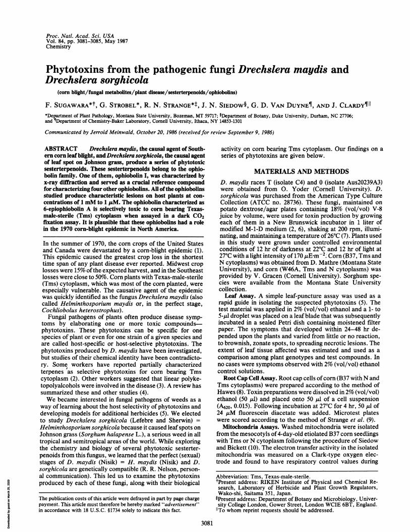
The structural analysis of the series began with ophiobolinI (1). The natural compound (1.2 mg) was converted to thep-bromobenzoate by dissolving it in 1.0 ml of dry pyridineand adding 3.0 mg of p-bromobenzoyl chloride. After astandard workup, the residue was chromatographed onpreparative TLC plates with CHCl3/MeOH, 50:1 (vol/vol)and toluene/ethyl acetate, 9:1 (vol/yol). This gave 2.0 mg ofa crystalline derivative, mp 215-217'C. This derivative wascharacterized by single-crystal x-ray diffraction (5). Crystalsformed in the monoclinic space group P21 with a =
13.759(12), b = 6.445(8), c = 16.655(13) A, and = 92.07(7)0with one molecule of composition C31H3904Br in the asym-metric unit. All unique diffraction maxima with 20 < 1140were collected using graphite monochromated CuKa radia-tion (1.54178 A) and variable speed, 10 )-scans. Only 1094 ofthe 1699 reflections collected in this fashion (64%) werejudged observed [F. > 3of(F0)] after correction for Lorentz,polarization, and background effects. A phasing model was
found easily. Block diagonal least squares refinements withanisotropic heavy atoms and fixed hydrogens have con-verged to a current residual of 0.085 for the observedreflections. A computer generated drawing of the final x-raymodel for the derivative is given in Fig. 1 and a chemicaldrawing of ophiobolin I is given as structure 1. The absoluteconfiguration was set from earlier studies on the ophiobolins,and the standard ophiobolin numbering scheme was used(13).The important point about this structure was that the
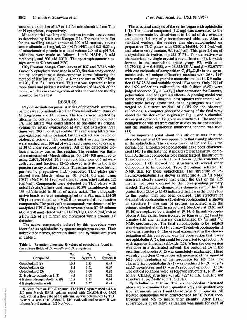
stereochemistry at C6 was opposite to that usually observedin the ophiobolins. The cis-ring fusion at C2 and C6 is thenormal one, although 6-epiophiobolins have been character-ized (14). To illustrate the standard stereochemistry, ophio-bolin A, the first ophiobolin to be characterized, has structure2, and ophiobolin C is structure 3. Securing the structure ofophiobolin I (1) allowed the structures of several otherophiobolins to be deduced. Table 2 contains selected 'HNMR data for these ophiobolins. The structure of 25-hydroxyophiobolin I is shown as structure 4. Its 1H NMRspectrum clearly showed that either the C24 or the C25methyl had been oxidized to the corresponding primaryalcohol. The dramatic change in the chemical shift of the C18proton from 65.14 to 85.43 indicated that it was the methyl cisto this proton that had been oxidized. The structure of6-epianhydroophiobolin A (21-dehydroophiobolin I) is shownas structure 5. The pair of protons associated with theprimary alcohol at C21 in structure 1 (doublets at 63.91 and84.16) are replaced by a singlet at 69.24. 6-Epianhydroophi-obolin A had earlier been isolated by Kim et al. (1$) and byCanales (16) and tentatively characterized by 1H and 13CNMR spectroscopy. The final epiophiobolin to be isolatedwas 6-epiophiobolin A (3-hydroxy-21-dehydroophiobolin I)shown as structure 6. The crucial experiment in the charac-terization of this compound was the observation that it wasnot ophiobolin A (2), but could be cohverted to ophiobolin Awith aqueous dimethyl sulfoxide (15). When the conversionwas done in a deuterated solvent, the proton at C6 in theresulting ophiobolin A (2) was completely exchanged. Therewas also a nuclear Overhauser enhancement of the signal ofH10 upon irradiation of the resonance for H6 (16). Thecharacterized ophiobolin A (2) was produced by D. maydisand D. sorghicola, and D. maydis produced ophiobolin C (3).The optical rotations were as follows: structure 1, [a]22+480(c 3.8, CHCl3); structure 4, [aI2 +220 (c 1.6, CHCl3); andstructure 6, [a] , +460 (c 5.3, CHCl3).
Ophiobolins in Culture. The six ophiobolins discussedabove were examined both quantitatively and qualitativelyfrom D. maydis races T and 0 and from D. sorghicola. Allisolated ophiobolins were characterized by 'H NMR spec-troscopy and MS to insure their identity. After HPLCseparation, a quantitative estimation was made for each of
(vol/vol) at a flow rate of 1.0 ml/min. Rf was determined by TLC.System A was CHC13/MeOH, 14:1 (vol/vol) and system B wastoluene/ethyl acetate, 1:2 (vol/vol).
3082 Chemistry: Sugawara et A
Dow
nloa
ded
by g
uest
on
Mar
ch 2
5, 2
020
Proc. Natl. Acad. Sci. USA 84 (1987) 3083
A
C21
BR
Cl
B 23
cH3C 1
OH 1 18 ~~~~~~~~25
20 lo1
H H0HO1C13 CH
3
20 C ;H
H53
3x
5~~~~~2
H
21 CH
CH0~~~~
X-CH3
Ho3
HOCH3~~~
0 6 24~~
FIG. 1. (A) A computer generated perspective drawing ofthe final x-ray model ofthep-bromobenzoate derivative ofophiobolin I. Hydrogensare omitted for clarity, and the absolute configuration was set from earlier studies on the ophiobolins (B).
the ophiobolins based on the dry weight of the fungus. Theseresults are given in Table 3. The maximum production ofthese compounds occurred after 14 days of incubation, andone compound, 6-epiophiobolin A (6), was not detected untilday 10. If cultures were incubated over 3 weeks, the extractswere no longer completely separable under our HPLCconditions.As can be seen in Table 3, there was a noticeable difference
in the quality and quantity of ophiobolins in D. sorghlcola incomparison to D. maydis races T and 0. For example,25-hydroxyophiobolin I (4) was a major metabolite in D.sorghicola, but occurred only in minor amounts in D. maydisrace 0. Ophiobolin C occurred in roughly equal amounts in
both races of D. maydis, but was not detected in D.sorghicola. Finally, race T of D. maydis made five timesmore 25-hydroxyophiobolin I (4) than race 0.
Plant Assays. Since a number of phytotoxic effects hadbeen attributed to D. maydis extracts, it was crucial to testthe isolated compounds in a variety of assays. All of theophiobolins discussed in this report were tested by the leafpuncture method on both Tms and N corn (B37 and W64Agenetic backgrounds), Johnson grass, and sorghum. All wereactive, and a summary of the activity is given in Table 4. At1 mM, they were all capable of causing brownish lesions.However, the most bioactive compound was 6-epiophiobolinA (6), which produced large lesions with reddish runners on
Chemistry: Sugawara et al.
Dow
nloa
ded
by g
uest
on
Mar
ch 2
5, 2
020
Proc. Natl. Acad. Sci. USA 84 (1987)
Table 2. Summary of proton NMR data for the ophiobolins isolated from Drechslera spp
Ophiobolin I (1) 25-Hydroxyophiobolin I (4) 6-Epianhydroophiobolin A (5) 6-Epiophiobolin A (6)
H2 2.53 m 2.54 m 2.30 m 2.11 mH4 5.96 d 1.1 5.96 s 6.04 s 2.41 d 16.7H4 3.06 d 16.7H6 3.67 d 2.6 3.62 d 2.6 3.43 d 4.1 3.36 d 10.5H8 5.78 d 3.8 5.77 d 4.0 6.82 dd 2.3, 6.6 6.88 d 2.0, 6.8H17 4.58 dd 7.2, 15.8 4.63 dd 7.2, 15.4 4.60 dd 7.1, 15.7 4.62 dd 8.5, 15.6H18 5.14 ddd 1.0, 1.0, 7.2 5.43 dd 0.8, 7.2 5.14 ddd 1.0, 1.0, 7.2 5.14 ddd 1.0, 1.0, 7.2H20 2.08 s 2.08 s 2.05 s 1.42 sH21 3.91 d 11.0 3.17 d 11.9 9.32 s 9.22 sH21 4.16 d 11.0 3.91 d 11.9H22 1.00 s 0.99 s 0.88 s 0.85 sH23 1.02 d 6.9 1.03 d 6.9 1.05 d 7.0 1.04 d 6.8H24 1.70 d 1.0 1.69 d 0.8 1.71 d 1.0 1.71 d 1.0H25 1.66 d 1.0 4.00 s 1.66 d 1.0 1.67 d 1.0
The proton assignments are given on the left. Each entry is the chemical shift (8), multiplicity, and coupling constant(s) (Hz).
Tms corn. These appeared to be very similar to lesions foundon diseased corn in the field. On N corn, 6-epiophiobolin Aproduced only sunken lesions similar in size to those on Tmscorn but lacking colored runners. Furthermore, Tms cornwith a W64A genetic background appeared to be more
sensitive than Tms with a B37 background, which agrees withthe field observations of Lim (17). In contrast to all of theother compounds, 6-epiophiobolin A (6) produced noticeablelesions on both Tms and N corn at micromolar concentra-tions.
All ophiobolins were tested on isolated root cap cells ofboth B37 N and Tms corn. All produced 50% cell deathbetween 0.18 and 0.60 mM; however, in this assay, 6-epianhydroophiobolin A (5) was significantly more effective(0.18 mM) than any of the others (P < 0.05). There were nodifferences in response between corn with Tms and Ncytoplasm. These experiments were repeated three times,and the data were analyzed according to Student's t test. Theleast effective compounds in this assay were ophiobolin I (1)and 25-hydroxyophiobolin 1 (4) that required at least 0.60mMto cause 50% cell death. All of the other compounds wereeffective in the 0.30 to 0.38 mM range.
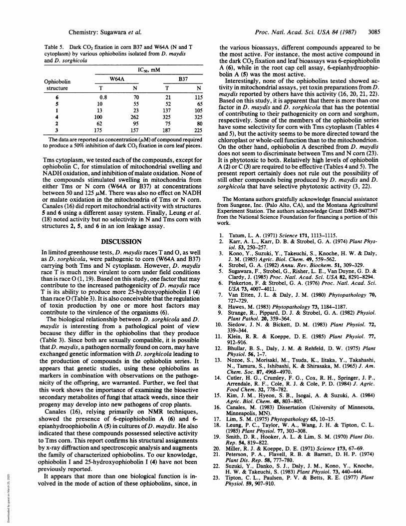
Since ophiobolin A as well as crude toxin preparationsfrom D. maydis were known to inhibit dark CO2 fixation, wetested each of the purified ophiobolins in this assay. Theseresults are given in Table 5. As in the leaf puncture assay,
6-epiophiobolin A (6) was the most effective compound indisrupting dark CO2 fixation in Tms corn. The effect was
much more pronounced in corn with a W64A nuclear back-ground. Comparable tests with the other ophiobolins, withthe exception of ophiobolin C (3), generally showed a
tendency toward greater sensitivity of corn with Tms cyto-
Table 3. Quantitative and qualitative determinations of theophiobolins present in 2-week-old culture fluids ofD. maydis and D. sorghicola
Ophiobolin concentration,mmol/g (dry weight)
D. maydis
Ophiobolin Race T Race 0 D. sorghicola
Ophiobolin I (1) 0.3 0.7 0.525-Hydroxyophiobolin 1 (4) 0.3 0.06 3.16-Epianhydroophiobolin A (5) Trace Trace Not detected6-Epiophiobolin A (6) 1.3 1.5 1.7Ophiobolin A (2) 8.0 8.4 5.2Ophiobolin C (3) 1.4 1.7 Not detected
Concentrations are in mmol/g for the dry weight of the fungus. Allexperiments were carried out at least twice with a variation notexceeding 10% of the average value recorded.
plasm than N cytoplasm (Table 5). Crude toxin preparations,the ethyl acetate fraction, at concentrations up to 100 Ag/mldid not cause any greater reduction in CO2 fixation than acombination of purified ophiobolins in their naturally occur-ring D. maydis ratios at the same concentration.
Since crude culture filtrates ofD. maydis have traditionallybeen noted to affect mitochondrial activities in corn bearing
Table 4. Effects of the ophiobolins from D. maydis and D.sorghicola in the leaf puncture wound test on corn (Zeamays), Johnson grass (Sorghum halepense), andsorghum (Sorghum bicolor)
Lesion size*
Corn(W64A and
Ophiobolin Concentration, B37) Johnsonstructure M T corn N corn grass Sorghum
1 10-3 + + + +10-4 + + + +
-0- - + +10-6 _ _ _ _
4 10-3 + + + +10-4 - - - +
-5 - - - -
10-6 _ - _
5 10-3 + + + +10-4 + + _ _
10-6 _ - _
6 10-3 +++t +++t +++§ ++¶10-4 + ++ +++ ++
10-5 + + ++ +10-6 + + - -
2 10-3 ++ ++ ++ ++10-4 + + ++ ++i-S _ _ _ +
10-6 _ _
3 10-3 + + + +10-4 + + _ +
10-6 _ - -_
*Lesion size: +++, >5 x 3 mm; ++, 2 x 2 to 5 x 3 mm; +, 0.5 x0.5 to 2 x 2 mm; -, <0.5 x 0.5 mm.tReddish-brown lesion with several 5-mm reddish runners. Therunners were more apparent in W64A-Tms corn than in B37-Tmscorn.*Sunken lesion.§Necrotic lesion with brown runner.$Dark, necrotic lesion.
3084 Chemistry: Sugawara et al.
Dow
nloa
ded
by g
uest
on
Mar
ch 2
5, 2
020
Proc. Natl. Acad. Sci. USA 84 (1987) 3085
Table 5. Dark CO2 fixation in corn B37 and W64A (N and Tcytoplasm) by various ophiobolins isolated from D. maydisand D. sorghicola
IC50, mM
Ophiobolin W64A B37structure T N T N
6 0.8 70 21 1155 10 55 52 651 13 23 137 1054 100 262 325 3252 62 95 75 803 175 157 187 225
The data are reported as concentration (AM) ofcompound requiredto produce a 50% inhibition of dark CO2 fixation in corn leaf pieces.
Tms cytoplasm, we tested each of the compounds, except forophiobolin C, for stimulation of mitochondrial swelling andNADH oxidation, and inhibition ofmalate oxidation. None ofthe compounds stimulated swelling in mitochondria fromeither Tms or N corn (W64A or B37) at concentrationsbetween 50 and 125 AM. There was also no effect on NADHor malate oxidation in the mitochondria of Tms or N corn.Canales (16) did report mitochondrial activity with structures5 and 6 using a different assay system. Finally, Leung et al.(18) noted activity but no selectivity in N and Tms corn withstructures 2, 5, and 6 in an ion leakage assay.
DISCUSSIONIn limited greenhouse tests, D. maydis races T and 0, as wellas D. sorghicola, were pathogenic to corn (W64A and B37)carrying both Tms and N cytoplasm. However, D. maydisrace T is much more virulent to corn under field conditionsthan is race 0 (1, 19). Based on this study, one factor that maycontribute to the increased pathogenicity of D. maydis raceT is its ability to produce more 25-hydroxyophiobolin I (4)than race 0 (Table 3). It is also conceivable that the regulationof toxin production by one or more host factors maycontribute to the virulence of the organisms (6).The biological relationship between D. sorghicola and D.
maydis is interesting from a pathological point of viewbecause they differ in the ophiobolins that they produce(Table 3). Since both are sexually compatible, it is possiblethat D. maydis, a pathogen normally found on corn, may haveexchanged genetic information with D. sorghicola leading tothe production of compounds in the ophiobolin series. Itappears that genetic studies, using these ophiobolins asmarkers in combination with observations on the pathoge-nicity of the offspring, are warranted. Further, we feel thatthis work shows the importance of examining the bioactivesecondary metabolites of fungi that attack weeds, since theirprogeny may develop into new pathogens of crop plants.
Canales (16), relying primarily on NMR techniques,showed the presence of 6-epiophiobolin A (6) and 6-epianhydroophiobolin A (5) in cultures ofD. maydis. He alsoindicated that these compounds possessed selective activityto Tms corn. This report confirms his structural assignmentsby x-ray diffraction and spectroscopic analysis and augmentsthe family of characterized ophiobolins. To our knowledge,ophiobolin I and 25-hydroxyophiobolin I (4) have not beenpreviously reported.
It appears that more than one biological function is in-volved in the mode of action of these ophiobolins, since, in
the various bioassays, different compounds appeared to bethe most active. For instance, the most active compound inthe dark CO2 fixation and leaf bioassays was 6-epiophiobolinA (6), while in the root cap cell assay, 6-epianhydroophio-bolin A (5) was the most active.
Interestingly, none of the ophiobolins tested showed ac-tivity in mitochondrial assays, yet toxin preparations from D.maydis reported by others have this activity (16, 20, 21, 22).Based on this study, it is apparent that there is more than onefactor in D. maydis and D. sorghicola that has the potentialof contributing to their pathogenicity on corn and sorghum,respectively. Some of the members of the ophiobolin serieshave some selectivity for corn with Tms cytoplasm (Tables 4and 5), but the activity seems to be more directed toward thechloroplast or whole-cell function than to the mitochondrion.On the other hand, ophiobolin A described from D. maydisdoes not seem to discriminate between Tms and N corn (23).It is phytotoxic to both. Relatively high levels of ophiobolinA (2) or C (3) are required to be effective (Tables 4 and 5). Thepresent report certainly does not rule out the possibility ofstill other compounds being produced by D. maydis and D.sorghicola that have selective phytotoxic activity (3, 22).
The Montana authors gratefully acknowledge financial assistancefrom Sungene, Inc. (Palo Alto, CA), and the Montana AgriculturalExperiment Station. The authors acknowledge Grant DMB-8607347from the National Science Foundation for financing a portion of thiswork.
1. Tatum, L. A. (1971) Science 171, 1113-1115.2. Karr, A. L., Karr, D. B. & Strobel, G. A. (1974) Plant Phys-
iol. 53, 250-257.3. Kono, Y., Suzuki, Y., Takeuchi, S., Knoche, H. W. & Daly,
J. M. (1985) Agric. Biol. Chem. 49, 559-562.4. Strobel, G. A. (1982) Annu. Rev. Biochem. 51, 309-329.5. Sugawara, F., Strobel, G., Risher, L. E., Van Duyne, G. D. &
Clardy, J. (1985) Proc. Natl. Acad. Sci. USA 82, 8291-8294.6. Pinkerton, F. & Strobel, G. A. (1976) Proc. Natl. Acad. Sci.
USA 73, 4007-4011.7. Van Etten, J. L. & Daly, J. M. (1980) Phytopathology 70,
727-729.8. Hawes, M. (1983) Phytopathology 73, 1184-1187.9. Strange, R., Pippard, D. J. & Strobel, G. A. (1982) Physiol.
Plant Pathol. 20, 359-364.10. Siedow, J. N. & Bickett, D. M. (1983) Plant Physiol. 72,
339-344.11. Klein, R. R. & Koeppe, D. E. (1985) Plant Physiol. 77,
912-916.12. Bhullar, B. S., Daly, J. M. & Rehfeld, D. W. (1975) Plant
Physiol. 56, 1-7.13. Nozoe, S., Morisaki, M., Tsuda, K., Iitaka, Y., Takahashi,
N., Tamura, S., Ishibashi, K. & Shirasaka, M. (1965) J. Am.Chem. Soc. 87, 4968-4970.
14. Cutler, H. G., Crumley, F. G., Cox, R. H., Springer, J. P.,Arrendale, R. F., Cole, R. J. & Cole, P. D. (1984) J. Agric.Food Chem. 32, 778-782.
15. Kim, J. M., Hyeon, S. B., Isogai, A. & Suzuki, A. (1984)Agric. Biol. Chem. 48, 803-805.
16. Canales, M. (1983) Dissertation (University of Minnesota,Minneapolis, MN).
17. Lim, S. M. (1975) Phytopathology 65, 10-15.18. Leung, P. C., Taylor, W. A., Wang, J. H. & Tipton, C. L.
(1985) Plant Physiol. 77, 303-308.19. Smith, D. R., Hooker, A. L. & Lim, S. M. (1970) Plant Dis.
Rep. 54, 819-822.20. Miller, R. J. & Koeppe, D. E. (1971) Science 173, 67-69.21. Peterson, P. A., Flavell, R. B. & Barratt, D. H. P. (1974)
Plant Dis. Rep. 58, 777-780.22. Suzuki, Y., Danko, S. J., Daly, J. M., Kono, Y., Knoche,
H. W. & Takeuchi, S. (1983) Plant Physiol. 73, 440-444.23. Tipton, C. L., Paulsen, P. V. & Betts, R. E. (1977) Plant
Physiol. 59, 907-910.
Chemistry: Sugawara et al.
Dow
nloa
ded
by g
uest
on
Mar
ch 2
5, 2
020
Proc. Natl. Acad. Sci. USA 85 (1988)
Correction. In the article "Phytotoxins from the pathogenicfungi Drechslera maydis and Drechslera sorghicola" by F.Sugawara, G. Strobel, R. N. Strange, J. N. Siedow, G. D.Van Duyne, and J. Clardy, which appeared in number 10,May 1987, ofProc. Natl. Acad. Sci. USA (84, 3081-3085), theauthors request that the following corrections be noted. InTable 3 on p. 3084, ophiobolin concentration, mmollg (dryweight), should be ophiobolin concentration, /Lmol/g (dryweight), and in the first line of the legend mmol/g should be,umol/g. In Table 5 on p. 3085, IC50, mM, should be IC50, uM.
Table 3. Quantitative and qualitative determinations of theophiobolins present in 2-week-old culture fluids ofD. maydis and D. sorghicola
Ophiobolin concentration,/.mol/g (dry weight)
D. maydis
Ophiobolin Race T Race 0 D. Sorghicola
Ophiobolin I (1) 0.3 0.7 0.525-Hydroxyophiobolin 1 (4) 0.3 0.06 3.16-Epianhydroophiobolin A (5) Trace Trace Not detected6-Epiophiobolin A (6) 1.3 1.5 1.7Ophiobolin A (2) 8.0 8.4 5.2Ophiobolin C (3) 1.4 1.7 Not detected
Concentrations are in /mol/g for the dry weight of the fungus. Allexperiments were carried out at least twice with a variation notexceeding 10% of the average value recorded.
Table 5. Dark CO2 fixation in corn B37 and W64A (N and Tcytoplasm) by various ophiobolins isolated from D. maydisand D. sorghicola
IC50,o M
Ophiobolin W64A B3structure T N T N
6 0.8 70 21 1155 10 55 52 651 13 23 137 1054 100 262 325 3252 62 95 75 803 175 157 187 225
The data are reported as concentration (jiM) ofcompound requiredto produce a 50% inhibition of dark CO2 fixation in corn leaf pieces.
3276 Chemistry: Correction


![Sulfuric Acid is[1]](https://img.pdfslide.net/doc/110x75/552847e14a7959c93d8b4684/sulfuric-acid-is1.jpg)