Embed Size (px)
Citation preview
Yeditepe Medical Journal 2009;(11):231-234
231
Pigmented Villonodular Tenosynovitis at Wrist:Presentation of Two Cases Reports Case Report
El Bileğinde Pigmente Villonodüler Tenosinovit: Đki Olgu Sunumu Olgu Sunumu
Nevzat Selim GÖKAY
Department of Orthopaedics and Traumatology, Namık Kemal University, Tekirdağ
Mehmet Burak YALÇIN
Department of Orthopaedics and Traumatology, Nusaybin State Hospital, Mardin
A.Erdem BAGATUR
Department of Orthopaedics and Traumatology, Medical Park Hospital, Bahçelievler/Istanbul
Corresponding Author
Mehmet Burak YALÇIN
Department of Orthopaedics and Traumatology, Nusaybin State Hospital, Mardin E-mail: [email protected]
ABSTRACT Pigmented villonodular synovitis (PVNS) is a benign, of unknown etiology, proliferate disease of synovial membrane. The most frequent observed areas are the joints of knee, hip, shoulder and ankle. The wrist is an unusual anatomical region regarding the PVNS and only few related subjects have been reported in the literature. Even that PVNS is generally progressing benign, rarely it may progress destructively. We have the belief that in the treatment of PVNS of wrist the large tenosynovectomy and when necessary the bone debridement are singly sufficient for the treatment. Keywords: Pigmented villonodular synovitis, wrist, surgical treatment ÖZET Pigmente villonodüler sinovit (PVNS) iyi huylu, etiyolojisi bilinmeyen, sinoviyal membranın proliferatif bir hastalığıdır. En sık görüldüğü bölgeler diz, kalça, omuz ve ayak bileği eklemleridir. El bileği, PVNS açısından alışılmadık bir anatomik bölgedir ve literatürde bununla ilgili az sayıda olgu bildirilmiştir. PVNS genellikle selim seyretmekle beraber, nadiren destrüktif seyredebilir. El bileğinin PVNS'nin tedavisinde geniş tenosinovektomi ve gerektiğinde kemik debridmanının tedavi için tek başına yeterli olacağı kanısındayız. Anahtar Kelimeler:Pigmente villonodüler sinovit, el bileği, cerrahi tedavi
Yeditepe Medical Journal 2009;(11):231-234 Gökay N.S. et al
232
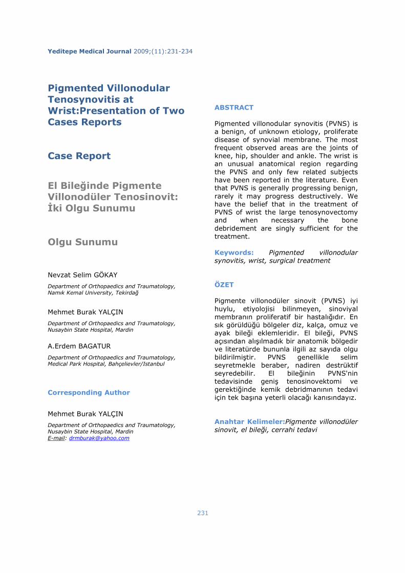
INTRODUCTION Pigmented villonodular synovitis (PVNS) is a benign, of unknown etiology, proliferate disease of synovial membrane (1). It has been first described in 1941 by Jaffe (2). It has got two forms, respectively localized and frequently. The localized form is the focal proliferation characterized with the nodules of the synovial tissue. At the frequent form all the synovial tissue from the joint is retained (3). The most frequent observed areas are the joints of knee, hip, shoulder and ankle. The wrist is an unusual anatomical region regarding the PVNS and only few related subjects have been reported in the literature. The patient applies in generally with complaints of distention and pain at wrist. It may be seen at every age but it is mostly seen at the young adults. The woman and man distribution is similar (4). The recommended treatment is marginal excision or total synovectomy (5). Adjuvant radiotherapy may be applied at the treatment. PRESENTATIONS OF CASE CASE 1 Patient of age 28, male, has applied with complaints of pain and distention at wrist of left hand. In the volar surface of left wrist there was a diffused distention and complication of flexion at the writs and fingers. The radiographies and laboratory investigation were without properties. The frequent tenosynovial mass tissue which was encircling the flexor tendons lying to distal of the carpal tunnel from the level of distal radioulnar joint has been monitored with magnetic resonance viewing (MR) (Figure 1). Large flexor tenosynovectomy has been made to patient. The pathological evaluation result of the extracted material has been conforming to PVNS. An additional treatment has not been applied. There was not any complaint in the 8 years history of the patient and by means of physical consultation it has been seen that hand
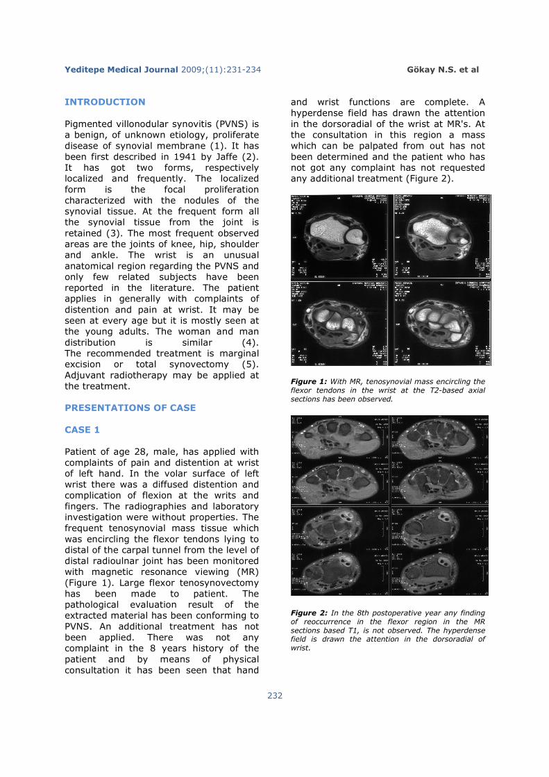
and wrist functions are complete. A hyperdense field has drawn the attention in the dorsoradial of the wrist at MR's. At the consultation in this region a mass which can be palpated from out has not been determined and the patient who has not got any complaint has not requested any additional treatment (Figure 2).
Figure 1: With MR, tenosynovial mass encircling the flexor tendons in the wrist at the T2-based axial sections has been observed.
Figure 2: In the 8th postoperative year any finding of reoccurrence in the flexor region in the MR sections based T1, is not observed. The hyperdense field is drawn the attention in the dorsoradial of wrist.
Yeditepe Medical Journal 2009;(11):231-234
233
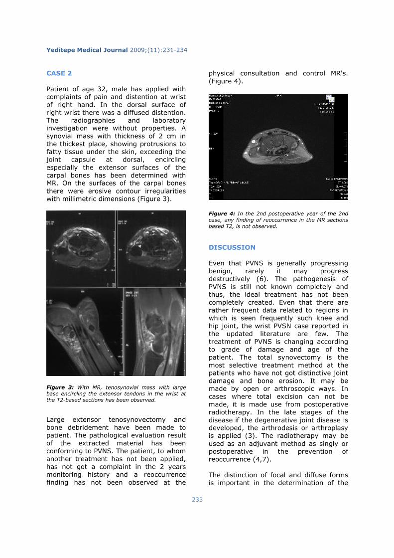
CASE 2 Patient of age 32, male has applied with complaints of pain and distention at wrist of right hand. In the dorsal surface of right wrist there was a diffused distention. The radiographies and laboratory investigation were without properties. A synovial mass with thickness of 2 cm in the thickest place, showing protrusions to fatty tissue under the skin, exceeding the joint capsule at dorsal, encircling especially the extensor surfaces of the carpal bones has been determined with MR. On the surfaces of the carpal bones there were erosive contour irregularities with millimetric dimensions (Figure 3).
Figure 3: With MR, tenosynovial mass with large base encircling the extensor tendons in the wrist at the T2-based sections has been observed.
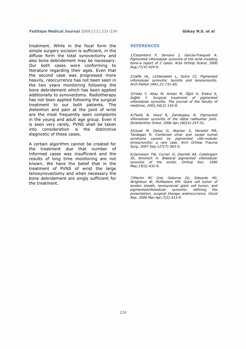
Large extensor tenosynovectomy and bone debridement have been made to patient. The pathological evaluation result of the extracted material has been conforming to PVNS. The patient, to whom another treatment has not been applied, has not got a complaint in the 2 years monitoring history and a reoccurrence finding has not been observed at the
physical consultation and control MR's. (Figure 4). Figure 4: In the 2nd postoperative year of the 2nd case, any finding of reoccurrence in the MR sections based T2, is not observed.
DISCUSSION Even that PVNS is generally progressing benign, rarely it may progress destructively (6). The pathogenesis of PVNS is still not known completely and thus, the ideal treatment has not been completely created. Even that there are rather frequent data related to regions in which is seen frequently such knee and hip joint, the wrist PVSN case reported in the updated literature are few. The treatment of PVNS is changing according to grade of damage and age of the patient. The total synovectomy is the most selective treatment method at the patients who have not got distinctive joint damage and bone erosion. It may be made by open or arthroscopic ways. In cases where total excision can not be made, it is made use from postoperative radiotherapy. In the late stages of the disease if the degenerative joint disease is developed, the arthrodesis or arthroplasy is applied (3). The radiotherapy may be used as an adjuvant method as singly or postoperative in the prevention of reoccurrence (4,7). The distinction of focal and diffuse forms is important in the determination of the
Yeditepe Medical Journal 2009;(11):231-234 Gökay N.S. et al
234
treatment. While in the focal form the simple surgery excision is sufficient, in the diffuse form the total synovectomy and also bone debridement may be necessary. Our both cases were conforming to literature regarding their ages. Even that the second case was progressed more heavily, reoccurrence has not been seen in the two years monitoring following the bone debridement which has been applied additionally to synovectomy. Radiotherapy has not been applied following the surgical treatment to our both patients. The distention and pain at the joint of wrist are the most frequently seen complaints in the young and adult age group. Even it is seen very rarely, PVNS shall be taken into consideration is the distinctive diagnostic of these cases. A certain algorithm cannot be created for the treatment due that number of informed cases was insufficient and the results of long time monitoring are not known. We have the belief that in the treatment of PVNS of wrist the large tenosynovectomy and when necessary the bone debridement are singly sufficient for the treatment.
REFERENCES 1)Carpintero P, Serrano J, García-Frasquet A. Pigmented villonodular synovitis of the wrist invading bone-a report of 2 cases. Acta Orthop Scand. 2000 Aug;71(4):424-6.
2)Jaffe HL, Lichtenstein L, Sutro CJ. Pigmented villonodular synovitis: bursitis and tenosynovitis. Arch Pathol 1941;31:731-65.
3)Yıldız Y, Altay M, Arıkan M, Öğüt H, Erekul S, Sağlık Y. Surgical treatment of pigmented villonodular synovitis. The journal of the faculty of medicine, 2001;54(2):143-8.
4)Tselis N, Heyd R, Zamboglou N. Pigmented villonodular synovitis of the distal radioulnar joint. Strahlenther Onkol. 2006 Apr;182(4):247-51. 5)Uysal M, Ozkoç G, Akpinar S, Hersekli MA, Tandogan R. Combined ulnar and carpal tunnel syndrome caused by pigmented villo-nodular tenosynovitis: a rare case. Arch Orthop Trauma Surg. 2007 Sep;127(7):563-5. 6)Jamieson TW, Curran JJ, Desmet AA, Cotelingam JD, Kimmich H. Bilateral pigmented villonodular synovitis of the wrists. Orthop Rev. 1990 May;19(5):432-6. 7)Martin RC 2nd, Osborne DL, Edwards MJ, Wrightson W, McMasters KM. Giant cell tumor of tendon sheath, tenosynovial giant cell tumor, and pigmentedvillonodular synovitis: defining the presentation, surgical therapy andrecurrence. Oncol Rep. 2000 Mar-Apr;7(2):413-9.


![Clínica Universitária de Reumatologia – Faculdade de ...§ões_Reumatologia_CHUC... · Apr 30. [Epub ahead of print] • Pigmented Villonodular Synovitis: a recurrent case with](https://img.pdfslide.net/doc/110x75/5c65198c09d3f2916e8c4235/clinica-universitaria-de-reumatologia-faculdade-de-oesreumatologiachuc.jpg)


![Index [link.springer.com]978-0-85729-507-1/1.pdf · High school personality questionnaire (HSPQ), 130 Hoffa’s fat pad extraskeletal osteosarcoma, 148 localized pigmented villonodular](https://img.pdfslide.net/doc/110x75/5be095d809d3f2de4d8c5b79/index-link-978-0-85729-507-11pdf-high-school-personality-questionnaire.jpg)