Embed Size (px)
Citation preview
Date: 08 May 2017 Sources Searched: Medline, Embase, Oxford Medicine Online
DISCLAIMER: Results of database and or Internet searches are subject to the
limitations of both the database(s) searched, and by your search request. It is the
responsibility of the requestor to determine the accuracy, validity and interpretation of
the results.
Placental Mesenchymal Dysplasia
See full search strategy
1. Placental mesenchymal dysplasia: What every radiologist needs to know
Author(s): Mittal D.; Anand R.; Sisodia N.; Singh S.; Biswas R.
Source: Indian Journal of Radiology and Imaging; 2017; vol. 27 (no. 1); p. 62-64
Publication Date: 2017
Publication Type(s): Article
Available in full text at Indian Journal of Radiology and Imaging - from Free Access Content
Abstract:Placental mesenchymal dysplasia (PMD) is an uncommon vascular anomaly of the placenta characterized by placentomegaly with multicystic placental lesion on ultrasonography and mesenchymal stem villous hyperplasia on histopathology. Placental mesenchymal dysplasia should be considered in the differential diagnosis of cases of multicystic placental lesion such as molar pregnancy, chorioangioma, subchorionic hematoma, and spontaneous abortion with hydropic placental changes. However, lack of high-velocity signals inside the lesion and a normal karyotype favor a diagnosis of PMD. PMD must be differentiated from gestational trophoblastic disease because management and outcomes differ. We report the case of an 18-year-old female at 15 weeks of gestation with sonographic findings suggestive of placental mesenchymal dysplasia. The diagnosis was confirmed on histopathology.
Database: EMBASE
2. Angiogenesis-related biomarkers (sFlt-1/PlGF) in placental mesenchymal dysplasia.
Author(s): Herraiz, Ignacio; Simón, Elisa; Toldos, Óscar; Rodríguez, Yolanda; Gómez-Arriaga, Paula I; Galindo, Alberto
Source: The journal of maternal-fetal & neonatal medicine : the official journal of the European Association of Perinatal Medicine, the Federation of Asia and Oceania Perinatal Societies, the International Society of Perinatal Obstetricians; Apr 2017; vol. 30 (no. 8); p. 958-961
Publication Date: Apr 2017
Publication Type(s): Journal Article
Abstract:Determination of the soluble fms-like tyrosine kinase-1 to placental growth factor ratio (sFlt-1/PlGF) in the maternal serum is expected to aid in the monitoring and decision-making process of women at risk for placental dysfunction. We report two cases of placental mesenchymal dysplasia (PMD) with sFlt-1/PlGF correlation. The first case is a dichorionic twin pregnancy with one fetus affected by PMD and Beckwith-Wiedemann syndrome in which a high value of sFlt-1/PlGF was found, coinciding with acute maternal and fetal wellbeing decline at 31 weeks. The second case corresponds to a singleton pregnancy diagnosed of PMD with normal sFlt-1/PlGF and favorable outcome.
Database: Medline
3. We can Diagnose it if we Consider it. Diagnostic Pitfall for Placenta: Placental Mesenchymal Dysplasia.
Author(s): Toru, Havva Serap; Aytekin, Esra Çobankent; Sanhal, Cem Yaşar; Yakut, Sezin; Çetin, Zafer; Mendilcioğlu, İbrahim İnanç; Peştereli, Hadice Elif
Source: Turk patoloji dergisi; Feb 2017
Publication Date: Feb 2017
Publication Type(s): Journal Article
Abstract:Placental mesenchymal dysplasia is an increasingly recognizable abnormality. Early cases have been confused with partial hydatidiform mole. Placental mesenchymal dysplasia is probably under-diagnosed because of being an unfamiliar clinical entity and also mistaken for gestational trophoblastic disease due to the similar sonographic findings of two entities. In this report, we describe the clinical, gross, and histopathological findings of placental mesenchymal dysplasia in two cases. The 33-week-preterm baby of a 26-year-old woman with cardiovascular disease and 342 gram placenta and the 19-week fetus with trisomy 21 of a 40 year-old woman were terminated. Macroscopically thick-walled vessels and microscopically hydropic villous with peripherally localized thick-walled vessels without trophoblastic cell proliferation were observed in both cases. These two cases represent a rare placental anomaly that is benign but it is challenging to distinguish placental mesenchymal dysplasia from an incomplete mole. Placental mesenchymal dysplasia should be included in the differential diagnosis of sonographic findings that show a normal appearing fetus and a placenta with cystic lesions. Placental mesenchymal dysplasia is associated with pregnancy-related hypertension. In conclusion, the most important point is "you can diagnose it if you consider it".
Database: Medline
4. Placental mesenchymal dysplasia: A clinicopathological study of 12 cases with gestational ages less than 20 weeks
Author(s): Fukunaga M.
Source: Laboratory Investigation; Feb 2017; vol. 97
Publication Date: Feb 2017
Publication Type(s): Conference Abstract
Abstract:Background: Placental mesenchymal dysplasia (PMD) is characterized by placentomegaly and can be mistaken for partial hydatidiform mole (PM) or complete hydatidiform mole (CM) with twin both clinically and macroscopically because of the presence of "grapelike vesicles". It may be associated with Beckwith-Wiedemann syndrome (BWS). However, PMD is both underdiagnosed and underreported. Cases of PMD with gestational ages less than 20 weeks are rarely reported. Design: In order to elucidate the clinical presentation, complications, microscopic features, and differential diagnoses, 12 cases of PMD with gestational ages less than 20 weeks were clinicopathologically analyzed. An immunohistochemical study of p57 (Kip2) (p57) and TSSC3, both of which are products of paternally imprinted, maternally expressed genes, was also done. Results: The patients'ages ranged from 23 to 40 years. Five patients each were initially diagnosed as PM or missed abortion, and 2 were diagnosed as CM with twin on ultrasound examinations. The gestational ages ranged from 7 to 19 weeks. One case was associated with BWS. In 10 cases, early stage PMD was histologically characterized by moderate swelling of stem villi with cistern formation, myxoid change, dilated veins, stromal cell proliferation, and the absence of trophoblastic hyperplasia. No dilated subchorionic vascular vessels with or without luminal thrombosis, or chorangiosis, which are observed in 3rd-trimester cases of PMD, were found. In all cases, cytotrophoblasts were positive for p57 and TSSC3. Regarding p57, villous stromal cells were diffusely positive in 3 cases, focally positive in 5, and uniformly negative in 2. The remaining 2 cases were diagnosed as PMD with CM, and the stromal cells of PMD component were negative for p57. Conclusions: PMD presents with a wide spectrum of clinicopathologic findings. Early stage PMD can be clinically or pathologically misdiagnosed as missed abortion, PM, or CM with twin. Two cases were diagnosed as PMD with CM. The histology of early stage PMD are less distinctive than those of PMD in the 3rd trimester. The diagnostic clues that can be used to identify such cases include moderate swelling of stem villi with cistern formation, myxoid change, dilated veins, mild stromal cell proliferations, and the absence of trophoblastic hyperplasia. Immunohistochemical examinations of p57 expression are useful for differential diagnosis in equivocal cases. It is important to identify PMD at an early stage to reduce the risk of fetal morbidity or mortality. Patients with PMD with CM should be followed-up with HCG monitoring.
Database: EMBASE
5. A case of placental mesenchymal dysplasia with one year follow-up
Author(s): Cheng M.; Peng B.; You Y.
Source: Clinical and Experimental Obstetrics and Gynecology; 2016; vol. 43 (no. 5); p. 777-779
Publication Date: 2016
Publication Type(s): Article
Abstract:Placental mesenchymal dysplasia (PMD) is a rare placental disease with unknown etiology. Because of the malformation of the placenta, the incidence of the obstetric complications and the poor perinatal outcomes are higher than usual. PMD usually accompanies with Beckwith-Wiedemann syndrome. In this article, the authors report a case of PMD with a live delivery in 35+5 gestational weeks. The phenotype of the neonate was normal. During the one-year follow-up, the development of the child was normal.
Database: EMBASE
6. Anemia in a neonate with placental mesenchymal dysplasia.
Author(s): Ishikawa, Satoshi; Morikawa, Mamoru; Umazume, Takeshi; Yamada, Takahiro; Kanno, Hiromi; Takakuwa, Emi; Minakami, Hisanori
Source: Clinical case reports; May 2016; vol. 4 (no. 5); p. 463-465
Publication Date: May 2016
Publication Type(s): Journal Article
Available in full text at Clinical Case Reports - from National Library of Medicine
Abstract:Causes of intrauterine fetal death (IUFD) are uncertain in most placental mesenchymal dysplasia (PMD) cases. Our case showed high α-fetoprotein levels in the maternal circulation, markedly dilated subchorionic vessels, and neonatal hemoglobin concentration of 8.4 g/dL, suggesting that fetal anemia may explain some adverse outcomes in PMD pregnancies.
Database: Medline
7. Prenatal imaging and pathology of placental mesenchymal dysplasia: A report of three cases
Author(s): Tanuma A.; Kawaguchi R.T.; Tanaka T.; Yanaihara N.; Okamoto A.; Yanagisawa H.
Source: Case Reports in Perinatal Medicine; Mar 2016; vol. 5 (no. 1); p. 9-14
Publication Date: Mar 2016
Publication Type(s): Article
Available in full text at Case Reports in Perinatal Medicine - from ProQuest
Abstract:Objective: Placental mesenchymal dysplasia (PMD) is a rare vascular anomaly characterized by mesenchymal stem villous hyperplasia. Accurate differential diagnosis of PMD is crucial to predict fetal outcomes associated with serious obstetrical complications. Methods: We reviewed the clinical and pathological features and immunohistochemical and imaging findings of three patients with PMD. Results: First trimester sonographic cystic findings identified molar pregnancy or PMD. However, PMD was highly suspected according to the maternal serum human chorionic gonadotropin (hCG) titers, fetal karyotypes, and imaging findings. The outcome of patient 1, in whom placental multicystic areas decreased as pregnancy progressed, was a live birth. In contrast, the babies of patients 2 and 3 were stillborn, and multicystic formations detected during the first trimester completely and consistently occupied the placentas. Pathological and immunohistochemical analyses using anti-CD34 and anti-D2-40 antibodies distinguished the
cisternae from multiple small vessels in the villi. Immunohistochemical analyses using anti-CK7 and anti-Ki-67 antibodies did not detect excessive proliferation of trophoblasts. Most abnormal villi associated with PMD comprised stromal cells that did not react with an anti-p57kip2 antibody. Conclusion: In patients with PMD, if the percentage of the normal placental area decreases as pregnancy progresses, the possibility of fetal growth restriction and intrauterine fetal demise should always be considered. The immunostaining pattern of CD34 and D2-40 may represent a unique feature of PMD and can provide supporting evidence for the differential diagnosis of PMD.Copyright © 2016 by De Gruyter 2016.
Database: EMBASE
8. Beckwith-wiedemann syndrome with placental mesenchymal dysplasia in a male (46,XY) infant: Unique findings at autopsy
Author(s): Laib A.; Stanek J.
Source: Laboratory Investigation; Feb 2016; vol. 96
Publication Date: Feb 2016
Publication Type(s): Conference Abstract
Available in full text at Laboratory Investigation - from ProQuest
Abstract:Background: Beckwith-Wiedemann syndrome (BWS) is a fetal overgrowth syndrome often presenting with: enlarged tongue/body/organs; omphalocele; ear pitting; hemihypertrophy; and placental mesenchymal dysplasia (PMD), a rare (0.02%) placental anomaly. PMD shows strong female predominance (8:1) and usually presents with: large multivesicular placenta, elevated maternal alpha-fetoprotein; fetal growth restriction. Different from partial molar pregnancy (triploid non-viable fetus, hydatid intermediate villi) and twin chorioma (diploid fetus with complete molar twin, hydatid terminal villi), PMD features a diploid viable fetus and hydatid stem villi. 25% of PMD cases are associated with BWS. Design: We present an autopsy case of a 21-week-EGA male infant delivered by termination pregnancy due to: prenatal diagnosis of partial molar pregnancy and multiple malformations (omphalocele, cleft lip/palate, ambiguous genitalia), maternal hCG 444,862mIU/ml, and early-onset severe pre-eclampsia, despite normal 46,XY karyotype on amniocentesis. The infant died minutes after delivery. Results: At autopsy, the appropriate-for-gestational-age infant (351.8g) showed omphalocele, small mandible, and ear pitting, but no cleft lip/palate or ambiguous genitalia. Microscopic examination revealed many findings consistent with BWS (adrenal cytomegaly, nesidioblastosis, Leydig cell hyperplasia, pituitary amphophil hyperplasia). Unexpected findings included: cardiac cartilaginous choristoma, cochlear dysplasia of the inner ear, perihepatic paraganglion, and pituitary pseudoglandular transformation. The placenta demonstrated PMD, despite male gender, with multiple thin-walled grape-like vesicles (grossly) and hydatid stem villi with cisterns (microscopically). Fetal genetic testing confirmed 46,XY karyotype by microarray/chromosome analysis/aneuploidy FISH. Conclusions: This case illustrates the difficulties in prenatal differential diagnosis of congenital malformations in general, and gestational trophoblastic disease and PMD, in particular, especially in the case of a 46,XY male. This unusual case of BWS (without overgrowth features probably due to extreme immaturity) demonstrated multiple unexpected findings. (Figure Presented).
Database: EMBASE
9. Mesenchymal dysplasia of placenta.
Author(s): Balachandran Nair, Krishna G; Srinish, Minu; Balan, Preesha; Sadasivan, Santha
Source: Indian journal of pathology & microbiology; 2015; vol. 58 (no. 3); p. 371-373
Publication Date: 2015
Publication Type(s): Case Reports Journal Article
Available in full text at Indian Journal of Pathology and Microbiology - from Free Access Content
Abstract:A rare case of placental mesenchymal dysplasia (PMD) in a 26-year-old patient is reported. Ultrasound scan at 17 weeks of gestation showed placenta with multiple cystic spaces and a normal appearing fetus. Following delivery of a term live baby, histological examination of the placenta was suggestive of PMD. The early recognition of this rare condition by characteristic ultrasonographic findings is herein emphasized and hence that PMD is distinguished from molar pregnancy.
Database: Medline
10. Placental mesenchymal dysplasia without fetal development in a twin gestation: a case report and review of the spectrum of androgenetic biparental mosaicism.
Author(s): Linn, Rebecca L; Minturn, Lucy; Yee, Lynn M; Maniar, Kruti; Zhang, Yanming; Fritsch, Michael K; Kashireddy, Papreddy; Kapur, Raj; Ernst, Linda M
Source: Pediatric and developmental pathology : the official journal of the Society for Pediatric Pathology and the Paediatric Pathology Society; 2015; vol. 18 (no. 2); p. 146-154
Publication Date: 2015
Publication Type(s): Case Reports Twin Study Journal Article Review
Available in full text at Pediatric and Developmental Pathology - from ProQuest
Abstract:We report a dichorionic twin gestation with diffuse placental mesenchymal dysplasia (PMD) and androgenetic biparental mosaicism (ABM) involving one twin's placenta with complete absence of fetal development for that twin. To our knowledge, this is the 1st reported case of PMD without fetal development. We discuss the gross, histologic, and genetic hallmarks of PMD and the spectrum of variability depending on degree and distribution of ABM.
Database: Medline
11. Prospective risk of stillbirth in women with placental mesenchymal dysplasia.
Author(s): Ishikawa, Satoshi; Morikawa, Mamoru; Yamada, Takahiro; Akaishi, Rina; Kaneuchi, Masanori; Minakami, Hisanori
Source: The journal of obstetrics and gynaecology research; Oct 2015; vol. 41 (no. 10); p. 1562-1568
Publication Date: Oct 2015
Publication Type(s): Case Reports Journal Article
Available in full text at Journal of Obstetrics and Gynaecology Research - from John Wiley and Sons
Abstract:AIMThe aim of this study was to provide better counsel to pregnant women with suspected placental mesenchymal dysplasia (PMD) regarding the risks of preterm birth and intrauterine fetal death.MATERIAL AND METHODSWe reviewed the outcomes of 109 PMD pregnancies with gestational week (GW) ≥ 24 abstracted from 63 reports in the English-language published reports, including two cases that we encountered recently. The prospective risk of stillbirth at GW N was defined as the number of women with stillbirth at GW ≥ N divided by the number of women giving birth at GW ≥ N.RESULTSA total of 32 (29.4%) women experienced stillbirth at a median GW of 31
(range, 24-38). Preterm birth (GW < 37) occurred in 52 (67.5%) of the 77 live-born infants. Only 25 (22.9%) women had full-term (GW ≥ 37) live-born infants. The prospective risks of stillbirth were 29.4% (32/109), 27.5% (25/91), 20.9% (14/67) and 13.0% (6/46) for women who reached GW 24(+0) , 28(+0) , 32(+0) and 36(+0) respectively.CONCLUSIONAs women with PMD are at markedly elevated risk of intrauterine fetal death, early admission to the hospital and intensive monitoring of fetal status should be considered, although whether this policy improves outcome has not been validated.
Database: Medline
12. Intrauterine Growth Restriction Associated with Hematologic Abnormalities: Probable Manifestations of Placental Mesenchymal Dysplasia.
Author(s): Martinez-Payo, Cristina; Bernabeu, Rocio Alvarez; Villar, Isabel Salas; Goy, Enrique Iglesias
Source: AJP reports; Oct 2015; vol. 5 (no. 2); p. e085
Publication Date: Oct 2015
Publication Type(s): Journal Article
Available in full text at AJP Reports - from National Library of Medicine
Abstract:Introduction Placental mesenchymal dysplasia is a rare vascular disease associated with intrauterine growth restriction, fetal demise as well as Beckwith-Wiedemann syndrome. Some neonates present hematologic abnormalities possibly related to consumptive coagulopathy and hemolytic anemia in the placental circulation. Case report We present a case of placental mesenchymal dysplasia in a fetus with intrauterine growth restriction and cerebellar hemorrhagic injury diagnosed in the 20th week of pregnancy. During 26th week, our patient had an intrauterine fetal demise in the context of gestational hypertension. We have detailed the ultrasound findings that made us suspect the presence of hematologic disorders during 20th week. Discussion We believe that the cerebellar hematoma could be the consequence of thrombocytopenia accompanied by anemia. If hemorrhagic damage during fetal life is found, above all associates with an anomalous placental appearance and with intrauterine growth restriction, PMD should be suspected along other etiologies.
Database: Medline
13. Selective fetal growth restriction due to placental mesenchymal dysplasia in twin pregnancy
Author(s): Delgado Sanchez E.; Marin Camacho S.; Quereda Bernabeu B.; De La Calle M.; Rigojo R.M.; Basrtha J.L.
Source: Journal of Perinatal Medicine; Oct 2015; vol. 43
Publication Date: Oct 2015
Publication Type(s): Conference Abstract
Abstract:Background: First described in 1991 by Moscoso et al, placental mesenchymal dysplasia (PMD) is a rare lesion (0.02% of all pregnancies). It is a vascular anomaly of the placenta, and its aspect can be confused with a partial hydatiform mole. PMD is more frequent between female fetuses. It is associated with Beckwith-Widemann syndrome (BWS) in 20-30% of the cases, and 50% with intrauterine fetal growth restriction. Approximately 40% of the fetuses have intrauterus or neonatal demise. Case: We present a 34 year-old woman, pregnant preceded by in vitro fertilization, bychorialbiamniotic pregnancy. On the morphologic echography done in the twentieth week, second twin was diagnosed of early selective fetal growth restriction and hydropic-cyst placenta. TORCH serology was requested, being negative. In twenty-first week a genetic amniocentesis was
performed. The result was 46XX with normal karyotype. Array technique showed three copies of Xp13.23, which do not justify the selective fetal growth restriction. Parent's karyotype was normal. On twenty-first week and 5 days, the patient get into the hospital because of premature rupture of membranes and symptoms of clinical chorioamnionitis, and the spontaneous expulsion of the two fetuses occurred two hours later. The necropsy showed that the first twin had an augmented weigh for gestational age (519 g) with organomegaly and diffuse bilateral adrenal cytomegalia. The placental weight was 188 g (percentile 10-25), and its pathological study showed intense acute chorioamnionitis and distal villous hypoplasia. The necropsy of the second twin showed low weight and size for gestational age (249 g), pseudocysticchanges in adrenal cortex. The placental weight was 246 (percentile 50-75). Its pathological study showed placental mesenchymal dysplasia. Both fetuses had a negative genetic study for BWS. Conclusion: For our knowledge, this is the first case in the literature of placental mesenchymal dysplasia in a bichorial-biamniotic twin pregnancy. Its pathogenesis and cause is not well known. Genetic studies shows that it may be associated with genetic imprinting of chromosome Xp15, BWS and other genetic mutations of chromosome X. Awareness the ultrasongraphyc similarity of this entity to the partial hydatiform mole, will help to do a correct prenatal monitoring. The cooperation between obstetrician, pathologist and neonatologist will help to advance in this field of research. There are several cases described in the literature of unique pregnancies in with the child evolution wasfavorable. However, there is no data of long term follow-up.
Database: EMBASE
14. Preterm delivery due to a rare clinicopathologic entity
Author(s): Stefanescu C.V.
Source: Journal of Perinatal Medicine; Oct 2015; vol. 43
Publication Date: Oct 2015
Publication Type(s): Conference Abstract
Abstract:Rates of preterm delivery have increased in the last years, yet mechanisms elude us still. The author report the case of an IUI obtained pregnancy (male factor) complicated with growth restriction and premature delivery due to a rare placental abnormality - placental mesenchymal dysplasia (PMD). PMD is characterised by placentomegaly and grape-like vesicles but with a viable fetus. Prenatal diagnosis is challenging and could be presumed by ultrasound placentomegaly and elevated levels of HCG. A molar pregnancy or a triploidy should be ruled out and the final diagnosis is achieved by histological examination of the placental tissue. The fetal karyotype is normal but it has an increased risk for premature rupture of membrame, preterm delivery, IUGR, intrauterine fetal demise, perinatal death, postnatal fetal sever anemia and thrombocytopenia. About 20% of fetuses may also be affected by Beckwith-Wiedemann syndrome.
Database: EMBASE
15. Placental mesenchymal dysplasia without fetal tissue: Differentiating it from complete mole: A case study
Author(s): Peng Y.-C.; Shukla P.
Source: American Journal of Clinical Pathology; Oct 2015; vol. 144
Publication Date: Oct 2015
Publication Type(s): Conference Abstract
Available in full text at American Journal of Clinical Pathology - from ProQuest
Available in full text at American Journal of Clinical Pathology - from Oxford University Press ; Collection notes: To access please select Login with Athens and search and select NHS England as your institution before entering your NHS OpenAthens account details.
Abstract:Placental mesenchymal dysplasia (PMD), a rare condition of pregnancy, is characterized by placentomegaly and grape-like vesicles similar to molar pregnancy. It is associated with a fetus and therefore often clinically misdiagnosed as a partial mole. There is strong association of PMD with fetal growth restriction and Beckwith- Wiedemann syndrome (BWS), but it is also seen with live birth of normal-appearing fetuses. Maternal serum alpha- fetoprotein is elevated and beta-HCG levels are normal to increased in PMD. There are occasional case reports where a twin pregnancy consisted of 1 complete mole and 1 fetus with PMD. There are no case reports of a PMD without fetus. We present the first case of PMD that was not associated with fetus and was clinically diagnosed as complete hydatidiform mole. A 17-year-old girl with 10-week intrauterine pregnancy had beta-HCG level of 49,000 mIU/mL and a characteristic "snowstorm" pattern without any fetus on ultrasound. Complete mole was diagnosed and D&C was performed. Macroscopically red-brown tissue with grape-like vesicles without identifiable fetal tissue was seen. Entire specimen was submitted for microscopic examination. Characteristic histologic features of PMD, namely enlarged chorionic villi with central cisterns and fibromuscular vessels at the periphery without any trophoblast hyperplasia, were seen. No fetal or embryonic tissue was seen microscopically. No fetal RBCs were seen in the chorionic villous vessels. Immunostain for p57 was positive in trophoblast and villous stromal cells. Diagnosis of PMD was made based on histologic findings and positive p57 immunostain. The patient's HCG came down to 22 mIU/mL 1 week post D&C. PMD is associated with a fetus and clinically presents as partial mole. Ours is the first case in literature in which PMD did not have associated fetal tissue and was clinically diagnosed as complete mole. Complete mole was ruled out by positive p57 immunostaining and lack of trophoblast hyperplasia. Diagnosis of PMD was based on characteristic histologic findings.
Database: EMBASE
16. Severe fetal growth restriction caused by placental mesenchymal dysplasia
Author(s): Lin J.; Foster S.; Soundararajan S.
Source: American Journal of Clinical Pathology; Oct 2015; vol. 144
Publication Date: Oct 2015
Publication Type(s): Conference Abstract
Available in full text at American Journal of Clinical Pathology - from ProQuest
Available in full text at American Journal of Clinical Pathology - from Oxford University Press ; Collection notes: To access please select Login with Athens and search and select NHS England as your institution before entering your NHS OpenAthens account details.
Abstract:Placental mesenchymal dysplasia (PMD) is a rare complication characterized by placentomegaly and grape-like vesicles that resemble molar pregnancy. PMD is associated with fetal growth restriction (FGR), intrauterine fetal death, Beckwith-Wiedemann syndrome, androgenetic biparental mosaicism and other cytogenetic abnormalities. Here we report on the diagnostic challenges associated with this pathology in an otherwise healthy 20-year-old G3P1011 woman. A multicystic heterogeneous placenta concerning for molar pregnancy was discovered on a first trimester viability ultrasound and persisted throughout the pregnancy. The patient was monitored closely and counseled accordingly throughout her pregnancy. Growth restriction was detected at 30 weeks' gestation with an abdominal circumference <3rd percentile, and by 36 weeks, overall growth was <5th percentile. Uterine artery Doppler studies were within normal limits. A full term infant boy was delivered by repeat C-section. APGAR scores were 9/9. The infant's weight was 2245 g, and no morphologic abnormalities were appreciated. The placenta measured 20 x 14 x 3 cm, weighed 707 g, and revealed dilated fetal chorionic vessels with minimal branching. Parenchyma had multiple subchorionic cystic dilatations measuring 4.5 x 2.6 x 2.5 cm, which constituted up to 25% of the placental volume. The cysts microscopically revealed cistern formation within large abnormal stem villi, which were hypo to avascular, and had abundant stroma with focal myxoid changes. No trophoblast proliferation was present. The distal villi appeared relatively normal with slight variability in maturation. The morphologic changes are consistent with PMD. Severe FGR of this infant may be explained by potentially decreased maternalfetal gas exchange due to an insufficient amount of normal chorionic villi and shunting of blood from the dysplastic villi caused by PMD.
Database: EMBASE
17. A rare case of placenta mesenchymal dysplasia
Author(s): Evangelista M.R.; Cruz-Javier G.; Arcellana-Nuqui E.; Tolentino-Molina M.K.
Source: International Journal of Gynecology and Obstetrics; Oct 2015; vol. 131
Publication Date: Oct 2015
Publication Type(s): Conference Abstract
Available in full text at Intl Jrnl Gynecology and Obstet - from John Wiley and Sons
Abstract:Objectives: To increase awareness of clinicians regarding placental mesenchymal dysplasia (PMD), and that it should be considered in the differential diagnosis of every placental abnormality, especially in specific sonologic findings of enlarged cystic placenta. Method: This is a case of an 18 year old Gravida 2 Para 0 (0-0-1-0), with an ultrasonographic finding of an enlarged cystic placenta, with pregnancy complicated with intrauterine growth restriction (IUGR) and oligohydramnios. TORCH panel was positive for cytomegalovirus (CMV) IgG. The prenatal diagnosis only included molar pregnancy with a live twin fetus and CMV infection. Postnatal evaluation of the placenta was done to confirm the diagnosis. Results: Grossly, cut sections of the placenta revealed multiple cystic spaces filled with watery fluid. Microscopically, there were large hydropic villi and dilated thick-
walled vessels without trophoblastic proliferation. Immunohistochemical study was further done and showed dysplastic villi that were immunoreactive to desmin and negative to smooth muscle actin (SMA) and Ki-67. These findings were consistent with placental mesenchymal dysplasia. Conclusions: Placental mesenchymal dysplasia is a rare placental abnormality associated with adverse pregnancy outcome. Patients should be counselled regarding complications. Heightened surveillance with assessment of fetal well-being should be always be considered. A detailed histologic, immunohistochemical and also genetic analyses are essential for accurate diagnosis.
Database: EMBASE
18. Phaeochromocytoma in placental mesenchymal dysplasia: Who should we screen and for how long?
Author(s): White M.; Zacharin M.; McGillivray G.; White S.
Source: Hormone Research in Paediatrics; Sep 2015; vol. 84 ; p. 321
Publication Date: Sep 2015
Publication Type(s): Conference Abstract
Available in full text at Hormone Research in Paediatrics - from ProQuest
Abstract:Background: Beckwith-Wiedemann syndrome (BWS) characterised by a group of clinical abnormalities (macrosomia, macroglossia, neonatal hypoglycaemia, omphalocoele and umbilical hernia) results from dysregulation of imprinted genes due to mosaic paternal uniparental isodisomy (patUPD) of 11p15.5. Its association with tumours of embryonic origin is well documented and screening guidelines largely aim to detect hepatoblastoma and Wilm's tumours during the first decade of life. BWS features have been noted in 25-30% of infants with placental mesenchymal dysplasia (PMD), a distinct condition with cystic placental histology and mosaicism for genome-wide patUPD. One previous case of bilateral phaeochromocytoma has been reported in PMD. Case presentation: We describe the case of a 12 year old female diagnosed with PMD in infancy on the basis of cystic placental histology, BWS-like clinical features and confirmatory genetic analyses. Her early years were complicated by severe congenital hyperinsulinism requiring subtotal pancreatectomy and hepatoblastoma. Antenatal sonography at 18, 22, and 28 weeks gestation documented bilateral cystic adrenomegaly which had regressed on postnatal imaging by 5 months of age. During prolonged screening a unilateral asymptomatic right sided adrenal cystic lesion was noted at age 11 which demonstrated no appreciable uptake on targeted imaging but which had increased in size over 12 months of surveillance to 3.7x3.1x3.6 cm and was secretory of noradrenaline and dopamine; this was confirmed as a phaeochromocytoma after an uneventful surgical removal of the lesion. Regular surveillance for a recurrent or left sided lesion is ongoing. Conclusion: BWS and PMD share clinical and genetic features and an increased risk of malignancy. Their association with phaeochromocytoma, while rare, may not be co-incidental given that there is a known association between these tumours and maternal loss of 11p15 genes. Due consideration of continued screening for phaeochromocytomas beyond the first decade of life may be appropriate for individuals with PMD.
Database: EMBASE
19. A clinicopathologic study of early stage Placental Mesenchymal Dysplasia (PMD)
Author(s): Fukunaga M.
Source: Virchows Archiv; Sep 2015; vol. 467 (no. 1)
Publication Date: Sep 2015
Publication Type(s): Conference Abstract
Available in full text at Virchows Archiv - from Springer Link Journals
Available in full text at Virchows Archiv - from ProQuest
Abstract:Objective: To elucidate the clinical presentation, complications, microscopic features, and differential diagnoses of early stage (gestational age less than 20 weeks) PMD. Method: 10 cases of PMD were clinicopathologically analyzed. Immunohistochemical study of p57 (Kip2) (p57), which is products of a paternally imprinted, maternally expressed gene, was also done. Results: Maternal ages ranged from 23 to 40 years. Gestational periods ranged from 12 to 19 weeks. Five patients were initially diagnosed as partial mole (PM) and one was as complete mole (CM) with twin on ultrasound examination. One case was associated with BWS. Histologically, early stage PMD was characterized by moderate swelling of stem villi with cistern formation, myxoid change, dilated veins, mild stromal cell proliferations, and the absence of trophoblastic hyperplasia. Dilated subchorionic vascular vessels with or without luminal thrombosis, or chorangiosis, which were observed in the third trimester PMD, were not found. Cytotrophoblasts were positive for p57 in all cases, and villous stromal cells were diffusely positive in 3 cases, focally positive in 5, and uniformly negative in 2. Conclusion: Early stage PMD can be clinically or pathologically misdiagnosed as abortion, PM, or CM with a twin. Histologic features in early stage PMD are less distinctive compared with those of PMD in the third trimester. The diagnostic clues aremoderate swelling of stemvilli with cistern formation, myxoid change, dilated veins, and mild stromal cell proliferations and the absence of trophoblastic hyperplasia. The p57 immunohistochemical study is useful for differential diagnoses in equivocal cases. It is important to identify PMD cases prenatally to reduce fetal morbidity and mortality.
Database: EMBASE
20. Placental mesenchymal dysplasia: Important differential diagnosis for partial moles and frequently associated with Beckwith-Wiedemann syndrome
Author(s): Mau-Holzmann U.A.; Singer S.; Eggermann T.; Mackensen-Haen S.; Goelz R.; Tzschach A.; Kagan K.O.
Source: Chromosome Research; Jun 2015; vol. 23 (no. 1)
Publication Date: Jun 2015
Publication Type(s): Conference Abstract
Available in full text at Chromosome Research - from Springer Link Journals
Available in full text at Chromosome Research - from ProQuest
Abstract:Placental mesenchymal dysplasia (PMD) is a rare placental anomaly. Prenatal ultrasound indicates placentomegaly and cystic structures resembling a partial mole, but with apparently normal fetal morphology. PMD is associated with intra-uterine growth restriction (IUGR), intra-uterine mortality, prematurity and in about 20 % of the cases with Beckwith- Wiedemann syndrome (BWS), respectively. The underlying cause is unusual and interesting as PMD is a special type of mosaicism. One part of the placenta is normal with a biparental diploid genotype (preponderance of females). The dysplastic part is also diploid, but of paternal uniparental origin. We present a case of a prematurely born female with typical signs of BWS. PMD was suspected prenatally (confirmed pathologically). Molecular studies confirmed the diagnosis of BWS (UPD11pat) and showed a

genome wide paternal uniparental disomy in the majority of cells, with a low degree of biparental cells in blood. PMD should be considered in cases with partial mole-especially if there are no major anomalies indicating triploidy. Gynaecologists, cytogeneticists and pathologists should be aware of this rare differential diagnosis and initiate respective studies.
Database: EMBASE
21. Placental mesenchymal dysplasia (PMD) in association with beckwith-wiedemann syndrome identified by first trimester sonography
Author(s): Lindsley W.; Haeri S.
Source: Journal of Diagnostic Medical Sonography; May 2015; vol. 31 (no. 3); p. 181-185
Publication Date: May 2015
Publication Type(s): Article
Abstract:Placental mesenchymal dysplasia (PMD) has commonly been identified on second trimester ultrasound in association with Beckwith-Wiedemann syndrome (BWS). In this report, a case of PMD later confirmed as Beckwith-Wiedemann is presented, which was identified by sonography in the first trimester. When faced with a first trimester finding of an enlarged cystic placenta, it is suggested that BWS be considered as a possible diagnosis and accordingly, genetic testing with methylation studies offered to the parents.Copyright © The Author(s) 2015.
Database: EMBASE
22. Placental mesenchymal dysplasia associated with spontaneous ovarian hyperstimulation syndrome.
Author(s): Davoudian, Payam
Source: BMJ case reports; Apr 2015; vol. 2015
Publication Date: Apr 2015
Publication Type(s): Case Reports Journal Article
Available in full text at BMJ Case Reports - from Highwire Press
Abstract:Placental mesenchymal dysplasia (PMD) is a rare disorder of unknown aetiology characterised by placentomegaly, serpiginous surface blood vessels and large cystic villi. Although a mimic of molar pregnancies and other entities, it can be distinguished from them by its association with intrauterine growth restriction or death of a relatively normal fetus and its characteristic histopathology. We report the case of a 20-year-old primigravida who presented at 16 weeks with heavy clotty vaginal bleeding and subsequent miscarriage. The placenta was grossly abnormal and resembled a partial molar pregnancy. PMD was confirmed by microscopic examination. Postmiscarriage, ultrasound revealed enlarged and multicystic ovaries similar to those seen in ovarian hyperstimulation syndrome (OHSS), which returned to normal a few weeks after miscarriage. This is the first report of PMD associated with OHSS, and we hypothesise that the most likely pathogenesis is ovarian stimulation from PMD-derived vascular endothelial growth factor.
Database: Medline
23. Late diagnosis of hepatic mesenchymal hamartoma and placental mesenchymal dysplasia-case report and review of the literature
Author(s): Divya G.; Joung S.; Ryder L.; Nayyar R.
Source: Journal of Paediatrics and Child Health; Apr 2015; vol. 51 ; p. 109
Publication Date: Apr 2015
Publication Type(s): Conference Abstract
Available in full text at Journal of Paediatrics and Child Health - from John Wiley and Sons
Abstract:Background: Placental mesenchymal dysplasia (PMD) is a rare condition characterised by placental enlargement, oedematous stem villi and multiple anechoic cysts. Hepatic mesenchymal hamartoma (HMH) is an uncommon, benign, proliferation of mesenchymal tissue, commonly seen in infants under the age of two years. While the aetiology is poorly understood, the increased incidence of HMH with PMD and the morphological similarities of the oedematous, myxoid changes seen in both the placenta and liver, suggests a possible common developmental mechanism. HMH should be monitored for closely in all cases of PMD, because although rare, in combination it is known to carry a poorer prognosis. Only 11 other cases of this concurrent pathology have been reported so far. Results/Method: We illustrate the case of a 28-year-old lady, G4P2, with two previous normal vaginal deliveries (NVD), who had a normal morphology scan at 19 weeks gestation and an uneventful early pregnancy. At 34 weeks, a growth scan revealed a well-circumscribed, septated cyst on the right liver lobe (4 x 3 x 4 cm), suggestive of HMH and a large, thickened placenta, with multiple anechoic cysts (largest 3 cm in diameter), consistent with PMD. There were no other structural abnormalities noted. At 38 weeks she underwent an induction of labour with a NVD of a live female infant. Histopathology confirmed PMD with the placenta weighing 1.97 kg (>97 th percentile for gestation). Conclusion: We will also present a review of the literature including an evaluation of antenatal diagnosis, obstetric management and long-term prognosis of this rare placental and fetal anomaly.
Database: EMBASE
24. Rare fetal complications associated with placental mesenchymal dysplasia: a report of two cases.
Author(s): Jimbo, Tomoka; Fujita, Yasuyuki; Yumoto, Yasuo; Fukushima, Kotaro; Kato, Kiyoko
Source: The journal of obstetrics and gynaecology research; Feb 2015; vol. 41 (no. 2); p. 304-308
Publication Date: Feb 2015
Publication Type(s): Case Reports Journal Article
Available in full text at Journal of Obstetrics and Gynaecology Research - from John Wiley and Sons
Abstract:Placental mesenchymal dysplasia (PMD) is a rare disease that may be difficult to distinguish from molar pregnancy. The disease is associated with major fetal complications, including Beckwith-Wiedemann syndrome, fetal growth restriction and intrauterine fetal death. Rarely, fetal hematological disorders and liver tumors also may occur. Two patients were referred to our hospital during their second trimesters because of suspected molar pregnancies. Fetal karyotyping and maternal serum human chorionic gonadotropin level determinations led to the PMD diagnoses. In one case, the maternal clinical course was normal, but the neonate suffered from disseminated intravascular coagulation and needed a platelet transfusion. In the second case, the PMD decreased during pregnancy, but a gradually increasing fetal liver tumor appeared. The tumor was diagnosed as mesenchymal hamartoma, based on ultrasound and magnetic resonance imaging studies. The neonate was delivered without cardiovascular compromise. Due to the difficulty of immediate surgical treatment, expectant management, with close follow-up, was chosen.
Database: Medline
25. Placental mesenchymal dysplasia with fetal gastroschisis.
Author(s): Kim, Binnari; Hyeon, Jiyeon; Lee, Minju; Hwang, Hyewon; Shin, Yooju; Choi, Suk-Joo; Kim, Jung-Sun
Source: Journal of pathology and translational medicine; Jan 2015; vol. 49 (no. 1); p. 71-74
Publication Date: Jan 2015
Publication Type(s): Journal Article
Available in full text at Journal of Pathology and Translational Medicine - from National Library of Medicine
Database: Medline
26. Management of placental mesenchymal dysplasia associated with fetal anemia and IUGR.
Author(s): Simeone, Serena; Franchi, Chiara; Marchi, Laura; Rambaldi, Marianna Pina; Serena, Caterina; Vitagliano, Amerigo; Mecacci, Federico
Source: European journal of obstetrics, gynecology, and reproductive biology; Jan 2015; vol. 184 ; p. 132-134
Publication Date: Jan 2015
Publication Type(s): Letter Case Reports
Database: Medline
27. Placental mesenchymal dysplasia: a case of a normal-appearing fetus with intrauterine growth restriction.
Author(s): Li, Hui; Li, Lei; Tang, Xiao; Yang, Fan; Yang, Kai-Xuan
Source: International journal of clinical and experimental pathology; 2014; vol. 7 (no. 8); p. 5302-5307
Publication Date: 2014
Publication Type(s): Case Reports Journal Article
Available in full text at International Journal of Clinical and Experimental Pathology - from National Library of Medicine
Available in full text at International Journal of Clinical and Experimental Pathology - from Free Access Content
Abstract:In this paper, we described a placenta with vesicular lesions in a 23-year-old woman (1-gravid) who visited our hospital at 13 weeks of gestation on prenatal routine examination. Ultrasound findings showed multiple vesicular lesions which gradually increased as the pregnancy advanced, and a live normal-appearing fetus which was confirmed of IUGR at 30 weeks of gestation in her uterus. Throughout gestation, the maternal serum β-human chorionic gonadotropin level keeps normal, but the serum alpha-fetoprotein was higher than average. The patient delivered an 1800-g female without obvious anomalies at 35 weeks 5 days of gestation due to premature rupture of membrane. The diagnosis of placental mesenchymal dysplasia was determined on the pathological examination and androgenetic/biparental mosaicism in the placenta was identified by immunohistochemical staining of p57kip2.
Database: Medline
28. Early ultrasonographic diagnosis of placental mesenchymal dysplasia.
Author(s): Gerli, S; Giordano, C; Del Sordo, R; Fratini, D; Di Renzo, G C
Source: European review for medical and pharmacological sciences; 2014; vol. 18 (no. 17); p. 2419-2423
Publication Date: 2014
Publication Type(s): Case Reports Journal Article
Available in full text at European Review for Medical and Pharmacological Sciences - from Free Access Content
Abstract:Placental mesenchymal dysplasia (PMD) is a rare pathology characterized by vascular anomalies, placentomegaly and grapelike vesicles resembling partial molar pregnancy. PMD is often associated with fetal growth restriction or intrauterine fetal demise. We report a case of an early diagnosis of PMD at 10 weeks' gestation, with a regular intrauterine growth and a fetal demise occurring at 31 week's gestation. The placenta showed aneurysmally dilated and tortuous vessels with luminal thrombosis. Even in presence of a regular fetal growth, a fetal demise may always occur, suggesting the option of an early heparin administration to reduce the risk of thrombosis of chorionic vessels.
Database: Medline
29. Placental mesenchymal dysplasia.
Author(s): Pawoo, Nogba; Heller, Debra S
Source: Archives of pathology & laboratory medicine; Sep 2014; vol. 138 (no. 9); p. 1247-1249
Publication Date: Sep 2014
Publication Type(s): Journal Article Review
Available in full text at Archives of Pathology & Laboratory Medicine - from EBSCOhost
Available in full text at Archives of Pathology and Laboratory Medicine - from ProQuest
Abstract:Placental mesenchymal dysplasia is a rare placental lesion characterized by stem villous cystic dilation and vesicle formation, placentomegaly, and vascular abnormalities. It can be associated with growth restriction, stillbirth, Beckwith-Wiedemann syndrome, and some chromosomal abnormalities, and needs to be distinguished from its main differential diagnosis, hydatidiform mole.
Database: Medline
30. Imaging analysis and pathological features of placental mesenchymal dysplasia (PMD)
Author(s): Tanuma A.; Kawaguchi R.; Tanemoto T.; Yanaihara N.; Okamoto A.; Yanagisawa H.
Source: Placenta; Sep 2014; vol. 35 (no. 9)
Publication Date: Sep 2014
Publication Type(s): Conference Abstract
Abstract:Objective: Placental mesenchymal dysplasia (PMD) is a rare disease caused by mesenchymal stem villous forming hyperplasia which result in placental vascular anomaly. The differential diagnosis of PMD including molar pregnancy is clinically important, since it is highly associated with serious obstetrical complications. Method: We reviewed here the clinical and pathological features of three PMD cases in our institution. Result: All cases were screened as either molar pregnancy or PMD at first trimester because of sonographic cystic findings. However, these cases were highly suspected as PMD by serum hCG-b titer, fetal karyotype, and Magnetic Resonance Imaging (MRI) findings. MRI study showed that multicystic area was partially observed in thick and hydropic placenta of case 1. In contrast, placenta of case 2 was occupied by the multicystic area. Case 1 had a normal female infant (36wm, 2010g) with the placental weight of 1055g. A half of the placenta showed multicystic and grape-like vesicles distributer changes, which is characterized by PMD. Case 2 and 3 were to be stillbirth (28wm/940g, 24wm /550g) and the placental weight were 1062g and 535g, respectively. Only 10% of these placentae showed solid parts as normal portion. In immunochemical analyses for placental tissue, thickness of vascular wall and multiple small vessels in PMD area were stained with CD34, while cisternae were positive for D2-40. There was less trophoblastic proliferation in the immunostaining of CK-7, Mib-1 and P57kip2 of cases compared with molar placenta. Conclusion: Prenatal diagnosis and management should be carefully considered in case of PMD that have huge multicystic area in the placenta. Immunostaining of CD34 and D2-40 can be supportive for differential diagnosis of PMD.
Database: EMBASE
31. Placental mesenchymal dysplasia and fetal hematologic disorder.
Author(s): Koga, Hiroshi; Makimura, Mika; Tanaka, Hiromasa; Sumioki, Hisao
Source: Journal of pediatric hematology/oncology; Aug 2014; vol. 36 (no. 6); p. e389
Publication Date: Aug 2014
Publication Type(s): Case Reports Journal Article
Available in full text at Journal of Pediatric Hematology/Oncology - from Ovid
Abstract:Placental mesenchymal dysplasia (PMD) is a rare, recently recognized placental vascular anomaly. About 20% of patients with this placental anomaly have Beckwith-Wiedemann syndrome. We report a case of a phenotypically normal neonate with anemia and thrombocytopenia associated with PMD. Histologic examination of the placenta showed findings consistent with PMD, including chorangioma. The patient's hematologic abnormalities resolved during the week following birth. Normal phenotypic fetuses with PMD seem to exhibit hematologic disorders at birth in some cases, especially in the presence of chorangioma.
Database: Medline
32. Diagnostic pitfall for placeta: Placental mesenchymal dysplasia
Author(s): Cobankent Aytekin E.; Sanhal C.Y.; Yakut S.; Cetin Z.; Mendilcioglu I.; Pestereli H.E.; Toru H.S.
Source: Virchows Archiv; Aug 2014; vol. 465 (no. 1)
Publication Date: Aug 2014
Publication Type(s): Conference Abstract
Available in full text at Virchows Archiv - from Springer Link Journals
Available in full text at Virchows Archiv - from ProQuest
Abstract:Objective: Placental mesenchymal dysplasia (PMD) is an increasingly recognizable abnormality. Early cases of PMD have been confused with partial hydatidiform mole. PMD first described in 1991 as a rare lesion of placenta which is also known as mesenchymal stem villous hyperplasia. PMD is probably under-diagnosed because of being an unfamiliar clinical entity and also mistaken for gestational trophoblastic disease because of similar sonographic findings of two entities. Because of relatively recent recognition of PMD, the data is limited about the effected pregnancies. Method: In this report we describe clinical, macroscopical, histopathological findings of PMD among two cases. Results: Thirythree-week-preterm baby of a 26-year-old women with cardovascular disease had 342 g placenta. And 19-week fetus with trisomy 21 of a 40 year-old women was terminated. In both cases macroscopically thick walled vessels and microscopically hydropic villous with peripherically localised thick walled vessels, without trophoblastic cell proliferation were observed. Conclusion: These two cases represents a rare placental anomaly which is benign but it is challenging to distinguish PMD from a complete mole. PMD should be included in differatial diagnosis for sonographic findings that show a normal appearing fetus with cystic lesions of placenta. PMD has a relation-ship with pregnancy related hypertension.
Database: EMBASE
33. Prenatal differential diagnosis of complete hydatidiform mole with a twin live fetus and placental mesenchymal dysplasia by magnetic resonance imaging.
Author(s): Himoto, Yuki; Kido, Aki; Minamiguchi, Sachiko; Moribata, Yusaku; Okumura, Ryosuke; Mogami, Haruta; Nagano, Tadayoshi; Konishi, Ikuo; Togashi, Kaori
Source: The journal of obstetrics and gynaecology research; Jul 2014; vol. 40 (no. 7); p. 1894-1900
Publication Date: Jul 2014
Publication Type(s): Multicenter Study Journal Article
Available in full text at Journal of Obstetrics and Gynaecology Research - from John Wiley and Sons
Abstract:AIMTo assess the use of magnetic resonance imaging (MRI) for prenatal differentiation between complete hydatidiform mole with a twin live fetus (CHMTF) and placental mesenchymal dysplasia (PMD).METHODSThree CHMTF cases and three PMD cases, from two institutions over a 6-year period, were retrospectively included in this study. Clinical findings including age, pregnancy history, serum hCG level, ultrasonography findings, complications of the mother, outcome of the fetus, and results of chromosomal study of fetus, amniotic fluid and lesion, if possible, were noted. MRI findings were evaluated by two radiologists with respect to the location of the disease (intra- or extra-fetal sac), the presence of multicystic component, and presence of intra- or extra-lesional hemorrhage.RESULTSIn all six cases, the diseases were recognized as multicystic lesions by ultrasonography and MRI. In two of three CHMTF cases, patients continued with the pregnancy, which resulted in spontaneous abortion. In one case of CHMTF, the patient underwent artificial abortion, after which the mole progressed into an invasive mole with lung metastases. All three PMD patients had live births, and two of the three babies had fetal growth restriction. By MRI, CHMTF
was located within an extra-fetal sac accompanied by intra- and/or extra-lesional hemorrhage, while PMD was located within the placenta in the fetal sac without hemorrhage.CONCLUSIONMRI could provide important information about the prenatal differential diagnosis of CHMTF and PMD, based on the pathophysiology and characteristics of the diseases.
Database: Medline
34. Placental mesenchymal dysplasia, a case of intrauterine sudden death.
Author(s): Yamada, Ai; Sakaida, Noriko; Okamura, Akiharu; Yamada, Takashi; Ota, Takehito; Bo, Masaki
Source: The Kobe journal of medical sciences; Jun 2014; vol. 60 (no. 1); p. E1
Publication Date: Jun 2014
Publication Type(s): Case Reports Journal Article
Available in full text at Kobe Journal of Medical Sciences - from Free Access Content
Abstract:Placental mesenchymal dysplasia (PMD) is a rare condition presenting with enlarged, multicystic placenta like molar changes. Although PMD usually features a normal fetus and the pregnancy often extends into the third trimester, PMD is clinically significant lesion with high rates of FGR, IUFD, and is associated with Beckwith-Wiedemann syndrome (BWS). We report a 30-year old woman at her first pregnancy with intrauterine sudden death at 31 weeks of gestation. The vesicular lesion in her uterus was detected at 10 weeks on ultrasound. The fetus was normal size without any anomaly on ultrasound and normal trophoblastic vascularization by Doppler study during the pregnancy. As the pregnancy advanced, the vesicular lesion decreased in size and no fetal abnormalities were detected. At 28 weeks of gestation an ultrasound detected dilated periumbilical chorionic vessels. We didn't detect severe FGR or abnormal trophoblastic vascularization. At 31 weeks of gestation an intrauterine sudden death of a normal-sized fetus without any anomaly occurred. The placenta was enlarged, and microscopic morphology confirmed a diagnosis of PMD. The chorionic vessels were cirsoid, dilated and tortuous. We determined the rupture of expanded periumbilical chorionic vessels led to fetal death.
Database: Medline
35. Placental mesenchymal dysplasia: A case of success
Author(s): Mouraz M.; Cominho J.; Saramago S.; Proenca S.; Reis M.I.; Martins L.; Pedroso S.; Colaco J.; Rodrigues R.; Nunes F.
Source: Journal of Maternal-Fetal and Neonatal Medicine; Jun 2014; vol. 27 ; p. 104-105
Publication Date: Jun 2014
Publication Type(s): Conference Abstract
Abstract:Brief Introduction: Placental mesenchymal dysplasia (PMD) is a rare benign condition of placentomegaly and abnormal chorionic villi, with high incidence of intrauterine growth restriction and fetal demise. Differential diagnosis from gestational trophoblastic disease is important because different management and outcomes. The etiology is unknown. Materials & Methods:. Clinical Cases or Summary Results: We reported a case of a 32-year-old woman, gravida 2, para 0, referred to our department at 17 weeks of gestation, presenting an abnormal ultrasound in the first trimester with an enlarged placenta with multicystic, anechoic regions - impression of partial hydatidiform mole. We performed an amniocentesis and a normal karyotype was found. The ultrasound revealed a normal female fetus with an enlarged, cystic placenta with dilated chorionic vessels. PMD was suspected. An heightened surveillance with serial growth scans and assessment of fetal wellbeing in
the third trimester followed by an elective induction of labor at 35 weeks of gestation was performed. There were no apparently anomalies in the newborn or in the umbilical cord. PMD was histopathologically confirmed. Conclusions: PMD is a rare placental vascular malformation commonly confused with partial mole antenatally. Despite its rarity, this condition should be kept in mind to rigorous follow-up and to avoid unnecessary termination of pregnancy. (Figure Presented).
Database: EMBASE
36. Changes in expression of vascular endothelial growth factor D-related genes in placental mesenchymal dysplasia.
Author(s): Takahashi, Hironori; Matsubara, Shigeki; Kuwata, Tomoyuki; Saruyama, Miyuki; Usui, Rie; Ohkuchi, Akihide; Takizawa, Toshihiro; Suzuki, Mitsuaki
Source: The journal of obstetrics and gynaecology research; Apr 2014; vol. 40 (no. 4); p. 1145-1149
Publication Date: Apr 2014
Publication Type(s): Case Reports Journal Article
Available in full text at Journal of Obstetrics and Gynaecology Research - from John Wiley and Sons
Abstract:A recent report indicated that vascular endothelial growth factor (VEGF)-D, regulating cell proliferation and/or differentiation, may be associated with the development of placental mesenchymal dysplasia (PMD), a disorder characterized by cell proliferation/differentiation. In PMD placenta, we examined the expression of five cell-proliferation/differentiation-associated genes, namely, Wnt3a, Wnt5a, β-catenin, VEGF-D and Dickkopf-1 (DKK-1). In PMD, expressions of Wnt3a, Wnt5a and β-catenin were decreased, whereas those of VEGF-D and DKK-1 were increased. These abnormal expressions suggest a relationship between these genes and PMD pathogenesis/pathophysiology.
Database: Medline
37. Case study: Placental mesenchymal dysplasia-partial hydatidiform mole masquerader
Author(s): Bindu A.; Jaiman S.
Source: BJOG: An International Journal of Obstetrics and Gynaecology; Apr 2014; vol. 121 ; p. 193
Publication Date: Apr 2014
Publication Type(s): Conference Abstract
Available in full text at BJOG: An International Journal of Obstetrics and Gynaecology - from John Wiley and Sons
Abstract:Background Placental mesenchymal dysplasia (PMD) is a rare benign condition characterised by placentomegaly and grapelike vesicles that resemble a molar pregnancy. PMD remains unfamiliar to many because prenatal detection is difficult and the final diagnosis is usually achieved by postpartum histological examination of the placenta. The estimated incidence is 0.02% with a strong female predominance (female:male ratio of 4:1). The most common ultrasonographic features include enlarged (50%) and cystic (80%) placenta, resembling a partial mole along with dilated chorionic vessels. Pregnancy complications include fetal growth restriction (FGR) in 33-50%, intrauterine fetal death (IUFD) in 13-43% and preterm labor (33%). Maternal complications occur in 9% and consist of gestational hypertension, pre-eclampsia (PE), and eclampsia. Fetal associations include Beckwith-Wiedemann syndrome (BWS in 20%), and fetal/neonatal mesenchymal hamartomas of viscera and skin. Karyotyping is normal in 88% of the fetuses. Case A case series with histopathologic diagnosis of PMD is presented. Case 1: 24 years, G2A1 with anencephalic fetus in the previous pregnancy, delivered a 750 g still born female fetus at 24 weeks of gestation. Parental and
fetal karyotypes were normal. Case 2: 36 years, G5P4 with PE in all previous pregnancies developed recurrent PE associated with oligohydramnios, FGR and absent end diastolic flow (AEDF) in umbilical artery. A live female baby of 1050 gm was delivered by caesarean section at 32 weeks. Case 3: 26 years, G1 with PE delivered an 850 gm still born male fetus at 32 weeks. Antenatal sonography had suggested FGR, AEDF, bilateral ventriculomegaly and pericardial effusion. Case 4: 28 years, G2P1 developed PE associated with AEDF in umbilical artery. The neonate delivered by caesarean section at 32 weeks weighed 1760 gm and had ambiguous genitalia. Case 5: 24 years, G1 underwent termination of pregnancy at 21 weeks in view of severe PE. The 444 gm growth-restricted still born female fetus had features of BWS in the form of vermian agenesis, omphalocele and postaxial polydactyly. Conclusion PMD masquerading as partial mole is clinically significant since pregnancy would mandatorily be terminated in partial mole while not so in PMD. PMD is also associated with infant mesenchymal hamartomas of the liver and lung and hemangiomas of liver and /or skin warranting a close postnatal monitoring, notwithstanding a structurally normal fetus/infant. Finally, awareness of this entity - its association with FGR and BWS and high rate of IUFD - is important for prenatal counseling.
Database: EMBASE
38. Detection of altered methylation status at 11p15.5 and 7q32 in placental mesenchymal dysplasia.
Author(s): Chen, Chih-Ping; Su, Yi-Ning; Lin, Ming-Huei; Wang, Tao-Yeuan; Chern, Schu-Rern; Kuo, Yu-Ling; Chen, Yu-Ting; Wang, Wayseen
Source: Taiwanese journal of obstetrics & gynecology; Mar 2014; vol. 53 (no. 1); p. 68-73
Publication Date: Mar 2014
Publication Type(s): Research Support, Non-u.s. Gov't Case Reports Journal Article
Available in full text at Taiwanese Journal of Obstetrics and Gynecology - from Free Access Content
Abstract:OBJECTIVEThis paper aims to present molecular cytogenetic and epigenetic evaluation of placental mesenchymal dysplasia (PMD).MATERIALS AND METHODSA 33-year-old woman was referred to the hospital at 18 weeks of gestation because of a multicystic mass in the placenta. Ultrasound showed a normal amount of amniotic fluid and a normal singleton fetus. Amniocentesis revealed a karyotype of 46,XX. Array comparative genomic hybridization analysis of amniocytes revealed no genomic imbalance. Preterm labor and premature rupture of the membranes occurred, and a female fetus was delivered with no structural abnormality. The placenta was enlarged and filled with many grape-like vesicles. In the placental cystic mass, interphase fluorescence in situ hybridization revealed diploidy and array comparative genomic hybridization revealed no genomic imbalance. Quantitative fluorescent polymerase chain reaction (QF-PCR), methylation-specific multiplex ligation-dependent probe amplification (MS-MLPA), and methylation-specific PCR were performed in the placental cystic mass.RESULTSMS-MLPA analysis showed hypermethylation (methylation index = 0.8) at H19 differentially methylated region (DMR) [imprinting center 1 (IC1)] at 11p15.5 and hypomethylation (methylation index = 0.2) at KvDMR1(IC2) at 11p15.5. Methylation-specific PCR assay identified hypomethylation of PEG1/MEST at 7q32, and hypermethylation at H19DMR and hypomethylation at KvDMR1 at 11p15.5. QF-PCR analysis identified androgenetic/biparental mosaicism in the placenta. The placental cystic mass was consistent with the diagnosis of PMD.CONCLUSIONMS-MLPA and methylation-specific PCR are useful methods for rapid detection of epigenetic alternations in PMD, and QF-PCR is useful in the diagnosis of androgenetic/biparental mosaicism.
Database: Medline
39. Early stage placental mesenchymal dysplasia: A clinicopathologic study of 10 cases with gestational age less than 20 weeks
Author(s): Fukunaga M.
Source: Laboratory Investigation; Feb 2014; vol. 94
Publication Date: Feb 2014
Publication Type(s): Conference Abstract
Available in full text at Laboratory Investigation - from ProQuest
Available in full text at Laboratory Investigation - from Nature Publishing Group
Abstract:Background: Placental mesenchymal dysplasia (PMD) is characterized by placentomegaly and may be mistaken for partial hydatidiform mole (PM) or complete mole (CM) with co-twin both clinically and macroscopically because of the presence of "grape-like vesicles". It may be associated with a normal fetus, a fetus with intrauterine growth restriction, or a fetus with features of Beckwith-Wiedemann syndrome (BWS). However, PMD is both underdiagnosed and underreported. PMD with gestational age less than 20 weeks is rarely reported. Design: In order to elucidate the clinical presentation, complications, microscopic features, and differential diagnoses, 10 cases of PMD with gestational age less than 20 weeks were clinicopathologically analyzed. Immunohistochemical study of p57 (Kip2) (p57) and TSSC3, both are products of a paternally imprinted, maternally expressed gene, was also done. Results: Maternal ages ranged from 23 to 40 years (mean age, 32 years). Five patients were initially diagnosed as PM and one was as CM with twin on ultrasound examination. The gestational periods ranged from 12 to 19 weeks. One case was associated with BWS. Histologically, early stage PMD was characterized by moderate swelling of stem villi with cistern formation, myxoid change, dilated veins, mild stromal cell proliferations, and the absence of trophoblastic hyperplasia. Dilated subchorionic vascular vessels with or without luminal thrombosis, or chorangiosis, which were observed in the third trimester PMD, were not found. Cytotrophoblasts were positive for p57 and TSSC3 in all cases. Regarding p57, villous stromal cells were diffusely positive in 3 cases, focally positive in 5, and uniformly negative in 2. TSSC3 was negative in stromal cells in all cases. Conclusions: PMD presents with a wide spectrum of clinicopathologic findings. It is important to identify PMD cases prenatally to reduce fetal morbidity and mortality. Early stage PMD can be clinically or pathologically misdiagnosed as abortion, PM, or CM with a twin. Histologic features in early stage PMD are less distinctive compared with those of PMD in the third trimester. The diagnostic clues are moderate swelling of stem villi with cistern formation, myxoid change, dilated veins, and mild stromal cell proliferations, and the absence of trophoblastic hyperplasia. The p57 and TSSC3 immunohistochemical study is useful for differential diagnoses in equivocal cases. It is important to identify PMD cases in early stage to reduce fetal morbidity and mortality.
Database: EMBASE
40. Placental mesenchymal dysplasia differentially diagnosed from molar pregnancy by 3-D inversion mode rendering: a case report.
Author(s): Minekawa-Mehandjiev, Ryoko; Masuda, Kumi; Yamamoto, Kasumi; Miura, Kiyonori; Nakayama, Masahiro; Murata, Yuji
Source: The journal of obstetrics and gynaecology research; Jan 2014; vol. 40 (no. 1); p. 284-287
Publication Date: Jan 2014
Publication Type(s): Case Reports Journal Article
Available in full text at Journal of Obstetrics and Gynaecology Research - from John Wiley and Sons
Abstract:Placental mesenchymal dysplasia (PMD) is a rare placental vascular anomaly which resembles partial molar pregnancy by 2-D ultrasonography. It is challenging but clinically important to distinguish between them in order to avoid unnecessary termination of pregnancy. A patient was referred to our centre at 13 weeks of gestation and 2-D ultrasound of the placenta showed a widespread vesicular pattern mixed with normal appearing placenta. Amniotic fluid volume was normal, and the fetus appeared to be an appropriate size for gestation without obvious structural abnormalities. 3-D reconstruction imaging of the placenta showed a large multi-cystic area arising from the chorionic plate which was adjacent to normal-appearing placenta. 3-D imaging rendered with 'inversion mode' revealed multiple fluid-filled structures with different sizes and appearances. Her serum hCG level was slightly elevated. All findings taken together, we suspected PMD rather than partial molar pregnancy. Histological examinations of the placenta after termination at 15 weeks confirmed the diagnosis.
Database: Medline
41. Co-occurrence of multifocal chorioangiomatosis and mesenchymal dysplasia in preeclampsia.
Author(s): Rosefort, Audrey; Cordier, Anne-Gael; Kaddioui, Sahar; Beaumont, Brigitte; Baergen, Rebecca; Benachi, Alexandra; Martinovic, Jelena
Source: Pediatric and developmental pathology : the official journal of the Society for Pediatric Pathology and the Paediatric Pathology Society; 2013; vol. 16 (no. 3); p. 206-209
Publication Date: 2013
Publication Type(s): Case Reports Journal Article
Available in full text at Pediatric and Developmental Pathology - from ProQuest
Abstract:Chorioangioma is the most common benign vascular placental tumor. It is often small and has no clinical significance. Large chorioangiomata are rarer and can lead to fetal or maternal complications. Chorioangiomatosis is even rarer and is defined as a focal or multifocal proliferation of placental capillaries permeating villous tissue. Placental mesenchymal dysplasia (PMD) is characterized by the overgrowth of placental mesenchymal tissue and can be associated with fetal or obstetrical complications. We report a case associated with preeclampsia and intrauterine growth restriction, in which both chorioangiomatosis and PMD were present.
Database: Medline
42. Placental mesenchymal dysplasia and fetal renal-hepatic-pancreatic dysplasia: androgenetic-biparental mosaicism and pathogenesis of an autosomal recessive disorder.
Author(s): Kapur, Raj P; Cole, Bonnie; Zhang, Min; Lin, Jingxian; Fligner, Corinne L
Source: Pediatric and developmental pathology : the official journal of the Society for Pediatric Pathology and the Paediatric Pathology Society; 2013; vol. 16 (no. 3); p. 191-200
Publication Date: 2013
Publication Type(s): Research Support, Non-u.s. Gov't Case Reports Journal Article
Available in full text at Pediatric and Developmental Pathology - from ProQuest
Abstract:Androgenetic-biparental mosaicism (ABM) denotes an embryo in which a subset of cells contains a diploid chromosomal complement derived entirely from the father. Such embryos have a high incidence of placental mesenchymal dysplasia (PMD) and paternal imprinting disorders because the androgenetic cells have pangenomic paternal uniparental disomy. Uniparental disomy also poses a theoretical risk for paternally transmitted autosomal recessive disorders, if both chromosomes of each autosomal pair are identical (isodisomy). We present the 1st example of a recessive disorder, renal-hepatic-pancreatic dysplasia, in a pregnancy complicated by PMD and ABM. Androgenetic-biparental mosaicism was demonstrated in fetal DNA, extracted from multiple organs, by quantitative polymerase chain reaction-based methods that detected allelic imbalances at the differentially methylated SNRPN locus (chromosome 15); polymorphic short tandem repeat microsatellite markers located on chromosomes 4, 7, 8, 13, 18, and 21; and single nucleotide polymorphisms on chromosomes 1 and 19. Laser capture microdissection was performed to isolate specific placental and renal cell populations and document selective enrichment of androgenetic cells in the stroma of PMD and the epithelium of renal cysts. Mutational analysis of coding sequences did not reveal any mutations in NPHP3, a ciliopathy gene implicated in some cases of renal-hepatic-pancreatic dysplasia. Nonetheless, the fetal phenotype and laser capture data support the model of a paternally transmitted autosomal recessive disorder, which occurred because of ABM.
Database: Medline
43. Placental mesenchymal dysplasia: a report of two cases with review of literature.
Author(s): Sheeja, Sainulabdeen; Usha, Poothiode; Shiny, Mohan P; Renu, Thambi
Source: Indian journal of pathology & microbiology; 2013; vol. 56 (no. 1); p. 57-59
Publication Date: 2013
Publication Type(s): Case Reports Journal Article
Available in full text at Indian Journal of Pathology and Microbiology - from Free Access Content
Abstract:Placental mesenchymal dysplasia (PMD) is a recently recognized, rare placental vascular anomaly characterized by placentomegaly and grape-like vesicles mimicking partial molar pregnancy. It is associated with significant fetal morbidity and mortality. We describe the histologic features of PMD in two different cases with different disease outcomes, one in a preterm intrauterine death (IUD) and another in a live birth. Placental examination in both the cases revealed large placenta with multiple vesicles and mesenchymal dysplasia.
Database: Medline
44. Placental mesenchymal dysplasia: chronological observation of placental images during gestation and review of the literature.
Author(s): Ohira, Satoshi; Ookubo, Nao; Tanaka, Kyoko; Takatsu, Akiko; Kobara, Hisanori; Kikuchi, Norihiko; Ohya, Ayumi; Kanai, Makoto; Shiozawa, Tanri
Source: Gynecologic and obstetric investigation; 2013; vol. 75 (no. 4); p. 217-223
Publication Date: 2013
Publication Type(s): Journal Article Review
Available in full text at Gynecologic and Obstetric Investigation - from ProQuest
Abstract:Placental mesenchymal dysplasia (PMD) is characterized by multiple hypoechoic vesicles which are similar to molar changes in the placenta; however, the process of such morphological changes of PMD during pregnancy has not been fully understood. We performed a review of all PMD cases published in English and identified 49 articles including 110 cases. With regard to the gestational age at which the multicystic pattern was seen, approximately 70% of cases were diagnosed at 13-20 weeks of gestation. Another characteristic feature of PMD is varicose dilation of fetal chorionic vessels. As many as 90% of cases were diagnosed as placenta with dilated fetal chorionic vessels in the third trimester. We also report a case of PMD which was found at 10 weeks of gestation according to ultrasonic molar patterns. Serial observations of the placenta using ultrasound and magnetic resonance imaging revealed that multicystic lesions became smaller after 23 weeks. In contrast, dilated placental vessels on the fetal side became apparent at 38 weeks. The present review highlights that placental vesicular lesions of PMD may precede dilation of fetal chorionic vessels during pregnancy. It also indicates the potential of a gradual reduction in size of PMD's placental vesicular lesions by serial study of placental images.
Database: Medline
45. Pregnancy with concomitant chorioangioma and placental mesenchymal dysplasia: a rare placental abnormality.
Author(s): Qichang, Wu; Wenbo, Wang; Liangkai, Zheng; Hui, Kong; Xiaoqin, He; Li, Sun; Yasong, Xu
Source: Case reports in obstetrics and gynecology; 2013; vol. 2013 ; p. 591956
Publication Date: 2013
Publication Type(s): Journal Article
Available in full text at Case Reports in Obstetrics and Gynecology - from ProQuest
Available in full text at Case Reports in Obstetrics and Gynecology - from National Library of Medicine
Abstract:Background. Pregnancy with concomitant chorioangioma and placental mesenchymal dysplasia (PMD) coexisting with a normal viable fetus is very rare. The literature was reviewed to explore the incidence and genetic origin of this condition. Case. The case was first identified by prenatal ultrasonography, but the prenatal diagnosis only included chorioangioma. PMD was then confirmed during postnatal evaluation, which included gross and histologic examination of the placenta. The macroscopic and microscopic findings were consistent with concomitant chorioangioma and placental mesenchymal dysplasia during pregnancy. Genetic findings confirmed genetic similarity of the chorioangioma and vesicle-like villi with the fetus. Conclusions. The case represents a rare placental abnormality whose pathogenesis and molecular basis need further research. Detailed histologic and genetic analyses are essential for accurate and differential diagnosis.
Database: Medline
46. A case of placental mesenchymal dysplasia.
Author(s): Taga, Shigeki; Haraga, Junko; Sawada, Mari; Nagai, Aya; Yamamoto, Dan; Hayase, Ryoji
Source: Case reports in obstetrics and gynecology; 2013; vol. 2013 ; p. 265159
Publication Date: 2013
Publication Type(s): Journal Article
Available in full text at Case Reports in Obstetrics and Gynecology - from ProQuest
Available in full text at Case Reports in Obstetrics and Gynecology - from National Library of Medicine
Abstract:Placental mesenchymal dysplasia (PMD) rarely complicates with pregnancy. A 30-year-old woman, gravida 3, para 3, presenting with placentomegaly, was referred to our department at 18 weeks of gestation. An ultrasonography revealed a normal fetus with a large multicystic placenta, measuring 125 × 42 × 80 mm. The border between the lesion and normal region was not clear. Color doppler revealed little blood flow in the lesion. Magnetic resonance imaging revealed normal fetus and a large multicystic placenta. Serum human chorionic gonadotropin level was 20124.97 U/L, which was normal at 20 weeks of gestation. Thus, placental mesenchymal dysplasia rather than hydatidiform mole with coexistent fetus was suspected. Then, routine checkup was continued. Because she had the history of Cesarean section, an elective Cesarean section was performed at 37 weeks of gestation, and 2520 g female infant with apgar score 8/9 was delivered. The baby was normal with no evidence of Beckwith-Wiedemann syndrome. Placenta of 20 × 16 × 2 cm, weighing 720 g, was bulky with grape like vesicles involving whole placenta. Microscopic examination revealed dilated villi and vessels with thick wall which was lacking trophoblast proliferation. Large hydropic stem villi with myxomatous struma and cistern formation were seen. PMD was histopathologically confirmed.
Database: Medline
47. Placental mesenchymal dysplasia with hepatic mesenchymal hamartoma: a case report and literature review.
Author(s): Harris, Kemoy; Carreon, Chrystalle Katte; Vohra, Nidhi; Williamson, Alex; Dolgin, Stephen; Rochelson, Burton
Source: Fetal and pediatric pathology; Dec 2013; vol. 32 (no. 6); p. 448-453
Publication Date: Dec 2013
Publication Type(s): Case Reports Journal Article Review
Abstract:Placental mesenchymal dysplasia (PMD) is characterized by placentomegaly and grapelike vesicles resembling a partial molar pregnancy and in most cases, a phenotypically normal fetus. Hepatic mesenchymal hamartoma (HMH) is a benign hamartomatous proliferation of mesenchymal liver tissue. PMD has been associated with HMH. Although rare, in combination, it is known to carry a poorer prognosis than in fetuses without structural abnormalities. There are only a few reported cases of PMD and associated HMH with varying management strategies and outcomes, precluding ascertainment of the most appropriate treatment plan. We present a case of PMD with associated cystic HMH resulting in fetal death. We also reviewed the published literature on this issue and explored possible management strategies to prevent adverse fetal and neonatal outcomes.
Database: Medline
48. Placental mesenchymal dysplasia complicated by hydrops fetalis and fetal death: a case report.
Author(s): Akbarzadeh-Jahromi, Mojgan; Sari Aslani, Fatemeh; Parvari, Shams
Source: Archives of Iranian medicine; Sep 2013; vol. 16 (no. 9); p. 551-554
Publication Date: Sep 2013
Publication Type(s): Case Reports Journal Article
Available in full text at Archives of Iranian Medicine - from ProQuest
Available in full text at Archives of Iranian Medicine - from Free Access Content
Abstract:Placental mesenchymal dysplasia is a rare condition of the placenta and its true incidence and underlying cause has remained unknown till now due to its rarity. Its accurate diagnosis is essential, because placental mesenchymal dysplasia is usually compatible with a good fetal and maternal outcome. A precise ultrasonographic evaluation can contribute to the identification of characteristic features, particularly to discriminate it from partial hydatidiform mole, its main differential diagnosis. We report an early third-trimester pathologically- diagnosed case of placental mesenchymal dysplasia. It was complicated by fetal hydrops and death.
Database: Medline
49. Placental mesenchymal dysplasia associated with androgenetic/biparental mosaicism
Author(s): Hichijo A.; Morine M.
Source: Placenta; Sep 2013; vol. 34 (no. 9)
Publication Date: Sep 2013
Publication Type(s): Conference Abstract
Abstract:Objectives: Placental mesenchymal dysplasia (PMD) is a distinct syndrome of unknown etiopathogenesis associated with significant fetal morbidity and mortality. We report two cases of PMD with trisomy and tetrasomy mosaic cells in the placenta. Case 1: The case was referred at 14 weeks of gestation because of suggesting molar placenta with a live fetus. The amniocentesis detected the diploid karyotype of the fetus. As the result of breach presentation, cesarean section was performed at 37 weeks of gestation. Chromosomal analysis of cord blood and placenta confirmed karyotype of 46,XY and 48,XY,+7,+16/46,XY, respectively. Case 2: The case was referred at 15 weeks of gestation because ultrasound scan showed molar like placenta with a live fetus. At 17 weeks of gestation, the amniocentesis showed a karyotype of 47,XX,+5/46,XX. Cordocentesis performed at 20 weeks of gestation revealed a normal female karyotype. Subsequent ultrasound examinations revealed the fetus associated with fetal intra-abdominal umbilical vein (FIUV) varix and two large hepatic tumors. At 32 weeks of gestation, as the results of previous cesarean birth and the possibility of intrauterine demise due to FIUV varix, cesarean section was performed at 32 weeks of gestation after antenatal corticosteroid therapy for fetal lung maturation. Chromosome analysis of cord blood and placenta confirmed karyotype of 46,XX and 47,XX,+5/46,XX, respectively. In the both cases, molecular genetic analysis using a single nucleotide polymorphism (SNP) microarray was performed on placental samples from case 2 and confirmed an excess of placental alleles, consistent with the presence of confined placental androgenetic/biparental mosaicism for paternal uniparental isodisomy (UPiD) for chromosome 5. Conclusion: The abnormal expression of imprinted genes has also been raised as a possible etiology for PMD and similar imbalance may occasionally affect fetal tissue. Such pregnancies offer a rare opportunity to consider the imprinting effects associated with uniparental inheritance in different tissues.
Database: EMBASE
50. Clinical difficulties and forensic diagnosis: histopathological pitfalls of villus mesenchymal dysplasia in the third trimester causing foetal death.
Author(s): Ventura, Francesco; Rutigliani, Mariangela; Bellini, Carlo; Bonsignore, Alessandro; Fulcheri, Ezio
Source: Forensic science international; Jun 2013; vol. 229 (no. 1-3); p. e35
Publication Date: Jun 2013
Publication Type(s): Case Reports Journal Article
Available in full text at Forensic Science International - from ProQuest
Abstract:In this article, the authors present a case of intrauterine foetal death (IUFD). The post-mortem histologic examination revealed placental mesenchymal dysplasia (PMD), a rare human placental disorder. Moreover, cases of PMD are often misdiagnosed as partial mole. The mother was a 26-year-old Italian, whose pregnancy, her first, had been uneventful until week 34⁺⁴ of gestation when IUFD suddenly occurred. The 2350 g male foetus showed no external abnormalities and the karyotype was 46, XY. The placenta weighed 450 g, the chorionic disk was round shaped, measuring 19.5-20.5 cm in diameter and had many enlarged villous structures. Histologically, the parenchyma showed abnormally enlarged and focally hydropic stem villi. Many of them were also surrounded by a fibrinoid material. Neither abnormal trophoblastic proliferation nor inclusion was observed in the examined sections. Causes and pathogenesis of PMD are still unclear and it is difficult to make a diagnosis solely on prenatal ultrasound during pregnancy. Generally, the correct diagnosis is reached only after the histological analysis of the placenta. However, obstetricians and gynaecologists should consider PMD also when a normal looking foetus is accompanied by a molar placenta (index factor for placentomegaly). The authors stress the importance of cooperation and information exchange among clinical and forensic pathologists, neonatologists, obstetricians and gynaecologists to avoid medical malpractice court proceedings in cases of IUFD.
Database: Medline
51. Placental mesenchymal dysplasia-the potential for an incorrect diagnosis of triploidy
Author(s): Burvill-Holmes L.; Moore J.-A.; Eileen R.; Grant S.; Platt C.
Source: Chromosome Research; Jun 2013; vol. 21 (no. 1)
Publication Date: Jun 2013
Publication Type(s): Conference Abstract
Available in full text at Chromosome Research - from Springer Link Journals
Available in full text at Chromosome Research - from ProQuest
Abstract:Placental mesenchymal dysplasia (PMD) is a complication of pregnancy in which an abnormal placenta resembling that of a partial hydatidiform mole may support a karyotypically normal fetus, with the possibility of a favourable pregnancy outcome. We have identified cases in which results of prenatal QFPCR analysis may have led to an incorrect diagnosis of triploidy. We describe two cases in which ultrasound scanning identified a placenta with an appearance of a partial hydatidiform mole with a subsequent possible or confirmed diagnosis of PMD. In one patient a chorionic villus sample was taken at 11 weeks gestation and in the second an amniotic fluid sample was taken at 18 weeks. QF-PCR analysis was undertaken for the rapid detection of aneuploidy for chromosomes 13, 18 and 21; the results were highly suggestive of triploidy. Triploidy was not confirmed by further testing by in situ hybridization on uncultured material or chromosome analysis of cultured cells. Subsequent follow up samples showed the fetuses to have a normal karyotype with no evidence of a second cell line. In the first patient three separate QFPCR assays were undertaken on the available villus material, one showed a completely homozygous result due to tissue which
was entirely paternal in origin, the other two assays showed results suggestive of triploidy due to the presence of two diploid cell lines, one of uniparental paternal origin and the other of biparental inheritance. This is consistent with a proposed mechanism for PMD in which there is a mosaic placenta in which one cell line is wholly paternal in origin and the other has a normal biparental inheritance. These cases highlight the potential for an incorrect triploid diagnosis and the possibility of termination of a karyotypically normal fetus.
Database: EMBASE
52. Systematic review of sonographic findings of placental mesenchymal dysplasia and subsequent pregnancy outcome.
Author(s): Nayeri, U A; West, A B; Grossetta Nardini, H K; Copel, J A; Sfakianaki, A K
Source: Ultrasound in obstetrics & gynecology : the official journal of the International Society of Ultrasound in Obstetrics and Gynecology; Apr 2013; vol. 41 (no. 4); p. 366-374
Publication Date: Apr 2013
Publication Type(s): Case Reports Journal Article Review
Available in full text at Ultrasound in Obstetrics and Gynecology - from John Wiley and Sons
Abstract:OBJECTIVETo describe the sonographic features and pregnancy outcomes of placental mesenchymal dysplasia (PMD), an entity often misdiagnosed as molar pregnancy.METHODSWe reviewed PMD cases from our institution and performed a systematic review of the existing literature. Inclusion criteria for the review were diagnosis of PMD as defined by placental pathology, description of placental morphology on antenatal ultrasound and reporting of pregnancy outcomes.RESULTSWe found three cases of PMD at our institution. Patient 1 had elevated human chorionic gonadotropin (hCG) and an enlarged, hydropic placenta at 13 weeks, suggestive of a molar pregnancy. Patient 2 also had elevated hCG with large, vascular placental lakes on ultrasound suggesting placenta accreta or molar pregnancy. Case 3 involved placentomegaly and fetal anomalies suggestive of Beckwith-Wiedemann syndrome. From the literature review, 61 cases met the inclusion criteria. The most common sonographic features included enlarged (50%) and cystic (80%) placenta with dilated chorionic vessels. Biochemical aneuploidy screening abnormalities were relatively common as were fetal anomalies, Beckwith-Wiedemann syndrome and other genetic abnormalities. Pregnancy complications included intrauterine growth restriction (IUGR; 33%), intrauterine fetal death (IUFD; 13%), and preterm labor (33%). Pregnancies without fetal anomalies, IUGR, IUFD or preterm labor had normal neonatal outcomes despite PMD (9%).CONCLUSIONSThe differential diagnosis of PMD includes molar pregnancy and other placental vascular anomalies. PMD is associated with adverse pregnancy outcome, so heightened surveillance with genetic evaluation, serial growth scans and third-trimester assessment of wellbeing should be considered. PMD must be differentiated from gestational trophoblastic disease because management and outcomes differ.
Database: Medline
53. Placental mesenchymal dysplasia: a rare clinicopathologic entity confused with molar pregnancy.
Author(s): Ulker, V; Aslan, H; Gedikbasi, A; Yararbas, K; Yildirim, G; Yavuz, E
Source: Journal of obstetrics and gynaecology : the journal of the Institute of Obstetrics and Gynaecology; Apr 2013; vol. 33 (no. 3); p. 246-249
Publication Date: Apr 2013
Publication Type(s): Case Reports Journal Article Review
Abstract:Placental mesenchymal dysplasia (PMD) is a rare placental abnormality characterised by placentomegaly and grape-like vesicles resembling partial mole by ultrasonography, but in contrast to partial mole can co-exist with a viable fetus. Although the karyotype is normal, the fetus is at increased risk for intrauterine growth restriction, intrauterine fetal demise or perinatal death and Beckwith-Wiedemann syndrome. Prenatal diagnosis is difficult and the final diagnosis is usually achieved by postpartum histological examination of the placenta. We present two recent cases of placental mesenchymal dysplasia with poor obstetric outcome. One fetus presented with reduced growth parameters, while the other fetus showed hepatosplenomegaly and early hydropic changes that appear to be associated with Beckwith-Wiedemann syndrome. In this report, the clinico-pathological features of two cases of PMD are discussed and the differentiation from a partial mole is highlighted. This study also supports the utility of cytogenetic ploidy analysis and p57KIP2 protein staining in the evaluation of pregnancies with PMD.
Database: Medline
54. Placental mesenchymal dysplasia: A case with hellp syndrome, dic and placental abruption
Author(s): McKay H.; Dekker G.; Moore L.
Source: Journal of Paediatrics and Child Health; Apr 2013; vol. 49 ; p. 125
Publication Date: Apr 2013
Publication Type(s): Conference Abstract
Available in full text at Journal of Paediatrics and Child Health - from John Wiley and Sons
Abstract:Background: Placental mesenchymal dysplasia (PMD) is a rare placental condition characterized by placentomegaly and can resemble a molar pregnancy due to 'grape-like vesicles' observed macroscopically and on ultrasound. It has been described in association with Beckwith- Wiedemann syndrome, intrauterine growth restriction and intrauterine fetal demise. Method: We describe PMD in a 36 year old gravid-4 para-2 transferred to our care at 34 weeks' gestation with HELLP syndrome and DIC. She underwent an emergency caesarean section, at which time a placental abruption was discovered, however the female neonate was delivered in good condition. The placenta appeared grossly abnormal and was sent for histopathological examination and cytogenetic testing. Results: The trimmed placenta weighed 570 g, greater than the 90th centile for gestation. Fetal surface vessels were markedly distended by blood clot. Half of the maternal surface was a normal colour, the other half was pale with a depressed surface showing extensive fibrin and several cysts up to 10 mm in diameter. Microscopically there were areas of old thrombus with secondary cystic degenerative changes and some vessels had pale staining walls with a degenerative mucoid appearance. Some villi showed stromal cystic changes, however features of molar disease were not observed. Cytogenetics showed karyotype 46 XX and no chromosomal abnormality was detected. Postpartum investigations revealed no thrombophilia. Conclusion: PMD is a rare condition which has not previously been described in the context of HELLP and DIC. In this case, early delivery led to a favourable outcome for mother and baby.
Database: EMBASE
55. Placental mesenchymal dysplasia associated with antepartum hemorrhage, subchorionic hematoma, and intrauterine growth restriction.
Author(s): Chen, Chih-Ping; Hsu, Chin-Yuan; Su, Yi-Ning; Wang, Tao-Yeuan; Chern, Schu-Rern; Su, Jun-Wei; Wang, Wayseen
Source: Taiwanese journal of obstetrics & gynecology; Mar 2013; vol. 52 (no. 1); p. 154-156
Publication Date: Mar 2013
Publication Type(s): Research Support, Non-u.s. Gov't Case Reports Journal Article
Available in full text at Taiwanese Journal of Obstetrics and Gynecology - from Free Access Content
Database: Medline
56. Placental mesenchymal dysplasia, a case of intrauterine sudden death in a normal-sized fetus.
Author(s): Sudano, Maria Chiara; D'Emidio, Laura; Mangiafico, Lucia; Mobili, Luisa; Giorlandino, Claudio
Source: Journal of prenatal medicine; Jan 2013; vol. 7 (no. 1); p. 9-11
Publication Date: Jan 2013
Publication Type(s): Journal Article
Available in full text at Journal of Prenatal Medicine - from ProQuest
Available in full text at Journal of Prenatal Medicine - from National Library of Medicine
Abstract:INTRODUCTIONplacental mesenchymal dysplasia (PMD) is a rare placental anomaly characterized by placentomegaly and grape-like vesicles which resemble molar pregnancy.CASEwe report the case of 33-year-old woman (1-gravid) who visited our clinic at 11 weeks of gestation due to a suspected molar pregnancy. Ultrasound examination showed an enlarged placenta with multiple vesicular lesions. Maternal human chorionic gonadotropin level was normal and chorionic villus sampling showed a normal male karyotype (46 XY). The fetus exhibited no specific anomalies and fetal growth was normal during pregnancy with no signs of fetal suffering. At 31 weeks, the pregnancy ended owing to intrauterine fetal death (IUFD). The patient delivered a normal-sized male fetus (1800 g) with no definite anomalies. A pathological examination led to a diagnosis of placental mesenchymal dysplasia.CONCLUSIONin the presence of placental ultrasound anomalies with no other sign of fetal suffering, the pregnancy should be considered at risk and, therefore, should be monitored carefully including the option of hospitalization.
Database: Medline
57. Placental mesenchymal dysplasia: a case report.
Author(s): Agarwal, Rachna; Khatuja, Ritu; Sharma, Lipi; Singh, Alpana
Source: Case reports in obstetrics and gynecology; 2012; vol. 2012 ; p. 202797
Publication Date: 2012
Publication Type(s): Journal Article
Available in full text at Case Reports in Obstetrics and Gynecology - from ProQuest
Available in full text at Case Reports in Obstetrics and Gynecology - from National Library of Medicine
Abstract:Introduction. A rare case of histologically proven placental mesenchymal dysplasia (PMD) with fetal omphalocele in a 22-year-old patient is reported. Material and Methods. Antenatal ultrasound of this patient showed hydropic placenta with a live fetus of 17 weeks period of gestation associated with omphalocele. Cordocentesis detected the diploid karyotype of the fetus. Patient, when prognosticated, choose to terminate the pregnancy in view of high incidence of fetal and placental anomalies. Subsequent histopathological examination of placenta established the diagnosis to be placental mesenchymal dysplasia. Conclusion. On clinical and ultrasonic grounds, suspicion of P.M.D. arises when hydropic placenta with a live fetus presents in second trimester of pregnancy. Cordocentesis can detect the diploid karyotype of the fetus in such cases. As this condition is prognostically better than triploid partial mole, continuation of pregnancy can sometimes be considered after through antenatal screening and patient counseling. However, a definite diagnosis of P.M.D. is made only on placental histology by absence of trophoblast hyperplasia and trophoblastic inclusions.
Database: Medline
58. Placentomegaly and placental mesenchymal dysplasia.
Author(s): Rohilla, Minakshi; Siwatch, Sujata; Jain, Vanita; Nijhawan, Raje
Source: BMJ case reports; Dec 2012; vol. 2012
Publication Date: Dec 2012
Publication Type(s): Case Reports Journal Article
Available in full text at BMJ Case Reports - from Highwire Press
Abstract:A 23-year-old primigravida presented to the labour ward at 37 weeks gestation referred with intrauterine growth restriction, oligohydramnios and placentomegaly. Differential diagnoses of placentomegaly were considered. Her antenatal blood screening tests were normal. There were no fetal malformations. However, triple screen and fetal karyotype were not done as patient presented late in pregnancy. The patient soon went into spontaneous labour and delivered a girl weighing 2.15 kg with a normal Apgar score. The cord was long and twisted; placenta was bulky, 1.7 kg, with prominent grape-like vesicles involving whole placenta with a rim of normal placenta at the periphery. Microscopy showed some areas of multiple villi with marked hydropic changes and myxoid degeneration, preserved vasculature and no trophoblastic proliferation. Placental mesenchymal dysplasia was thus diagnosed. The baby had no evidence of Beckwith-Wiedemann syndrome. The child is now 3 years old with normal development and is doing well.
Database: Medline
59. A case report of placental mesenchymal dysplasia with an increased VEGF-D expression.
Author(s): Kotani, T; Sumigama, S; Tsuda, H; Mano, Y; Yamamoto, E; Iwase, A; Shimoyama, Y; Nagasaka, T; Hayakawa, H; Yamamoto, T; Ino, K; Kikkawa, F
Source: Placenta; Oct 2012; vol. 33 (no. 10); p. 888-891
Publication Date: Oct 2012
Publication Type(s): Case Reports Journal Article
Abstract:The pathogenesis of placental mesenchymal dysplasia (PMD) remains unclear. This report presents a case of PMD with a female fetus complicated with intrauterine growth restriction (IUGR). The ultrasound findings were similar to molar pregnancies, but PMD was suspected based on the presence of low β-hCG levels and a normal karyotype. After delivery, pathological examination of the placenta showed dilated villi and thick-walled vessels lacking trophoblast proliferation, which thus led to a diagnosis of PMD. The VEGF-D (Xp22.31) mRNA expression was found to have increased in the abnormal villi. Whether this is an incidental or X-linked gene specific event in, IUGR complicated, PMD pathogenesis warrants further investigation of VEGF-D expression in PMD.
Database: Medline
60. The placenta in Beckwith-Wiedemann syndrome: genotype-phenotype associations, excessive extravillous trophoblast and placental mesenchymal dysplasia.
Author(s): Armes, Jane E; McGown, Ivan; Williams, Mark; Broomfield, Amy; Gough, Karen; Lehane, Fiona; Lourie, Rohan
Source: Pathology; Oct 2012; vol. 44 (no. 6); p. 519-527
Publication Date: Oct 2012
Publication Type(s): Journal Article
Available in full text at Pathology - from Ovid
Abstract:AIMSPlacental mesenchymal dysplasia (PMD) is a rare condition which is associated with the disparate fetal outcomes of Beckwith-Wiedemann syndrome (BWS), fetal growth restriction or intrauterine and neonatal death. We aimed to investigate the potential epigenetic/genetic anomalies associated with PMD and their relationship with the different causes of BWS.METHODSEight archival cases in which PMD, BWS or both were diagnosed were investigated by correlating morphology with p57 Kip2 expression, XY fluorescence in situ hybridisation (FISH) analysis and DNA genotyping.RESULTSPlacentae from BWS cases caused by aberrant IC2 methylation, leading to abnormal p57 Kip2 expression, did not show PMD but had a striking excess of extravillous trophoblast. PMD in the absence of BWS was caused by androgenetic/biparental mosaicism. The single case of BWS with PMD was due to mosaic uniparental disomy of 11p15.5. In the latter two aetiologies, our results indicate that the uniparental disomy is confined to the villous mesenchyme.CONCLUSIONSThese results suggest that the link between PMD and BWS is uniparental disomy of genes confined to the telomeric IC1 region of 11p15.5. A strong candidate gene is IGF2, a known growth factor of placental mesenchyme.
Database: Medline
61. Placental mesenchymal dysplasia: Clinical features, histopathological findings, and diagnosis
Author(s): Okajima M.; Sakaguchi I.; Ohba T.; Katabuchi H.; Higashimoto K.; Soejima H.; Fukunaga M.
Source: Placenta; Sep 2012; vol. 33 (no. 9)
Publication Date: Sep 2012
Publication Type(s): Conference Abstract
Abstract:Objectives: Placental mesenchymal dysplasia (PMD) is an unusual condition characterized by an enlarged, hydropic placenta. Its diagnostic criteria have not been defined. In this study, we investigated the clinical features, histopathological findings, and diagnosis of PMD in Japanese women. Methods: We accumulated twenty-four cases reported or identified as PMD-complicating pregnancies between 2000 and 2010 in Japan. Attending physicians were asked to complete a questionnaire that helped us gather clinical details for each case and to provide us with paraffinembedded placenta specimens. Histopathological findings and the P57kip2 immunohistochemistry of these samples were examined. Results: Of 24 cases, artificial abortion was performed in 1 because of a suspected hydatidiform mole. In the subjects, PMD was strongly associated with female infants, small-for-date babies, preterm births, and increased incidences of intrauterine fetal demise, but not with increased maternal age or assisted reproductive technology. In 17 of 23 cases, placental gross weight was greater than mean+2SD of each gestational week; the placental/birth weight ratio exceeded the mean+2SD in 19 cases. Histopathological findings for the placentas of 12 cases were as enlarged edematous stem villi, dilated thick-walled chorionic plate vessels with fibromuscular hyperplasia, and fresh or organized thromboi. These findings were consistent with PMD. The immunohistochemistry of P57kip2 revealed that 9 of 12 cases were stained as PMD patterns, but other 3 showed the coexistence of both normal and PMD patterns. Conclusions: PMD is strongly associated with adverse pregnancy outcomes. Although placentomegary has been reported as a common feature of PMD, enlarged placentas were not always found because of premature birth. P57kip2 immunohistochemistry is utilized as a diagnostic marker of PMD. Further research is required to understand PMD's clinical features and to establish its diagnostic criteria.
Database: EMBASE
62. Placental mesenchymal dysplasia: can early diagnosis ensure a good materno-foetal outcome? A case report.
Author(s): Gizzo, Salvatore; Di Gangi, Stefania; Patrelli, Tito Silvio; Saccardi, Carlo; D'Antona, Donato; Nardelli, Giovanni Battista
Source: Archives of gynecology and obstetrics; Jul 2012; vol. 286 (no. 1); p. 15-17
Publication Date: Jul 2012
Publication Type(s): Case Reports Journal Article
Available in full text at Archives of Gynecology and Obstetrics - from Springer Link Journals
Abstract:Placental mesenchymal dysplasia is a rare disorder characterized by an increased size placenta with cystic villi and ectasic vessels. The correct diagnosis is very important, because placental mesenchymal dysplasia is usually compatible with a normal foetal morphology and a good materno-foetal outcome. An accurate ultrasound evaluation can help in the identification of characteristic patterns associated to this trophoblastic disease, particularly to distinguish it from its main differential diagnosis, i.e. hydatidiform mole. We report an early second-trimester ultrasound diagnosis of placental mesenchymal dysplasia complicated by foetal growth restriction, but with normal female karyotype and good healthy baby.
Database: Medline
63. Placental mesenchymal dysplasia and an estimation of the population incidence.
Author(s): Zeng, Xing; Chen, Moy Fong; Bureau, Yves-André; Brown, Richard
Source: Acta obstetricia et gynecologica Scandinavica; Jun 2012; vol. 91 (no. 6); p. 754-757
Publication Date: Jun 2012
Publication Type(s): Journal Article
Available in full text at Acta Obstetricia et Gynecologica Scandinavica - from John Wiley and Sons
Available in full text at Acta Obstetricia Et Gynecologica Scandinavica - from John Wiley and Sons
Abstract:Placental mesenchymal dysplasia (PMD) is a rare placental malformation of as yet undetermined etiology. We report a single center's experience of this diagnosis and present an estimation of the population incidence. Within our institution, all placentae are examined within a pathology department that provides a dedicated perinatal service. In this study, we evaluated the incidence of PMD over a period of 18 years following the description and recognition of PMD as a pathological diagnosis. During the period 1991-2009, only two cases were identified amongst over 95 000 deliveries at our institution. This series of placental examinations is by far the largest in a normal population within which the occurrence of PMD is reported, and the resulting incidence of only 0.02 per 1000 deliveries is some 10 times less than that which has previously been estimated.
Database: Medline
64. Prenatal presentation and postnatal outcome of placental mesenchymal dysplasia with chorangioma and hepatic mesenchymal hamartoma
Author(s): Niederhoffer K.; Senger C.; Kent N.; Tessier F.; McFadden D.; Gagnon A.
Source: Prenatal Diagnosis; Jun 2012; vol. 32 ; p. 115
Publication Date: Jun 2012
Publication Type(s): Conference Abstract
Available in full text at Prenatal Diagnosis - from John Wiley and Sons
Abstract:OBJECTIVES: Identify key ultrasonographic and histological features of placental mesenchymal dysplasia (PMD).Describe the proposed embryologic origin of PMD and associated hepatic mesenchymal hamartoma (HMH). METHOD: Case report and literature search with keywords: "placental mesenchymal dysplasia", "hepatic hamartoma". RESULTS: Case: A primigravida was referred for antenatal scan at 21+1 weeks gestational age. Fetal anatomy was normal but the placenta appeared thick and cystic. Ultrasound at 22+6 weeks, identified a new 1.1x1.7cm hypoechoic liver lesion. Neither lesion appeared vascular. Serial ultrasounds demonstrated progressive enlargement of the liver lesion to 6.9x5.6x5.4cm and a well circumscribed placental mass to 7.7x8.5x6.7cm by 33+1 weeks. Fetal echocardiogram and amniocentesis were normal. MCA Dopplers were elevated at 60-70cm/sec from 29+5 until delivery. Because of possible fetal malignancy, maternal imaging was performed, ruling out maternal metastases. Postnatal placental examination demonstrated large stem villi with cistern formation and myxoid stroma with no trophoblastic hyperplasia, consistent with PMD. A separate mass contained capillary type vessels with large feeding vessels, in keeping with chorangioma. Neonatal imaging and open liver biopsy confirmed HMH. Thrombocytopenia complicated the postnatal course. The child was discharged with plans for monthly abdominal ultrasounds.Literature Search: On ultrasound, PMD can resemble isolated chorangioma, partial mole, twin gestation with complete mole, spontaneous abortion with hydropic changes, or subchorionic hematoma. Coexistence of a normal fetus and absence of trophoblastic hyperplasia suggests PMD. HMH has been reported in some cases of PMD.
CONCLUSIONS: PMD should be suspected when a cystic abnormal appearing placenta is visualized by ultrasound and can be confirmed postnatally by characteristic histopathological features. PMD can occur in association with other anomalies such as hamartomas of the placenta and fetal liver. It is important to rule out other differential diagnoses as pregnancy management and outcome significantly differ. Further, when placental and liver lesions are seen, rarer diagnoses, including malignancy, should be considered.
Database: EMBASE
65. A rare fetal aneuploidy associated with placenta mesenchymal dysplasia
Author(s): Kim M.H.; Han J.Y.; Lee S.W.; Ryu H.M.; Park S.Y.; Kim D.J.; Lee D.E.; Lee B.Y.; Lee Y.H.; Hong S.R.
Source: Prenatal Diagnosis; Jun 2012; vol. 32 ; p. 84
Publication Date: Jun 2012
Publication Type(s): Conference Abstract
Available in full text at Prenatal Diagnosis - from John Wiley and Sons
Abstract:OBJECTIVES: Placental mesenchymal dysplasia (PMD) is a rare condition of placenta associated with placentomagaly and abnormal chorionic villi often clinically mistaken as partial hydatidiform mole. PMD is associated with high rate of fetal death and intrauterine growth restriction. We describe a case of PMD associated with abnormal karyotype of fetus. METHOD: A 35 year-old pregnant woman was referred for suspected partial hydatidiform mole. We performed ultrasound, karyotype of fetus and placenta, and confirmed placental pathology. RESULTS: Ultrasound scan showed thick placenta with so-called "Swiss cheese appearance". Amniocentesis was performed. The fetal karyotype was diploid, XX by quantitative fluorescent polymerase chain reaction (QF-PCR) analysis. However, fetal karyotype was 47,XY,+15/ 46,XX by analysis of cultured amniocytes. We performed second amniocentesis, cordocentesis and placental biopsy at the same time. The karyotypes of amniocytes were diploid, XX by QF-PCR and 47,XY,+15/46,XX by culture method, respectively. The karyotypes of placenta were also diploid, XX by QF-PCR and 47,XY,+15/46,XX by culture method. The karyotype of fetal blood cell was 46,XX. Intrauterine fetal death occurred at 36th of gestational weeks. Microscopic examination of placenta identified prominent villous enlargement mainly involving stem or intermediate villi and mixofibroblastic proliferation of villous stromal cells, cystic degeneration, and some portion of normal villi. PMD was confirmed histologically. CONCLUSIONS: PMD has similar appearance with placenta in molar pregnancy regardless of fetal aneuploidy. Clinical suspicion of PMD is important for prenatal counseling and monitoring owing to high rate of fetal death.
Database: EMBASE
66. Placental mesenchymal dysplasia-a case report and review of literature
Author(s): Jayaram P.M.; Fernandes N.; Ifaturoti O.
Source: Archives of Disease in Childhood: Fetal and Neonatal Edition; Apr 2012; vol. 97
Publication Date: Apr 2012
Publication Type(s): Conference Abstract
Available in full text at Fetal and Neonatal - from Highwire Press
Abstract:Case This 32 year old primigravida with no risk factors had ultrasound scan at 15 weeks which showed abnormally thick placenta with cystic spaces suggestive of partial molar pregnancy with normal fetus. Her AFP was elevated and chromosomal analysis of placental biopsy showed 46XX/XY with only paternal alleles in 46XX, and amniotic cells had normal 46XY karyotype. Pregnancy was continued with close monitoring. Live male infant of 1.02 kg was delivered by caesarean section at 31+2 days in view of fetal growth restriction, oligohydramnios, pre-eclampsia with abnormal liver function tests. Placental section showed significant admixture of abnormal focal enlarged and stem villi with focal cystic degeneration and hydropic changes with no evidence of trophoblastic hyperplasia, consistent with diagnosis of placental mesenchymal dysplasia. Chromosomal analysis of multiple placental biopsies confirmed two cell lines with variable ratio and cord and cord blood cells had normal male karyotype. Comments Placental mesenchymal dysplasia is rare with only 87 reported cases. Most recent theory of etiology is androgenetic/biparental mosacism as in our case. It is associated with growth restriction, Beckwith-wiedemann syndrome and stillbirth. There is no risk of persistent trophoblastic disease. These pregnancies should be closely monitored for development of possible fetal and maternal complications. Placenta should be examined by experienced histopathologist and cytogenetic analysis should be performed. Neonates should be monitored for possible complications and due to uncertainties about long term prognosis particularly in presence of placental mosaicism and chimerism children should have long term follow-up.
Database: EMBASE
67. Placental mesenchymal dysplasia with SMA positivity and pleomorphic stromal cells
Author(s): Brookes C.; Gradhand E.; Armstrong R.; Platt C.; Abdel-Fattah S.
Source: Journal of Pathology; Mar 2012; vol. 226
Publication Date: Mar 2012
Publication Type(s): Conference Abstract
Available in full text at Journal of Pathology, The - from John Wiley and Sons
Abstract:Placental mesenchymal dysplasia (PMD) is an uncommon condition, one study suggesting an incidence of 0.02%. There is a female predominance of 3.6:1 and PMD has also been shown to have an association with Beckwith-Wiedemann syndrome. SMA positivity has been thought not to be a typical feature of PMD. We describe the placenta from a fetus of 21 weeks gestation with a dysmorphic facies and low set misshapen ears. Internal malformations were not identified. X-rays showed no skeletal abnormality.The placenta weighed 146 g (Approximately 50th centile). It showed a normal fetal surface but on slicing numerous collapsed grape-like villous structures were seen. Histologically, the villi were of second trimester type. Stem villi and other villi appeared focally expanded. A number of stem villi contained very large dilated, largely empty spaces together with a few vessels with prominent muscular walls. In those villi some of the stroma cells appeared plump and pleomorphic and were positive by immunohistochemistry for SMA, DESMIN but negative for CK7 and CK20. Small numbers of CD68 positive cells where seen in the stroma. Cytogenetics showed a normal male karyotype. No abnormality of methylation at ICR1 and ICR2 on chromosome 11p15.5

was detected in the sample. The results did not support a diagnosis of Beckwith Wiedemann Syndrome. Also excluded were uniparental disomy and 11p15 duplication. Conclusion: We present histological, radiological, autopsy and cytogentic findings in a case of PMD. This case showed some characteristic macroscopic features but not placentomegaly. Some of the microscopic features are not well described previously and in this case include pleomorphic stromal cells and SMA positivity.
Database: EMBASE
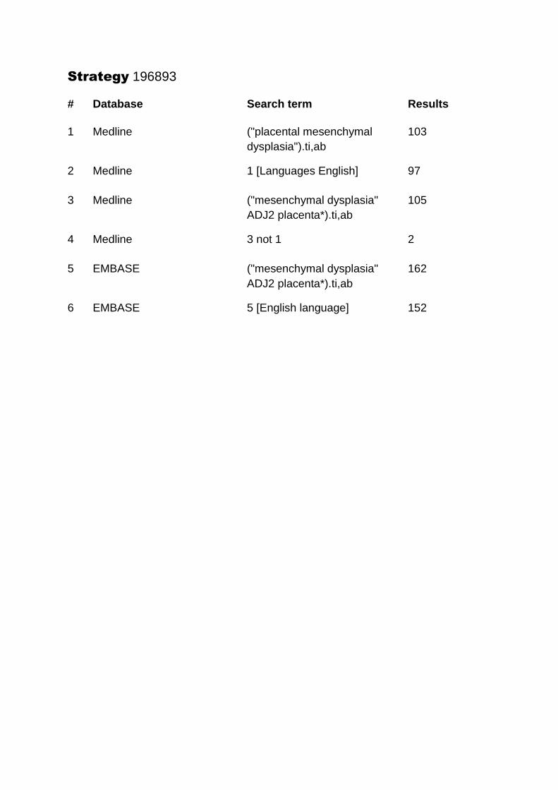
Strategy 196893
# Database Search term Results
1 Medline ("placental mesenchymal
dysplasia").ti,ab
103
2 Medline 1 [Languages English] 97
3 Medline ("mesenchymal dysplasia"
ADJ2 placenta*).ti,ab
105
4 Medline 3 not 1 2
5 EMBASE ("mesenchymal dysplasia"
ADJ2 placenta*).ti,ab
162
6 EMBASE 5 [English language] 152