Embed Size (px)
Citation preview
ENG
LISH
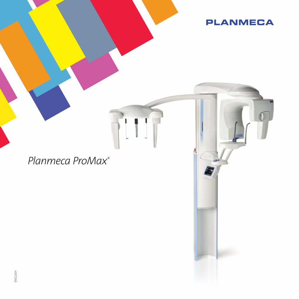
Planmeca ProMax®
2 3
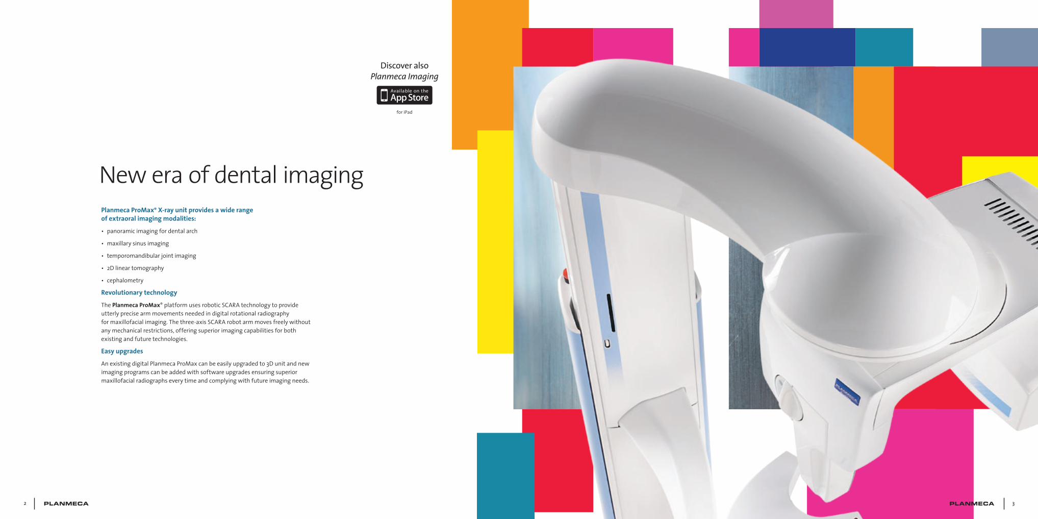
Planmeca ProMax® X-ray unit provides a wide range of extraoral imaging modalities:
• panoramicimagingfordentalarch
• maxillarysinusimaging
• temporomandibularjointimaging
• 2Dlineartomography
• cephalometry
Revolutionary technology
ThePlanmeca ProMax®platformusesroboticSCARAtechnologytoprovideutterlyprecisearmmovementsneededindigitalrotationalradiographyformaxillofacialimaging.Thethree-axisSCARArobotarmmovesfreelywithoutanymechanicalrestrictions,offeringsuperiorimagingcapabilitiesforbothexistingandfuturetechnologies.
Easy upgrades
AnexistingdigitalPlanmecaProMaxcanbeeasilyupgradedto3Dunitandnewimagingprogramscanbeaddedwithsoftwareupgradesensuringsuperiormaxillofacialradiographseverytimeandcomplyingwithfutureimagingneeds.
New era of dental imaging
Discover also Planmeca Imaging
foriPad
4 5
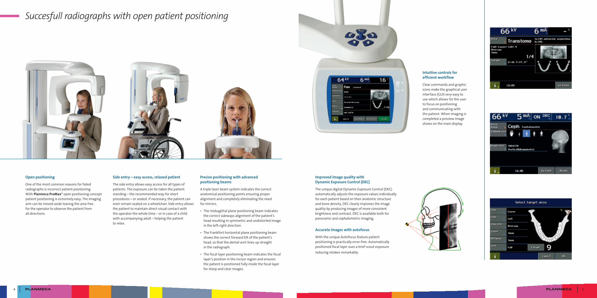
Succesfull radiographs with open patient positioning
Open positioning
Oneofthemostcommonreasonsforfailedradiographsisincorrectpatientpositioning.WithPlanmeca ProMax®openpositioningconceptpatientpositioningisextremelyeasy.Theimagingarmcanbemovedasideleavingtheareafreefortheoperatortoobservethepatientfromalldirections.
Side entry – easy access, relaxed patient
Thesideentryallowseasyaccessforalltypesofpatients.Theexposurecanbetakenthepatientstanding–therecommendedwayforshortprocedures–orseated.Ifnecessary,thepatientcanevenremainseatedonawheelchair.Sideentryallowsthepatienttomaintaindirectvisualcontactwiththeoperatorthewholetime–orincaseofachildwithaccompanyingadult–helpingthepatienttorelax.
Precise positioning with advanced positioning beams
Atriplelaserbeamsystemindicatesthecorrectanatomicalpositioningpointsensuringproperalignmentandcompletelyeliminatingtheneedformirrors.
• Themidsagittalplanepositioningbeamindicatesthecorrectsidewaysalignmentofthepatient’sheadresultinginsymmetricandundistortedimageintheleft-rightdirection.
• TheFrankforthorizontalplanepositioningbeamshowsthecorrectforwardtiltofthepatient’shead,sothatthedentalarchlinesupstraightintheradiograph.
• Thefocallayerpositioningbeamindicatesthefocallayer’spositionintheincisorregionandensuresthepatientispositionedfullyinsidethefocallayerforsharpandclearimages.
Improved image quality with Dynamic Exposure Control (DEC)
TheuniquedigitalDynamicExposureControl(DEC)automaticallyadjuststheexposurevaluesindividuallyforeachpatientbasedontheiranatomicstructureandbonedensity.DECclearlyimprovestheimagequalitybyproducingimagesofmoreconsistentbrightnessandcontrast.DECisavailablebothforpanoramicandcephalometricimaging.
Accurate images with autofocus
WiththeuniqueAutofocusfeaturepatientpositioningispracticallyerror-free.Automaticallypositionedfocallayerusesabriefscoutexposurereducingretakesremarkably.
Intuitive controls for efficient workflow
Clearcommandsandgraphiciconsmakethegraphicaluserinterface(GUI)veryeasytousewhichallowsfortheusertofocusonpositioningandcommunicatingwiththepatient.Whenimagingiscompletedapreviewimageshowsonthemaindisplay.
6 7
Special programs for various diagnostic needs
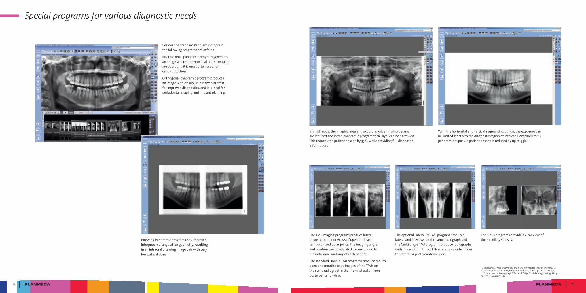
BesidestheStandardPanoramicprogramthefollowingprogramsareoffered:
Interproximalpanoramicprogramgeneratesanimagewhereinterproximalteethcontactsareopen,anditismostoftenusedforcariesdetection.
Orthogonalpanoramicprogramproducesanimagewithclearlyvisiblealveolarcrestforimproveddiagnostics,anditisidealforperiodontalimagingandimplantplanning.
BitewingPanoramicprogramusesimprovedinterproximalangulationgeometry,resultinginanintraoralbitewingimagepairwithverylowpatientdose.
Inchildmode,theimagingareaandexposurevaluesinallprogramsarereducedandinthepanoramicprogramfocallayercanbenarrowed.Thisreducesthepatientdosageby35%,whileprovidingfulldiagnosticinformation.
Withthehorizontalandverticalsegmentingoption,theexposurecanbelimitedstrictlytothediagnosticregionofinterest.Comparedtofullpanoramicexposurepatientdosageisreducedbyupto93%.*
TheTMJimagingprogramsproducelateralorposteroanteriorviewsofopenorclosedtemporomandibularjoints.Theimagingangleandpositioncanbeadjustedtocorrespondtotheindividualanatomyofeachpatient.
ThestandardDoubleTMJprogramsproducemouthopenandmouthclosedimagesoftheTMJsonthesameradiographeitherfromlateralorfromposteroanteriorview.
TheoptionalLateral-PATMJprogramproduceslateralandPAviewsonthesameradiographandtheMulti-angleTMJprogramsproduceradiographswithimagesfromthreedifferentangleseitherfromthelateralorposteroanteriorview.
Thesinusprogramsprovideaclearviewofthemaxillarysinuses.
*Absorbed dose reduced by sliced exposure using sector selector system with rotational panoramic radiography,Y.Hayakawa,N.Kobayashi,Y.Kousuge,H.FujimoriandK.Kuroyanagi,BulletinofTokyoDentalCollege,Vol.35,No.3,pp.127–131,August,1994
8 9
New opportunities with tomography
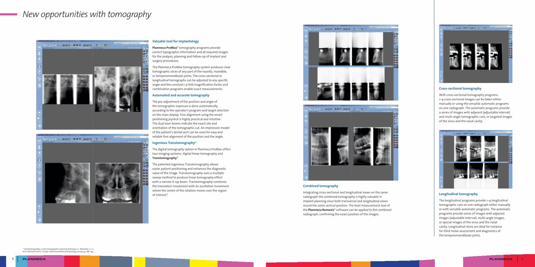
Valuable tool for implantology
Planmeca ProMax®tomographyprogramsprovidecorrecttopographicinformationandallrequiredimagesfortheanalysis,planningandfollow-upofimplantandsurgeryprocedures.
ThePlanmecaProMaxtomographysystemproducescleartomographicslicesofanypartofthemaxilla,mandible,ortemporomandibularjoints.Thecross-sectionalorlongitudinaltomographscanbeadjustedtoanyspecificangleandtheconstant1.5-foldmagnificationfactorandcombinationprogramsenableexactmeasurements.
Automated and accurate tomography
Thepre-adjustmentofthepositionandangleofthetomographicexposureisdoneautomaticallyaccordingtotheoperator’sprogramandtargetselectiononthemaindisplay.Fine-alignmentusingthesmartpositioningjoystickishighlypracticalandintuitive.Theduallaserbeamsindicatetheexactsiteandorientationofthetomographiccut.Animpressionmodelofthepatient’sdentalarchcanbeusedforeasyandreliablefine-alignmentofthepositionandtheangle.
Ingenious Transtomography®
ThedigitaltomographyoptioninPlanmecaProMaxofferstwoimagingsystems:digitallineartomographyandTranstomography®.
ThepatentedingeniousTranstomographyallowseasierpatientpositioningandenhancesthediagnosticvalueoftheimage.TranstomographyusesamultiplesweepmethodtoproducelineartomographyeffectwithanarrowX-raybeam.Transtomographycombinesthetranslationmovementwithanoscillationmovementwherethecentreoftherotationmovesovertheregionofinterest.*
Combined tomography
Integratingcross-sectionalandlongitudinalviewsonthesameradiographthecombinedtomographyishighlyvaluableinimplantplanningsincebothtransversalandlongitudinalviewsrecordthesameverticalposition.ThelevelmeasurementtoolofthePlanmeca Romexis®softwarecanbeappliedtothecombinedradiograph,confirmingtheexactpositionoftheimages.
Cross-sectional tomography
Withcross-sectionaltomographyprograms,1–4cross-sectionalimagescanbetakeneithermanuallyorusingtheversatileautomaticprogramsononeradiograph.Theautomaticprogramsprovideaseriesofimageswithadjacent(adjustableinterval)andmulti-angletomographiccuts,ortargetedimagesofthesinusandthenasalcavity.
Longitudinal tomography
Thelongitudinalprogramsprovide1–4longitudinaltomographiccutsononeradiographeithermanuallyorwithversatileautomaticprograms.Theautomaticprogramsprovideseriesofimageswithadjacentimages(adjustableinterval),multi-angleimages,orspecialimagesofthesinusandthenasalcavity.Longitudinalslicesareidealforinstanceforthirdmolarassessmentanddiagnosticsofthetemporomandibularjoints.
* Transtomography: a new tomographic scanning technique,U.Welander,G.Li,W.D.McDavidandG.Tronje,DentomaxillofacialRadiology(2004)33,188–195
10 11
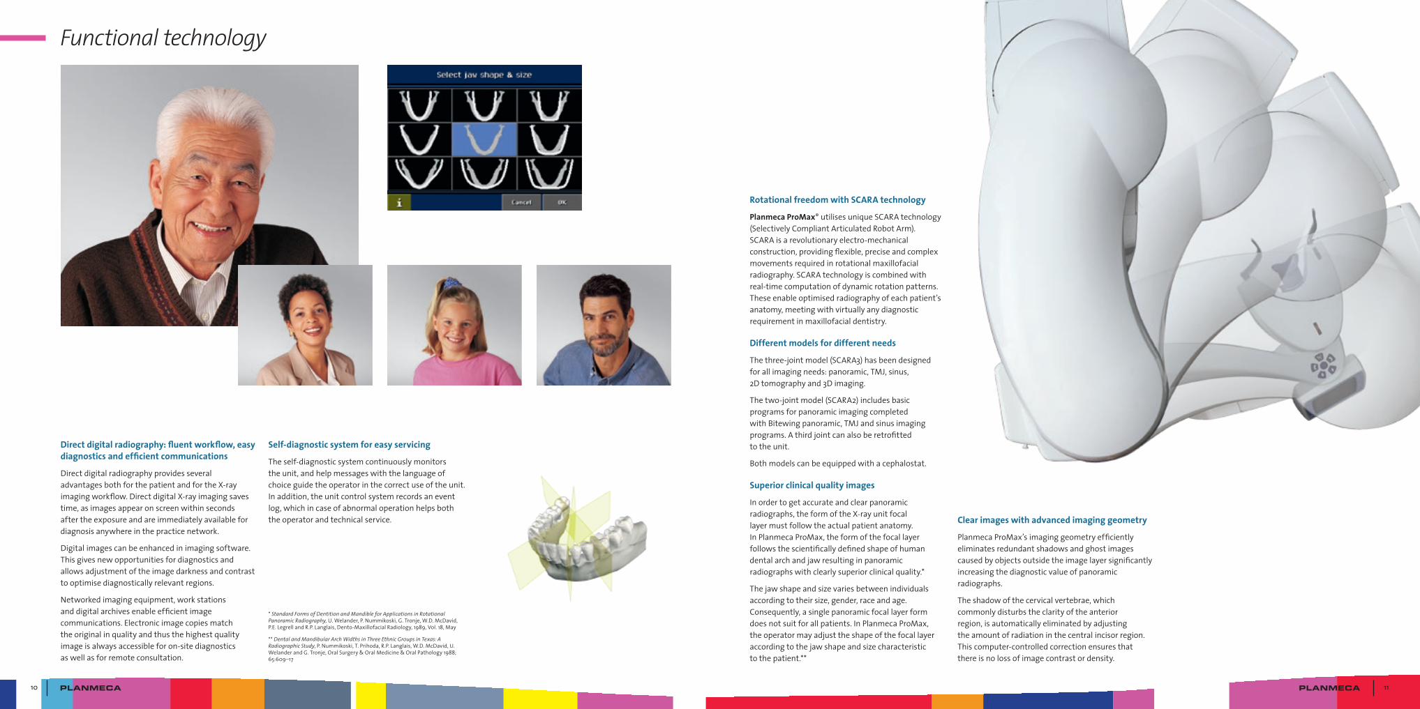
Functional technology
Direct digital radiography: fluent workflow, easy diagnostics and efficient communications
DirectdigitalradiographyprovidesseveraladvantagesbothforthepatientandfortheX-rayimagingworkflow.DirectdigitalX-rayimagingsavestime,asimagesappearonscreenwithinsecondsaftertheexposureandareimmediatelyavailablefordiagnosisanywhereinthepracticenetwork.
Digitalimagescanbeenhancedinimagingsoftware.Thisgivesnewopportunitiesfordiagnosticsandallowsadjustmentoftheimagedarknessandcontrasttooptimisediagnosticallyrelevantregions.
Networkedimagingequipment,workstationsanddigitalarchivesenableefficientimagecommunications.Electronicimagecopiesmatchtheoriginalinqualityandthusthehighestqualityimageisalwaysaccessibleforon-sitediagnosticsaswellasforremoteconsultation.
Rotational freedom with SCARA technology
Planmeca ProMax®utilisesuniqueSCARAtechnology(SelectivelyCompliantArticulatedRobotArm).SCARAisarevolutionaryelectro-mechanicalconstruction,providingflexible,preciseandcomplexmovementsrequiredinrotationalmaxillofacialradiography.SCARAtechnologyiscombinedwithreal-timecomputationofdynamicrotationpatterns.Theseenableoptimisedradiographyofeachpatient’sanatomy,meetingwithvirtuallyanydiagnosticrequirementinmaxillofacialdentistry.
Different models for different needs
Thethree-jointmodel(SCARA3)hasbeendesignedforallimagingneeds:panoramic,TMJ,sinus,2Dtomographyand3Dimaging.
Thetwo-jointmodel(SCARA2)includesbasicprogramsforpanoramicimagingcompletedwithBitewingpanoramic,TMJandsinusimagingprograms.Athirdjointcanalsoberetrofittedtotheunit.
Bothmodelscanbeequippedwithacephalostat.
Superior clinical quality images
Inordertogetaccurateandclearpanoramicradiographs,theformoftheX-rayunitfocallayermustfollowtheactualpatientanatomy.InPlanmecaProMax,theformofthefocallayerfollowsthescientificallydefinedshapeofhumandentalarchandjawresultinginpanoramicradiographswithclearlysuperiorclinicalquality.*
Thejawshapeandsizevariesbetweenindividualsaccordingtotheirsize,gender,raceandage.Consequently,asinglepanoramicfocallayerformdoesnotsuitforallpatients.InPlanmecaProMax,theoperatormayadjusttheshapeofthefocallayeraccordingtothejawshapeandsizecharacteristictothepatient.**
*Standard Forms of Dentition and Mandible for Applications in Rotational Panoramic Radiography,U.Welander,P.Nummikoski,G.Tronje,W.D.McDavid,P.E.LegrellandR.P.Langlais,Dento-MaxillofacialRadiology,1989,Vol.18,May
**Dental and Mandibular Arch Widths in Three Ethnic Groups in Texas: A Radiographic Study,P.Nummikoski,T.Prihoda,R.P.Langlais,W.D.McDavid,U.WelanderandG.Tronje,OralSurgery&OralMedicine&OralPathology1988;65:609–17
Self-diagnostic system for easy servicing
Theself-diagnosticsystemcontinuouslymonitorstheunit,andhelpmessageswiththelanguageofchoiceguidetheoperatorinthecorrectuseoftheunit.Inaddition,theunitcontrolsystemrecordsaneventlog,whichincaseofabnormaloperationhelpsboththeoperatorandtechnicalservice. Clear images with advanced imaging geometry
PlanmecaProMax’simaginggeometryefficientlyeliminatesredundantshadowsandghostimagescausedbyobjectsoutsidetheimagelayersignificantlyincreasingthediagnosticvalueofpanoramicradiographs.
Theshadowofthecervicalvertebrae,whichcommonlydisturbstheclarityoftheanteriorregion,isautomaticallyeliminatedbyadjustingtheamountofradiationinthecentralincisorregion.Thiscomputer-controlledcorrectionensuresthatthereisnolossofimagecontrastordensity.
12 13
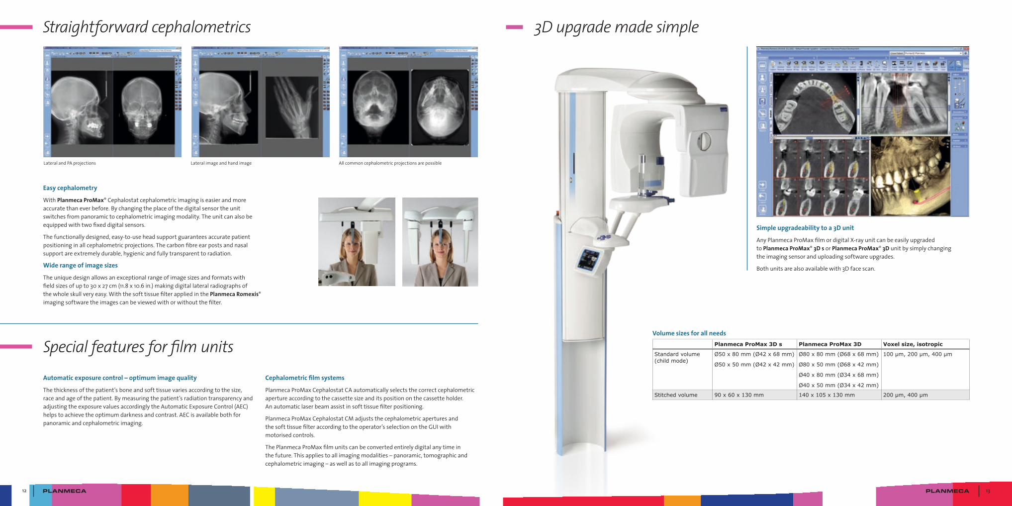
Straightforward cephalometrics
Automatic exposure control – optimum image quality
Thethicknessofthepatient’sboneandsofttissuevariesaccordingtothesize,raceandageofthepatient.Bymeasuringthepatient’sradiationtransparencyandadjustingtheexposurevaluesaccordinglytheAutomaticExposureControl(AEC)helpstoachievetheoptimumdarknessandcontrast.AECisavailablebothforpanoramicandcephalometricimaging.
Special features for film units
3D upgrade made simple
Cephalometric film systems
PlanmecaProMaxCephalostatCAautomaticallyselectsthecorrectcephalometricapertureaccordingtothecassettesizeanditspositiononthecassetteholder.Anautomaticlaserbeamassistinsofttissuefilterpositioning.
PlanmecaProMaxCephalostatCMadjuststhecephalometricaperturesandthesofttissuefilteraccordingtotheoperator’sselectionontheGUIwithmotorisedcontrols.
ThePlanmecaProMaxfilmunitscanbeconvertedentirelydigitalanytimeinthefuture.Thisappliestoallimagingmodalities–panoramic,tomographicandcephalometricimaging–aswellastoallimagingprograms.
Simple upgradeability to a 3D unit
AnyPlanmecaProMaxfilmordigitalX-rayunitcanbeeasilyupgradedtoPlanmeca ProMax® 3D sorPlanmeca ProMax® 3Dunitbysimplychangingtheimagingsensoranduploadingsoftwareupgrades.
Bothunitsarealsoavailablewith3Dfacescan.
Easy cephalometry
WithPlanmeca ProMax®Cephalostatcephalometricimagingiseasierandmoreaccuratethaneverbefore.Bychangingtheplaceofthedigitalsensortheunitswitchesfrompanoramictocephalometricimagingmodality.Theunitcanalsobeequippedwithtwofixeddigitalsensors.
Thefunctionallydesigned,easy-to-useheadsupportguaranteesaccuratepatientpositioninginallcephalometricprojections.Thecarbonfibreearpostsandnasalsupportareextremelydurable,hygienicandfullytransparenttoradiation.
Wide range of image sizes
Theuniquedesignallowsanexceptionalrangeofimagesizesandformatswithfieldsizesofupto30x27cm(11.8x10.6in.)makingdigitallateralradiographsofthewholeskullveryeasy.WiththesofttissuefilterappliedinthePlanmeca Romexis®imagingsoftwaretheimagescanbeviewedwithorwithoutthefilter.
Lateralimageandhandimage AllcommoncephalometricprojectionsarepossibleLateralandPAprojections
Volume sizes for all needsPlanmeca ProMax 3D s Planmeca ProMax 3D Voxel size, isotropic
Standard volume (child mode)
Ø50 x 80 mm (Ø42 x 68 mm)
Ø50 x 50 mm (Ø42 x 42 mm)
Ø80 x 80 mm (Ø68 x 68 mm)
Ø80 x 50 mm (Ø68 x 42 mm)
Ø40 x 80 mm (Ø34 x 68 mm)
Ø40 x 50 mm (Ø34 x 42 mm)
100 µm, 200 µm, 400 µm
Stitched volume 90 x 60 x 130 mm 140 x 105 x 130 mm 200 µm, 400 µm
14 15
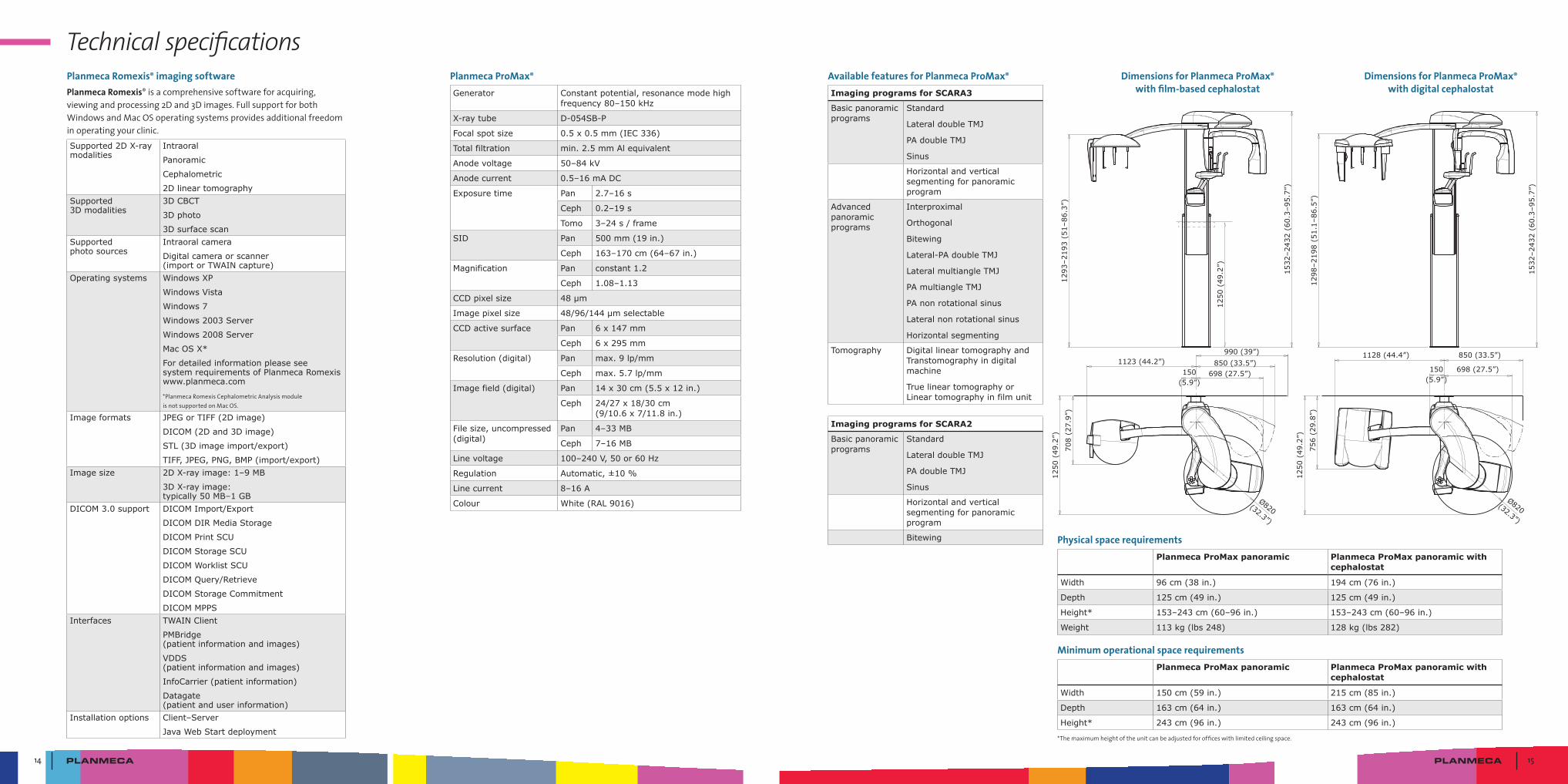
Technical specificationsDimensions for Planmeca ProMax®
with film-based cephalostatDimensions for Planmeca ProMax®
with digital cephalostat
1293
–219
3 (5
1–86
.3”)
1250
(49
.2”)
1532
–243
2 (6
0.3–
95.7
”)
1298
–219
8 (5
1.1–
86.5
”)
1532
–243
2 (6
0.3–
95.7
”)
708
(27.
9”)
698 (27.5”)150(5.9”)
1123 (44.2”) 850 (33.5”)
Ø820(32.3”)
1250
(49
.2”)
1250
(49
.2”)
756
(29.
8”)
698 (27.5”)150(5.9”)
1128 (44.4”) 850 (33.5”)
Ø820(32.3”)
990 (39”)
Planmeca ProMax®
Generator Constant potential, resonance mode high frequency 80–150 kHz
X-ray tube D-054SB-P
Focal spot size 0.5 x 0.5 mm (IEC 336)
Total filtration min. 2.5 mm Al equivalent
Anode voltage 50–84 kV
Anode current 0.5–16 mA DC
Exposure time Pan 2.7–16 s
Ceph 0.2–19 s
Tomo 3–24 s / frame
SID Pan 500 mm (19 in.)
Ceph 163–170 cm (64–67 in.)
Magnification Pan constant 1.2
Ceph 1.08–1.13
CCD pixel size 48 µm
Image pixel size 48/96/144 µm selectable
CCD active surface Pan 6 x 147 mm
Ceph 6 x 295 mm
Resolution (digital) Pan max. 9 lp/mm
Ceph max. 5.7 lp/mm
Image field (digital) Pan 14 x 30 cm (5.5 x 12 in.)
Ceph 24/27 x 18/30 cm (9/10.6 x 7/11.8 in.)
File size, un compressed (digital)
Pan 4–33 MB
Ceph 7–16 MB
Line voltage 100–240 V, 50 or 60 Hz
Regulation Automatic, ±10 %
Line current 8–16 A
Colour White (RAL 9016)
Available features for Planmeca ProMax®
Imaging programs for SCARA3
Basic panoramic programs
Standard
Lateral double TMJ
PA double TMJ
Sinus
Horizontal and vertical segmenting for panoramic program
Advanced panoramic programs
Interproximal
Orthogonal
Bitewing
Lateral-PA double TMJ
Lateral multiangle TMJ
PA multiangle TMJ
PA non rotational sinus
Lateral non rotational sinus
Horizontal segmenting
Tomography Digital linear tomography and Transtomography in digital machine
True linear tomography or Linear tomography in film unit
Imaging programs for SCARA2
Basic panoramic programs
Standard
Lateral double TMJ
PA double TMJ
Sinus
Horizontal and vertical segmenting for panoramic program
Bitewing Physical space requirementsPlanmeca ProMax panoramic Planmeca ProMax panoramic with
cephalostat
Width 96 cm (38 in.) 194 cm (76 in.)
Depth 125 cm (49 in.) 125 cm (49 in.)
Height* 153–243 cm (60–96 in.) 153–243 cm (60–96 in.)
Weight 113 kg (lbs 248) 128 kg (lbs 282)
Minimum operational space requirementsPlanmeca ProMax panoramic Planmeca ProMax panoramic with
cephalostat
Width 150 cm (59 in.) 215 cm (85 in.)
Depth 163 cm (64 in.) 163 cm (64 in.)
Height* 243 cm (96 in.) 243 cm (96 in.)
*Themaximumheightoftheunitcanbeadjustedforofficeswithlimitedceilingspace.
Planmeca Romexis® imaging softwarePlanmeca Romexis®isacomprehensivesoftwareforacquiring,viewingandprocessing2Dand3Dimages.FullsupportforbothWindowsandMacOSoperatingsystemsprovidesadditionalfreedominoperatingyourclinic.Supported 2D X-ray modalities
Intraoral
Panoramic
Cephalometric
2D linear tomographySupported 3D modalities
3D CBCT
3D photo
3D surface scanSupported photo sources
Intraoral camera
Digital camera or scanner (import or TWAIN capture)
Operating systems Windows XP
Windows Vista
Windows 7
Windows 2003 Server
Windows 2008 Server
Mac OS X*
For detailed information please see system requirements of Planmeca Romexis www.planmeca.com
*PlanmecaRomexisCephalometricAnalysismoduleisnotsupportedonMacOS.
Image formats JPEG or TIFF (2D image)
DICOM (2D and 3D image)
STL (3D image import/export)
TIFF, JPEG, PNG, BMP (import/export)Image size 2D X-ray image: 1–9 MB
3D X-ray image: typically 50 MB–1 GB
DICOM 3.0 support DICOM Import/Export
DICOM DIR Media Storage
DICOM Print SCU
DICOM Storage SCU
DICOM Worklist SCU
DICOM Query/Retrieve
DICOM Storage Commitment
DICOM MPPSInterfaces TWAIN Client
PMBridge (patient information and images)
VDDS (patient information and images)
InfoCarrier (patient information)
Datagate (patient and user information)
Installation options Client–Server
Java Web Start deployment
10016
041/0
712
/en
Planmeca Oy designs and manufactures a full line of high technology dental equipment, including dental care units, panoramic and intraoral X-ray units, and digital imaging products. Planmeca Oy, the parent company of the Finnish Planmeca Group,
is strongly committed to R&D, and is the largest privately held company in the field.
Asentajankatu 6 | 00880 Helsinki | Finland | tel. +358 20 7795 500 | fax +358 20 7795 555 | [email protected] | www.planmeca.com
Images may contain optional items not included in standard delivery. Available configurations and features may have country or area specific variations. Some products displayed above may not be available in all countries or areas. Rights for changes reserved. All in one, Anatomat Plus, Comfy, DentroVac, Digital perfection, Economat Plus, Elegant, Mini-dent, PlanEasyMill, Planmeca Chair, Planmeca Compact, Planmeca Intra, Planmeca iRomexis, Planmeca Lumion, Planmeca Minea, Planmeca Minendo,
Planmeca Minetto, Planmeca Online, Planmeca PlanScan, Planmeca ProCeph, Planmeca ProFace, Planmeca ProMax, Planmeca ProModel, Planmeca ProOne, Planmeca ProSensor, Planmeca ProX, Planmeca Romexis, Planmeca SingLED, Planmeca Sonea, Planmeca Sovereign, Planmeca Vision, Planmeca Waterline Cleaning System, Proline Dental Stool, Saddle Stool, SmartPan, Trendy and Ultra Relax are registered or non-registered trademarks of Planmeca in various countries.