Embed Size (px)
Citation preview
461N. Tuteja and S. Singh Gill (eds.), Plant Acclimation to Environmental Stress, DOI 10.1007/978-1-4614-5001-6_17, © Springer Science+Business Media New York 2013
1 Introduction
Plants are important natural resources source which can convert the solar energy into various usable forms including energy substance glucose and storage carbohydrate like starch. However, time to time, the potential of plants is challenged by many environmental fl uctuations including various biotic and abiotic stresses. Excess and de fi cit of any physical or biological factor can cause stress to the plant. Recently, the world is witnessing climatic changes such as increase in temperature, irregular rains, drought, excessive salt, fl ooding, excessive chilling, cyclones, tornedos, and various other natural calamities.
Stress causes changes in plant cellular functioning; as it prepares the plant for the incoming dangers. All the inactive protector genes become active due to sudden shift in the biochemistry and molecular biology of plant cell. The key organ in plants that acts as a fi rst line of defense is cell wall. Which is useful to protect plant from pest or pathogen invasion and chilling or mechanical stress. However, it is unable to secure plants from stresses like water de fi cit, salt stress etc.
D. Bhardwaj International Centre for Genetic Engineering and Biotechnology (ICGEB), Aruna Asaf Ali Marg , New Delhi , 110 067 , India
Department of Botany , University of Delhi , New Delhi , India
S. Lakhanpaul Department of Botany , University of Delhi , New Delhi , India
N. Tuteja (*) Plant Molecular Biology, International Centre for Genetic Engineering and Biotechnology (ICGEB), Aruna Asaf Ali Marg , New Delhi , India e-mail: [email protected]
Chapter 17 Can G-Proteins be the Key Proteins for Overcoming Environmental Stresses and Increasing Crop Yield in Plants?
Deepak Bhardwaj , Suman Lakhanpaul, and Narendra Tuteja
462 D. Bhardwaj et al.
Recently, G-proteins and related machinery has been noted for its role in regulating stress related pathways, including ROS production, stomatal regulation and process related to plant water relations. We hereby present the role of G-proteins in all aspects of plant life and its metabolism (AM Jones 1994 ).
2 What is Stress? Perception and Signal Transduction Pathway
Any object that is affected by stress whether living or non-living undergoes de fi nite change in response to stress which is known as strain. In case of plants it is hard to measure the propensity of stress. However, during stressed conditions plants show many molecular and cellular changes.
Water is a primary requirement of plant, and its excess as well as de fi cit affect the growth of plants. Water is absorbed by roots, and root hairs serve as channels to increase the surface area of absorption. The whole physiology of stress can only be understood if one knows the parameters that decide the water status of plant. These parameters are water potential and relative water content.
Presence of cell wall in case of plants protects them from outer dangers including pathogens and adverse conditions. Plant cell membrane is a semi-permeable, quasi- fl uid structure which can allow only selective substances to pass through it; they serve as a second wall having many gates in the form of receptors and ion channels. Receptors being sensitive in nature are vulnerable to several elicitors that direct plant cell to respond against various stimuli. The cellular responses are initiated primarily by interaction of the extracellular material with a plasma membrane pro-tein. This extracellular molecule is called a ligand (or an elicitor) and the plasma membrane protein, which binds and interacts with this molecule, is called a recep-tor. Interestingly, various stress factors, both abiotic as well as biotic, act as elicitors for the plant cell. Plant growth and development are mediated by complex array of signaling pathways co-ordinated by exogenous factors, which regulate all phases of growth including cell division, differentiation, and cell death. During evolution all living organisms including plants have been subjected to continuous variation of external environmental stimuli such as heat, cold, light, darkness, and other rhyth-mic parameters. In response to these external stimuli cells start many reactions to regulate their physiological processes. The cells of multicellular organisms also need to communicate with each other in order to co-ordinate their growth and dif-ferentiation. All fundamental processes of biology including growth and develop-ment rely on the proper response to environmental signals (Bowler and Chua 1994 ). The molecular mechanism by which these signals are perceived by cells, transduced toward the proper targets, and integrated into a biological response is known as signal transduction. Basically, two functional principles are involved in the extracel-lular signal (primary stimulus) as the fi rst messenger itself penetrates the cell (prob-ably through speci fi c receptors) and fi nds its way to the nucleus. Alternatively, the signal remains outside the cell and is converted at plasma membrane into intracellular
46317 Can G-Proteins be the Key Proteins for Overcoming Environmental Stresses…
signals called second messenger (Hucho and Buchner 1997 ). Subsequent, sites are in the cytoplasm, at organelle membranes such as nuclear envelope and inside the nucleus.
Cell is like a universe where proteins like humans interact with each other, and one protein can interact with several and so on. These connections and interactions create a network or web of many signaling cascades which fi nally lead to the dif-ferential regulation of genes. Receptors are like sensors that sense the external envi-ronment and send the information to the cell machinery through various effectors, carriers, modulators, transcription factors, secondary messengers, enzymes etc. No information can be transcribed into response until receptor sends the signals to nucleus which is harboring plethora of dormant genes. Many plant receptors have been reported till date.
Stress is fi rst recognized by the receptors present on the plasma membranes. Many membrane receptors are characterized till date which includes serine threo-nine kinase (Afzal et al . 2008 ) , receptor-like protein kinase (Walker 1994 ) , cal-cineurin B-like sensors (Cheong et al . 2007 ) , G-protein-coupled receptor (Jones and Assmann 2004 ) , etc. After recognition, there is an induction of the relay of various proteins along with the generation of second messengers including cal-cium, calmodulin, calcium-induced protein kinases, calcium-dependent protein kinases, G-proteins, MAP kinase, (ROS), and inositol phosphates. Various pro-teins in a given cell may or may not work in isolation; they interact with several other proteins to bring about certain changes that could be the need of time. This initiates the activity of various kinases and phosphatases which ultimately targets various genes and trans-elements. The up-regulation of certain dormant genes is required for the survival of cell under stressed conditions. This gives plant the potential to adapt or acclimatize to the particular environment. There are many hormones related to stress that add to this effect and activate many stress-induced genes. Their level generally increases during stressed conditions and they further start different signaling pathways to resist stressed conditions.
There is a different group of molecules that initiates and catalyses the modi fi cation of certain signaling proteins. They help in the modi fi cation or assembly of signaling components. Myristoylation, glycosylation, methylation and ubiquitination are some common modi fi cations that give stress-related proteins some special features so that they can perform functions under alternate situations. Stress response could be early or late, and, on the basis of this, various stress-responsive genes can be broadly categorized as early and late induced genes. Early genes are induced within minutes and these genes belong to transcription factors, whereas other genes like RD (responsive to dehydration) (Kariola et al . 2006 ) and KIN (cold induced)/COR (cold responsive) (Thomashow 1998 ) , which encode and modulate the proteins needed for synthesis of LEA-like proteins (late embryogenesis abundant) ( Hundertmark 2008 ) , antioxidants, membrane-stabilizing proteins. However syn-thesis of osmolytes take hours to get induced.
There are many signature elements on the promoter of genes that also tell the importance of particular gene in the alleviation of various stresses such as. DRE (dehydration-responsive elements) (Sun et al . 2008 ) or CRT (C-repeats) and some
464 D. Bhardwaj et al.
of them which contain ABRE (ABA-responsive element) ( Gómez-Porras et al . 2007 ) . Stress-induced transcription factors bind to this element and their overex-pression brings about the tolerance against various stresses. Same way, there are many nuclear genes whose overexpression can give genetic potential to the plant against any kind of environmental challenges.
Description of all the genes of all the genes, transcription factors, chaperones and related proteins are beyond the scope of this chapter and we will only focus on the G-proteins and their role in various plant processes including stress.
3 What are G-proteins and Their Role in Plant Metabolism, Growth, and Development?
3.1 GPCR and G-proteins
Plant membranes like animal membranes are studded by G-protein-coupled receptor (GPCR), RGS and heterotrimeric G-proteins (G a , G b /G g subunits) that are often link to the GPCR, constituting one of the most important components of cell signal-ing cascade. Signals are perceived by GPCR or RGS and later on transduced to inner part of the cell through G-proteins. This constitutes GPCR/G-protein-induced signaling cascade. Alfred G. Gilman and Martin Rodbell were awarded Nobel prize in 1994 for their contribution in the fi eld of cell signal transduction; they proved the role of G-proteins as signaling molecules. Martin Rodbell and his collaborators linked the transducer with hormones, whereas Alfred G. Gilman and his co-worker characterized and puri fi ed the G-protein. Hundreds of chemicals and physical sig-nals that can be stress-causing elicitors constantly bombard the surface of all cells. Some of these do not enter the cell, but, instead, bind to receptors at the cell surface and initiate a fl ow of information that moves to the cell interior. The receptors for many hormones (such as catecholamine, gonadotropin, parathyroid hormone, glu-cagon and ABA), odorants and light are heptahelical structures. Stimulation of these receptors causes activation of special proteins that are linked to the G-proteins. G-protein performs various functions including regulating few types of enzymes and ion channel. The target enzymes or ion channels are called effectors and they are generally proteinaceous in nature; G-protein after activation becomes effector itself and it interacts with other proteins to bring about changes in their activity causing alterations in ionic composition or in second messenger level that ultimately leads to the cellular response (Neer 1995 ) .
GPCR is an integral membrane protein receptor that contains seven-trans-membrane a -helical regions and this portion of receptor binds to a wide range of ligands like hormones. Interestingly, binding of ligands can bring about confor-mational changes in the structure of GPCR activating G-proteins. Binding of ligands is a switching on of various steps that happen in tandem. This includes the
46517 Can G-Proteins be the Key Proteins for Overcoming Environmental Stresses…
exchange of GDP/GTP associated with the G a subunit and disassociation of alpha subunit from beta gamma dimer. The separated subunits individually act as effec-tors and interact with other intracellular proteins. This whole process can be switched off by simply replacing GTP attached to alpha subunit by GDP. This creates the earlier situation when the heterotrimer of G-proteins was attached to GPCR as a dormant unit. Nevertheless, GPCR can also work independent of G-proteins (AM Jones 2002 ) .
3.2 Historical Background of G-proteins
Historically, it is work on adenylyl cyclase since 1950 that fi nally resulted in the discovery of G-protein. Now, it is well known that G-proteins are involved in broad range of cellular regulatory activities. Nobel Prizes has been awarded in this fi eld to Sutherland and Rall ( 1958 ) , who discovered the cAMP and adenylyl cyclase as a second messenger; Fischer and Krebs (1992), whose extensive studies of reversible protein phosphorylation began with cAMP-dependent protein kinase; and Gilman and Rodbell (1994) for their work on heterotrimeric G-proteins (Vaugham 1998 ) . Here, a brief history of G-proteins has been described.
1957–1958 The G-proteins fi eld originated. Sutherland and Rail described the basic properties of the enzyme adenylyl cyclase (AC), which is involved in hormone (epinephrine)-regulated synthesis of second messenger (cAMP) (Rall 1957 ; Sutherland and Rall 1958 )
1969 Fat cell adenylyl cyclase was reported to be activated by multiple hormones: the hormone receptors were distinct from the enzyme. ATP could reverse the binding action of glucagon to the rat liver cell membrane receptor and thus dissociate the glucagon from the cell (Birnbaumer and Rodbell 1969 )
1971–1974 Role of GTP was fi rst reported by Martin Rodbell. GTP required for glucagon stimulated adenylyl cyclase in liver. GTP enhances disso-ciation of glucagon from its receptor (Rodbell et al. 1971 )
1975 Henery Bourne and co-workers isolated S49 mouse lymphoma cell line de fi cient in adenylyl cyclase activity, named cyc. These cells were later used as assay tool to isolate coupling factors (receptor, G-proteins, adenylyl cyclase) (Bourne et al. 1975 )
Pfeuffer and Helmreish ( 1975 ) separated a GTP-binding protein from the adenylyl cyclase complex
1976 Orly and Schramm ( 1976 ) directly demonstrated the independence of receptor and cyclase enzyme
466 D. Bhardwaj et al.
1977 E.M. Robs and A.G. Gilman in 1977 demonstrated the resolution of some components of adenylyl cyclase necessary for catalytic activity. They reported the 4G kDa GTP-binding protein, now, known as G
a s
Rodbell discovered that G-proteins at the cell receptor could both inhibit and activate transduction, often at the same time
1978 Robert Lefkowitz in 1978 demonstrated the conformational change occurred to receptor after binding to ligands, and GTP regulated the association with macromolecule
Cassel and Selingen in 1978 showed that activation of a GTPase (GDP was replaced by GTP) in membrane is important in hormonal activation of adenylyl cyclase
1979 Rodbell reported that G-proteins can carry out primary and second-ary processes of signal transduction
1981 R.G.L. Shorr and co-workers in 1981 reported puri fi cation of a b -adrenergic receptor, the fi rst G-protein-coupled receptor with seven-membrane-spanning a -helices
1983 A.F. Gilman resolved Gas from G b g . G a binds GTP and activates adenylyl cyclase. G b g deactivates G a -GTP
1984 Gilman identi fi ed G a i protein (inhibitory G-protein), which inhibits the activity of adenylyl cyclase
1986 cDNAs encoding Gas, Gczt (transducin), G a i, and G a q were cloned
Stryer and Bourne predicted the imminent rewards from X-ray crystallographic analysis of G-protein structure and site-speci fi c mutagenesis for functional studies
G-protein-coupled receptor (GPCR) was cloned from hamster and terkey (Dixon et al. 1986 ; Yarden et al. 1986 )
1990 First plant G a was isolated from Arabidopsis thaliana (Ma et al. 1990 )
Large G-proteins, Gha (74 kDa) and Gh7a (78 kDa), were reported in mammals (Im and Graham 1990 )
Potential G-protein-coupled receptor was shown in barely ( Devoto and Turner 2003 )
1994 First plant G b gene was isolated from Arabidopsis thaliana and maize ( Weiss et al. 1993 )
1995 AGS (activator of G-protein signaling), which activated heterotri-meric G-protein signaling in the absence of a cell surface receptor, was reported (Sato et al. 1995 )
1999 Extra-large G-protein (AtXLG 1, 99 kDa) was isolated from plant (Lee and Assmann 1999 )
ATDRG1, Arabidopsis thaliana developmentally regulated G-proteins, was reported (Etheridge et al. 1999 )
46717 Can G-Proteins be the Key Proteins for Overcoming Environmental Stresses…
2000 The fi rst high-resolution structure of a GPCR , Rhodopsin, the visual light receptor, was solved (Palczewski et al. 2000 )
First plant G g gene was isolated from Arabidopsis thaliana (Mason and Botella 2000 )
2002 Plant G a , b , and g subunits interact with each other in vitro and in vivo, and also interact with PLC- d protein
2003 A seven-transmembrane RGS protein function in plant was discovered
2004 Putative GPCR called GCR1 and its interaction with G-alpha with regard to ABA was discovered
2005 Role of G-protein in Arabidopsis seed germination
Role of G-proteins in giving resistance against rice blast fungus
2006 Role of G-proteins in regulating cell divisions in roots of Arabidopsis
Interaction between G-alpha and THF1 was shown in Arabidopsis
Interaction between G-alpha and prephenatedehydratase 1 was shown in Arabidopsis
2007 GPCR was proved to be a receptor of ABA
G a and G b subunits of Pisum sativum and their roles in salt and heat stress, respectively
2008 Role of G-protein in regulating ion channels of stomata guard cells
2009 NDL is shown as interacting partners of beta subunit of G-protein of Arabidopsis
2010 G-protein alpha subunit involved in controlling TE transpiration ef fi ciency
2011 G-protein intractome in Arabidopsis was elucidated
3.3 G-Proteins and Various Plant Processes
Life of all plants starts from seed germination which is supposed to be a complex mechanism involving many genes, enzymes and proteins. No seed can germinate properly without the acquisition and availability of nutrients stored in the seed. Seed reserve mobilization is largely dependent on cells which in turn are controlled by gibberellin and abscisic acid (ABA). Gibberellic acid (GA) being a phytohor-mone is involved in so many biological processes including stem elongation, fl owering, senescence, and seed germination. GA helps in the induction of de novo synthesis of a amylase and other enzymes in the aleurone layer of barley. GA is supposed to be perceived by the membranes of aleurone cell, from where it induces the transcription of genes of a -amylase (Mirbahar and Laidman 1982 ) . Role of
468 D. Bhardwaj et al.
G-protein in gibberellin-induced a -amylase gene expression was shown by Huw et al. in 1998. G-proteins are connected with membrane-bound GPCR through G a subunit which contains intrinsic GTPase activity (Misra et al. 2007 ) . In the acti-vated form G a -GTP acts as a signaling molecule which can catalyze several down-stream actions. Interestingly, Jones used mastoparan analogue Mas7 which stimulates GDP-to-GTP exchange in the same fashion as activated GPCR performs when elicited by ligands. It was found that when the protoplast of aleurone was incubated with Mas7 it can secrete a -amylase. Further, this was con fi rmed by using a -Amy2/54: GUS promoter: reporter construct and the hydrolysis-resistant guanine nucleotide analogues GTP- g -S and GDP- b -S. GTP- g -S and GDP- b -S bind to G a subunits and keep them in either the activated (GTP- g -S-bound) or inactivated (GDP- b -S-bound) form. Expectedly, when GDP- b -S was introduced into aleurone protoplasts along with reporter gene constructs, the GUS activity was found to be completely abolished. In the other case, GTP- g -S caused the slight expression of reporter gene construct. Therefore, G-protein has been shown to be clearly linked with GA signaling and germination. Next to the germination is development of the seedling which requires sunlight and pigments like chlorophylls, phytochrome, and cryptochrome.
In early 1991, it was reported G-protein is associated with the plasma mem-branes of the apical bud of etiolated peas and the GTPase activity is induced by low in fl uences of blue light (Warpeha et al . 1991 ) . G-protein mediation of blue-green light perception by rhodopsin in fl agellate green algae also has been proposed (Calenberg et al . 1998 ) .
Deng’s group (Okamoto et al . 2001 ) generated transgenic Arabidopsis express-ing the G a subunit under the control of a glucocorticoid-inducible promoter. With the conditional overexpression of either the wild type or a constitutively active ver-sion of Arabidopsis G a , transgenic seedlings exhibited a hypersensitive response to light. This enhanced light sensitivity was more exaggerated at a relatively lower intensity of light and was observed in white as well as far-red, red, and blue light conditions.
Many research workers suggested the involvement of G-proteins in phytochrome-mediated light. In the early 1990s it was shown that dark grown soybean cells when treated with cholera or pertussis toxins could uncouple phytochrome-dependent expression of chlorophyll a/b-binding protein (cab) (Romero and Lam 1993 ) . Interestingly, tomato photo-morphogenetic mutant can mock the effect of phyto-chrome-mediated responses like anthocyanin production and chloroplast develop-ment, when treated with elicitors of G-proteins (Neuhaus et al. 1993 ; Bowler et al. 1994a, b ) . Cross-talk between G-protein could be Ca 2+ dependent or Ca 2+ indepen-dent (Neuhaus et al . 1993 ; Bowler et al. 1994a, b ) . The link between G-protein in phyA signaling supported the fact that various other proteins are also involved to fi nally convey message from photoreceptors to these two proteins (Neuhaus et al . 1993 ; Bowler et al. 1994a, b ) . Chua and colleagues (Neuhaus et al . 1993 ; Bowler et al . 1994a, b ) combined all biological, molecular, and genetic approaches to more thoroughly address the role of heterotrimeric G-protein in phytochrome responses.
46917 Can G-Proteins be the Key Proteins for Overcoming Environmental Stresses…
They used the phytochrome A (Phy A)-de fi cient tomato mutant aurea to investigate whether G-protein activation could initiate known phytochrome responses like chloroplast development, anthocyanin biosynthesis, and CAB gene expression in a Phy-de fi cient genetic background. Microinjection of puri fi ed oat PhyA into indi-vidual hypocotyl cells was shown to partially restore this response in a cell-autono-mous and red/far-red light-reversible manner. Co-injection of either GDP b S or PTX with PhyA eliminated the responses, whereas injection of GTP g S alone initiated them. Analogue manipulations implicated calcium/calmodulin as acting down-stream of the G-protein.
G-proteins are also involved in brassinosteroids signaling. In 1997, Wang et al.characterized G-alpha subunit of a heterotrimer G-protein from rice. This subunit was found to be involved in disease resistance. Later on, the same subunit was dis-covered to be involved in brassinosteroids signaling (Wang et al . 2006 ; Oki et al. 2009a, b ) . Rice harbors one alpha (RGA1) and beta (RGB1) subunit and two gamma subunits (RGG1 and RGG2). The dwarf mutant of rice, often known as D1 mutant, is actually identi fi ed as mutant of RGA1 (Izawa et al. 2010 ).
Arabidopsis G-proteins were studied in great detail owing to the availability of its diverse natural and mutant genetic resources. It has one alpha subunit (GPA1), one beta (AGB1) and three gamma subunits (AGG1, AGG2, and AGG3). Interestingly, the results of rice have been con fi rmed in Arabidopsis though later is a dicotyledonous model system. The G-proteins of Arabidopsis remain intact till the GCR/RGS gets activated by a ligand. ABA was found to bring about the conforma-tional changes in GCR thus causing the release of activated GPA1 and AGB1 and AGG1 or AGG2 or AGG3 dimer. ABA plays an important role in seed germination, seedling development, and guard cell regulation including various environmental stresses. In one of the study, the G-protein knock-out mutants were used to see the effect on ABA signaling on seed germination; interestingly, it was found that agb1 mutant is more sensitive to ABA than gpa1 (Pandey and Assmann 2004 ; Pandey et al . 2006 ) . Null mutants of GPA1 were found to be insensitive to gibberellin and BRs and highly sensitive to glucose (Ullah et al . 2002 ) . The same mutants behaved as wild type in response to ABA and ethylene.
Cytokinin, one of the plant hormones, is shown to induce stomata opening, cause cell division, and release plant from the effect of apical dominance. They are also involved in the photosensory responses. In fact, it was thought at one time that light-induced effectors activated by cytokinin have some interaction with G-proteins or related effectors. Though it is not yet proved that whether GCR1 is a receptor for cytokinin or not, some reports of late 90s proved the probable connection between GCR- and cytokinin-based signaling. GCR1 mutant of Arabidopsis was found insensitive to benzyl adenine, which is synthetic cytokinin (Plakidou-Dymock and Dymock 1998 ; Mark Estelle 1998 ) . G-proteins are also in fl uenced by Methyl jas-monate and related compounds (Trusov et al. 2006 ; Bhardwaj et al . 2011 ) as a subunit of Arabidopsis G-protein affects the jasmonate responses and its mutants are not responsive to JA (Okamoto et al . 2009 ) .
470 D. Bhardwaj et al.
3.4 Organ Development
G-protein regulates cell growth, differentiation, and transformation in animal cells (Gutkind 1998 ) . In Drosophila, distinct mechanism orients asymmetric cell division along the apical basal axis in neuroblasts and along the anterior-posterior axis in sensory organ precursor (SOP) cells. The G-protein subunit G a I localized apically in neuroblasts and anteriorly in SOP cells before and during mitosis. Interfering with G-protein function by G a I overexpression or depletion of heterotrimeric G-protein complexes causes defects in spindle orientation and asymmetric localization of determinants (Schaffer et al. 2001 ) . In C. elegans, a G b g subunit is required for cor-rect orientation of mitotic spindles during early development (Gotta and Ahringer 2001 ; Zwaal et al . 1996 ) , and two G a subunits function redundantly in asymmetric spindle positioning and generation of daughter cells of different sizes (Gotta and Ahringer 2001 ) . Signaling by heterotrimeric G-protein is also involved in the control of cell polarity in unicellular organisms such as yeast and Dictyostelium (Bähler and Peter 2000 ; Weiner et al. 2000). Taken together, it suggests that G-protein regulates cell polarity and asymmetric cell division. Additionally, not only G a but G b g sub-units were involved in Golgi organization (Jamora et al . 1997 , 1999).
In plants, G-proteins play role in the development of roots, leaves, and repro-ductive organs (Lease et al . 2001 ; Chakravorty et al. 2011; Shengjun et al. 2012). G-protein OE lines and mutant lines are different from control plants in terms of morphology and physiological processes, yet these mutants are not lethal. This suggests the signi fi cance but not the necessity of G-proteins in plants. Mutants of G-beta are distinguishable from rest of the mutants of G-protein subunits and GCR mutants. AGB1 mutants have round spatulate leaves, the plant and fl oral bud are shorter and their siliques are different in shape from the normal wild-plant siliques, causing increased weight of fruit and seed (Lease et al . 2001 ) . Interestingly, the sepals are longer in case of gpa1 mutant when they are compared with wild plants but sepals of agb1 mutants are longer than both wild-type and gpa1 mutant. G-beta mutants are abnormal in terms of fl ower development and in fl orescence architecture as evident from the mutants in case of Arabidopsis, rice, and Tobacco. Interestingly, the effect was species speci fi c. In Arabidopsis, it was the female gametophyte where the changes were conspicuous and visible, whereas in tobacco the male gametophyte was affected in terms of shape and viability of pollen grains. In tobacco, the beta null mutants were dysfunctional in anther develop-ment (Peskan-Berghofer et al . 2005 ) . The plants raised by antisense technology were found to have aberrant anther shape with inviable pollen grains. Mutant of beta gene also affected vegetative organs including the panicle which was branched and stem that was short and branched unlike Arabidopsis in which beta mutant was not smaller and highly branched. These studies prove the role of beta subunit in plant morphology which is directly related to plant’s growth and devel-opment. In rice, the well-studied RGA1 gene regulates shape and size of seed and also controls the length of internode through GA signaling (Ashikari et al . 1999 ; Oki et al . 2009a ) .
47117 Can G-Proteins be the Key Proteins for Overcoming Environmental Stresses…
Mutants of all the three subunits have distinct phenotypes, especially gpa1 and agb1, in which gpa1 mutant lines have fewer lateral roots but normal primary roots, whereas agb1 mutant is hypersensitive to d -glucose, causing increased cell division and thus have longer primary roots with numerous lateral roots (Ullah et al. 2002 ) . Auxin plays an important role in root growth and development and its signaling is linked with G-protein and glucose. Recently, role of glucose was discovered in rela-tion to AtRGS and G-proteins. Glucose serves as a food for yeast and animals and plants are the direct source of this important organic compound. It is also a signaling molecule in Arabidopsis and yeast. NDL1 which interacts with AGB1 increases the basipetal movement of auxin, whereas AGB1 is considered to attenuate this effect by binding with NDL1 (Mudgil et al. 2009). In essence, understanding the role of G-proteins in the development of root can greatly bene fi t agriculture in many ways.
Recently, new gamma subunit was discovered (Chakravorty et al. 2011) which has DNA/protein sequence similarity with gamma subunit and is functionally analo-gous to AGB1. This was termed as AGG3. Phenotypes of mutants of both AGB1 and AGG3 are similar in terms of shape of leaves and reproductive organs, and their wider siliques are more or less similar and blunt from the tip unlike tapering in case of mutants of GPA1 and GCR1.
4 Interacting Partners of G-proteins
a , b , and g subunits of G-proteins regulate the activities of a structurally diverse group of effector molecules. These include enzymes engaged in the synthesis and degradation of intracellular second messengers, as well as ion-selective channels.
The fi rst effector that was discovered long ago was adenylyl cyclase (AC), an enzyme (AC) which catalyzes the formation of cAMP from the substrate Mg 2+ -ATP (Tang and Gilman 1991 ) . The G-protein-regulated AC isoform identi fi ed in higher eukaryotes share a common motif. These enzymes are large, single polypeptides (molecular mass of 120 kDa). Generally, all forms of AC-identi fi ed mammalian systems are stimulated by GTP-ligand G a s.
In case of animals eye being a sensory organ was always favorite for the research on GPCR- and G-protein-regulated pathways. It harbors distinct cyclic phosphodi-esterase (PDE) which is found in the vertebrate photoreceptor cells (rods and cones). Phosphodiesterase catalyzes the degradation of cyclic cGMP in response to light which in turn causes the closure of cyclic GMP-gated cation channels. This retinal PDE is stimulated by the active form of Gt (transducin), which, in turn, is activated by the photobleached (meta II) form of rhodopsin, a receptor whose ligand is cova-lently bound to the light-sensing chromopore of retina. Gt-sensitive PDE is a het-erotetramer of a b g catalytic dimer and two inhibitory g subunits, to which activated Gt binds. In contrast to adenylyl cyclase, which is activated by the binding of G a s, the PDE is stimulated by the dissociation of the g subunit when it is sequestered by G a t-GTP. The rhodopsin-Gt-phosphodiesterase pathway is also unusual in that its components are expressed at extraordinarily high levels in photoreceptors, where
472 D. Bhardwaj et al.
rhodopsin represents about 70 % of the total membrane protein and Gt is about half of the remainder (Arshavsky et al . 1992 ) .
The other special class of enzyme that serves as an effector for G-proteins is PIP2-speci fi c phospholipase C- b (PLC- b ). Isozymes are peripheral members stimu-lated either by G a q family membranes, by G b g , or by both, depending on the PLC- b isoform (Montell 2000 ) . The PLC- b s contain a long, C-terminal extension that binds G a q and mediates its stimulatory interaction, and G b g subunits bind at sites in the N-terminal half of the molecule. Both in vitro and in vivo, PLC b -1 are pri-marily G a q-sensitive; PLC- b -2 and b -3 are also markedly stimulated by G b y. PLC d , another isoform of phospholipase C, was reported as an effector of Gh, high-molecular-weight G-protein.
G-protein-activated pathways operating via Phospholipase C and Phospholipase D have been reported in animals and plants. Often the two are activated together, with PLC producing a short pulse (seconds) and PLD a more prolonged pulse (min-utes) of lipid-derived second messengers (Munnik et al . 1995 ) . Recently, both G a and PLC genes were cloned and proteins were overexpressed in bacteria. When the G-protein was included in the PLD assay, a strong dosage-dependent inhibition of the PLD activity was observed (Lein and Saalbach 2001 ) .
Direct regulation of protein kinases by G-protein was fi rst demonstrated in the yeast S. cerevisiae , where G b g (Step4.Ste 18p) released from the G a subunit (Gpa1p) in response to a pheromone receptor activates an ERK signaling cascade to control the mating response. An increasing number of protein kinase is now known to be regulated by G-protein; for example, Phosphoinositide 3-kinase (P1 3-kinase), tyrosine kinase, C-terminal Src kinase, and MAP kinase have been reported as effectors of G-protein (Neves et al . 2002 ) .
G-protein-gated, inward recti fi er K + channels (GIRKs), which are found in heart, neurons, atrial myocytes, and endocrine cells, cause a cellular hyperpolarization that is particularly important in the control of neurons, cardiac muscle, and smooth muscle (Medina et al . 2000 ) . GIRKs are stimulated by G b g subunits that in most case are released by stimulation of Gi, so that GIRK activation is usually sensitive to pertussis toxin. A G a i-regulated K + channel is either a homotetramer or heterote-tramer of members of the GIRK promoter family. Free G b g binds directly to the GIRK channel, and addition of GDP-bound (inactive) G a can inhibit GIRK stimu-lation by binding G b g and blocking its action (Leaney and Tinker 2000 ; Lei et al . 2000 ) . It has been reported that activated G a i can inhibit GIRK-mediated currents, although it is not clear how its inhibition relates to the ability of non-activated G a i to chelate b g and thus terminate its activating effect.
In plants, evidence of G-protein regulation of inward K + channel was reported very early (Fairley-Grenot and Assmann 1991 ; Armstrong and Blatt 1995 ; Kelly et al . 1995 ) . Using Arabidopsis as model system, mutation of G a subunit showed lacks both ABA inhibitions of guard cell inward K + channel and pH-independent ABA activation of anion channels. Stomatal opening in mutant plants is insensitive to inhibition by ABA, and the rate of water loss from mutants is greater than that from wild-type plants ( Wang et al. 2006 ) . It has been known for years that the rate of intrinsic GTPase activity of G a in vitro is much lesser than the rate of termination
47317 Can G-Proteins be the Key Proteins for Overcoming Environmental Stresses…
of some physiological responses. Therefore, it has been proposed that additional factors accelerate GTPase activity in vivo . One class of G-protein GTPase-activating proteins (GAPs) is G-protein effectors such as cGMP phosphodiesterase and phos-pholipase C. Most of the G-protein effector molecules, however, do not possess GAP activity. In the recent years, a new class of GAPs, termed regulators of G-protein signaling (RGS), has emerged ( Guan 1999 ; De Vries et al. 2000 ; Ross and Wilkie 2000 ; Cowan et al . 2000 ) .
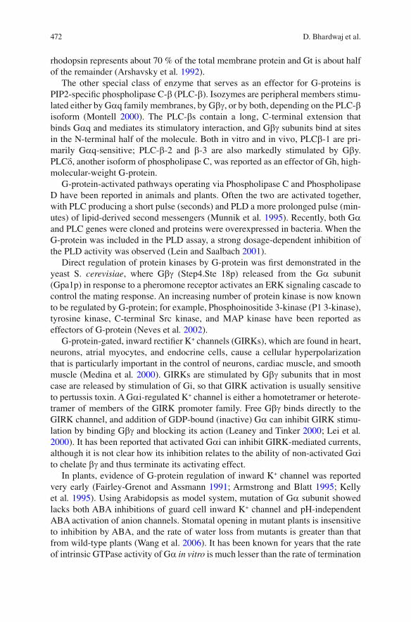
RGS protein family consists of at least 20 mammalian gene products that act as GTPase activating proteins on the a -subunits of heterotrimetric G-protein (Zhang et al. 1999 ) . By accelerating the inactivation of GTP-bound G a subunits, RGS serve as negative regulators of G-protein-mediated signaling pathways (Fig. 17.1 ). Additionally, two RGS proteins, p115-RhoGEF and PDZ (PSD-95, Disc-large, and
Fig. 17.1 A hypothetical model showing the various functions performed by GPCR, RGS, and G-proteins (G a , G b , and G g ). Elicitors like ABA, methyl jasmonate MeJA, brassinosteroids, and pathogens sensitize 7TM seven-transmembrane receptors like GCR (Pandey and Assmann 2004 ; Chen et al . 2004 ; Liu et al . 2007 ) and RGS (Plakidou-Dymock and Dymock 1998 ; Colucci et al . 2002 ; Chen et al . 2003, 2004 ) , causing the G-protein heterotrimer to disintegrate forming GTP-G a and G b -G g monomer and dimer, respectively. Each subunit plays signi fi cant role in various meta-bolic processes including environmental stress. G a whose role has been shown in signaling cas-cades related to Gibberellins (Ashikari et al . 1999 ; Oki et al. 2009a, b ) , glucose (Grigston et al . 2008 ) , salt (Misra et al . 2007 ) , ROS (Zhang et al . 2011 ) , water use ef fi ciency (WUE) (Nilson and Assmann 2010), and stomatal regulation (Fan et al. 2008) (G.C) also serves as a interacting unit for enzyme like phospholipase (PLC). G b -G g dimer also plays signi fi cant role in maintaining shapes of petal and fl oral organs (Lease et al . 2001 ) , root stature, number of stomata (Zhang et al. 2008a, b ) , defense against necrotrophic fungi (Trusov et al. 2006, 2009 ; Delgado-Cerezo et al. 2012 ) , and heat stress (Misra et al . 2007 )
474 D. Bhardwaj et al.
Z0-1)-RhoGEF, can also act as effectors (Cowan et al . 2000 ) . On the other hand, RGS can also interact with targets other than G-proteins, for example the phospho-serine-binding protein, 14-3-3 (Niu et al . 2002 ) .
RGS vary dramatically in size (from 23 to 160 kDa) and sequence, but they all have a common “RGS” domain (=125 aa), which is responsible for binding to the G a subunits and is suf fi cient for the GAP activity. Several RGS proteins contain a G-protein g -subunit-like (GGL) domain; work on RGS6, RGS7, RGS11, and RGS9 showed GGL domain to be important for RGS/G b 5 association (Snow et al . 1998, 1999 ; Makino et al . 1999 ) .
The GAP activity of RGS proteins has been demonstrated for all G a subgroups except G a s. RGS proteins appear to enhance the GTPase activity by binding to and stabilizing the transition state (Berman et al . 1996 ) . The three-dimensional structure of an RGS and G a complex demonstrates that RGS stabilizes the fl exible switch region of G to resemble the transition state, thereby facilitating GTP hydrolysis (Tesmer et al . 1997 ) .
The expression of more than 20 members of the mammalian family of RGS pro-teins suggests that they are likely to display marked selectivity of expression pattern and/or function, for example, RGS4 selectively enhances the GTPase activity of GO1 and Gi2 (Cavalli et al . 2000 ) , and RGS11 selectively interacts with G b 5 to act as GAPs on G a o (Snow et al . 1998 ) . Among the higher eukaryotes, certain RGS have been cloned, for example human (21 RGS genes), yeast ( S. cerevisiae; 2 RGS genes), fruit fl y ( Drosophila; 5 RGS genes), and round worms ( C. elegans; 12 RGS genes). Many studies on RGS are also available in plants including Arabidopsis thaliana and Glycine max. However, among the monocots only Setaria italica has been reported to possess RGS protein homolog (Chen & jones 2004 ; Urano et al. 2012 ; Choudhury et al. 2012 ).
Heterotrimers of G-proteins are like proteins that can be kept inactivated or acti-vated depending upon the requirements of the cell at a given condition. Once acti-vated, heterotrimer dissociates into single G-alpha subunit with GTPase activity and G-beta-gamma dimer. These uncoupled subunits then act as secondary mes-sengers and they further catalyze the downstream processes. GPA1 physically inter-acts with plasidial protein called Thylakoid formation1 (THF1), which has been considered signi fi cant in the sugar signaling (Zhang et al . 2009 ) . Arabidopsis plant grown on high level of glucose has expanded root growth which proves the role of sugar in cell division and cell expansion. Role of G-proteins has already been shown in root cell division. This supports the link between G-proteins and sugar signaling in plant root cells. Interestingly, the phenotypes of mutants of G-protein are highly distinguishable on the grounds of root and shoot stature.
GPA1 is also known to interact with PLD a 1 at the DRY motif, and this interac-tion helps in increasing the ef fi cacy of G-protein by in fl uencing the GTPase activity of G a and modulating its intracellular location (Zhao and Wang 2004 ) . There are reports that support the involvement of G a and Phospholipase A
2 in the biosynthe-
sis of phytoelexin in Eschscholzia californica (Viehweger et al. 2006). G-proteins are supposed to be involved in retrograde signaling through the interaction of G a and THF1, and any damage to the photosynthetic machinery specially D1 protein of
47517 Can G-Proteins be the Key Proteins for Overcoming Environmental Stresses…
PS II can be renewed by the involvement of FtsH protease which in turn degrades the defective D1 protein. This FtsH protease is shown to be regulated by G-proteins with the involvement of other secondary messengers (Zhang et al . 2009 ) .
5 Involvement of G-protein in Stomatal Functioning Suggests its Role in Environmental Stresses
Stomata are often considered as nostrils of plants. Present on the leaves, they perform the function of gaseous exchange and transpiration. Their role becomes evident during temperature and drought stress in order to prevent the excessive loss of water. Transpiration is a necessary evil; it is required to transport the nutrient from soil to the higher parts of the plant. Nutrients travel through the transpiration stream; indi-rectly it also maintains the turgidity and causes the cooling effect, saving plants from heat stress. Excessive transpiration can be dangerous as it leads to the wilting ulti-mately causing senescence of the plant. Plants have acquired smart mechanism to control the excessive loss of water by regulating the opening and closing of stomata. Role of ABA was reported in stomatal closure, as it induces the signal transduction pathway that fi nally causes the loss of certain osmolytes that causes the turgidity of guard cell thereby resulting in closing of stomata. The interplay between the mem-brane of guard cells and ion channels present on them is crucial in this regard. GCR, the seven-trans-membrane proteins, were proved to be the receptor for ABA (Chen et al . 2004 ) . It transduces the signal by uncoupling G-proteins and activating several downstream pathways including activation of MAP kinase pathway and Ca 2+ signal-ing pathways. Moreover, Pisum sativum G b subunit was shown to play a role in heat stress when overexpressed in Tobacco lines (Misra et al . 2007 ) . In fact, there are many gaps in related to G-protein and other guard cell signaling proteins that need to be bridged to decipher a meaningful pathway out of it. In the wake of global warm-ing, it is required to understand the exact mechanism of regulation of stomata. Guard cells are sensitive to high temperature and low CO
2 levels and they operate in con-
nection with several molecular cues including K + channels. They act as balloons that can be swelled or diffused depending upon the concentration of K + ion inside them. In fl ux of K + ion through K + channels causes stomatal opening but there are several environmental factors like light, low CO
2 level and humidity that can cause the open-
ing of stomata. Overall, stomatal closure is more or less dependent on factors like high temperature, drought, pathogen attack, and levels of phytohormones like ABA and MeJA. ROS are one of the byproducts of oxidative stress in plants; heat can also result in the release of these reactive oxygen free radicals. These radicals not only damage the cell membrane but they also can lead to cell death. G-proteins are also involved in the regulation of ROS signaling (Zhang et al . 2011 ) .
Stomatal functioning is a complex phenomenon which includes several molecu-lar proteins and secondary messengers. It was suggested that G-protein can be a link between receptor of ABA and ion channels on the guard cells (Fan et al. 2008); several researches in the past have put some light on the involvement of G-proteins
476 D. Bhardwaj et al.
in the stomatal functioning and related processes such as immunity against patho-gens and relief against oxidative stress (Zhang et al. 2008a, b ) . Role of G-proteins was showed in the regulation of inward K + channel in fava bean (Fairley-Grenot and Assmann 1991 ) .
In 1998, it was reported that beta subunit is involved in the voltage-dependent modulation of N-type Ca 2+ channels (García et al. 1998 ) . The direct proof of interac-tion of beta subunit with inward recti fi ed K + channel Kir3 was given in 2002; this interaction was found to be signi fi cant for slow hyperpolarization of cardiac myo-cytes and neurons. On the same line, plant scientists also looked for the involvement of beta subunit in modulating K + channels and in this regard the fi rst breakthrough was reported in the mutant lines of Arabidopsis. It was found that unlike in the case of animals, G b g has no direct control over the K + channels in plants and it may require the involvement of other proteins to in fl uence the channel proteins (Fan et al. 2008). Indirect involvement of G-proteins in ion channel control was further sup-ported by one of the fi ndings in which the null mutant of G a failed to show fl g22-regulated stomatal responses. Flg22 is a bacterial protein which causes the inhibition of light-induced stomatal opening ( Zhang et al. 2008a, b ) . Interestingly, agb1 mutants have more number of stomata per unit area, whereas gpa1 mutants have low number as compared to control plant (Zhang et al. 2008a, b ) . This property could be the rea-son of gpa1 mutant being more ef fi cient with regard to transpiration especially dur-ing drought stress (Nilson and Assmann 2010 ). Pisum sativum beta subunit PsG b showed interaction with PsMPK3, further strengthening the possibility of involve-ment of G-proteins with stomatal functioning though more experimentation is required to prove this fact (Bhardwaj et al . 2011 ) . Recently, it was shown that one of the interacting proteins AGB1 in Arabidopsis named AGG3 is involved in the regula-tion of guard cell K + channels. AGG3 is predicted to be the third gamma subunit in Arabidopsis, with AGG1 and AGG2 being the other two and mostly involved in the developmental processes (Chakravorty et al. 2011 ; Shengjun et al. 2012 ).
All these reports indicate the involvement of G-protein in stomatal regulations which seem to be operating through the interactions with other proteins.
6 G-proteins and their Role in Pathogenesis and Biotic Stress
Evidence is accumulating for the importance of plant heterotrimeric G-protein in response to both bacterial and fungal pathogens. Beffa et al . ( 1995 ) showed that stable transformation of tobacco plants with the A1 subunit of CTX resulted in reduced susceptibility to Pseudomonas tabaci, accumulation of salicylic acid, and constitutive expression of pathogenesis-related genes. The role of plant G-proteins in response to fungal pathogens was suggested by the work of Legendre and Heinstein ( 1992 ) who showed that mastoparan elicits an oxidative burst in soybean cell suspensions, whereas CTX enhances the burst initiated by the elicitor Verticillium dahliae. They demonstrated subsequently that both the elicitor polygalacturonic acid and the G-protein activator mastoparan stimulate PLC activity in this prepara-tion, leading to an increase in inositol triphosphate (Legendre et al . 1993 ) .
47717 Can G-Proteins be the Key Proteins for Overcoming Environmental Stresses…
Many studies using inhibitors and mutants have pinpointed the role of G-proteins in biotic stress and defense-related signaling pathway. G a mutant of rice which is commonly known as D1 mutant was found ineffective in fi ghting against blast disease due to delayed response of pathogenesis-related genes (Suharsono et al . 2002 ) .
G-protein, especially G b g dimer, provides resistance against necrotrophic fungi through jasmonate-mediated pathway in Arabidopsis, while their mutants are sus-ceptible to pathogenesis (Trusov et al . 2009 ; Trusov et al. 2006 ) . The reduced resis-tance of mutant plants could be because of difference in cell wall composition, especially xylose content ( Delgado-Cerezo et al. 2012 ) . Recently, up-regulation of gene of G b subunit of Pisum sativum was also reported after treating the plant with methyl jasmonate (Bhardwaj et al . 2011 ) .
7 Conclusions
Climate change is a major concern of every nation but no country is in the position of cutting down their greenhouse emissions due to the challenges of accelerated development. Climate change has adverse effects on the production and yield of various crops, in turn creating alarming situations for everyone including agricul-tural scientists. Plant breeders and genetic engineers have to look for useful genes and genetic systems whose fundamentally correct manipulations can help amelio-rate the status quo. Responding to the situation, G-proteins have been identi fi ed as a genetic tool whose manipulations could be useful for protecting plants from vari-ous abiotic and biotic stresses under the heat of climate change. G-proteins are smart molecular machines whose structure and functions are already well known in animals but their functions are still to be fully understood in plants. Recently, their overexpression in Arabidopsis and tobacco has given these plants genetic potential to overcome many abiotic and biotic stresses, and the same technology is now applied on several economically important plants such as rice, pea, tomato, and soybean. The cell machinery is endowed in its ability to allow various pathways to work simultaneously under various stimuli of nature, some act independently and some open the gates for cross-talks. Interestingly, G-proteins linked to the GPCRs the receptors fi nd their way in after getting proper signals. They interact with sev-eral other proteins to bring about desirable changes in the plant cell environment. G-proteins also interact with several other proteins in the cellular environment. Based on the above-mentioned information we can conclude that GCR, RGS, and G-proteins play signi fi cant role in the biological processes with GCR and RGS acting as a sensor protein and G-protein as the molecular machines that execute the downstream cascade.
Therefore, studies leading to their effective manipulations can result in plants that are genetically better equipped to perceive and combat biotic as well as abiotic stresses thereby leading to increased crop productivity.
Acknowledgements The authors acknowledge the University Grant Commission and Department of Science and Technology, New Delhi, India, for funding their research.
478 D. Bhardwaj et al.
References
Afzal AJ, Wood AJ, Lightfoot DA (2008) Plant receptor-like serine threonine kinases: roles in signaling and plant defense. Mol Plant Microbe Interact 21:507–517
Armstrong F, Blatt MR (1995) Evidence for K+ channel control in Vicia guard cells coupled by G-proteins to a 7TMS receptor mimetic The. Plant J 8:187–198
Arshavsky VY, Dumke CL, Bownds MD (1992) Noncatalyticc GMP binding sites of amphibian rod cGMP phosphodiesterase control interaction with its inhibitory g -subunits. A putative regu-latory mechanism of the rod photoresponse. J Biol Chem 267:24501–24507
Ashikari M, Wu J, Yano M, Sasaki T, Yoshimura A (1999) Rice gibberellin insensitive dwarf mutant gene Dwarf 1 encodes the alpha-subunit of GTP-binding protein. Proc Natl Acad Sci USA 96:10284–10289
Bähler J, Peter M (2000) Cell polarity in yeast. In: Drubin DG (ed) Frontiers in molecular biology: cell polarity. Oxford University Press, Oxford
Beffa R, Szell M, Meuwly P, Pay A, Vogeli-Lange R, Metraux JP, Neuhaus G, Meins F Jr, Nagy F (1995) Cholera toxin elevates pathogen resistance and induces pathogenesis-related gene expression in tobacco. EMBO J 14:5753–5761
Berman DM, Kozasa T, Gilman AG (1996) The GTPase activating protein RGS4 stabilizes the transition state for nucleotide hydrolysis. J Biol Chem 271:27209–27212
Bhardwaj D, Sheikh AH, Sinha AK, Tuteja N (2011) Stress induced b subunit of heterotrimeric G-proteins from Pisum sativum interacts with mitogen activated protein kinase. Plant Signal Behav 6:287–292
Birnbaumer L, Rodbell M (1969) Adenyl cyclase in fat cells. II. Hormone receptors. J Biol Chem 224:3477–3482
Bourne HR, Cof fi no P, Tompkins GM (1975) Selection of a variant lymphoma cell de fi cient in adenylate cyclase. Science 187:750–752
Bowler C, Chua NH (1994) Emerging themes of plant signal transduction. Plant Cell 6:1529–1541
Bowler C, Neuhaus G, Yamagata H, Chua NH (1994a) Cyclic GMP and calcium mediate phyto-chromephototransduction. Cell 77:73–81
Bowler C, Yamagata H, Neuhaus G, Chua NH (1994b) Phytochrome signal transduction pathways are regulated by reciprocal control mechanisms. Genes Dev 8:2188–2202
Calenberg M, Brohsonn U, Zedlacher M, Kreimer G (1998) Light and Ca 2+ −modulated GTPase in the eyespot apparatus of a fl agellate green alga. Plant Cell 10:91–103
Cassel D, Selinger Z (1978) Mechanism of adenylate cyclase activation through the b -adrenergic receptor: catecholamine-induced displacement of bound GDP by GTP. Proc Natl Acad Sci USA 75:4155–4159
Cavalli A, Druey KM, Milligan G (2000) The regulator of G protein signalling RGS4 selectively enhances alpha 2A-adreoreceptor stimulation of the GTPase activity of Go1alpha and Gi2alpha. J Biol Chem 275:23693–23699
Chakravorty D, Trusov Y, Zhang W, Acharya BR, Sheahan MB, McCurdy DW, Assmann SM, Botella JR (2011) An atypical heterotrimeric G-protein γ-subunit is involved in guard cell K-channel regulation and morphological development in Arabidopsis thaliana. http://www.ncbi.nlm.nih.gov/pubmed/21575088 Plant J 67:840–51
Chen JG, Jones AM (2004) AtRGS1 function in Arabidopsis thaliana. Methods Enzymol 389:338–350
Chen JG, Willard FS, Huang J, Liang J, Chasse SA, Jones AM, Siderovski DP (2003) A seven-Tran membrane RGS protein that modulates plant cell proliferation. Science 301:1728–1731
Chen JG, Pandey S, Huang J, Alonso JM, Ecker JR, Assmann SM, Jones AM (2004) GCR1 can act independently of heterotrimeric G-protein in response to brassinosteroids and gibberellins in Arabidopsis seed germination. Plant Physiol 13:907–915
Cheong YH, Pandey GK, Grant JJ, Batistic O, Li L, Kim BG, Lee SC, Kudla J, Luan S (2007) Two calcineurin B-like calcium sensors, interacting with protein kinase CIPK23, regulate leaf tran-spiration and root potassium uptake in Arabidopsis. Plant J 52:223–239
47917 Can G-Proteins be the Key Proteins for Overcoming Environmental Stresses…
Choudhury SR, Westfall CS, Pandey S (2012) The RGS proteins add to the diversity of soybean heterotrimeric G-protein signaling. Plant Signal Behav. 1; http://www.ncbi.nlm.nih.gov/pubmed/22899066 [Epub ahead of print]
Colucci G, Apone F, Alyeshmerni N, Chalmers D, Chrispeels MJ (2002) GCR1, the putative Arabidopsis G protein-coupled receptor gene is cell cycle regulated, and its overexpression abolishes seed dormancy and shortens time to fl owering. Proc Natl Acad Sci USA 99:4736–4741
Cowan CW, Wensel TG, Arshavsky VY (2000) Enzymology of GTPase acceleration in phototrans-duction. Methods Enzymol 315:524–538
De Vries L, Zheng B, Fischer T, Elenko E, Farquhar MG (2000) The regulator of G protein signal-ing family. Annu Rev Pharmacol Toxicol 40:235–271
Delgado-Cerezo M, Sánchez-Rodríguez C, Escudero V, Miedes E, Fernández PV, Jordá L, Hernández-Blanco C, Sánchez-Vallet A, Bednarek P, Schulze-Lefert P, Somerville S, Estevez JM, Persson S, Molina A (2012) Arabidopsis heterotrimeric G-protein regulates cell wall defense and resistance to necrotrophic fungi. Mol Plant 5:98–114
Devoto A, Turner JG (2003) Regulation of jasmonate-mediated plant responses in Arabidopsis. Ann Bot 92:329–337
Dixon RA, Kobilka BK, Strader DJ, Benovic JL, Dohlman HG, Frielie T, Bolanowski MA, Bennett CD, Rands E, Diehl RE (1986) Cloning of the gene and c DNA for mammalian b -adrenergic receptor and homology with rhodopsin. Nature 321:75–79
Estelle M (1998) Cytokinin action: two receptors better than one? Curr Biol 8:539–541 Etheridge N, Trusov Y, Verbelen JP, Botella JR (1999) Characterization of ATDRG1, a member of
a new class of GTP-binding proteins in plants. Plant Mol Biol 39:1113–1126 Fairley-Grenot KA, Assmann SM (1991) Evidence for G-protein regulation of inward K+ channel
current in guard cells of fava bean. Plant Cell 3:1037–1044 García DE, Li B, García-Ferreiro RE, Hernández-Ochoa EO, Yan K, Gautam N, Catterall WA,
Mackie K, Hille B (1998) G-protein beta-subunit speci fi city in the fast membrane-delimited inhibition of Ca 2+ channels. J Neurosci 18:9163–9170
Gómez-Porras JL, Riaño-Pachón DM, Dreyer I, Mayer JE, Mueller-Roeber B (2007) Genome-wide analysis of ABA-responsive elements ABRE and CE3 reveals divergent patterns in Arabidopsis and rice. BMC Genomics 8:260
Gotta M, Ahringer J (2001) Distinct roles for Galpha and G-beta-gamma in regulating spindle position and orientation in Caenorhabditis elegans embryos. Nat Cell Biol 3:297–300
Grigston JC, Osuna D, Scheible WR, Liu C, Stitt M, Jones AM (2008) D-Glucose sensing by a plasma membrane regulator of G signaling protein, AtRGS1. FEBS Lett 582:3577–3584
Guan KL, Han M (1999) G-protein signaling network mediated by an RGS protein. Genes Dev 13:1763–1767
Gutkind JS (1998) The pathways connecting G protein-coupled receptors to the nucleus through divergent mitogen-activated protein kinase cascades. J Biol Chem 273:1839–1842
Hucho F and Buchner K (1997) Signal transduction and protein kinases: the long way from the plasma membrane into the nucleus. Naturwissenschaften. 84, 281–90
Hundertmark M, Dirk KH (2008) LEA (late embryogenesis abundant) proteins and their encoding genes in Arabidopsis thaliana . BMC Genomics 9:118
Huw D, Jones HD, Smith SJ, Desikan R, Plakidou-Dymock S, Alison Lovegrove A, Hooley R (1998) Heterotrimeric G proteins are implicated in gibberellin induction of a -amylase gene expression in wild oat aleurone. Plant Cell 10:245–253
Im MJ, Graham RM (1990) A novel guanine nucleotide-binding protein coupled to the a1-adrenergic receptor. I. Identi fi cation by photolabeling of membrane and ternary complex preparations. J Biol Chem 265:18952–18960
Jamora C, Takizawa PA, Zaarour RF, Denesvre C, Faulkner DJ, Malhotra V (1997) Regulation of Golgi structure through heterotrimeric G proteins. Cell 91:617–626
Jones AM (2002) G-protein-coupled signaling in Arabidopsis. Curr Opin Plant Biol 5(5):402–407
Jones AM, Assmann SM (2004) Plants: the latest model system for G-protein research. EMBO Rep 5:572–578
480 D. Bhardwaj et al.
Kariola T, Brader G, Helenius E, Li J, Heino P, Palva ET (2006) EARLY RESPONSIVE TO DEHYDRATION 15 , a Negative Regulator of Abscisic Acid Responses in Arabidopsis. Plant Physiol 142:1559–1573
Kelly WB, Esser JE, Schroeder JI (1995) Effects of cytosolic calcium and limited, possible dual, effects of G protein modulators on guard cell inward potassium channels. Plant J 8:479–489
Leaney JL, Tinker A (2000) The role of members of the pertussis toxin sensitive family of G pro-teins in coupling receptors to the activation of the G protein-gated inwardly rectifying potas-sium channel. Proc Natl Acad Sci USA 97:5651–5656
Lease KA, Wen J, Li J, Doke JT, Liscum E, Walker JC (2001) A mutant Arabidopsis heterotrimeric G-protein beta subunit affects leaf, fl ower, and fruit development. Plant Cell 13:2631–2641
Lee YRJ, Assmann SM (1999) Arabidopsis thaliana ‘extra-large GTP-binding protein (AtXLG1): A new class of G-protein. Plant Mol Biol 40:55–64
Legendre L, Heinstein PF, Low PS (1992) Evidence for participation of GTP-binding proteins in elicitation of the rapid oxidative burst in cultured soybean cells. J Biol Chem 267: 20140–22014
Legendre L, Heinstein PF, Crain RC, Low PS (1993) Phospholipase C activation during elicitation of the oxidative burst in cultured plant cells. J Biol Chem 268:24559–24563
Lei Q, Jones MB, Talley EM, Schrier AD, McIntire WE, Garrison JC, Bayliss DA (2000) Activation and inhibition of G protein-coupled inwardly rectifying potassium (Kir3) channels by G pro-tein beta gamma subunits. Proc Natl Acad Sci USA 97:9771–9776
Lein W, Saalbach G (2001) Cloning and direct G-protein regulation of phospholipase D from tobacco. Biochim BiophyS Acta 1530:172–183
Liu X, Yue Y, Li B, Nie Y, Li W, Wu WH, Ma L (2007) A G protein-coupled receptor is a plasma membrane receptor for the plant hormone abscisic acid. Science 315:1712–1716
Ma H, Yanofsky M, Meyerowitz EM (1990) Molecular cloning and characterization of GPA1 , a G protein a subunit gene from Arabidopsis thaliana . Proc Natl Acad Sci USA 87:3821–3825
Makino ER, Handy JW, Li T, Arshavsky VY (1999) The GTPase activating factor for transducin in rod photoreceptors is the complex between RGS9 and type 5G protein subunit. Proc Natl Acad Sci USA 96:1947–1952
Mason MG, Botella JR (2000) Completing the heterotrimer: isolation and characterization of an Arabidopsis thaliana G protein gamma-subunit cDNA. Proc Natl Acad Sci USA 97: 14784–14788
Medina JF, Garcia KS, Nores WL, Taylor NM, Mauk MD (2000) Timing mechanisms in the cer-ebellum: testing predictions of a large-scale computer simulation. J Neurosci 20:5516–5525
Mirbahar RB, Laidman DL (1982) Gibberellic acid-stimulated alpha-amylase secretion and phos-pholipid metabolism in wheat aleurone tissue. Biochem J 208:93–100
Misra S, Wu Y, Venkataraman G, Sopory S, Tuteja N (2007) Heterotrimeric G-protein complex and G-protein-coupled receptor from a legume (Pisum sativum): role in salinity and heat stress and cross-talk with Phospholipase C. Plant J 52:656–669
Montell C (2000) PLC fi lls a GAP in G-protein-coupled signalling. Nat Cell Biol 2:82–83 Munnik T, Arisz SA, de Vrije T, Musgrave A (1995) G protein activation stimulates phospholipase
D signaling in plants. Plant Cell 7:2197–2210 Neer EJ (1995) Heterotrimeric G-proteins: organizers of transmembrane signals. Cell 80:249–257 Neuhaus G, Bowler C, Kern R, Chua NH (1993) Calcium/calmodulin-dependent and -independent
phytochrome signal transduction pathways. Cell 73:937–952 Neves SR, Ram PT, Iyengar R (2002) G protein pathways. Science (Wash DC) 296:1636–1639 Nilson SE, Assmann SM (2010) The alpha-subunit of the Arabidopsis heterotrimeric G protein,
GPA1, is a regulator of transpiration ef fi ciency. http://www.ncbi.nlm.nih.gov/pubmed/20200073 Plant Physiol.152:2067–77
Niu J, Scheschonka A, Druey KM, Davis A, Reed E, Kolenko V, Bodnar R, Voyno-Yasenetskaya T, Du X, Kehrl J, Dulin NO (2002) RGS3 interacts with 14-3-3 via the N-terminal region distinct from the RGS (regulator of G-protein signaling) domain. Biochem J 365:677–684
Okamoto H, Matsui M, Deng XW (2001) Overexpression of the heterotrimeric G-protein a -subunit enhances phytochrome-mediated inhibition of hypocotyl elongation. Plant Cell 13:1639–1652
48117 Can G-Proteins be the Key Proteins for Overcoming Environmental Stresses…
Okamoto H, Gobel C, Capper RG, Saunders N, Feussner I, Knight MR (2009) The a -subunit of the heterotrimeric G-protein affects jasmonate responses in Arabidopsis thaliana. J Exp Bot 60:1991–2003
Oki K, Inaba N, Kitagawa K, Fujioka S, Kitano H, Fujisawa Y, Kato H, Iwasaki Y (2009a) Function of the alpha subunit of rice heterotrimeric G-protein in brassinosteroid signaling. Plant Cell Physiol 50:161–172
Orly J, Schramm M (1976) Coupling of catecholamine receptor from one cell with adenylate cyclase from another cell by cell fusion. Proc Natl Acad Sci USA 73:4410–4414
Palczewski K, Polans AS, Baehr W, Ames JB (2000) Ca (2+)-binding proteins in the retina: struc-ture, function, and the etiology of human visual diseases. Bioessays 22:337–350
Pandey S, Assmann SM (2004) The Arabidopsis putative G protein-coupled receptor GCR1 inter-acts with the G protein a subunit GPA1 and regulates abscisic acid signaling. Plant Cell 16:1616–1632
Pandey S, Chen JG, Jones AM, Assmann SM (2006) G-protein complex mutants are hypersensi-tive to abscisic acid regulation of germination and post germination development. Plant Physiol 141:243–256
Peskan-Berghofer T, Neuwirth J, Kusnetsov V, Oelmeller R (2005) Suppression of heterotrimeric G-protein b -subunit affects anther shape, pollen development and in fl orescence architecture in tobacco. Planta 220:737–746
Pfeuffer T, Helmreish EJM (1975) Activation of pigeon erythrocytes membrane adenylate cyclase by guanyl nucleotide analogues and separation of nucleotide-binding protein. J Biol Chem 250:867–876
Plakidou-Dymock S, Dymock D, Hooley R (1998) A higher plant seven-transmembrane receptor that in fl uences sensitivity to cytokinins. Curr Biol 8:315–324
Rall K, Sutherland EW, Berthet J (1957) The relationship of epinephrine and glucagon to liver phosphorylase. IV. Effect of epinephrine and glucagons on the reactivation of phosphorylase in liver homogenates. J Biol Chem 224:463–3475
Rodbell M, Birnbaumer L, Pohl SL, Krans HMJ (1971) The glucagons sensitive adenylyl cyclase in plasma membrane of rat liver. V. An obligatory role of guanyl nucleotides in glucagons action. J Biol Chem 246:1877–1882
Romero LC, Lam E (1993) Guanine nucleotide binding protein involvement in early steps of phy-tochrome regulated gene expression. Proc Natl Acad Sci USA 90:1465–1469
Ross EM, Wilkie TM (2000) GTPase-activating proteins for heterotrimeric G proteins: regulators of G protein signaling (RGS) and RGS-like proteins. Annu Rev Biochem 69:795–827
Sato M, Kataoka R, Dingus J, Wilcox M, Hildebrandt J, Lanier SM (1995). Factors determining speci fi city of signal transduction by G-protein-coupled receptors. J. Biol. Chem. 270, 15269–15276
Schaffer R, Landgraf J, Accerbi M, Simon V, Larson M, Wisman E (2001) Microarray analysis of diurnal and circadian-regulated genes in Arabidopsis. Plant Cell 13:113–123
Shorr RGL, Lefkowitz RJ, Caron MG (1981) Puri fi cation of the b -adrenergic receptor. Identi fi cation of the hormone-binding subunit. J Biol Chem 256:5820–5826
Snow BE, Krumins AM, Brothers GM, Lee S, Wall MA, Chung S, Mangion J, Arya S, Gilman AG, Siderovske DP (1998) A G protein g subunit-like domain shared between RGS11 and other RGS proteins speci fi es binding to Gb5 subunits. Proc Natl Acad Sci USA 95:13307–13312
Snow BE, Betts L, Mangion J, Sondek J, Siderovski DP (1999) Fidelity of G protein b-subunit association by the G protein g-subunit-like domains of RGS6, RGS7, and RGS11. Proc Natl Acad Sci USA 96:6489–6494
Suharsono U, Fujisawa Y, Kawasaki T, Iwasaki Y, Satoh H, Shimamoto K (2002) The heterotri-meric G protein alpha subunit acts upstream of the small GTPase Rac in disease resistance of rice. Proc Natl Acad Sci USA 99:13307–13312
Sun S, Yu JP, Chen F, Zhao TJ, Fang XH, Li YQ, Sui SF (2008) TINY, a dehydration-responsive element (DRE)-binding protein-like transcription factor connecting the DRE- and ethylene-responsive element-mediated signaling pathways in Arabidopsis. J Biol Chem 283:6261–6271
482 D. Bhardwaj et al.
Sutherland EW, Rall TW (1958) Fractionation and characterization of a cyclic adenine ribonucle-otide formed by tissue particle. J Biol Chem 232:1077–1091
Tang WJ, Gilman AG (1991) Type-speci fi c regulation of adenylyl cyclase by G protein b g subunits. Science (Wash DC) 254:1500–1503
Tesmer JJ, Sunahara RK, Gilman AG, Sprang SR (1997) Crystal structure of the catalytic domains of adenylyl cyclase in a complex with Gsa-GTPgS. Science 278:1907–1916
Thomashow MF (1998) Role of cold-responsive genes in plant freezing tolerance. Plant Physiol 118:1–8
Trusov Y, Rookes JE, Chakravorty D, Armour D, Schenk PM, Botella JR (2006) Heterotrimeric G proteins facilitate Arabidopsis resistance to necrotrophic pathogens and are involved in jasmonate signaling. Plant Physiol 140:210–220
Trusov Y, Sewelam N, Rookes JE, Kunkel M, Nowak E, Schenk PM, Botella JR (2009) Heterotrimeric G proteins-mediated resistance to necrotrophic pathogens includes mechanisms independent of salicylic acid-, jasmonic acid/ethylene- and abscisic acid-mediated defense signaling. Plant J 58:69–81
Ullah H, Chen JG, Wang S, Jones AM (2002) Role of a heterotrimeric G protein in regulation of Arabidopsis seed germination. Plant 129:897–907
Urano D, Jones JC, Wang H, Matthews M, Bradford W, Bennetzen JL, Jones AM (2012) G protein activation without a GEF in the plant kingdom. http://www.ncbi.nlm.nih.gov/pubmed/22761582 PLoS Genet. 8:e1002756.
Vaugham M (1998) Signaling by heterotrimeric G proteins minireview series. J Biol Chem 273:667–668
Walker JC (1994) Structure and function of the receptor-like protein kinases of higher plants. Plant Mol Biol 26:1599–1609
Wang L, Xu YY, Ma QB, Li D, Xu ZH, Chong K (2006) Heterotrimeric G protein a subunit is involved in rice brassinosteroid response. Cell Res 16:916–922
Warpeha KMF, Hamm HE, Rasenick MM, Kaufman LS (1991) A blue-light activated GTP binding protein in the plasma membrane of etiolated pea. Proc Natl Acad Sci USA 88:8925–8929
Weiner OD, Neilsen PO, Prestwich GD, Kirschner MW, Cantley LC, Bourne HR (2002) A PtdInsP(3)- and Rho-GTPase-mediated positive feedback loop regulates neutrophil polarity. Nat Cell Biol 4:509–513
Weiss CA, Huang H, Ma H (1993) Immunolocalization of the G-protein alpha subunit encoded by the GPA1 gene in Arabidopsis. Plant Cell 5:1513–1528
Yarden Y, Rodriguez H, Wong SK, Brandt DR, May DC, Burnier JR, Harkinns RN, Chen EY, Ramachandran J, Ullrich A (1986) The avian beta-adrenergic receptor: primary structure and membrane topology. Proc Natl Acad Sci 83:6795–6799
Zhang Y, Neo SY, Han J, Yaw LP, Lin SC (1999) RGS16 attenuates G- a q-dependent p38 mitogen-activated protein kinase activation by platelet-activating factor. J Biol Chem 274:2851–2857
Zhang L, Hu G, Cheng Y, Huang J (2008a) Heterotrimeric G protein alpha and beta subunits antagonistically modulate stomatal density in Arabidopsis thaliana. Dev Biol 324:68–75
Zhang W, He SY, Assmann SM (2008b) The plant innate immunity response in stomatal guard cells invokes G-protein-dependent ion channel regulation. Plant J 56:984–996
Zhang L, Wei Q, Wu W, Cheng Y, Hu G, Hu F, Sun Y, Zhu Y, Sakamoto W, Huang J (2009) Activation of the heterotrimeric G-protein a -subunit gpa1 suppresses the ftsh-mediated inhibi-tion of chloroplast development in Arabidopsis. Plant J 58:1041–1053
Zhang W, Jeon BW, Assmann SM (2011) Heterotrimeric G-protein regulation of ROS signaling and calcium currents in Arabidopsis guard cells. J Exp Bot 62:2371–2379
Zhao J, Wang X (2004) Arabidopsis phospholipase D a 1 interacts with the heterotrimeric g-protein a -subunit through a motif analogous to the dry motif in G-protein-coupled receptors. J Biol Chem 279:1794–1800
Zwaal RR, Ahringer J, van Luenen HGAM, Rushfort A, Anderson P, Plasterk RHA (1996) G proteins are required for spatial orientation of early cell cleavage in C. elegans embryos. Cell 86:1–20