Embed Size (px)
Citation preview
Open Journal of Pediatrics, 2018, 8, 221-237 http://www.scirp.org/journal/ojped
ISSN Online: 2160-8776 ISSN Print: 2160-8741
DOI: 10.4236/ojped.2018.83024 Sep. 5, 2018 221 Open Journal of Pediatrics
Plasma Asymmetric Dimethylarginine Levels in Neonates with Bronchopulmonary Dysplasia Associated with Pulmonary Hypertension
Safaa Abd Elhamid EL Meneza1, Seham Mohamed Bahgat2, Asmaa EL Saudi Nasr1
1Department of Pediatrics, Faculty of Medicine for Girls, Al-Azhar University, Cairo, Egypt 2Clinical Pathology Department, Faculty of Medicine for Girls, Al-Azhar University, Cairo, Egypt
Abstract Background: Bronchopulmonary dysplasia (BPD) continues to be an impor-tant problem in neonates especially premature infants despite improved facil-ities of care, monitoring and treatment. Pulmonary hypertension (PH) is a major complicating factor and key cause of mortality in this population. Al-tered vascular and alveolar growth particularly in canalicular and early sac-cular stages of lung development following mechanical ventilation and oxy-gen therapy result in arrest of the lung development leading to BPD with PH. Early recognition of PH in infants with these risk factors is important for op-timal management. We tested the hypothesis that asymmetric dimethylargi-nine, would be greater in infants with bronchopulmonary dysplasia asso-ciated pulmonary hypertension than in infants with BPD alone. The Aim: The aim of the current study was to measure the Asymmetric dimethylargi-nine (ADMA) levels, arginine levels & the plasma arginine-to-ADMA ratio in newborn infants with broncho-pulmonary dysplasia, to evaluate echocardio-graphic parameters among neonates with bronchopulmonary dysplasia, to correlate between plasma ADMA & arginine-to-ADMA ratio and echocardi-ographic (ECHO) parameters in those patients and to compare full term & preterm neonates with bronchopulmonary dysplasia as regard to plasma ADMA level. Methods: A case-control study was carried out of ninety (90) newborns selected from those admitted to Neonatal Intensive Care Unit at Maternity & Children Hospital and Alzhraa University hospital during the period from October 2015 to March 2018. Neonates were divided into 2 groups: Patient with BPD with PH (cases group): It included 45 neonates with BPD & PH, 35 preterm neonates and 10 full term neonates. Patient with BPD only (Control group): It included 45 neonates with BPD without PH. These 45 neonates were divided as 22 preterm neonates and 23 full term neonates.
How to cite this paper: EL Meneza, S.A.E., Bahgat, S.M. and Nasr, A.E.S. (2018) Plas-ma Asymmetric Dimethylarginine Levels in Neonates with Bronchopulmonary Dyspla-sia Associated with Pulmonary Hyperten-sion. Open Journal of Pediatrics, 8, 221-237. https://doi.org/10.4236/ojped.2018.83024 Received: July 31, 2018 Accepted: September 2, 2018 Published: September 5, 2018 Copyright © 2018 by authors and Scientific Research Publishing Inc. This work is licensed under the Creative Commons Attribution International License (CC BY 4.0). http://creativecommons.org/licenses/by/4.0/
Open Access
S. A. E. EL Meneza et al.
DOI: 10.4236/ojped.2018.83024 222 Open Journal of Pediatrics
Laboratory work was done in Alzhraa University hospital. Asymmetric di-methylarginine (ADMA) levels & arginine levels were measured using com-petitive enzyme linked immune-assay (ELISA). Results: Patients with both BPD and PH had greater plasma levels of ADMA than patients with BPD alone (P value 0.000). ADMA level > 186 ng/dl can predict development of PH in patient with BPD with sensitivity 100% and specify 100%. Preterm neonates with BPD had greater level of ADMA than full term neonates (P value 0.002). There was no statically significance difference between level of ADMA if withdrawn before or after 28 days of age (range of age at time of sampling in our study was 23 - 40 days) (P value 0.878), even ADMA level increased above the cut point early in the disease before we screened some cases by ECHO. There was no statically significance difference between level of arginine in cases and control groups with P value 0.530. The plasma argi-nine-to-ADMA ratio was lower in cases than in controls suggesting a greater likelihood of inhibition of nitric oxide production in patients with both BPD and PH than in patients with BPD alone (P value 0.000). ADMA level can predict severity of pulmonary hypertension in patient with BPD, as it was po-sitively correlated with the grade of pulmonary hypertension (P value 0.006). ADMA level is higher in neonates with BPD and PH who died than those who survived; it can predict death in neonates with BPD &PH at cut off point > 643 ng/dl. Conclusion: ADMA increased in newborn infants with BPD, who developed PH. ADMA may have diagnostic and prognostic values. ADMA level was higher in preterm neonates than full term neonates and its level was correlated positively with severity of PH. ADMA levels were signifi-cant higher in infants with BPD with PH who died later than those who sur-vived. There was no statically significance difference between levels of ADMA, whether it was drawn before or after 28 days of age (range 23 - 40 days). Echocardiographic screening and ADMA measurement could help in prevention of PH, diagnosis and early treatment of newborn infants suffering from BPD.
Keywords Asymmetric Dimethylarginine, Bronchopulmonary Dysplasia, Pulmonary Hypertension
1. Introduction
Bronchopulmonary dysplasia is the most common chronic lung disease in in-fants. Pulmonary hypertension is a complication of BPD, with a prevalence es-timated between 25% and 37%. PH is associated with an increase in morbidity and mortality [1].
PH in BPD is likely the result of abnormal vasculature development in the preterm lung. Both the decreased surface area and vasoconstriction of the pul-monary vasculature can contribute to the increased vascular resistance and greater pulmonary arterial pressures in patients with both BPD and PH [2].
S. A. E. EL Meneza et al.
DOI: 10.4236/ojped.2018.83024 223 Open Journal of Pediatrics
Currently, not only is it difficult to diagnose PH in BPD, but there are no clinical tests for predicting which patients with BPD will develop PH [2].
Nitric oxide (NO) is produced from L-arginine and it is central in maintaining the normal low pulmonary vasculature resistance seen. In patients with certain forms of PH, endogenous NO production is decreased [3]. Therefore, the regu-lation of NO is potentially both a biomarker and a therapeutic target in BPD- associated PH. The production of NO can be inhibited by ADMA. Little is known regarding the role of ADMA in neonatal disease [4]. ADMA is formed by the methylation of arginine residues contained in proteins by the protein argi-nine methyltransferases, and subsequent proteolysis results in the release of me-thylated arginines, including ADMA [5].
ADMA competes with L-arginine for the active site of NO synthase (NOS), and when ADMA is bound it, NO production by NOS is inhibited. Normally, the balance between production of ADMA and its degradation results in low le-vels of ADMA and relatively little inhibition of NOS [6].
Research question: Could the early detection of PH among infants with BPD improve the outcome among these infants?
Hypothesis: ADMA, would be greater in infants with bronchopulmonary dys-plasia associated pulmonary hypertension than in infants with BPD alone.
2. Aim of the Work
To assess Dimethylarginine levels in newborn infants with bronchopulmonary dysplasia. To assess arginine levels & the plasma arginine-to-ADMA ratio in newborn infants with bronchopulmonary dysplasia. To evaluate echocardio-graphic parameters among neonates with bronchopulmonary dysplasia. To cor-relate between Plasma Asymmetric Dimethylarginine, arginine & arginine-to- ADMA ratio and Echocardiographic parameters in those patients. To compare full term & preterm neonates with bronchopulmonary dysplasia as regard to plasma ADMA level.
3. Subjects and Methods
This study (case-control study) was carried out on nighty (90) newborns selected from those admitted to Neonatal Intensive Care Unit at Maternity & Children Hospital and ALZhraa University Hospital, Cairo, Egypt from October 2015 to March 2018.
Cases were divided into two groups: Group I BPD with PH (cases group): It included 45 neonates with BPD & PH,
35 preterm neonates and 10 full term neonates 27 males & 18 females with mean GA = 33.47 ± 3.81 weeks, mean BW = 2.40 ± 0.95 kg.
Group II BPD only (control group): It included 45 neonates with BPD without PH. These 45 neonates were divided as 22 preterm neonates and 23 full term neonates, 24 males and 21 females, the mean GA = 35.40 ± 3.31 weeks, mean BW = 2.64 ± 0.84 kg.
Inclusion criteria: Neonates diagnosed as BPD. BPD was defined as a supple-
S. A. E. EL Meneza et al.
DOI: 10.4236/ojped.2018.83024 224 Open Journal of Pediatrics
mental oxygen requirement at 28 days of life [7]. Neonates with both BPD and PH they had evidence of increased pulmonary arterial pressure on ECHO with a structurally normal heart.
Exclusion criteria: Patients with congenital heart disease (except for patent ductus arteriosus and/or atrial septal defect). Patients with anatomical causes of PH, including diaphragmatic hernia or other causes of lung hypoplasia.
Ethical consideration: An informed consent was obtained from all parents or guardians of the participating neonates to be involved at this study. The study objectives and tools was explained to them. The study was done after approval of ethical committees of pediatric department & faculty of medicine for girls Al-Azhar University on 27/10/2015.
Methods: All patients were subjected to the followings full antenatal, natal and postnatal medical history taking including therapeutic history and through clin-ical examination.
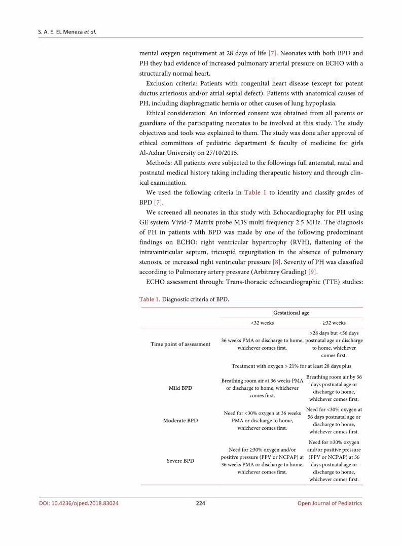
We used the following criteria in Table 1 to identify and classify grades of BPD [7].
We screened all neonates in this study with Echocardiography for PH using GE system Vivid-7 Matrix probe M3S multi frequency 2.5 MHz. The diagnosis of PH in patients with BPD was made by one of the following predominant findings on ECHO: right ventricular hypertrophy (RVH), flattening of the intraventricular septum, tricuspid regurgitation in the absence of pulmonary stenosis, or increased right ventricular pressure [8]. Severity of PH was classified according to Pulmonary artery pressure (Arbitrary Grading) [9].
ECHO assessment through: Trans-thoracic echocardiographic (TTE) studies: Table 1. Diagnostic criteria of BPD.
Gestational age
<32 weeks ≥32 weeks
Time point of assessment 36 weeks PMA or discharge to home,
whichever comes first.
>28 days but <56 days postnatal age or discharge
to home, whichever comes first.
Treatment with oxygen > 21% for at least 28 days plus
Mild BPD Breathing room air at 36 weeks PMA
or discharge to home, whichever comes first.
Breathing room air by 56 days postnatal age or discharge to home,
whichever comes first.
Moderate BPD Need for <30% oxygen at 36 weeks
PMA or discharge to home, whichever comes first.
Need for <30% oxygen at 56 days postnatal age or
discharge to home, whichever comes first.
Severe BPD
Need for ≥30% oxygen and/or positive pressure (PPV or NCPAP) at 36 weeks PMA or discharge to home,
whichever comes first.
Need for ≥30% oxygen and/or positive pressure (PPV or NCPAP) at 56 days postnatal age or discharge to home,
whichever comes first.
S. A. E. EL Meneza et al.
DOI: 10.4236/ojped.2018.83024 225 Open Journal of Pediatrics
TTE t M-Mode, 2D, Doppler (pulsed and continous wave), color flow mapping in the standard views from all accessible windows were obtained with ECG phy-sio signal displayed with all detected echo-Doppler study.
Two-dimensional echocardiography: Routine examination was done from the parasternal, apical and subcostal views focusing on: Exclusion of congenital heart disease & Guidance for M-Mode and color Doppler.
M-Mode echocardiography: Parameters were obtained by the guidance of two dimensional (2-D) echocardiography from the parasternal long axis view, at the level of the papillary muscle and at the level of aorta and left atrium using the leading edge technique [10].
Assessment of pulmonary artery pressure: The tricuspid regurgitant (TR) jet is used to estimate pulmonary artery pressure, and represents the most common and reliable method to evaluate for the presence and severity of PH [11]. Also assessment for flattening of interventricular septum and accelerated pulmonary regurgitation velocity, by continuous-wave doppler using the modified Bernoulli equation in the absence of RV outflow tract (RVOT) obstruction to determine the RV systolic pressure (RVSP) which is the same systolic pulmonary artery pressure (SPAP)RVSP = SPAP = 4(TR max)2 + mean RA pressure (mRAP). With a shunt lesion, such as VSD or PDA the peak systolic velocity across the shunt can be used to estimate systolic pressure in the RV or PA [12].
Radiological investigation including Cranium Ultrasonography & x-ray chest and heart.
Laboratory investigations include routine investigations as CBC, Electrolytes, liver and kidney function tests, CRP, Blood gases analysis.
Specific laboratory investigation: Plasma ADMA and arginine were done by using competitive enzyme linked immune-assay (ELISA) method, using both ADMA and arginine ELIZA kits. SinoGeneClon Biotech company, Hangzhou, China. Arginine-to-ADMA ratio
Laboratory techniques: Peripheral venous blood samples were taken and se-rum was examined by Enzyme-linked immunosorbent assay (ELISA) for quan-titative evaluation of Plasma Asymmetric Dimethyl arginine and arginine in all participants. This was done in Al Zahra University Hospital laboratories.
Statistical analysis of data: Data was analyzed using Statistical Package for the Social Sciences (SPSS) version 20, Data are reported as mean and SD, or as number and percent. Demographics and clinical characteristics of cases (BPD and PH) and controls (BPD alone) were compared using c2 test for categorical data and Student t test for continuous data. ADMA, Arginine & ADMA/Arginine ratio levels were compared between study populations by Student t test. p value < 0.05 was considered statistically significant. Mann-Whitney test was used for comparison between two groups with quantitative data and non-parametric dis-tribution [13].
4. Results
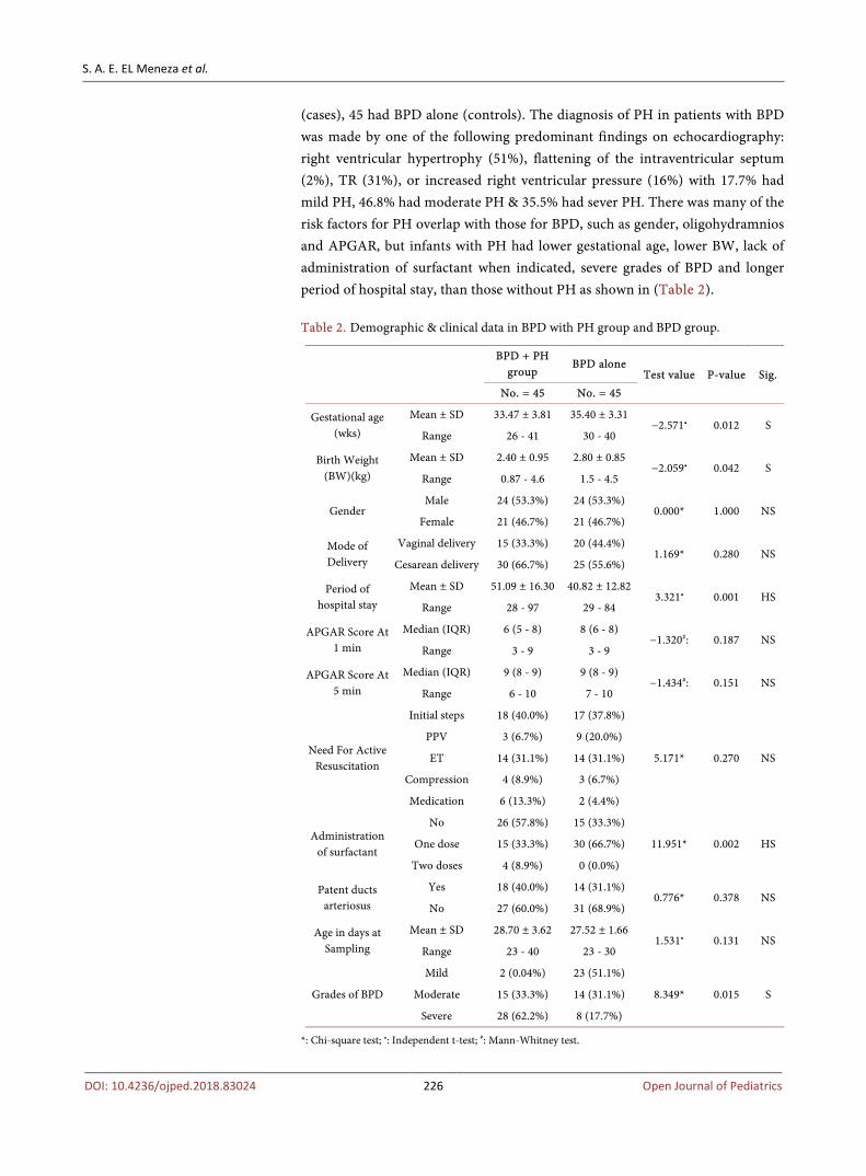
Among 90 patients with BPD enrolled in the study, 45 had both BPD and PH
S. A. E. EL Meneza et al.
DOI: 10.4236/ojped.2018.83024 226 Open Journal of Pediatrics
(cases), 45 had BPD alone (controls). The diagnosis of PH in patients with BPD was made by one of the following predominant findings on echocardiography: right ventricular hypertrophy (51%), flattening of the intraventricular septum (2%), TR (31%), or increased right ventricular pressure (16%) with 17.7% had mild PH, 46.8% had moderate PH & 35.5% had sever PH. There was many of the risk factors for PH overlap with those for BPD, such as gender, oligohydramnios and APGAR, but infants with PH had lower gestational age, lower BW, lack of administration of surfactant when indicated, severe grades of BPD and longer period of hospital stay, than those without PH as shown in (Table 2). Table 2. Demographic & clinical data in BPD with PH group and BPD group.
BPD + PH
group BPD alone
Test value P-value Sig. No. = 45 No. = 45
Gestational age (wks)
Mean ± SD 33.47 ± 3.81 35.40 ± 3.31 −2.571• 0.012 S
Range 26 - 41 30 - 40
Birth Weight (BW)(kg)
Mean ± SD 2.40 ± 0.95 2.80 ± 0.85 −2.059• 0.042 S
Range 0.87 - 4.6 1.5 - 4.5
Gender Male 24 (53.3%) 24 (53.3%)
0.000* 1.000 NS Female 21 (46.7%) 21 (46.7%)
Mode of Delivery
Vaginal delivery 15 (33.3%) 20 (44.4%) 1.169* 0.280 NS
Cesarean delivery 30 (66.7%) 25 (55.6%)
Period of hospital stay
Mean ± SD 51.09 ± 16.30 40.82 ± 12.82 3.321• 0.001 HS
Range 28 - 97 29 - 84
APGAR Score At 1 min
Median (IQR) 6 (5 - 8) 8 (6 - 8) −1.320#: 0.187 NS
Range 3 - 9 3 - 9
APGAR Score At 5 min
Median (IQR) 9 (8 - 9) 9 (8 - 9) −1.434#: 0.151 NS
Range 6 - 10 7 - 10
Need For Active Resuscitation
Initial steps 18 (40.0%) 17 (37.8%)
5.171* 0.270 NS
PPV 3 (6.7%) 9 (20.0%)
ET 14 (31.1%) 14 (31.1%)
Compression 4 (8.9%) 3 (6.7%)
Medication 6 (13.3%) 2 (4.4%)
Administration of surfactant
No 26 (57.8%) 15 (33.3%)
11.951* 0.002 HS One dose 15 (33.3%) 30 (66.7%)
Two doses 4 (8.9%) 0 (0.0%)
Patent ducts arteriosus
Yes 18 (40.0%) 14 (31.1%) 0.776* 0.378 NS
No 27 (60.0%) 31 (68.9%)
Age in days at Sampling
Mean ± SD 28.70 ± 3.62 27.52 ± 1.66 1.531• 0.131 NS
Range 23 - 40 23 - 30
Grades of BPD
Mild 2 (0.04%) 23 (51.1%)
8.349* 0.015 S Moderate 15 (33.3%) 14 (31.1%)
Severe 28 (62.2%) 8 (17.7%)
*: Chi-square test; •: Independent t-test; #: Mann-Whitney test.
S. A. E. EL Meneza et al.
DOI: 10.4236/ojped.2018.83024 227 Open Journal of Pediatrics
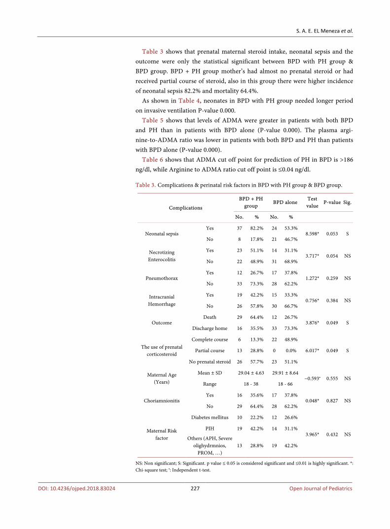
Table 3 shows that prenatal maternal steroid intake, neonatal sepsis and the outcome were only the statistical significant between BPD with PH group & BPD group. BPD + PH group mother’s had almost no prenatal steroid or had received partial course of steroid, also in this group there were higher incidence of neonatal sepsis 82.2% and mortality 64.4%.
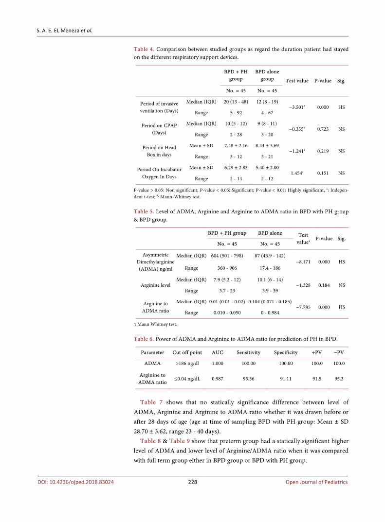
As shown in Table 4, neonates in BPD with PH group needed longer period on invasive ventilation P-value 0.000.
Table 5 shows that levels of ADMA were greater in patients with both BPD and PH than in patients with BPD alone (P-value 0.000). The plasma argi-nine-to-ADMA ratio was lower in patients with both BPD and PH than patients with BPD alone (P-value 0.000).
Table 6 shows that ADMA cut off point for prediction of PH in BPD is >186 ng/dl, while Arginine to ADMA ratio cut off point is ≤0.04 ng/dl. Table 3. Complications & perinatal risk factors in BPD with PH group & BPD group.
Complications
BPD + PH group
BPD alone Test value
P-value Sig.
No. % No. %
Neonatal sepsis Yes 37 82.2% 24 53.3%
8.598* 0.053 S No 8 17.8% 21 46.7%
Necrotizing Enterocolitis
Yes 23 51.1% 14 31.1% 3.717* 0.054 NS
No 22 48.9% 31 68.9%
Pneumothorax Yes 12 26.7% 17 37.8%
1.272* 0.259 NS No 33 73.3% 28 62.2%
Intracranial Hemorrhage
Yes 19 42.2% 15 33.3% 0.756* 0.384 NS
No 26 57.8% 30 66.7%
Outcome Death 29 64.4% 12 26.7%
3.876* 0.049 S Discharge home 16 35.5% 33 73.3%
The use of prenatal corticosteroid
Complete course 6 13.3% 22 48.9%
6.017* 0.049 S Partial course 13 28.8% 0 0.0%
No prenatal steroid 26 57.7% 23 51.1%
Maternal Age (Years)
Mean ± SD 29.04 ± 4.63 29.91 ± 8.64 −0.593• 0.555 NS
Range 18 - 38 18 - 66
Choriamnionitis Yes 16 35.6% 17 37.8%
0.048* 0.827 NS No 29 64.4% 28 62.2%
Maternal Risk factor
Diabetes mellitus 10 22.2% 12 26.6%
3.965* 0.432 NS PIH 19 42.2% 14 31.1%
Others (APH, Severe olighydrmnios,
PROM, …) 13 28.8% 19 42.2%
NS: Non significant; S: Significant. p value ≤ 0.05 is considered significant and ≤0.01 is highly significant. *: Chi-square test; •: Independent t-test.
S. A. E. EL Meneza et al.
DOI: 10.4236/ojped.2018.83024 228 Open Journal of Pediatrics
Table 4. Comparison between studied groups as regard the duration patient had stayed on the different respiratory support devices.
BPD + PH group
BPD alone group Test value P-value Sig.
No. = 45 No. = 45
Period of invasive ventilation (Days)
Median (IQR) 20 (13 - 48) 12 (8 - 19) −3.501# 0.000 HS
Range 5 - 92 4 - 67
Period on CPAP (Days)
Median (IQR) 10 (5 - 12) 9 (8 - 11) −0.355# 0.723 NS
Range 2 - 28 3 - 20
Period on Head Box in days
Mean ± SD 7.48 ± 2.16 8.44 ± 3.69 −1.241• 0.219 NS
Range 3 - 12 3 - 21
Period On Incubator Oxygen In Days
Mean ± SD 6.29 ± 2.83 5.40 ± 2.00 1.454• 0.151 NS
Range 2 - 14 2 - 12
P-value > 0.05: Non significant; P-value < 0.05: Significant; P-value < 0.01: Highly significant, •: Indepen-dent t-test; #: Mann-Whitney test. Table 5. Level of ADMA, Arginine and Arginine to ADMA ratio in BPD with PH group & BPD group.
BPD + PH group BPD alone Test
value• P-value Sig.
No. = 45 No. = 45
Asymmetric Dimethylarginine (ADMA) ng/ml
Median (IQR) 604 (501 - 798) 87 (43.9 - 142) −8.171 0.000 HS
Range 360 - 906 17.4 - 186
Arginine level Median (IQR) 7.9 (5.2 - 12) 10.1 (6 - 14)
−1.328 0.184 NS Range 3.7 - 23 3.9 - 39
Arginine to ADMA ratio
Median (IQR) 0.01 (0.01 - 0.02) 0.104 (0.071 - 0.185) −7.785 0.000 HS
Range 0.010 - 0.050 0 - 0.984
•: Mann Whitney test. Table 6. Power of ADMA and Arginine to ADMA ratio for prediction of PH in BPD.
Parameter Cut off point AUC Sensitivity Specificity +PV −PV
ADMA >186 ng/dl 1.000 100.00 100.00 100.0 100.0
Arginine to ADMA ratio
≤0.04 ng/dL 0.987 95.56 91.11 91.5 95.3
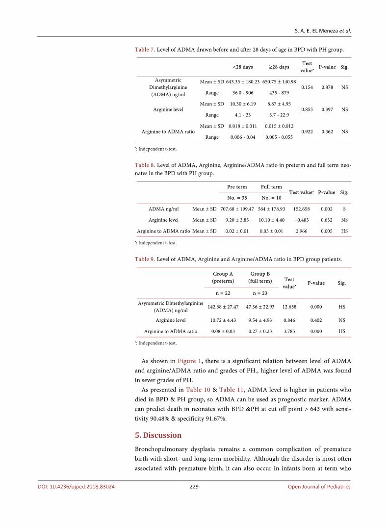
Table 7 shows that no statically significance difference between level of
ADMA, Arginine and Arginine to ADMA ratio whether it was drawn before or after 28 days of age (age at time of sampling BPD with PH group: Mean ± SD 28.70 ± 3.62, range 23 - 40 days).
Table 8 & Table 9 show that preterm group had a statically significant higher level of ADMA and lower level of Arginine/ADMA ratio when it was compared with full term group either in BPD group or BPD with PH group.
S. A. E. EL Meneza et al.
DOI: 10.4236/ojped.2018.83024 229 Open Journal of Pediatrics
Table 7. Level of ADMA drawn before and after 28 days of age in BPD with PH group.
<28 days ≥28 days Test
value• P-value Sig.
Asymmetric Dimethylarginine (ADMA) ng/ml
Mean ± SD 643.35 ± 180.23 650.75 ± 140.98 0.154 0.878 NS
Range 36 0 - 906 435 - 879
Arginine level Mean ± SD 10.30 ± 6.19 8.87 ± 4.95
0.855 0.397 NS Range 4.1 - 23 3.7 - 22.9
Arginine to ADMA ratio Mean ± SD 0.018 ± 0.011 0.015 ± 0.012
0.922 0.362 NS Range 0.006 - 0.04 0.005 - 0.055
•: Independent t-test.
Table 8. Level of ADMA, Arginine, Arginine/ADMA ratio in preterm and full term neo-nates in the BPD with PH group.
Pre term Full term
Test value• P-value Sig. No. = 35 No. = 10
ADMA ng/ml Mean ± SD 707.68 ± 199.47 564 ± 178.93 152.658 0.002 S
Arginine level Mean ± SD 9.20 ± 3.83 10.10 ± 4.40 −0.483 0.632 NS
Arginine to ADMA ratio Mean ± SD 0.02 ± 0.01 0.03 ± 0.01 2.966 0.005 HS
•: Independent t-test.
Table 9. Level of ADMA, Arginine and Arginine/ADMA ratio in BPD group patients.
Group A (preterm)
Group B (full term) Test
value• P-value Sig.
n = 22 n = 23
Asymmetric Dimethylarginine (ADMA) ng/ml
142.68 ± 27.47 47.36 ± 22.93 12.658 0.000 HS
Arginine level 10.72 ± 4.43 9.54 ± 4.93 0.846 0.402 NS
Arginine to ADMA ratio 0.08 ± 0.03 0.27 ± 0.23 3.785 0.000 HS
•: Independent t-test.
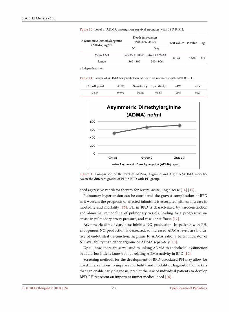
As shown in Figure 1, there is a significant relation between level of ADMA
and arginine/ADMA ratio and grades of PH., higher level of ADMA was found in sever grades of PH.
As presented in Table 10 & Table 11, ADMA level is higher in patients who died in BPD & PH group, so ADMA can be used as prognostic marker. ADMA can predict death in neonates with BPD &PH at cut off point > 643 with sensi-tivity 90.48% & specificity 91.67%.
5. Discussion
Bronchopulmonary dysplasia remains a common complication of premature birth with short- and long-term morbidity. Although the disorder is most often associated with premature birth, it can also occur in infants born at term who
S. A. E. EL Meneza et al.
DOI: 10.4236/ojped.2018.83024 230 Open Journal of Pediatrics
Table 10. Level of ADMA among non survival neonates with BPD & PH.
Asymmetric Dimethylarginine (ADMA) ng/ml
Death in neonates with BPD & PH Test value• P-value Sig.
No Yes
Mean ± SD 525.45 ± 100.46 769.03 ± 99.63 8.146 0.000 HS
Range 360 - 800 500 - 906
•: Independent t-test.
Table 11. Power of ADMA for prediction of death in neonates with BPD & PH.
Cut off point AUC Sensitivity Specificity +PV −PV
>634 0.940 90.48 91.67 90.5 91.7
Figure 1. Comparison of the level of ADMA, Arginine and Arginine/ADMA ratio be-tween the different grades of PH in BPD with PH group. need aggressive ventilator therapy for severe, acute lung disease [14] [15].
Pulmonary hypertension can be considered the gravest complication of BPD as it worsens the prognosis of affected infants, it is associated with an increase in morbidity and mortality [16]. PH in BPD is characterized by vasoconstriction and abnormal remodeling of pulmonary vessels, leading to a progressive in-crease in pulmonary artery pressure, and vascular stiffness [17].
Asymmetric dimethylarginine inhibits NO production. In patients with PH, endogenous NO production is decreased, so increased ADMA levels are indica-tive of endothelial dysfunction. Arginine to ADMA ratio, a better indicator of NO availability than either arginine or ADMA separately [18].
Up till now, there are serval studies linking ADMA to endothelial dysfunction in adults but little is known about relating ADMA activity in BPD [19].
Screening methods for the development of BPD-associated PH may allow for novel interventions to improve morbidity and mortality. Diagnostic biomarkers that can enable early diagnosis, predict the risk of individual patients to develop BPD-PH represent an important unmet medical need [20].
S. A. E. EL Meneza et al.
DOI: 10.4236/ojped.2018.83024 231 Open Journal of Pediatrics
Our study was a case control study conducted on ninety (90) newborns. In-fants were defined as suffering from BPD if they were on oxygen support ≥ 28 days. Demographic data in this study showed that infants with BPD and PH had a statistically significant lower gestational age, lower BW than those without PH.
Khemani et al. [21] and Check et al. [22] found that low gestational age and low birth weight significantly predict the development of PH in infants with BPD.
Bhat et al. [23] and an et al. [24] reported that infants with BPD and PH had lower BW, but in contrast to our result they found them had similar GA with no statistically significant with those who had BPD. We can explain this difference by the fact that their study was done on preterm babies only (median gestational age: 26 weeks), while in our study both preterms and full terms were included. Also Slaughter et al. [25], found that GA and BW were not different between those with and those without PH.
We found that gender and mode of delivery had no effect on development of PH in BPD as male to female ratio were in group I BPD with PH (27 males & 18 females) While Group II BPD only (24 males and 21 females) with a p value 1.000. Also mode of delivery in cases group (vaginal delivery 33.3% cesarean de-livery 66.7% while in control group (vaginal delivery 44.4% cesarean delivery 55.6% with a p value 0.280. This result confirmed the results reached by An et al. [24] and Stuart et al. [26] who found that there was no sex difference between infants with BPD and BPD with PH and mode of delivery had no effect, also Slaughter et al. [25] met the same results.
Patients in BPD and PH group had a longer period of hospital stay than those without PH with a p value 0.001. Also Bhat et al. [23] and Stuart et al. [26] found that PH in preterm infants with BPD was associated with longer hospitalizations than those with BPD only. In contrast to this, Al-Ghanem et al. [16] reported that there was no significant difference in duration of hospitalization between BPD-PH group and the BPD group.
We found that prenatal maternal steroid intake showed statistical significance between the studied groups as BPD + PH group mother’s had almost no prenatal steroid intake or had received partial course of steroid with a p value 0.049. This is similar to Rob et al. [18] and Slaughter et al. [25].
On studying the clinical data, we found most of patients who developed PH had severe grades of BPD (sever 62.2%, moderate 33.3% and mild 4.3%), while in group II BPD only (severe 17.7%, moderate 31.1% and mild 51.1%) with a p value 0.015 between both groups. It is in agreement with result of Al-Ghanem et al. [16] and del Cerro et al. [27]. However, Khemani et al. [21] reported that in-creasing BPD severity is not always associated with a higher incidence of PH, whereas other papers reported that infants with severe BPD are more likely to also develop PH [23] [24].
We observed a statically significant difference between BPD with PH group and BPD group, as regard administration of surfactant. Lack of administration
S. A. E. EL Meneza et al.
DOI: 10.4236/ojped.2018.83024 232 Open Journal of Pediatrics
of surfactant when indicated was mainly found in BPD and PH group with a p value 0.002. In contrast to this result, Slaughter et al. [25], found that no differ-ence between BPD with PH group and BPD only group as regard surfactant ad-ministration with a p value 0.231.
There was significant difference in sepsis and mortality between newborn in-fants with BPD and BPD with PH. These data agreed with others as Ali et al. [1] and Alfiero et al. [28]. Also Al-Ghanem et al. [16] and An et al. [24] met the same results and stated that no statically significant difference between group I (BPD with PH) and group II (BPD only), as regard complication like NEC, IVH. VAP is also among the complications detected by ELMeneza et al. [29]. Though post natal infection was higher in BPD with PH group (a p value 0.001).
Our study showed a statically significant difference concerning the period of invasive ventilation. BPD with PH group had stayed longer period on invasive ventilation. This result confirmed the results reached by other studies Ali et al. [1], An et al. [24], Kim et al. [30] and Alfiero et al. [28] who found that longer duration of mechanical ventilation may predict PH in patients with BPD. Bhat et al. [23]; Farquhar and Fitzgerald [31] met the same results.
Khemani et al. [21] and Berkelhamer et al. [32] stated that many of the risk factors for PH overlap with those for BPD, such as low GA, fetal growth restric-tion, oligohydramnios, prolonged mechanical ventilation as well as oxygen de-pendency.
W assessed severity of PH in BPD with PH group, we found 17.7% had mild PH, 46.8% had moderate PH and 35.5% had sever PH, then we found significant correlation between grades of BPD and severity of PH.PH was found mainly in sever and moderate cases of BPD. In accordance to this result, del Cerro et al. [27] reported that all of their patients with PH and BPD had moderate (24%) or severe BPD (76%).
Our study showed that higher levels of ADMA and lower level of arginine to ADMA ratio were found in BPD with PHgroup. Shao et al. [33] who studied ar-ginine metabolism in pulmonary hypertension in adult population had observed a direct association between dysregulated arginine methylation and elevated pulmonary artery pressures, they reported higher amounts of ADMA and dimi-nished global arginine bioavailability ratio were associated with higher systolic pulmonary artery pressure.
In our work we observed that there is no statically significance difference be-tween level of ADMA, Arginine and Arginine to ADMA ratio whether it was drawn before or after 28 days of age In contrast to our work, Jennifer et al. [8] in their studies demonstrated that greater levels of ADMA in BPD and PH were found in thoseplasma samples collected before 28 days of life. We can explain this difference that their study was done in small preterm neonates only with the biochemical reaction that appeared in response to the pathological changes in endothelium of pulmonary vessels appeared early before 28 days while in our study near term and full term neonates were included with those changes in en-
S. A. E. EL Meneza et al.
DOI: 10.4236/ojped.2018.83024 233 Open Journal of Pediatrics
dothelium can take more time to appear. We found the most predominant signs of PH in ECHO were RVH 51%, TR
31%, increased RT. ventricular pressure 16%, Flat intraventricular septum 2%. Paul H and Milenka [34] reported that TR jet velocity is the most common me-thod for determination of PH, We can explain that this method for detection of PH had has its limitation as the absence of a TR jet velocity does not rule out the presence of severe PH some patients had competent tricuspid valve despite the presence of sever PH, so in our study we depended on the most predominant signs of PH in ECHO.
In our study we found significant correlation between level of ADMA and grades of PH in our cases. Higher level of ADMA was found in sever grades of PH. This result is similar to, Shao et al. [33], Tang et al. [35] and Koro-Sajer et al. [36]
In this work we observed a statically significant difference between preterm neonates and full-term neonates as regard level of ADMA and Arginine/ADMA ratio. Higher level of ADMA is seen in preterm group and also this group had lower level of Arginine/ADMA ratio. Moreover we found significant negative correlation between ADMA level and BW, higher level of ADMA was seen in smaller birth weight. In accordance to these results, Tsukahara et al. [37] and Mittermayer et al. [38] who compared ADMA level in the umbilical veins of dif-ferent neonates. They reported that ADMA levels inversely correlated with ges-tational age and BW. ADMA levels in preterm and smaller birth weight were higher than in full term and appropriate BW. Moreover, Rob et al. [18] found a positive correlation between Arginine/ADMA ratio (AAR) with gestational age (p = 0.020) and BW, but no correlation with arginine or dimethylarginine levels.
Dzik et al. [39] found NO production was elevated in the perinatal period, particularly in preterm infants and they had the highest concentrations of ADMA.
We can explain this difference in ADMA between preterm and full term as it might be a consequence of increased ADMA synthesis, decreased metabolism by DDAH, decreased clearance by immature kidneys, or some combination of those factors in preterms.
In the current work we observed, no significant correlation between ADMA and gender with a P value 0.088. We met the result reached by Richir et al. [40] and Tsukahara et al. [37]. However, Mittermayer et al. [38] observed that ADMA levels were higher in male than in female preterm infants, they justified that reporting that males are at higher risk of prematurity as well as pulmonary, neurological, gastrointestinal, and cardiovascular prematurity-related condi-tions. This “male disadvantage” with respect to neonatal morbidity and mortali-ty has been recognized for more than three decades but the contributing biolog-ical mechanisms are poorly understood and likely to be multifactorial.
In our study there was significant positive correlation of ADMA level with pe-riod of invasive ventilation in BPD + PH group, higher level of ADMA level was
S. A. E. EL Meneza et al.
DOI: 10.4236/ojped.2018.83024 234 Open Journal of Pediatrics
seen in patient had longer period of invasive mechanical ventilation. Our result met the result reached by Richir et al. [40].
In our study we found significant positive correlation between ADMA level and development of necrotizing enterocolitis. In agreement with result Richir et al. [40], infants with NEC might present increased ADMA plasma concentra-tions. However, Moonen et al. [41] reported that ADMA and AAR were similar in infants with or without NEC.
In the current study, we observed that ADMA can predict death in neonates with BPD &PH at cut off point > 643 with sensitivity 90.48% &specificity 91.67%. In agreement with result, Judy L and Candice D [42] stated that it is possible that plasma ADMA levels could serve as a useful tool to identify those patients with BPD-PH with a poor prognosis and comparatively higher risk of mortality. Moreover, Brinkmann SJ et al. [43] reported that ADMA was a mark-er of mortality risk in adult population in intensive care, even for those patients with no underlying cardiovascular disease. S. Kavurt et al. [44]. found increased ADMA levels were associated with poor outcomes in preterm infants.
Avoid late and wrong diagnosis and treatment by using safe tools; updated guidelines and clinical path may decrease errors from prolonged/excessive oxy-gen use and invasive ventilation that lead to development of BPD and subse-quent complications as PH ELMeneza et al. [45].
6. Conclusion
Pulmonary hypertension development in BPD was found mainly in sever and moderate cases of BPD so screening for PH in those patients is a must. Neonates with BPD who developed PH by echocardiographic criteria had greater plasma levels of ADMA and lower plasma arginine to ADMA ratios than those patients with BPD that did not develop PH. ADMA level can predict severity of pulmo-nary hypertension in patient with BPD, also can be used as prognostic marker. Preterm neonates with BPD had greater level of ADMA than full term neonates. Studies are needed also to determine whether increased ADMA levels in BPD associated PH are caused by an increase in ADMA synthesis or a decrease in ADMA catabolism.
Conflicts of Interest
The authors declare no conflicts of interest regarding the publication of this pa-per.
References [1] Ali, Z., Schmidt, P., Dodd, J., et al. (2013) Predictors of Bronchopulmonary Dyspla-
sia and Pulmonary Hypertension in Newborn Children. The Danish Medical Jour-nal, 60, A468.
[2] Mourani, P.M. and Abman, S.H. (2013) Pulmonary Vascular Disease in Broncho-pul-Monary Dysplasia: Pulmonary Hypertension and Beyond. Current Opinion in Pediatrics, 25, 329-337. https://doi.org/10.1097/MOP.0b013e328360a3f6
S. A. E. EL Meneza et al.
DOI: 10.4236/ojped.2018.83024 235 Open Journal of Pediatrics
[3] Ghebremariam, Y.T., Erlanson, D.A. and Cooke, J.P. (2014) A Novel and Potent In-hibitor of Dimethylarginine Dimethylaminohydrolase: A Modulator of Cardiovas-cular Nitric Oxide. Journal of Pharmacology and Experimental Therapeutics, 348, 69-76 https://doi.org/10.1124/jpet.113.206847
[4] Klinger, J.R., Abman, S.H. and Gladwin, M.T. (2013) Nitric Oxide Deficiency and Endo-Thelial Dysfunction in Pulmonary Arterial Hypertension. American Journal of Respiratory and Critical Care Medicine, 188, 639-646. https://doi.org/10.1164/rccm.201304-0686PP
[5] Sanli, C., Oguz, D., Olgunturk, R., et al. (2012) Elevated Homocysteine and Asym-metric Dimethyl Arginine Levels in Pul-Monary Hypertension Associated with Congenital Heart Disease. Pediatric Cardiology, 33, 1323-1331. https://doi.org/10.1007/s00246-012-0321-9
[6] Latika, S., Sharad, C.A., Philip, D.H. and Rainer, H.B. (2010) The Role of Asymme-tric Dimethylarginine (ADMA) in Endothelial Dysfunction and Cardiovascular Disease. Current Cardiology Reviews, 6.
[7] Jobe, A.H. and Bancalari, E. (2001) Bronchopulmonary Dysplasia. American Jour-nal of Respiratory and Critical Care Medicine, 163, 1723. https://doi.org/10.1164/ajrccm.163.7.2011060
[8] Jennifer, K., Trittmann, F., Eric, P., et al. (2015) Plasma Asymmetric Dimethylargi-nine Levels Are Increased in Neonates with Bronchopulmonary Dysplasia-Associated Pulmonary Hypertension. Journal of Pediatric, 166, 230-233. https://doi.org/10.1016/j.jpeds.2014.09.004
[9] Goldstein, S. (2016) Echo in Pulmonary HTn, Systemic Review. Medstar Heart In-stitute, Washington Hospital Center, USA, American Society of Echocardiography, Vol. 29, No. 2, 93-102.
[10] Morikawa, T., Murata, M., Okuda, S., et al. (2011) Quantitative Analysis of Right Ventricular Function in Patients with Pulmonary Hypertension Using Three- Dimensional Echocardiography and a Two-Dimensional Summation Method Compared to Magnetic Resonance Imaging. American Journal of Cardiology, 107, 484-489. https://doi.org/10.1016/j.amjcard.2010.09.047
[11] Mertens, L. and Friedberg, M.K. (2016) Systolic Ventricular Function. In: Lai, W.W., Mertens, L.L. and Cohen, M.S., Eds., Echocardiography in Pediatric and Congenital Heart Disease: From Fetus to Adult, TalGeva, 2nd Edition, 96-102. https://doi.org/10.1002/9781118742440.ch7
[12] Nagiub, M., Lee, S. and Guglani, L. (2015) Echocardiographic Assessment of Pul-monary Hypertension in Infants with Bronchopulmonary Dysplasia: Systematic Re-view of Literature and a Proposed Algorithm for Assessment. Echocardiography, 32, 819. https://doi.org/10.1111/echo.12738
[13] Evans, J.D. (1996) Straight forward Statistics for the Behavioral Sciences. Brooks/Cole Publishing, Pacific Grove.
[14] Gien, J. and Kinsella, J.P. (2011) Pathogenesis and Treatment of Bronchopulmonary Dysplasia. Current Opinion in Pediatrics, 23, 305-313. https://doi.org/10.1097/MOP.0b013e328346577f
[15] El Meneza, S.A. and Gaber, A. (2014) Study of Pressure Volume Loop in Relation to Radiological Findings among Ventilated Newborn Infants. Journal of Neonatal Bi-ology, 3, 2.
[16] Al-Ghanem, G., Shah, P., Thomas, S., Banfield, L., Helou, S., et al. (2017) Broncho-pulmonary Dysplasia and Pulmonary Hypertension: A Meta-Analysis. Journal of Perinatology, 37, 414-419.
S. A. E. EL Meneza et al.
DOI: 10.4236/ojped.2018.83024 236 Open Journal of Pediatrics
[17] Ivy, D.D., Abman, S.H., Barst, R.J., et al. (2013) Pediatric Pulmonary Hypertension. Journal of the American College of Cardiology, 62, D117-D1126. https://doi.org/10.1016/j.jacc.2013.10.028
[18] Rob, M., Moonen, R., Maurice, J., et al. (2015) Plasma Levels of Dimethylarginines in Preterm Very Low Birth Weight Neonates: Its Relation with Perinatal Factors and Short-Term Outcome. International Journal of Molecular Sciences, 16, 19-39.
[19] Jennifer, K., Trittmann, F., Markus, V., et al. (2018) Arginase and α-Smooth Muscle Actin Induction after Hyperoxic Exposure in a Mouse Model of Bronchopulmonary Dysplasia. Clinical and Experimental Pharmacology and Physiology, 45, 556-562.
[20] Christine, B., Merrin, A., Arvind, S., et al. (2017) Pulmonary Hypertension Asso-ciated with Bronchopulmonary Dysplasia in Preterm Infants. Journal of Reproduc-tive Immunology, 24, 21-29.
[21] Khemani, E., McElhinney, D.B., Rhein, L., et al. (2007) Pulmonary Artery Hyper-tension in Formerly Premature Infants with Bronchopulmonary Dysplasia: Clinical Features and Outcomes in the Surfactant Era. Pediatrics, 120, 1260-1269. https://doi.org/10.1542/peds.2007-0971
[22] Check, J., Gotteiner, N., Liu, X., et al. (2013) Fetal Growth Restriction and Pulmo-nary Hypertension in Premature Infants with Bronchopulmonary Dysplasia. Jour-nal of Perinatology, 33, 553-557. https://doi.org/10.1038/jp.2012.164
[23] Bhat, R., Salas, A.A., Foster, C., et al. (2012) Prospective Analysis of Pulmonary Hypertension in Extremely Low Birth Weight Infants. Pediatrics, 129, e682-e689. https://doi.org/10.1542/peds.2011-1827
[24] An, H.S., Bae, E.J., Kim, G.B., Kwon, B.S., et al. (2010) Pulmonary Hypertension in Preterm Infants with Bronchopulmonary Dysplasia. Korean Circulation Journal, 40, 131-136. https://doi.org/10.4070/kcj.2010.40.3.131
[25] Slaughter, J.L., Pakrashi, T., Jones, D.E., et al. (2011) Echocardiographic Detection of Pulmonary Hypertension in Extremely Low Birth Weight Infants with Broncho-pulmonary Dysplasia Requiring Prolonged Positive Pressure Ventilation. Journal of Perinatology, 31, 635-640. https://doi.org/10.1038/jp.2010.213
[26] Stuart, B.D., Sekar, P., Coulson, J.D., et al. (2013) Health-Care Utilization and Res-piratory Morbidities in Preterm Infants with Pulmonary Hypertension. Journal of Perinatology, 33, 543-547. https://doi.org/10.1038/jp.2012.170
[27] Del Cerro, M.J., SabatéRotés, A., Cartón, A., et al. (2014) Pulmonary Hypertension in Bronchopulmonary Dysplasia: Clinical Findings, Cardiovascular Anomalies and Outcomes. Pediatric Pulmonology, 49, 49-59. https://doi.org/10.1002/ppul.22797
[28] Alfiero, M., Piva, D., Gangi, I.M., et al. (2011) Asymmetric Dimethylarginine in ELBW Newborns Exposed to Chorioamnionitis. Early Human Development, 87, 143-145. https://doi.org/10.1016/j.earlhumdev.2010.11.004
[29] Safaa A. El Meneza, Amelgaber, Awatef A Alrfaey, Enas E.EL Said (2010) VAP in NICU: Characteristics, Risk Factors and Outcome. Al Azhar Journal of Pediatrics, 13, 11.
[30] Kim, D.H., Kim, H.S., Choi, C.W., et al. (2012) Risk Factors for Pulmonary Artery Hypertension in Preterm Infants with Moderate or Severe Bronchopulmonary Dys-plasia. Neonatology, 101, 40-46. https://doi.org/10.1159/000327891
[31] Farquhar, M. and Fitzgerald, D.A. (2010) Pulmonary Hypertension in Chronic Neonatal Lung Disease. Paediatric Respiratory Reviews, 11, 149-153. https://doi.org/10.1016/j.prrv.2010.05.001
[32] Berkelhamer, S.K., Mestan, K.K. and Steinhorn, R.H. (2013) Pulmonary Hyperten-
S. A. E. EL Meneza et al.
DOI: 10.4236/ojped.2018.83024 237 Open Journal of Pediatrics
sion in Bronchopulmonary Dysplasia. Seminars in Perinatology, 37, 124-131. https://doi.org/10.1053/j.semperi.2013.01.009
[33] Shao, Z., et al. (2012) Pulmonary Hypertension Associated with Advanced Systolic Heart Failure Dysregulated Arginine Metabolism and Importance of Compensatory Dimethylarginine Dimethylaminohydrolase-1. Journal of the American College of Cardiology, 59, 1150-1158. https://doi.org/10.1016/j.jacc.2011.12.022
[34] Paul, H.D. and Milenka, C.G. (2018) Bronchopulmonary Dysplasia and Pulmonary Hypertension in Need of Cardiologist Innovation and Research. Pediatric Emer-gency Care and Medicine, 3, 1-5.
[35] Tang, W.H., Wang, Z., Cho, L., et al. (2008) Diminished Global Arginine Bioavaila-bility and Increased Arginine Catabolism as Metabolic Profile of Increased Cardi-ovascular Risk. Journal of the American College of Cardiology, 53, 2061-2067. https://doi.org/10.1016/j.jacc.2009.02.036
[36] Skoro-Sajer, N., et al. (2007) Asymmetric Dimethylarginine Is Increased in Chronic Thromboembolic Pulmonary Hypertension. American Journal of Respiratory and Critical Care Medicine, 176, 50-55.
[37] Tsukahara, H., Ohta, N., Tokuriki, S., et al. (2008) Determination of Asymmetric Dimethylarginine, an Endogenous Nitric Oxide Synthase Inhibitor, in Umbilical Blood. Metabolism, 57, 215-220. https://doi.org/10.1016/j.metabol.2007.09.003
[38] Mittermayer, F., Prusa, A.R., Pollak, A. and Wolzt, M. (2006) Umbilical Vein Plas-ma Concentrations of Asymmetrical Dimethylarginine Are Increased in Male But Not Female Neonates Delivered Preterm: A Pilot Study. Early Human Develop-ment, 82, 421-424. https://doi.org/10.1016/j.earlhumdev.2005.08.005
[39] Dzik, J.M., Dobrzanska, A., Gruszfeld, D. and Walajtys-Rode, E. (2002) Nitric Oxide Metabolites in the Urine of Full-Term and Preterm Infants. Pediatrics Inter-national, 44, 368-375. https://doi.org/10.1046/j.1442-200X.2002.01584.x
[40] Richir, M.C., Siroen, M.P., van Elburg, R.M., et al. (2007) Low Plasma Concentra-tions of Arginine and Asymmetric Dimethylarginine in Premature Infants with Ne-crotizing Enterocolitis. British Journal of Nutrition, 97, 906-911. https://doi.org/10.1017/S0007114507669268
[41] Moonen, R.M., Reyes, I., Cavallaro, G., et al. (2010) The T1405N Carbamoyl Phos-phate Synthetase Polymorphism Does Not Affect Plasma Arginine Concentrations in Preterm Infants. PLoS ONE, 5, e10792. https://doi.org/10.1371/journal.pone.0010792
[42] Aschner, J.L. and Fike, C.D. (2015) Plasma Asymmetric Dimethylarginine in Infants with Bronchopulmonary Dysplasia: A Promising Biomarker despite Uncertain Pa-thogenic Significance. The Journal of Pediatrics, 166, 222-224. https://doi.org/10.1016/j.jpeds.2014.10.039
[43] Brinkmann, S.J., de Boer, M.C., Buijs, N., et al. (2014) Asymmetric Dimethylargi-nine and Critical Illness. Current Opinion in Clinical Nutrition & Metabolic Care, 17, 90-97.
[44] Kavurt, S., Demirel, N., Yagmur, A., et al. (2017) Increased ADMA Levels Are As-sociated with Poor Pulmonary Outcome in Preterm Neonates. The Journal of Ma-ternal-Fetal & Neonatal Medicine, 30, 864-869.
[45] Elmeneza, S., et al. (2016) ENSTN Guidelines, Policies and Clinical Paths. Egyptian Neonatal Safety Training Network, 5-17. https://www.researchgate.net/publication/322831553