Embed Size (px)
Citation preview
Polymer-drug conjugates for intracellarmolecule-targeted photoinducedinactivation of protein and growthinhibition of cancer cellsBing Wang, Huanxiang Yuan, Chunlei Zhu, Qiong Yang, Fengting Lv, Libing Liu & Shu Wang
Beijing National Laboratory for Molecular Sciences, Key Laboratory of Organic Solids, Institute of Chemistry, Chinese Academy ofSciences, Beijing 100190, P. R. China.
For most molecule-targeted anticancer systems, intracellular protein targets are very difficult to be accessedby antibodies, and also most efforts are made to inhibit protein activity temporarily rather than inactivatethem permanently. In this work we firstly designed and synthesized multifunctional polymer-drugconjugates (polythiophene-tamoxifen) for intracellular molecule-targeted binding and inactivation ofprotein (estrogen receptor a, ERa) for growth inhibition of MCF-7 cancer cells. Small molecule drug wasconjugated to polymer side chain for intracellular signal protein targeting, and simultaneously thefluorescent characteristic of polymer for tracing the cellular uptake and localization of polythiophene-drugconjugates by cell imaging. Under light irradiation, the conjugated polymer can sensitize oxygen to producereactive oxygen species (ROS) that specifically inactivate the targeted protein, and thus inhibit the growth oftumor cells. The conjugates showed selective growth inhibition of ERa positive cancer cells, which exhibitslow side effect for our intracellular molecule-targeted therapy system.
Signal pathways are responsible for most biological processes such as cell growth, differentiation, migrationand apoptosis. Abnormal expression of proteins corresponding to specific signal pathway is closely relatedwith many serious diseases, especially cancer1–3. In recent decades, advances in genomics, proteomics,
chemistry, and protein engineering coalesce to accelerate the development of targeted anticancer drugs4–6. Inthese studies, small molecule drug-antibody conjugates that target specific proteins to retard cancer cell signaltransduction to arrest tumor growth are most favored as a result of relatively low side effect and strong selectivity.However most efforts are made to inhibit protein activity temporarily rather than inactivate them permanently.In the latter case, the protein remains inactive after the inhibitor diffuses away, and thus enhanced drug potencycan be achieved.
Photodynamic therapy (PDT) has been widely used for various cancer treatments to kill tumor cells7. Thecytotoxic agents, reactive oxygen species (ROS), are generated from the photosensitizer excited by appropriatelight exposure. With the interactions between ROS and biomacromolecules, tumor cell apoptosis and necrosiscan be initiated. Nevertheless, the surrounding healthy tissues are damaged at the same time. To minimize the sideeffects of conventional PDT, molecule-targeted PDT systems have been developed, yet with limited success8–12. Inthese systems, proteins targeted by antibodies or small molecules can be selectively inactivated by ROS generatedby photosentizers under light, without affecting the surrounding biomolecules. These systems combine thebenefits of targeting and inactivating and exhibit high spatial and temporal resolution in a noninvasive manner.However, these systems have two limitations: 1) proteomic or genetic modification is always needed9; 2) intra-cellular targets cannot be accessed by antibodies without microinject13. To the best of our knowledge, althoughselective photo-inactivation of proteins was studied8,9,13–17, selective inactivation of intracellar signal protein forcell growth inhibition and cancer treatment is seldom reported.
With the aim to develop a new molecule-targeted PDT system that can selectively target and kill the intracel-lular signal protein that tumor relies on efficiently with very low side effect, in this work, we designed multi-functional conjugated polymer-drug conjugates (PTD and PTDP, see their chemical structures in Figure 1A).Small molecule drug was conjugated to polymer side chain for intracellular signal protein targeting. The meanparticle sizes of PTD and PTDP are 31 and 83 nm from dynamic light scattering (DLS) experiments, respectively
SUBJECT AREAS:POLYMER CHEMISTRY
IMAGING TECHNIQUES
MATERIALS SCIENCE
SENSORS
Received21 August 2012
Accepted27 September 2012
Published24 October 2012
Correspondence andrequests for materials
should be addressed toQ.Y. (yangqiong@
iccas.ac.cn) or S.W.([email protected].
cn)
SCIENTIFIC REPORTS | 2 : 766 | DOI: 10.1038/srep00766 1
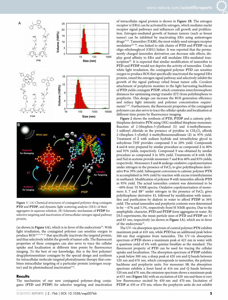
(as shown in Figure 1A), which is in favor of the endocytosis18. Withlight irradiation, the conjugated polymer can sensitize oxygen toproduce ROS12,17,19–23 that specifically inactivate the targeted protein,and thus selectively inhibit the growth of tumor cells. The fluorescentproperties of these conjugates can also serve to trace the cellularuptake and localization at different time points by fluorescenceimaging. To the best of our knowledge, this is the first polymer/drug/photosensitizer conjugate by the special design and synthesisfor intracellular molecule-targeted photodynamic therapy that com-bines intracellular targeting of a particular protein (estrogen recep-tor) and its photoinduced inactivation8,24.
ResultsThe mechanism of our new conjugated polymer-drug conju-gates (PTD and PTDP) for selective targeting and inactivation
of intracellular signal protein is shown in Figure 1B. The estrogenreceptor a (ERa) can be activated by estrogen, which mediates nucleireceptor signal pathways and influences cell growth and prolifera-tion. Estrogen-mediated growth of human tumors (such as breasttumor) can be inhibited by inactivating ERa using antiestrogendrugs24,25. Tamoxifen (TAM), the most widely used estrogen receptormodulator26–28, was linked to side chains of PTD and PTDP via anoligo-ethyleneglycol (OEG) linker. It was reported that the perma-nently charged tamoxifen derivatives can decrease side effects, dis-play good affinity to ERa and still modulate ERa-mediated tran-scription28. It is expected that similar modification of tamoxifen toPTD and PTDP would not deprive the activity of tamoxifen. Underwhite light irradiation, the conjugated polymer PTD can sensitizeoxygen to produce ROS that specifically inactivated the targeted ERaprotein, retard the estrogen signal pathway and selectively inhibit thegrowth of the signal pathway relied breast tumor cells. Covalentattachment of porphyrin moieties to the light harvesting backboneof PTD yields conjugate PTDP, which constrains interchromophoredistances for optimizing energy transfer (ET) from polythiophene toporphyrin. This design can increase the ROS generation efficiencyand reduce light intensity and polymer concentration require-ments12,22. Furthermore, the fluorescent properties of the conjugatedpolymers can also serve to trace the cellular uptake and localization atdifferent time points by fluorescence imaging.
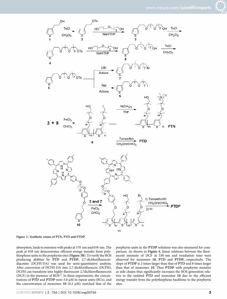
Figure 2 shows the synthesis of PTD, PTDP and a cationic poly-thiophene derivative PTN using OEG modified thiophene monomer.Reaction of 2-(thiophen-3-yl)ethanol (1) and 4-methylbenzene-1-sulfonyl chloride in the presence of pyridine in CH2Cl2 affords2-(thiophen-3-yl)ethyl 4-methylbenzenesulfonate (2) in 95% yield.Treatment of 2 with sodium hydride and tetraethylene glycol inanhydrous THF provides compound 3 in 20% yield. Compounds4 and 6 were prepared by similar procedure as compound 2 in 80%and 95% yields, respectively. Compound 5 was obtained by similarprocedure as compound 3 in 50% yield. Treatments of 6 with LiBrand NaI in acetone provide monomer 7 and 8 in 40% and 85% yields,respectively. Monomers 3 and 8 undergo oxidative copolymerizationunder nitrogen in the presence of FeCl3 to give polythiophene deriv-ative 9 in 39% yield. Subsequent conversion to cationic polymer PTNis accomplished in 94% yield by reaction with excess trimethylaminein methanol. Modification of polymer 9 with tamoxifen affords PTDin 95% yield. The actual tamoxifen content was determined to be,60% from 1H NMR spectra. Oxidative copolymerization of mono-mers 3, 7 and 1012 under nitrogen in the presence of FeCl3 givespolythiophene derivative 11, followed by modification with tamox-ifen and purification by dialysis in water to afford PTDP in 36%yield. The actual tamoxifen and porphyrin contents were determinedto be ,47% and 3.5%, respectively from1H NMR spectra. Due to theamphiphilic character, PTD and PTDP form aggregates in water. ByDLS experiments, the mean particle sizes of PTD and PTDP are 31and 83 nm, respectively (as shown in Figure 1A), which are in favorof the endocytosis18.
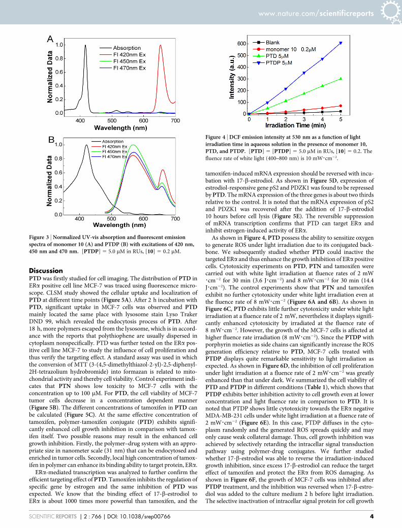
The UV-vis absorption spectrum of control polymer PTN exhibitsmaximum peak at 410 nm, while PTD has an additional peak below300 nm that originates from tamoxifen. The UV-vis absorptionspectrum of PTD shows a maximum peak at 423 nm in water witha quantum yield of 6% with quinine bisulfate as the standard. Thefluorescent property of PTD can be used for tracing the cellularuptake and localization. The absorption spectrum of PTDP exhibitsa peak below 300 nm, a sharp peak at 420 nm and Q bands between520 nm and 670 nm, which corresponds to tamoxifen, the polymerbackbone and porphyrin units. For monomer 10, the absorptionspectrum exhibits a Soret band at 416 nm and Q bands between520 nm and 670 nm; the emission spectrum shows a maximum peakat 653 nm (Figure 3A) with an excitation of 420 nm and there is verylow fluorescence excited by 450 nm and 470 nm. Excitation ofPTDP at 450 or 470 nm, where the porphyrin units do not exhibit
Figure 1 | (A) Chemical structures of conjugated polymer-drug conjugate
PTD and PTDP, and dynamic light scattering analysis (DLS) of their
aggregates in aqueous solution. (B) Schematic mechanism of PTDP for
selective targeting and inactivation of intracellular estregen signal pathway
protein.
www.nature.com/scientificreports
SCIENTIFIC REPORTS | 2 : 766 | DOI: 10.1038/srep00766 2
absorption, leads to emission with peaks at 578 nm and 658 nm. Thepeak at 658 nm demonstrates efficient energy transfer from poly-thiophene units to the porphyrin sites (Figure 3B). To verify the ROSproducing abilities by PTD and PTDP, 2,7-dichloriflurescin-diacetate (DCFH-DA) was used for semi-quantitative analysis.After conversion of DCFH-DA into 2,7-dichloriflurescin (DCFH),DCFH can transform into highly fluorescent 2,7dichlorofluorescein(DCF) in the presence of ROS22. In these experiments, the concen-trations of PTD and PTDP were 5.0 mM in repeat units (RUs), andthe concentration of monomer 10 (0.2 mM) matched that of the
porphyrin units in the PTDP solutions was also measured for com-parison. As shown in Figure 4, linear relations between the fluor-escent intensity of DCF at 530 nm and irradiation time wereobserved for monomer 10, PTD and PTDP, respectively. Theslope of PTDP is 2 times larger than that of PTD and 8 times largerthan that of monomer 10. Thus PTDP with porphyrin moietiesas side chains thus significantly increases the ROS generation rela-tive to the isolated PTD and monomer 10 due to the efficientenergy transfer from the polythiophene backbone to the porphyrinsites.
Figure 2 | Synthetic routes of PTN, PTD and PTDP.
www.nature.com/scientificreports
SCIENTIFIC REPORTS | 2 : 766 | DOI: 10.1038/srep00766 3
DiscussionPTD was firstly studied for cell imaging. The distribution of PTD inERa positive cell line MCF-7 was traced using fluorescence micro-scope. CLSM study showed the cellular uptake and localization ofPTD at different time points (Figure 5A). After 2 h incubation withPTD, significant uptake in MCF-7 cells was observed and PTDmainly located the same place with lysosome stain Lyso TrakerDND 99, which revealed the endocytosis process of PTD. After18 h, more polymers escaped from the lysosome, which is in accord-ance with the reports that polythiophene are usually dispersed incytoplasm nonspecifically. PTD was further tested on the ERa pos-itive cell line MCF-7 to study the influence of cell proliferation andthus verify the targeting effect. A standard assay was used in whichthe conversion of MTT (3-(4,5-dimethylthiazol-2-yl)-2,5-diphenyl-2H-tetrazolium hydrobromide) into formazan is related to mito-chondrial activity and thereby cell viability. Control experiment indi-cates that PTN shows low toxicity to MCF-7 cells with theconcentration up to 100 mM. For PTD, the cell viability of MCF-7tumor cells decrease in a concentration dependent manner(Figure 5B). The different concentrations of tamoxifen in PTD canbe calculated (Figure 5C). At the same effective concentration oftamoxifen, polymer-tamoxifen conjugate (PTD) exhibits signifi-cantly enhanced cell growth inhibition in comparison with tamox-ifen itself. Two possible reasons may result in the enhanced cellgrowth inhibition. Firstly, the polymer–drug system with an appro-priate size in nanometer scale (31 nm) that can be endocytosed andenriched in tumor cells. Secondly, local high concentration of tamox-ifen in polymer can enhance its binding ability to target protein, ERa.
ERa-mediated transcription was analyzed to further confirm theefficient targeting effect of PTD. Tamoxifen inhibits the regulation ofspecific gene by estrogen and the same inhibition of PTD wasexpected. We know that the binding effect of 17-b-estrodiol toERa is about 1000 times more powerful than tamoxifen, and the
tamoxifen-induced mRNA expression should be reversed with incu-bation with 17-b-estrodiol. As shown in Figure 5D, expression ofestrodiol-responsive gene pS2 and PDZK1 was found to be repressedby PTD. The mRNA expression of the three genes is about two thirdsrelative to the control. It is noted that the mRNA expression of pS2and PDZK1 was recovered after the addition of 17-b-estrodiol10 hours before cell lysis (Figure 5E). The reversible suppressionof mRNA transcription confirms that PTD can target ERa andinhibit estrogen-induced activity of ERa.
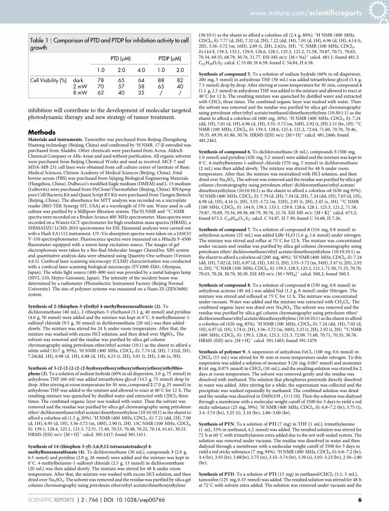
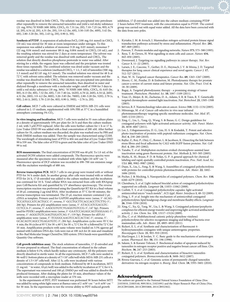
As shown in Figure 4, PTD possess the ability to sensitize oxygento generate ROS under light irradiation due to its conjugated back-bone. We subsequently studied whether PTD could inactive thetargeted ERa and thus enhance the growth inhibition of ERa positivecells. Cytotoxicity experiments on PTD, PTN and tamoxifen werecarried out with white light irradiation at fluence rates of 2 mW?cm22 for 30 min (3.6 J?cm22) and 8 mW?cm22 for 30 min (14.4J?cm22). The control experiments show that PTN and tamoxifenexhibit no further cytotoxicity under white light irradiation even atthe fluence rate of 8 mW?cm22 (Figure 6A and 6B). As shown inFigure 6C, PTD exhibits little further cytotoxicity under white lightirradiation at a fluence rate of 2 mW, nevertheless it displays signifi-cantly enhanced cytotoxicity by irradiated at the fluence rate of8 mW?cm22. However, the growth of the MCF-7 cells is affected athigher fluence rate irradiation (8 mW?cm22). Since the PTDP withporphyrin moieties as side chains can significantly increase the ROSgeneration efficiency relative to PTD, MCF-7 cells treated withPTDP displays quite remarkable sensitivity to light irradiation asexpected. As shown in Figure 6D, the inhibition of cell proliferationunder light irradiation at a fluence rate of 2 mW?cm22 was greatlyenhanced than that under dark. We summarized the cell viability ofPTD and PTDP in different conditions (Table 1), which shows thatPTDP exhibits better inhibition activity to cell growth even at lowerconcentration and light fluence rate in comparison to PTD. It isnoted that PTDP shows little cytotoxicity towards the ERa negativeMDA-MB-231 cells under white light irradiation at a fluence rate of2 mW?cm22 (Figure 6E). In this case, PTDP diffuses in the cyto-plasm randomly and the generated ROS spreads quickly and mayonly cause weak collateral damage. Thus, cell growth inhibition wasachieved by selectively retarding the intracellar signal transductionpathway using polymer-drug conjugates. We further studiedwhether 17-b-estrodiol was able to reverse the irradiation-inducedgrowth inhibition, since excess 17-b-estrodiol can reduce the targeteffect of tamoxifen and protect the ERa from ROS damaging. Asshown in Figure 6F, the growth of MCF-7 cells was inhibited afterPTDP treatment, and the inhibition was reversed when 17-b-estro-diol was added to the culture medium 2 h before light irradiation.The selective inactivation of intracellar signal protein for cell growth
Figure 3 | Normalized UV-vis absorption and fluorescent emissionspectra of monomer 10 (A) and PTDP (B) with excitations of 420 nm,450 nm and 470 nm. [PTDP] 5 5.0 mM in RUs, [10] 5 0.2 mM.
Figure 4 | DCF emission intensity at 530 nm as a function of lightirradiation time in aqueous solution in the presence of monomer 10,PTD, and PTDP. [PTD] 5 [PTDP] 5 5.0 mM in RUs, [10] 5 0.2. The
fluence rate of white light (400–800 nm) is 10 mW?cm22.
www.nature.com/scientificreports
SCIENTIFIC REPORTS | 2 : 766 | DOI: 10.1038/srep00766 4
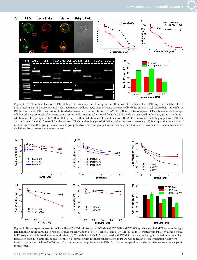
Figure 5 | (A) The cellular location of PTD at different incubation time: 2 h (upper) and 18 h (lower). The false color of PTD is green; the false color of
Lyso Tracker DND 99 (lysosome stain) is red; their merge is yellow. (B, C) Dose-response curves for cell viability of MCF-7 Cells treated with tamoxifen or
PTD as functions of PTD molar concentration (A) or mass concentration of effective TAM (B). (D) Reverse transcriptase-PCR analysis of mRNA. Images
of DNA gel electrophoresis after reverse transcription-PCR reaction. After seeded for 12 h, MCF-7 cells are incubated under dark, group 1: without
addition for 34 h; group 2: with PTD for 34 h; group 3: without addition for 24 h, and then with 10 nM 17-b-estrodiol for 10 h; group 4: with PTD for
24 h and then 10 nM 17-b-estrodiol added for 10 h. The housekeeping gene GAPDH is used as the internal reference. (E) Semi-quantitative analysis of
mRNA expression. Red: group 1 as control and group 2 as treated; green: group 3 as control and group 4 as treated. Error bars correspond to standard
deviations from three separate measurements.
Figure 6 | Dose-response curves for cell viability of MCF-7 cells treated with TAM (A), PTN (B) and PTD (C) by using a typical MTT assay under lightirradiation or in the dark. Dose-response curves for cell viability of MCF-7 cells (D) and MDA-MB-231 cells (E) treated with PTDP by using a typical
MTT assay under light irradiation or in the dark. (F) Cell viability of MCF-7 cells treated with PTDP in the dark, under light irradiation or under light
irradiation with 17-b-estrodiol added. The the 17-b-estrodiol with identical concentration to PTDP was added 2h before irradiation. Cells were
irradiated with white light (400–800 nm). The concentrations of polymer are in RUs. Error bars correspond to standard deviations from three separate
measurements.
www.nature.com/scientificreports
SCIENTIFIC REPORTS | 2 : 766 | DOI: 10.1038/srep00766 5
inhibition will contribute to the development of molecular targetedphotodynamic therapy and new strategy of tumor treatment.
MethodsMaterials and instruments. Tamoxifen was purchased from Beijing ZhongshengHuateng technology (Beijing, China) and confirmed by 1H NMR. 17-b-estrodiol waspurchased from Aladdin. Other chemicals were purchased from Acros, AldrichChemical Company or Alfa-Aesar and used without purification. All organic solventswere purchased from Beijing Chemical Works and used as received. MCF-7 andMDA-MB-231 cell lines were obtained from cell culture center of Institute of BasicMedical Sciences, Chinese Academy of Medical Sciences (Beijing, China). Fetalbovine serum (FBS) was purchased from Sijiqing Biological Engineering Materials(Hangzhou, China). Dulbecco’s modified Eagle medium (DMEM) and L-15 medium(Leibovitz) were purchased from HyClone/Thermofisher (Beijing, China). RNApreppure Cell/Bacteria Kit and Quant Script RT Kit were purchased from Tiangen Biotech(Beijing, China). The absorbance for MTT analysis was recorded on a microplatereader (BIO-TEK Synergy HT, USA) at a wavelength of 570 nm. Water used in cellculture was purified by a Millipore filtration system. The1H NMR and 13C NMRspectra were recorded on a Bruker Avance 400 MHz spectrometer. Mass spectra wererecorded on a Waters GCT spectrometer for high resolution mass spectra (HRMS), aSHIMADZU LCMS-2010 spectrometer for ESI. Elemental analyses were carried outwith a Flash EA1112 instrument. UV-Vis absorption spectra were taken on a JASCOV-550 spectrophotometer. Fluorescence spectra were measured on a Hitachi F-4500fluorometer equipped with a xenon lamp excitation source. The images of gelelectrophoresis were taken by a Bio-Rad Molecular Imager ChemiDoc XRS systemand quantitative analysis data were obtained using Quantity One software (Version4.6.5). Confocal laser scanning microscopy (CLSM) characterization was conductedwith a confocal laser scanning biological microscope (FV1000-IX81, Olympus,Japan). The white light source (400–800 nm) was provided by a metal halogen lamp(MVL-210, Mejiro Genossen, Japan). The intensity of the incident beam wasdetermined by a radiometer (Photoelectric Instrument Factory (Beijing NormalUniversity). The size of polymer systems was measured on a Nano ZS (ZEN3600)system.
Synthesis of 2-(thiophen-3-yl)ethyl 4-methylbenzenesulfonate (2). Todichloromethane (40 mL), 2-(thiophen-3-yl)ethanol (5.1 g, 40 mmol) and pyridine(4.0 g, 50 mmol) were added and the mixture was kept in 0uC. 4-methylbenzene-1-sulfonyl chloride (9.5 g, 50 mmol) in dichloromethane (20 mL) was then addedslowly. The mixture was stirred for 24 h under room temperature. After that, themixture was washed with excess HCl solution, and then dried over MgSO4. Thesolvent was removed and the residue was purified by silica gel columnchromatography using petroleum ether/ethyl acetate (10:1) as the eluent to afford awhite solid (10.7 g, 95%). 1H NMR (400 MHz, CDCl3, d): 7.74 (d, 2H), 7.32(d, 2H),7.24(dd, 1H), 6.98 (d, 1H), 6.88 (d, 1H), 4.23 (t, 2H), 3.01 (t, 2H), 2.46 (s, 3H).
Synthesis of 3-(2-(2-(2-(2-(2-hydroxyethoxy)ethoxy)ethoxy)ethoxy)ethylthio-phene (3). To a solution of sodium hydride (60% in oil dispersion, 3.0 g, 75 mmol) inanhydrous THF (60 ml) was added tetraethylene glycol (14.5 g, 75 mmol) drop bydrop. After stirring at room temperature for 30 min, compound 2 (7.0 g, 25 mmol) inanhydrous THF was added to the mixture and allowed to react at 40uC for 12 h. Theresulting mixture was quenched by distilled water and extracted with CHCl3 threetimes. The combined organic layer was washed with water. Then the solvent wasremoved and the residue was purified by silica gel chromatography using petroleumether/ dichloromethane/ethyl acetate/dimethoxyethylene (10:10:10:1) as the eluent toafford a colorless oil (1.5 g, 20%). 1H NMR (400 MHz, CDCl3, d): 7.21 (dd, 1H), 7.00(d, 1H), 6.95 (d, 1H), 3.56-3.72 (m, 18H), 2.90 (t, 2H). 13C NMR (100 MHz, CDCl3,d): 139.1, 128.4, 125.1, 121.1, 72.51, 71.40, 70.53, 70.48, 70.22, 70.14, 61.61, 30.52.HRMS (ESI) m/z: [M1H]1 calcd. 305.1417; found 305.1411.
Synthesis of 14-(thiophen-3-yl)-3,6,9,12-tetraoxatetradecyl 4-methylbenzenesulfonate (4). To dichloromethane (30 mL), compounds 3 (2.0 g,6.5 mmol) and pyridine (2.0 g, 26 mmol) were added and the mixture was kept in0uC. 4-methylbenzene-1-sulfonyl chloride (2.5 g, 13 mmol) in dichloromethane(20 mL) was then added slowly. The mixture was stirred for 48 h under roomtemperature. After that, the mixture was washed with excess HCl solution, and thendried over Na2SO4. The solvent was removed and the residue was purified by silica gelcolumn chromatography using petroleum ether/ethyl acetate/dimethoxyethylene
(30:10:1) as the eluent to afford a colorless oil (2.4 g, 80%). 1H NMR (400 MHz,CDCl3, d): 7.77 (d, 2H), 7.32 (d, 2H), 7.22 (dd, 1H), 7.01 (d, 1H), 6.96 (d, 1H), 4.14 (t,2H), 3.56–3.72 (m, 16H), 2.89 (t, 2H), 2.42(s, 3H). 13C NMR (100 MHz, CDCl3,d):144.9, 139.3, 133.1, 129.9, 128.6, 128.1, 125.3, 121.2, 71.58, 70.87, 70.71, 70.65,70.34, 69.35, 68.79, 30.76, 21.77. ESI-MS m/z: [M1Na]1 calcd. 481.1; found 481.2.C21H30O7S2: calcd. C 55.00, H 6.59; found C 54.84, H 6.58.
Synthesis of compound 5. To a solution of sodium hydride (60% in oil dispersion,200 mg, 5 mmol) in anhydrous THF (30 mL) was added tetraethylene glycol (1.4 g,7.5 mmol) drop by drop. After stirring at room temperature for 30 min, compound 4(1.1 g, 2.5 mmol) in anhydrous THF was added to the mixture and allowed to react at40uC for 12 h. The resulting mixture was quenched by distilled water and extractedwith CHCl3 three times. The combined organic layer was washed with water. Thenthe solvent was removed and the residue was purified by silica gel chromatographyusing petroleum ether/ethyl acetate/methanol/dimethoxyethylene (10:30:1:1) as theeluent to afford a colorless oil (600 mg, 50%). 1H NMR (400 MHz, CDCl3, d): 7.24(dd, 1H), 7.01 (d, 1H), 6.96 (d, 1H), 3.55–3.72 (m, 34H), 2.92 (t, 2H) 2.51 (br, 1H). 13CNMR (100 MHz, CDCl3, d): 139.3, 128.6, 125.4, 121.2, 72.64, 71.60, 70.70, 70.46,70.35, 69.59, 61.86, 30.76. HRMS (ESI) m/z: [M1H]1 calcd. 481.2466; found481.2462.
Synthesis of compound 6. To dichloromethane (8 mL), compounds 5 (500 mg,1.0 mmol) and pyridine (420 mg, 5.2 mmol) were added and the mixture was kept in0uC. 4-methylbenzene-1-sulfonyl chloride (570 mg, 3 mmol) in dichloromethane(2 mL) was then added slowly. The mixture was stirred for 48 h under roomtemperature. After that, the mixture was neutralized with HCl solution, and thendried over Na2SO4. The solvent was removed and the residue was purified by silica gelcolumn chromatography using petroleum ether/ dichloromethane/ethyl acetate/dimethoxyethylene (10:10:10:1) as the eluent to afford a colorless oil (630 mg 95%).1H NMR (400 MHz, CDCl3, d): 7.79 (d, 2H), 7.34 (d, 2H), 7.24 (dd, 1H), 7.03 (d, 1H),6.98 (d, 1H), 4.16 (t, 2H), 3.55–3.72 (m, 32H), 2.93 (t, 2H), 2.45 (s, 3H). 13C NMR(100 MHz, CDCl3, d): 144.9, 139.3, 133.1, 129.9, 128.6, 128.1, 125.3, 121.2, 71.58,70.87, 70.69, 70.34, 69.36, 68.79, 30.76, 21.76. ESI-MS m/z: [M1K]1 calcd. 673.2;found 673.3. C29H46O11S2: calcd. C 54.87, H 7.30; found C 54.68, H 7.26.
Synthesis of compound 7. To a solution of compound 6 (516 mg, 0.8 mmol) inanhydrous acetone (25 mL) was added LiBr?H2O (1.6 g, 1.6 mmol) under nitrogen.The mixture was stirred and reflux at 75uC for 12 h. The mixture was concentratedunder vacuum and residue was purified by silica gel column chromatography usingpetroleum ether/ dichloromethane/ethyl acetate/dimethoxyethylene (10:10:10:1) asthe eluent to afford a colorless oil (200 mg, 40%). 1H NMR (400 MHz, CDCl3, d): 7.24(dd, 1H), 7.02 (d, 1H), 6.97 (d, 1H), 3.81 (t, 2H), 3.55–3.72 (m, 34H), 3.47 (t, 2H), 2.92(t, 2H). 13C NMR (100 MHz, CDCl3, d): 139.3, 128.5, 125.2, 121.1, 71.50, 71.25, 70.70,70.63, 70.28, 30.70, 30.30. ESI-MS m/z: [M1NH4]1 calcd. 560.2; found 560.3.
Synthesis of compound 8. To a solution of compound 6 (530 mg, 0.8 mmol) inanhydrous acetone (40 mL) was added NaI (1.2 g, 8 mmol) under Nitrogen. Themixture was stirred and refluxed at 75uC for 12 h. The mixture was concentratedunder vacuum. Water was added and the mixture was extracted with CH2Cl2. Thecombined organic layer was dried over Na2SO4. The solvent was removed and theresidue was purified by silica gel column chromatography using petroleum ether/dichloromethane/ethyl acetate/dimethoxyethylene (10:10:10:1) as the eluent to afforda colorless oil (420 mg, 85%). 1H NMR (100 MHz, CDCl3, d): 7.24 (dd, 1H), 7.02 (d,1H), 6.97 (d, 1H), 3.74 (t, 2H), 3.56–3.72 (m, 30H), 3.25 (t, 2H), 2.92 (t, 2H). 13C NMR(400 MHz, CDCl3, d): 139.3, 128.6, 125.3, 121.3, 72.09, 71.60, 70.71, 70.35, 30.76.HRMS (ESI) m/z: [M1H]1 calcd. 591.1483; found 591.1479.
Synthesis of polymer 9. A suspension of anhydrous FeCl3 (100 mg, 0.6 mmol) inCHCl3 (15 mL) was stirred for 30 min at room temperature under nitrogen. To thissuspension was added a solution of monomer 3 (20 mg, 0.067 mmol) and monomer8 (44 mg, 0.075 mmol) in CHCl3 (10 mL), and the resulting solution was stirred for 2days at room temperature. The solvent was removed gently and the residue wasdissolved with methanol. The solution that phosphorus pentoxide directly dissolvedin water was added. After stirring for a while, the supernatant was collected and theprecipitate was washed repeatedly by methanol. The combined solution was driedand the residue was dissolved in DMSO/H 2 O (1:10). Then the solution was dialyzedthrough a membrane with a molecular weight cutoff of 3500 for 3 days to yield a redsticky substance (25 mg, 39%). 1H NMR (400 MHz, CDCl3, d): 6.8–7.2 (br), 3.75 (t),3.4–3.74 (br), 3.25 (t), 3.10 (br), 2.80–3.00 (br).
Synthesis of PTN. To a solution of PTI (7 mg) in THF (1 mL), trimethylamine(1 mL, 33% in methanol, 4.2 mmol) was added. The resulted solution was stirred for72 h at 60uC with trimethylamine extra added due to the not well-sealed system. Thesolution was removed under vacuum. The residue was dissolved in water and thendialyzed through a membrane with a molecular weight cutoff of 3500 for 3 days toyield a red sticky substance (7 mg, 94%). 1H NMR (400 MHz, CDCl3, d): 6.8–7.2 (br),4.4 (br), 3.93 (br), 3.88(br), 3.75 (m), 3.43–3.74 (br), 3.30 (s), 3.03–3.23 (br), 2.50–2.80(br).
Synthesis of PTD. To a solution of PTI (15 mg) in methanol/CHCl3 (1:1, 3 mL),tamoxifen (125 mg, 0.33 mmol) was added. The resulted solution was stirred for 48 hat 72uC with solvent extra added. The solution was removed under vacuum and the
Table 1 | Comparison of PTD and PTDP for inhibition activity to cellgrowth
PTD (mM) PTDP (mM)
1.0 2.0 4.0 1.0 2.0
Cell Viability (%) dark 78 65 64 88 822 mW 70 57 58 65 408 mW 62 40 35 / /
www.nature.com/scientificreports
SCIENTIFIC REPORTS | 2 : 766 | DOI: 10.1038/srep00766 6
residue was dissolved in little CHCl3. The solution was precipitated into petroleumether repeatedly to remove the unreacted tamoxifen and yield a red sticky substance(21 mg, 95%).1H NMR (400 MHz, CDCl3, d): 7.31 (m, 2H), 7.10–7.25 (m, 9H), 6.79(d, 2H), 6.54 (d, 2H), 4.31 (br, 2H), 3.9–4.2 (br, 6H), 3.50–3.85 (br, 40H), 3.42 (br,6H), 2.80–3.20 (br, 3H), 2.43 (q, 2H), 0.90 (t, 3H).
Synthesis of PTDP. A suspension of anhydrous FeCl3 (100 mg, 0.6 mmol) in CHCl3(10 mL) was stirred for 30 min at room temperature under nitrogen. To thissuspension was added a solution of monomer 3 (8 mg, 0.03 mmol), monomer 7(22 mg, 0.04 mmol) and monomer 10 (4 mg, 0.004 mmol) in CHCl3 (10 mL), andthe resulting solution was stirred for 2 days at room temperature. The solvent wasremoved gently and the residue was dissolved with methanol and CHCl3. Thesolution that directly dissolves phosphorus pentoxide in water was added. Afterstirring for a while, the organic layer was collected and the precipitate was treatedthree times repeatedly. The combined solution was dried under vacuum and theresidue was added to a solution (methanol:CHCl3 5 1:1, 3 ml) of tamoxifen (500 mg,1.3 mmol) and KI (45 mg, 0.3 mmol). The resulted solution was stirred for 48 h at72uC with solvent extra added. The solution was removed under vacuum and theresidue was dissolved in little CHCl3. The solution was precipitated into petroleumether repeatedly to remove the unreacted tamoxifen, then dissolved in water anddialyzed through a membrane with a molecular weight cutoff of 3500 for 3 days toyield a red sticky substance (18 mg, 36%). 1H NMR (400 MHz, CDCl3, d): 8.83 (br,8H), 8.13 (br, 8H), 7.52 (br, 6H), 7.10–7.40 (m, 162H), 6.78 (d, 26H), 6.54 (d, 26H),4.31 (br, 28H), 3.9–4.2 (br, 80H), 3.20–3.85 (br, 700H), 2.80–3.20 (br, 52H), 2.70 (s,9H), 2.44 (t, 26H), 1.70–2.10 (br, 8H), 0.90 (t, 39H), 22.76 (s, 2H).
Cell culture. MCF-7 cells were cultured in DMEM and MDA-MB-231 cells werecultured in L-15 medium, supplemented with 10% FBS at 37uC in a humidifiedatmosphere containing 5% CO2.
In vitro imaging and localization. MCF-7 cells were seeded in 35 mm culture platesat a density of approximately 10% per plate for 24 h and then the culture medium2 mL with 5 mM PTD replaced. After the cells were further culture for 1 h or 17 h,Lyso Traker DND 99 was added with a final concentration of 200 nM. After furtherculture for 1h, culture medium was discarded, the plate was washed once by PBS andfresh DMEM medium was added. Then the sample was characterized using CLSM.The wavelength of stimulating laser of PTD is 404 nm and that of Lyso Traker DND99 is 559 nm. The false color of PTD is green and the false color of Lyso Traker DND99 is red.
ROS measurements. The final concentration of DCFH was 40 mM. To 1.0 mL of theactivated DCFH solution were added compounds. The fluorescence spectra weremeasured after the specimens were irradiated with white light (10 mW?cm22).Fluorescence spectra of DCF solution was recorded in 498–700 nm emission rangewith the excitation wavelength of 488 nm.
Reverse transcription PCR. MCF-7 cells in one group were treated with or withoutPTD for 34 h under dark. In another group, after cells were treated with or withoutPTD for 24 h, 17-b-estrodiol was added to the culture medium and the incubationwas continued for 10 h. Total cellular RNA was extracted from cells with RNApreppure Cell/Bacteria Kit and quantified by UV absorbance spectroscopy. The reversetranscription reaction was performed using the QuantScript RT Kit in a final volumeof 20 mL containing 2 mg total RNA. After incubation at 37uC for 60 minutes, thereverse transcription was terminated. The 25 mL reaction mixture contained 2 mLcDNA as template. Primers used for GAPDH amplification were: (sense, 59-ACAG-TCCATGCCATCACTGCC-39; reverse, 59-GCCTGCTTCACCACCTTCTTG-39;266 bp). Primers for pS2 amplification were: (sense, 59-ATACCATCGACGTC-CCTCCA-39; reverse, 59-AAGCGTGTCTGAGGTGTCCG-39; 147 bp). Primersused for PDZK1 amplification were: (sense, 59-GAATCCAGAGCAGTGGGAAG-39;reverse, 59-AGGGTGTCAAGTGGATCAG-39; 119 bp). Primers for eEF1A1amplification were: (sense, 59-TCGGGCAAGTCCACCACTAC-39; reverse, 59-GCACAGTCAGCCTGAGATGTC-39; 284 bp). Amplification cycles were: 95uC for4 min, the 30 cycles at 95uC for 30 s, 60uC for 30 s, 72uC for 30 s; followed by 72uC for10 min. Amplification products with same volume were loaded on 3.5% agarose gelstained with Goldview DNA dye. Gels were run at 100 mA for 26 min and visualizedin Bio-Rad Molecular Imager ChemiDoc XRS system. DNA bands were quantified byQuantity One software.
Cell growth inhibition assay. The stock solutions of tamoxifen, 17-b-estrodiol and10 were prepared in ethanol. The final concentration of ethanol in the culturemedium is below 0.1%, which hardly induce any cytotoxicity. All the polymers weredirectly dissolved in sterile water. In all the experiments, MCF-7 cells were seeded in96-well U-bottom plates at a density of 73103 cells/well while MDA-MB-231 cells at adensity of 1.53104 cells/well. After 12 h, cells were incubated with variousconcentrations of compounds in fresh medium. Followed by 48 h incubation, MTT(5 mg mL21 in water, 10mL/well) was added to the wells by incubation at 37uC for 4 h.The supernatant was removed and 100 mL DMSO per well was added to dissolve theproduced formazan. After shaking the plates for 10 min, absorbance values of thewells were recorded with a microplate reader at 570 nm.
In the experiments of PDT, PDT treatment was performed 30 hours before MTTwas added by using white light source at fluence rates of 2 mW?cm22 or 8 mW?cm22
for 30 min. In the experiments to test the reverse ability to PDT-induced growth
inhibition, 17-b-estrodiol was added into the culture medium containing PTDP2 hours before PDT treatment, with the concentration equal to PTDP. The controlgroup was carried out with equal water added. All the data have been corrected withthe data from zero poles.
1. Kyriakis, J. M. & Avruch, J. Mammalian mitogen-activated protein kinase signaltransduction pathways activated by stress and inflammation. Physiol. Rev. 281,807–869 (2001).
2. Pawson, T. Protein modules and signaling networks. Nature 373, 573–580 (1995).3. Reya, T. & Clevers, H. Wnt signalling in stem cells and cancer. Nature 434,
843–850 (2005).4. Downward, J. Targeting ras signalling pathways in cancer therapy. Nat. Rev.
Cancer 3, 11–22 (2003).5. Larsen, J. E., Cascone, T., Gerber, D. E., Heymach, J. V. & Minna, J. D. Targeted
therapies for lung cancer clinical experience and novel agents. Cancer J. 17,512–527 (2011).
6. Hait, W. N. Targeted cancer therapeutics. Cancer Res. 69, 1263–1267 (2009).7. Moore, C. M., Pendse, D. & Emberton, M. Photodynamic therapy for prostate
cancer-a review of current status and future promise. Nat. Clin. Pract. Urol. 6,18–30 (2009).
8. Bugaj, A. Targeted photodynamic therapy - a promising strategy of tumortreatment. Photochem. Photobiol. Sci. 10, 1097–1109 (2011).
9. Tour, O., Meijer, R. M., Zacharias, D. A., Adams, S. R. & Tsien, R. Y. Geneticallytargeted chromophore-assisted light inactivation. Nat. Biotechnol. 21, 1505–1508(2003).
10. Service, R. F. Nanotechnology takes aim at cancer. Science 310, 1132–1134 (2005).11. Mitsunaga, M. et al. Cancer cell-selective in vivo near infrared
photoimmunotherapy targeting specific membrane molecules. Nat. Med. 17,1685–U210 (2011).
12. Xing, C., Liu, L., Yang, Q., Wang, S. & Bazan, G. C. Design guidelines forconjugated polymers with light-activated anticancer activity. Adv. Funct. Mater.21, 4058–4067 (2011).
13. Lee, J., Udugamasooriya, D. G., Lim, H.-S. & Kodadek, T. Potent and selectivephoto-inactivation of proteins with peptoid-ruthenium conjugates. Nat. Chem.Biol. 6, 258–260 (2010).
14. Rajfur, Z., Roy, P., Otey, C., Romer, L. & Jacobson, K. Dissecting the link betweenstress fibres and focal adhesions by CALI with EGFP fusion proteins. Nat. CellBiol. 4, 286–293 (2002).
15. Tanabe, T. et al. Multiphoton excitation-evoked chromophore-assisted laserinactivation using green fluorescent protein. Nat. Methods 2, 503–505 (2005).
16. Marks, K. M., Braun, P. D. & Nolan, G. P. A general approach for chemicallabeling and rapid, spatially controlled protein inactivation. Proc. Natl. Acad. Sci.USA 101, 9982–9987 (2004).
17. Duan, X., Liu, L., Feng, X. & Wang, S. Assemblies of conjugated polyelectrolyteswith proteins for controlled protein photoinactivation. Adv. Mater. 22, 1602–1606 (2010).
18. Pecher, J. & Mecking, S. Nanoparticles of conjugated polymers. Chem. Rev. 110,6260–6279 (2010).
19. Chemburu, S. et al. Light-induced biocidal action of conjugated polyelectrolytessupported on colloids. Langmuir 24, 11053–11062 (2008).
20. Corbitt, T. S. et al. Conjugated polyelectrolyte capsules: light-activatedantimicrobial micro ‘‘roach motels’’. ACS Appl. Mater. Interfaces 1, 48–52 (2009).
21. Ding, L. P. et al. Insight into the mechanism of antimicrobial conjugatedpolyelectrolytes: lipid headgroup charge and membrane fluidity effects. Langmuir26, 5544–5550 (2010).
22. Xing, C., Xu, Q., Tang, W., Liu, L. & Wang, S. Conjugated polymer/porphyrincomplexes for efficient energy transfer and improving light-activated antibacterialactivity. J. Am. Chem. Soc. 131, 13117–13124 (2009).
23. Zhu, C. et al. Multifunctional cationic poly(p-phenylene vinylene)polyelectrolytes for selective recognition imaging and killing of bacteria overmammalian cells. Adv. Mater. 23, 4805–4810 (2011).
24. Rickert, E. L. et al. Synthesis and characterization of fluorescent 4-hydroxytamoxifen conjugates with unique antiestrogenic properties.Bioconjugate Chem. 21, 903–910 (2010).
25. MacGregor, J. I. & Jordan, V. C. Basic guide to the mechanisms of antiestrogenaction. Pharmacol. Rev. 50, 151–196 (1998).
26. Salami, S. & Karami-Tehrani, F. Biochemical studies of apoptosis induced bytamoxifen in estrogen receptor positive and negative breast cancer cell lines. Clin.Biochem. 36, 247–253 (2003).
27. Rickert, E. L. et al. Synthesis and characterization of bioactive tamoxifen-conjugated polymers. Biomacromolecules 8, 3608–3612 (2007).
28. Rivera-Guevara, C. et al. Genomic action of permanently charged tamoxifenderivatives via estrogen receptor-a. Bioorg. Med. Chem. 18, 5593–5601 (2010).
AcknowledgmentsThe authors are grateful to the National Natural Science Foundation of China (Nos.21033010, 21003140, 90913014, 21021091) and the Major Research Plan of China (Nos.2012CB932600, 2011CB935800, 2011CB932302).
www.nature.com/scientificreports
SCIENTIFIC REPORTS | 2 : 766 | DOI: 10.1038/srep00766 7

Author contributionsB. W. designed experiments, conducted experiments and data analysis, and wrote the paper;H. Y. conducted experiments and data analysis; C. Z., Q. Y., L. L. and F. L. conducted dataanalysis; S.W. designed experiments, performed data analysis and wrote the paper.
Additional informationCompeting financial interests: The authors declare no competing financial interests.
License: This work is licensed under a Creative CommonsAttribution-NonCommercial-NoDerivative Works 3.0 Unported License. To view a copyof this license, visit http://creativecommons.org/licenses/by-nc-nd/3.0/
How to cite this article: Wang, B. et al. Polymer-drug conjugates for intracellarmolecule-targeted photoinduced inactivation of protein and growth inhibition of cancercells. Sci. Rep. 2, 766; DOI:10.1038/srep00766 (2012).
www.nature.com/scientificreports
SCIENTIFIC REPORTS | 2 : 766 | DOI: 10.1038/srep00766 8