Embed Size (px)
Citation preview
Abstracts
Poster Session and Competition Abstracts
Abstract Category: Laboratory Research
No. Title Competition
Category Presenting Author Affiliation
1 Expression of Bone-Sialoprotein (BSP) in Human Carious
Dentin: An Immunohistochemical Localization and Quantification Analysis
Student Scientist Maria Menetti
University of Bologna, Bologna, Italy
2 How Dental Pulp Stem Cells Behave in Presence of
Composites for Dental Clinical Use Junior
Scientist Iolanda Iezzi Polytechnic University of
Marche, Ancona, Italy
3 The Supremacy of Human Dental Pulp Stem Cells in Forming Hard Tissues: Comparison with Human Gingival and Foreskin
Fibroblasts
Junior Scientist
Riccardo Monterubbianesi
Polytechnic University of Marche, Ancona, Italy
4 Withdrawn
5 Enzymatic Activity and Bond Strength of a New Bioactive Material
Student Scientist
Antonella Covino University of Bologna, Bologna, Italy
6 The Influence of Different Bleaching Protocols on Dentinal Enzymatic Activity
Junior Scientist
Eric Mayer-Santos
University of Bologna, Bologna, Italy
7 The Effect of an Experimental Etchant on Dentinal Enzymatic Activity
Junior Scientist
Edoardo Mancuso
University of Bologna, Bologna, Italy
8 Influence of Carbodiimides on Bond Strength and Enzymatic
Activity Junior
Scientist Tatjana Maravić University of Bologna,
Bologna, Italy
9 Stress Analysis in Deep Adhesive Class I Restorations. A FEM
Study Student Scientist
Alessandra Di Rienzo
University of Naples Federico II, Naples, Italy
10 Effect of Curing Mode on Polymerization Characteristics of
Resin Cements Marcelo Giannini
University of Campinas, Piracicaba, SP, Brazil
11 Degree of Conversion of Luting Cements: Two Curing Protocols Compared
Junior Scientist
Giulia Orilisi Polytechnic University of Marche, Ancona, Italy
12 Three-step Etch-and-rinse Adhesive has Higher In-Situ Conversion than Self-Adhesive Composites
Junior Scientist
Antonio Delgado University College London, London, United Kingdom
13 Light-curing Effects on the µTBS of Simplified Adhesive Luting Materials
Claudia Mazzitelli University of Bologna, Bologna, Italy
1
Abstracts
Abstract Category: Laboratory Research (cont.)
No. Title Competition
Category Presenting Author Affiliation
14 How Finishing and Polishing Systems Influences on the Gloss Surface
Student Scientist
Vincenzo Tosco Polytechnic University of Marche, Ancona, Italy
15 Evaluation of Caredyne Restore on Shear-Bond Strength and Remineralization
Yasuyuki Nagano GC Corporation, Tokyo, Japan
16 Shear Bond Strength of G-Premio BOND to Dentin Under Moist Condition
Hiroaki Kakinuma GC Corporation, Tokyo, Japan
17 Repair of Indirect Composites Regarding Surface Pretreatments and Universal Adhesives
Hande Sar Sancakli
Istanbul University, Istanbul, Turkey
18 Modeling Liquids Effect on Translucency and Chromatic
Stability of Composites In-Vitro Giovanni Salvati
Università Vita-Salute San Raffaele, Milano, Italy
19 Roughness, Translucency, and Gloss Analyses of Dental
Ceramics After Polishing Antonio Arena
University of Bologna, Bologna, Italy
20 Interfacial Evaluation of Dentin-Cement Interface in Fiber-
Post Supported Restorations Student Scientist Andrea Querro
University of Turin, Turin, Italy
21 FIB: Focused Ion-Beam Milling. An Alternative to the Micro-
Indenter Test for the Evaluation of Dental Aging and Wear of Restorative Materials
Luca Maiolo Institute for
Microelectronics and Microsystems, Rome, Italy
22 CAD-CAM Materials Wear in Bruxism Patients: an In-Vitro Study
Student Scientist
Danilo Cavallaro University of Turin, Turin, Italy
23 Wear and Marginal Gap of Direct Composites on Endodontically Treated Teeth
Junior Scientist Andrea Baldi University of Turin, Turin,
Italy
24 Marginal Fit of Lithium Disilicate Overlays with Two
Preparation Designs Junior
Scientist Massimiliano
Lenhardt University of Trieste,
Trieste, Italy
25 Interfacial Gaps and Fracture Resistance of Indirect CAD-
CAM Restorations of Endodontically Treated Teeth Junior
Scientist Edoardo Alberto
Vergano University of Turin, Turin,
Italy
26 Interfacial Gap and Fracture Resistance of Ceramic Overlays
in Endodontically Treated Teeth Student Scientist Edoardo Italia
University of Turin, Turin, Italy
27 Fatigue Resistance of Monolithic Ceramic Crowns: In-Vitro Comparison with Teeth
Student Scientist
Francesca Gradara
University of Bologna, Bologna, Italy
28 Efficacy of Adhesive Crowns to Restore Endodontically Treated Molars: A Micro-CT Study
Student Scientist
Giorgio Ferrero University of Turin, Turin, Italy
29 Fracture Resistance of CAD/CAM-Crowns After Margin-relocation Using Two Different Composite Resins
Giulio Marchesi University of Trieste, Trieste Italy
30 Fracture Resistance of Endodontically Treated Premolars Restored with Different Posts
Student Scientist Irene Balla University of Bologna,
Bologna, Italy
31 Fracture Resistance of Endodontically Treated Premolars
Restored with Different Techniques Student Scientist Luca Bartoletti
University of Bologna, Bologna, Italy
32 Fracture Resistance of Teeth Restored with Lithium-Silicate
and Composite Endocrowns Junior
Scientist Matteo De Biasi University of Trieste,
Trieste, Italy
33 Root Canal Obturation Quality Evaluation Using Different Techniques: SEM Study
Marine Mamaladze
Tbilisi State Medical University, Tbilisi, Georgia
Abstract Category: Clinical Research
No. Title Competition Category Presenting Author Affiliation
34 Direct Composite Restorations of Anterior Teeth: a Retrospective Clinical Study
Giulia Massano University of Turin, Turin, Italy
35 Epidemiological Study on the Prevalence of Non-Carious
Cervical Lesions Clinician Salvatore
Emanuele Teresi University of Bologna,
Bologna, Italy
52 The White Spot Treatment Using an Infiltration Resin. A
Spectrophotometric Evaluation of Esthetic Improvement Durability
Giancarlo Bruno Messina University, Messina, Italy
2
Abstracts
Abstract Category: Clinical Report
No. Title Competition Category Presenting Author Affiliation
36 Clinical Experience of Indirect CAD/CAM Overlays Placed by Dental Students
Alvise Camurri Piloni
University of Trieste, Trieste, Italy
37 Full Mouth Adhesive Rehabilitation in a Severe Case of Erosion
Giacomo Dallari University of Bologna, Bologna, Italy
38 Wear from Acid Erosion: Case Report of a Minimally Invasive Rehabilitation
Clinician Andrea Toffoli University of Parma, Parma, Italy
39 Full-Mouth Adhesive Rehabilitation in a Bulimic Patient: a Case Report
Alberto Murri Dello Diago
University of Modena and Reggio Emilia, Modena,
Italy
40 Autologous Veneers Clinician Andrea Gerardi Private Practice, Creazzo,
Italy
41 Conservative Management of a Complicated Crown Fracture Yoo-Jin Ha Wonkwang University,
Iksan, Korea
42 Management of a Crown Fractured Tooth by Tooth Reattachment
Jiwon Jeong Wonkwang University, Iksan, Korea
43 Upper Molar Partial Fracture Treated with Indirect Technique: 2-yrs Follow-Up
Emanuele Bergantin
University of Bologna, Bologna, Italy
44 Reattachment of Tooth Fragment on a Crown-root Fractured Maxillary Incisor
Dong Hyun Huang
Wonkwang University, Iksan, Korea
45 The Biological Active Intrasulcular Restoration (BAIR) Technique
Luca Giachetti University of Florence, Florence, Italy
46 Combined Endo-Restorative Treatment of a Traumatized Central Incisor: a Five-Year Follow-Up
Riccardo
Michelotto Tempesta
University of Turin, Turin, Italy
47 Multiple Diastema Closure and Malposition Camouflage
Using Direct Composite Restorations Clinician Maria Rosaria
Ferraro Private Practice, Florence,
Italy
48 Resolution of a Dental Malposition by Indirect Restorations in Feldspathic Glass Ceramics
Massimo Manfredonia
Private Practice, Foggia, Italy
49 Ultra-Thin Disilicate Restorations Stabilizing Occlusion in a Unilateral Posterior Open-Bite
Stefano Gelli Complutense University of Madrid, Madrid, Spain
50 A Case-Report of Smile Improvement with Direct Technique: Two-year Follow-Up
Clinician Allegra Comba University of Bologna, Bologna, Italy
51 A Combined Minimal Invasive Treatment of Dental Fluorosis discolorations.
Giovanni Sammarco
Private Practice, Trento, Italy
53 Minimally Invasive Esthetic Treatment with Microabrasion
and Resin Infiltration Technique Seungjae Do
Wonkwang University, Daejon, Korea
54 Complete Digital Workflow: from Interim Denture to
Implant-Supported Fixed Prostheses Nicola Ragazzini
University of Bologna, Bologna, Italy
55 Digital Options for the Rehabilitation of the Atrophic Edentulous Maxilla
Leonardo Ciocca University of Bologna, Bologna, Italy
IAAD/AIC – Poster Competition Categories
Category Award Eligibility
Student Scientist Presidential IAAD Scientist Award
1st, 2nd, 3rd student or resident in undergraduate, BDS/DDS, or post-
graduate certificate/master
Junior Scientist Fusayama IAAD Scientist Award 1st, 2nd, 3rd
PhD program or be a researcher within five years after their last degree
Clinician IAAD Clinician Award 1st, 2nd, 3rd
any individual within five years after their last degree, students, residents, post-graduate certificate, master
and PhD students
3
Abstracts
No. 1
STUDENT
Expression of Bone-Sialoprotein (BSP) in Human Carious Dentin: an Immunohistochemical Localization and Quantification Analysis
Authors: Maria Menettia* / Gabriella Tetia / Dèsirèe Martinia / Alessandra Ruggeria
a University of Bologna, Bologna, Italy Introduction: Collagen Type 1 and Non-Collagenous Proteins (NCPs) are fundamental elements of the human dentin organic matrix. Among the NCPs Osteopontin (OPN), Bone SialoProtein (BSP), Dentin Matrix Protein 1 (DMP1), Dentin Sialo PhosfoProtein (DSPP) are the most represented. These acidic proteins are produced by odontoblasts and their role is essential during mineralization process. Recent studies have evaluated how the application of NCPs, or their analogs, added in the dental materials promote the process of re-mineralization in both interfibrillar and intrafibrillar space. The intrafibrillar re-mineralization process is fundamental for a long-lasting adhesive layer because it prevents the collagen fibers from the degradation by the Metalloproteinases (MMPs). DMP1 and the DSPP have been already evaluated, but few researches have focused on BSP, especially its expression in human carious dentin. Objective: The objective of this study was to compare, by an histomorphometric analysis, the different pattern and distribution of BSP in the human carious dentin vs sound dentin. Methods and Materials: Ten decayed teeth - test group - and ten sound teeth - control group - were processed and subsequently submitted to the immunolabeling process through BSP-sensitive antibody. Results: In the control samples BSP protein was not marked, except in predentinal layer. In the test group, however, the distribution pattern of the BSP protein was statistically significant both in the sclerotic dentin layer, underlying the carious lesion and in the reaction dentin layer too. The intensity of labeling decreased from the lesion towards the pulp and then intensified in reaction dentin front. Conclusion: The present study gives new considerations on the presence and distribution of BSP in carious dentin, indicating how the protein is major expressed in sclerotic and reaction dentin front. This pattern distribution is therefore compatible with a "self-remineralizing" response by dentin matrix in response to carious stimuli. Keywords: BSP, dentin remineralization, reactionary dentin, sclerotic dentin Funding/Conflict of interest: The authors declare no conflict of interest.
No. 2
JUNIOR How Dental Pulp Stem Cells Behave in Presence of Composites for Dental Clinical Use
Authors: Iolanda Iezzia* / Riccardo Monterubbianesia / Pierfrancesco Pagellab / Monica Mattioli Belmontea / Giovanna Orsinia / Thimios Mitsiadisb a Polytechnic University of Marche, Ancona, Italy b University of Zurich, Zurich, Switzerland
Objective: This in vitro study aims at investigating and comparing the effects of a commonly used restorative composites on human dental pulp stem cells (hDPSCs) and more specifically cell proliferation, differentiation, mineralization and apoptotic events. Methods and Materials: hDPSCs were cultured in a DMEM/F12 culture medium for up 18 days in presence of either dentine-tooth slices or with dental composites (Filtek Supreme, 3M). Proliferation was tested by cell counting and Ki67 immunohistochemistry. Differences in the expression levels of specific genes for odontogenic/osteogenic differentiation (DMP1, DSPP) were measured by qRT-PCR. Data were tested for significance (α=0.05) with one-way analysis of variance (ANOVA). In addition, to quantify mineralization, cells were stained with Von Kossa and Alizarin Red. DMP1 and DSPP antibodies were also used to confirm these previous analyses. Apoptosis was analyzed using the Tunel assay and the Caspase3 immunostaining. Results: Significant differences of the expression of DMP1, along with DSPP were observed (composite group vs. dentine group vs. control group). Less mineralized nodules were visible in the composite group compared to the dentine group. Apoptosis was more pronounced to the composite group. Conclusion: These data clearly demonstrate that the composites have a big impact on the physiology of DPSCs. Indeed, they affect and delay cell differentiation and mineralization processes when compared to the control group where the dentin-slice is added. This is of importance for the clinical application of dental composites, since in traumatic or carious lesions the pulp cells will have a decreased capability to regenerate the already vulnerable dental tissues. Therefore, these studies and other similar investigations are needed in order to validate the application, safety, and efficiency of the various new composites used for dental restorations. Keywords: dental materials, dental pulp, dentin, stem cells, tooth Funding/Conflict of interest: The authors declare no conflict of interest.
No. 3 JUNIOR
The Supremacy of Human Dental Pulp Stem Cells in Forming Hard Tissues: Comparison with Human Gingival and Foreskin Fibroblasts
Authors: Riccardo Monterubbianesia,b* / Pierfrancesco Pagellab / Angelo Putignanoa / Thimios Mitsiadisa / Giovanna Orsini a,b a Polytechnic University of Marche, Ancona, Italy b University of Zurich, Zurich, Switzerland Objective: The study aims to investigate and compare the in vitro differentiation capabilities of three human fibroblastic cell populations towards the osteogenic and adipogenic fates. Methods and Materials: Human dental pulp stem cells (hDPSCs), gingival fibroblasts (hGFs) and foreskin fibroblasts (hFFs) were cultured in both osteogenic and adipogenic media for 7, 14 and 21 days. Upon culture, RNA extraction
4
Abstracts
and Quantitative Real-Time Polymerase Chain Reaction (qRT-PCR) was performed to assess the expression of specific markers for osteogenic (RUNX2, ALP, SP7/OSX), odontogenic (DSPP) and adipogenic (PPAR-γ2, LPL) differentiation. Results: In osteogenic culture conditions, both hDPSCs and hFFs showed expression of RUNX2, during all stages of culture. SP7/OSX expression exhibited a moderate peak at day 14, while ALP showed a progressive upregulation during the period of culture. In contrast to hDPSCs and hFFs, RUNX2 expression was downregulated in hGFs, while the expression of ALP has not increased during the culture period. However, hGFs showed a striking peak in SP7/OSX expression at day 14. Interestingly, DSPP expression was increased in cultured hDPSCs, but not in hFFs and hGFs. In adipogenic culture conditions, although a significant upregulation of PPAR-γ2 and LPL expression was observed in all experimental groups at early time points, upregulation was most prominent in hFFs. Conclusion: The selection of fibroblastic cell populations to be used for the regeneration of the various elements of the craniofacial complex should be based on their multilineage differentiation potential. The present findings show that hDPSCs possess a strong osteogenic potential, and that hGFs and hFFs have also a certain capability to form bone hard tissue. However, these two cell populations are more prone towards adipogenic differentiation than hDPSCs. Furthermore, the results clearly establish hDPSCs as the only suitable source of mesenchymal cells originated from the orofacial area for the regeneration of dentin. Keywords: dental pulp, dentin, fibroblasts, regeneration, stem cells Funding/Conflict of interest: The authors declare no conflict of interest.
No. 4 WITHDRAWN
No. 5
STUDENT Enzymatic Activity and Bond Strength of a New Bioactive Material
Authors: Antonella Covinoa* / Allegra Combaa / Tatjana Maravica / Edoardo Mancusoa / Eric Mayer-Santosa,b / Lorenzo Breschia / Annalisa Mazzonia
a University of Bologna, Bologna, Italy b University of Sao Paulo, Sao Paulo, SP, Brazil Objective: Bioactive materials can adhere to the tooth structure, producing hydroxyapatite, releasing fluorides and protecting the teeth from secondary caries. The aim of this study was to test an experimental bioactive composite resin (Pulpdent) in terms of bond strength and endogenous enzymatic activity, employed with or without an adhesive resin, and compare it to Activa BioActive Restorative (Pulpdent). Methods and Materials: Sound molars inserted in acrylic resin (10 per group) were used for micro-shear bond strength test both on enamel and dentin. Specimens were divided into 4 groups: G1: experimental material; G2: experimental material+Scotchbond Universal adhesive (SBU,
3M) in self-etch mode; G3: Activa; G4: Activa+SBU in self-etch mode. Additional 3 molars per group were used for the optical microscope analysis of the nanoleakage, to evaluate infiltrations at the interface between the dentinal substrate and the material. Furthermore, 3 molars were employed in the in situ zymography, to estimate the MMPs endogenous activity in the hybrid layer. Results: Results of the micro-shear bond test showed influence on bond strength of the substrate, the material and the application of the adhesive (p<0.05). In particular, bonding to dentin resulted higher than enamel irrespective of the material tested. Activa BioActive Restorative showed higher bond strength (p<0.05) than the experimental material and the use of the adhesive systems showed higher bond strength than without the adhesive (p<0.05). Moreover, the experimental material inhibited the MMPs in the hybrid layer, but this property was lost when the resin was used with an adhesive. Conclusion: Even though the experimental biomaterial showed anti-enzymatic activity better than Activa BioActive Restorative, it presented a lower bond strength compared to other restorative resins available on the market. A bioactive adhesive would most likely provide better bond strength with the preservation of the anti-enzymatic potential. Keywords: adhesion, bond strength, dentin, enamel, MMPs Funding/Conflict of interest: The authors declare no conflict of interest.
No. 6
JUNIOR The Influence of Different Bleaching Protocols on Dentinal Enzymatic Activity
Authors: Eric Mayer-Santosa* / Tatjana Maravica / Allegra Combaa / Alexandra Stana / Edoardo Mancusoa / Patricia Moreira de Freitasb / Annalisa Mazzonia / Lorenzo Breschia a University of Bologna, Bologna, Italy b University of Sao Paulo, Sao Paulo, SP, Brazil Objective: Dentinal endogenous enzymes degrade the collagen fibrils exposed after bonding to dentin, impairing the longevity of the hybrid layer. The application of bleaching agents could be a factor that influences the activity of dentinal enzymes. The aim of this study was to evaluate the effects of in-office and at-home bleaching protocols on dentinal enzymatic activity.
Methods and Materials: Crowns of non-carious molars (n=3) were cut into four parts. Three parts of each tooth were used in the study and assigned to different treatment groups. The dentin exposed after the cut was protected with nail varnish and bleaching was performed on the enamel according to different protocols: G1–40% hydrogen peroxide (4 sessions per 50 min); G2–10% Carbamide peroxide (21 sessions per 3 h); G3–No treatment (control). Specimens were further cut into three slabs of 1 mm each, glued to glass slides and polished. In situ zymography was performed on the specimens immediately after the treatment in accordance with Mazzoni et al., 2014. Self-quenched fluorescein-conjugated gelatin mixture was placed on top of each specimen, protected with a cover slip and incubated in a
5
Abstracts
dark humidified chamber at 37°C overnight. Detection of endogenous enzymatic activity within the dentin was assessed with a multi-photon confocal laser scanning microscope and the integrated density of the fluorescent signal was quantified using ImageJ software. The intensity of fluorescence corresponds to the level of enzymatic activity. Statistical analysis was performed with one-way ANOVA and Bonferroni tests.
Results: Qualitatively, in situ zymographic assay presented a medium level of fluorescence in dentinal tubules of all tested groups. No statistically significant differences were observed in the integrated density of the signal of the tested groups (p>0.05), indicating that there are no differences in the enzymatic activity between them.
Conclusion: The different bleaching protocols tested in the present study did not modify the endogenous enzymatic activity of dentin. Further studies are needed to validate this investigation and define effective bleaching protocols able to preserve dentinal integrity.
Keywords: bleaching, dentin, enamel
Funding/Conflict of interest: The authors declare no conflict of interest.
No. 7
JUNIOR The Effect of an Experimental Etchant on Dentinal Enzymatic Activity
Authors: Edoardo Mancusoa* / Tatjana Maravića / Allegra Combaa / Sandra Ribeiro Cunhab / Eric Mayer-Santosb / Milena Cadenaroc / Annalisa Mazzonia / Lorenzo Breschia a University of Bologna, Bologna, Italy b University of Sao Paulo, Sao Paulo, SP, Brazil c University of Trieste, Trieste, Italy Objective: Traditional dentin demineralization with phosphoric acid can contribute to the activation of dentinal proteolytic enzymes. Dentinal matrix metalloproteinases (MMPs), particularly MMP-2 and -9 have been shown to contribute to the degradation of the hybrid layer. To address this issue, a novel, self-limiting etchant has been developed. The aim of this study was to investigate using gelatin zymography, the influence of an experimental etchant on the activity of the MMPs, compared to the traditional etching with phosphoric acid.
Methods and Materials: Aliquots of dentin powder, obtained from eight human third molars, were treated as follows: G1: Dentin powder left mineralized, as control; G2: Dentin powder demineralized with Ivoclar Vivadent Experimental Etchant; G3: Dentin powder demineralized with phosphoric acid. Protein content of the samples was extracted, concentrated and quantified. Sodium dodecyl sulfate-polyacrylamide gel electrophoresis (SDS-PAGE, 10%) was performed under non-reducing conditions. Proteolytic activity was registered under long-wave UV light scanner and evaluated using the ImageJ software.
Results: The qualitative and quantitative results showed low enzymatic activity in the mineralized dentin in the bands that correspond to molecular weight of pro- and active MMP-9.
In dentin demineralized with phosphoric acid, the active form of MMP-9 was more pronounced, and also a band in the molecular weight of the active MMP-2 could be noted. The experimental etchant showed enzymatic activity in the band that corresponds to molecular weight of pro-MMP-9. The activity is slightly lower than the corresponding band in the group demineralized with phosphoric acid. Conclusion: This in vitro experiment showed a lower enzymatic activity in the experimental etchant group. Hence, the new etchant demonstrated promising results in terms of potential inhibitory effects on dentin gelatinases. However, further studies on long-term influence of the novel etchant on the dentinal enzymatic activity and bond strength are required.
Keywords: adhesion, dentin, enamel
Funding/Conflict of interest: The authors declare no conflict of interest.
No. 8
JUNIOR Influence of Carbodiimides on Bond Strength and Enzymatic Activity
Authors: Tatjana Maravića* / Allegra Combaa / Eric Mayer-Santosa,b / Edoardo Mancusoa / Claudia Mazzitellia / Annalisa Mazzonia / Lorenzo Breschia
a University of Bologna, Bologna, Italy b University of Sao Paulo, Sao Paulo, SP, Brazil Objective: Cross-linkers, such as 1-Ethyl-3(3-dimethyl-aminopropyl) carbodiimide (EDC), and carbo-diimide Hydro-clhloride-Ethyl-3-[3-dimethylaminopropyl] (DCC), might en-hance the longevity of the hybrid layer and inhibit the endogenous dentinal enzymatic activity. The aim of this study was to investigate the influence of EDC and DCC on dentin bond strength and enzymatic activity using microtensile bond strength test (µTBS) and in-situ zymography. Methods and Materials: Non-carious teeth (N=8 per group) were cut to expose middle/deep dentin, randomly assigned to 3 groups, etched for 15 s and treated according to the manufacturer’s instructions: G1: DCC 0.5M ethanol solution for 1 min and bonded with Adper Scotchbond 1XT (SB1XT, 3M); G2: EDC 0.5M aqueous solution for 1 min and bonded with SB1XT; G3: bonded with SB1XT (control). Specimens were further subjected to µTBS and stressed until failure after 24 h (T0) or 12 months aging (T12) in artificial saliva at 37°C. Further, gelatine zymography was performed on dentin powder to examine the influence of cross-linkers on the activity of matrix metalloproteinases. The data were analyzed using the two-way ANOVA test with the significance set at α=0.05. Results: Treatment, as well as aging significantly influenced bond strength values (p<0.05). The highest bond strength was shown in the EDC-pretreated group (p<0.05). Lower bond strengths were recorded in all the groups after aging. However, the values were still the highest in the EDC-pretreated compared to other tested groups (p<0.05). The enzymatic activity of both MMP-2 and -9 was inhibited in all the groups treated with one of the tested cross-linkers.
6
Abstracts
Conclusion: The use of and EDC aqueous solution seems to have a positive influence on the longevity of the hybrid layer and a pronounced enzymatic activity. Despite the anti-MMP activity, the DCC primer did not exhibit positive influence on bond strength over time, possibly due to negative interactions with the adhesive resin. Keywords: adhesion, cross-linkers, dentin, hybrid layer, matrix metalloproteinases Funding/Conflict of interest: The authors declare no conflict of interest.
No. 9
STUDENT Stress Analysis in Deep Adhesive Class I Restorations. A FEM Study
Authors: Alessandra Di Rienzoa* / Gabriella Moscaa / Stefano Ciaramellaa / Amanda Dal Pivab / Joao Tribstb / Pietro Ausielloa
a University of Naples Federico II, Naples, Italy b ACTA-University of Amsterdam, Amsterdam, Netherlands Objective: Resin-based composites exhibit a volumetric polymerization shrinkage from 1% up to 4.5% depending on numerous chemical and physical factors. To examine the influence of different resin composite and GIC combinations under loading and shrinkage effects, a 3D finite element analysis (FEA) was conducted in differently deep class I adhesive restorations. Methods and Materials: Biomechanical responses in dental applications have been extensively investigated by means of modern CAD–FEM (Computer Aided Design and Finite Element Method) techniques. A 3D-CAD of the sound tooth was built-up from a CT (Bruker micro-CT, Kontich) scan dataset using reverse engineering techniques (Rhinoceros 6, Robert McNeel & Associate). Six restored tooth models with class I cavity were virtually created from a CAD model of a sound tooth. 3D-finite element (FE) models were created and analyzed starting from CAD models (HyperWorks® 14.0, Altair Engineering). Model A (5.5 mm depth) and B (4 mm depth) cavities were restored with a glass ionomer cement with as restoring lower layer (2.5 mm thick) and bulk-fill resin composite as restoring upper layer; model C (5.5 mm depth) and model D (4 mm depth) cavities were restored with flowable composite as restoring lower layer (2.5 mm thick) and bulk-fill resin composite as restoring upper layer; model E (5.5 mm depth) and F (4 mm depth) were restored with bulk fill only unique application. A 1% polymerization shrinkage was simulated with the thermal expansion approach. Physiologic masticatory loads were applied in combination with shrinkage effect. Nodal displacements on the lower surfaces of FE models were constrained in all directions. Static linear analyses were carried out. The maximum normal stress criterion was used to assess the influence of each factor (Von Mises). Results: In the present investigation, the biomechanical response of restored tooth models with class I cavities have been simulated. As these materials exhibit brittle behavior, the maximum normal stress criterion was adopted as a measure of potential damage. In E and F models the highest
stress redistribution was predicted than in C and D models. These showed a similar trend in dentin and in enamel but with lower stress peaks. Model A and B showed the best response because of a lower stress gradient along dentin-restoration interfaces, where a not shrinking material was used. Conclusion: Polymerization shrinkage and occlusal loading determined different levels of stress marginally and internally in class I. The present study indicates a beneficial effect of “multi-layer” restoration in these high C-factor cavities, more depending on the depth of the cavity itself compared to bulk fill placement (E=12 GPa). The combined use of shrinking (8 GPa) and non-shrink (8 GPa) materials, in association with a bulk fill composite (E= 12 GPa), may limit the stress peaks in tooth tissues and localize themselves within the restorative material combination. Keywords: dental materials, stress, polymerization shrinkage, occlusal loading, finite element analysis Funding/Conflict of interest: The authors declare no conflict of interest.
No. 10 Effect of Curing Mode on Polymerization Characteristics of Resin Cements
Authors: Marcelo Gianninia* / Bruna Marin Fronzaa / Vanessa Gallego Arias Pecorarib / Richard Bengt Thomas Pricec
a University of Campinas, Piracicaba, SP, Brazil b Paulista University, Indianópolis, SP, Brazil c Dalhousie University, Halifax, NS, Canada Objective: Evaluate the effect of the polymerizing mode and the priming application on degree of conversion (DC) and maximum rate of polymerization (Rpmax) of dual-cured cementing systems. Methods and Materials: Two commercial cementing systems (resin cement / bonding agent) were evaluated: Panavia V5 / Tooth Primer (Kuraray) and RelyX Ultimate / Scotchbond Universal (3M). To measure the DC and Rpmax (at 10 minutes), the bonding agents of respective resin cements were used or not. Resin cement were manipulated and applied to the horizontal diamond element in the optical bench of an infrared spectrometer. The materials were either light activated for 20 s (dual-polymerizing mode) or were allowed to auto-polymerize (auto-polymerizing mode). The DC and Rpmax were calculated using standard techniques of observing changes in aliphatic-to-aromatic peak ratios before and after polymerization. DC and Rpmax data (n=5) were analyzed by one-way ANOVA and Tukey’s post hoc test (α=0.05). Results: For both resin cements, auto-polymerizing groups exhibited lower DC and Rpmax values than the dual-polymerizing groups. The use of bonding agent increased the DC in both curing modes for Panavia V5 but affected the Rpmax only in dual-polymerizing mode. For RelyX Ultimate, the bonding agent did not influence the DC and Rpmax. Conclusion: The DC and Rpmax values of all auto-polymerized groups of resin cements were lower than those
7
Abstracts
of the dual-polymerized ones. However, the effects of bonding agent on polymerization kinetics of resin cement were product dependent. Keywords: adhesives, kinetics, polymerization, resin cements Funding/Conflict of interest: FAPESP No. 2017/50131-5
No.11
JUNIOR Degree of Conversion of Luting Cements: Two Curing Protocols Compared
Authors: Giulia Orilisia* / Vincenzo Toscoa / Riccardo Monterubbianesia / Giovanna Orsinia / Angelo Putignanoa
a Polytechnic University of Marche, Ancona, Italy Objective: Comparison of the degree of conversion (DC) of different luting agents, subjected to two different curing protocols. Methods and Materials: Six luting resin agents were tested: Hri Flow (MF) and pre-heated Hri Micerium light-cure (MH); Nexus Third Generation light-cure (NX3L) and dual-cure (NX3D); RelyX Ultimate dual-cure (RXU) and RelyX Veneers light-cure (RXL). For each material, ten samples (0.2 mm thick) were made and divided into two groups (n=5): in group P1 samples were cured for 40 s; in group P2 the were initially cured for for 5 s and after 20 s they were cured for another 40 s, using the light curing unit Elipar DeepCure S (1470 mW/cm2) in contact with the specimen. In curing phases, a pre-polymerized composite (Filtek Supreme) 2.0 mm thick disk was used. Kinetic of curing was evaluated, using FT-NIR Spectrometer. Spectra were obtained in the first 5 min of the curing phase and at day 1, 2, 7, 14 and 28. The heights of specific bands were calculated: peak at 6166 cm-1 (A), related to carbon double bonds, and at 5993 cm-1 (B) corresponding to the aromatic ring. Each band height ratio B/A was converted in DC. One-way ANOVA and Tukey HSD were performed, with α=0.05. Results: In the first 5 min, DC increases. At the end of 5 min, DC of P1 was in decreasing order as follows: MH>MF>NX3L>RXL>RXU>NX3D (p<0.05). In P2: MF=MH> NX3L=RXL>RXU>NX3D (p<0.05). All the luting agents reached more than 50% DC after 1 day, in both protocols. In each measurement, the light cured luting cements had higher DC values than the dual cured ones. P1 and P2 were not statistically different for each time point, but the DC significantly increased after 7 days and continued to increase up to 28 days. Conclusion: The tested P2 can be safely used by clinicians to lute indirect restoration, simplifying the removal of cement excesses, particularly in the interdental space. Keywords: resin cements; kinetics; luting agents; degree of conversion Funding/Conflict of interest: The authors declare no conflict of interest.
No.12 JUNIOR
Three-step Etch-and-rinse Adhesive has Higher In-Situ Conversion than Self-Adhesive Composites
Authors: António HS Delgadoa,b* / Wendy Xiaa / Paul Ashleya / Anne Younga
a University College London, London, United Kingdom b Centro de Investigação Interdisciplinar Egas Moniz, Almada, Portugal Objective To research the in-situ degree of conversion (DC%) of commercial and experimental self-adhesive materials comparing them to a three-step etch-and-rinse adhesive. Methods and Materials: Nine sound human teeth were sectioned parallel to the occlusal plane (middle dentin), polished (500SiC) and acid-etched (37.5% phosphoric acid, 10 s). They were subsequently restored (n=3) with an etch-and-rinse adhesive (Optibond FL, Kerr) and a flowable composite (G-aenial Universal Flo, GC); a novel resin-modified glass ionomer (Activa Kids, Pulpdent) or an experimental self-adhesive flowable resin composite (S1). S1 was made by combining urethane dimethacrylate (UDMA), poly(propylene glycol) dimethacrylate (PPGDMA) and 4-META with a hybrid filler phase. The filler phase had polylysine (PLS) and monocalcium phosphate monohydrate (MCPM) to promote antibacterial and self-repair features. Samples were light cured (20 s) with a blue-light LED unit (Demi Plus, Kerr) (1,100-1,200 mW/cm2). After storage in 20 ml deionized water (24 h) they were mesio-distally sectioned to expose the adhesive interface. The Raman laser (LabRam 300, Horiba) was focused on the dentin/composite interface and spectra along a straight line were obtained, every 1 µm, at a range of 800-1750 cm-1. Three random lines were selected in each sample. Spectra of the unpolymerized material were taken, to assign the reference/reaction peak for each material. The in-situ DC% was calculated using a ratio between the intensity of the reference peak (1,609 cm-1 for BisGMA or 1,457 cm-1 for UDMA) and the reaction peak (C=C at 1,639 cm-1) in the polymerized/unpolymerized sample. Results: Material choice influenced the in-situ DC%, as confirmed by One-Way ANOVA (p=0.01). Optibond FL/G-aenial registered the highest value (89±0.5 DC%), statistically significant to S1 (p=0.023) and Activa (p=0.01). The experimental S1 (80.3±3 DC%) also performed better than Activa (p<0.05). Conclusion: The three-step etch-and-rinse seems to be the gold standard regarding DC% at the interface. This may relate to better mechanical properties, explaining its adhesive performance. The novel self-adhesive composite showed promising results. Keywords: adhesion, dentin, Raman spectroscopy Funding/Conflict of interest: This research/project was funded and supported by the National Institute for Health Research University College London Hospitals Biomedical Research Centre.
8
Abstracts
No.13 Light-curing Effects on the µTBS of Simplified Adhesive Luting Materials
Authors: Claudia Mazzitellia* / Tatjana Maravica / Edoardo Mancusoa / Allegra Combaa / Annalisa Mazzonia / Lorenzo Breschia
a University of Bologna, Bologna, Italy Objective: To investigate the effects of light-curing on the microtensile bond strength (µTBS) and nanoleakage (NL) expression of a simplified universal adhesive system used in combination with dual-cure resin cements. Observations were performed at 24 h and after 1-year aging. Methods and Materials: Twenty composite overlays (made by layering 2 mm thick increments of Venus Diamond, shade A3) were luted to deep coronal dentin according to the following bonding procedures: 1) iBond Universal adhesive/RelyX Ultimate cement (UNI; Heraeus Kulzer/3M); 2) RelyX Unicem self-adhesive resin cement (RXU; 3M). Two subgroups were randomly formed according to the polymerization mode of the resin cements (n=5): self-cure mode (SC; 1 h at 37°C) or dual-cure mode (DC, light output > 500 mW/cm², wavelength 440-480 nm; 20 s light-cure followed by 15 min self-cure at 37°C). Each specimen was cut into microtensile sticks (1 mm²) which were either immediately stressed to failure under tension or aged in artificial saliva for 1 year before testing. The fracture pattern was evaluated under SEM. µTBS data were statistically analyzed with two-way ANOVA/Tukey’s test (α=0.05) including premature failures. Additional specimens were processed for quantitative interfacial NL expression using ammoniacal silver nitrate. Results: The factor “SC” statistically influenced the µTBS and nanoleakage of the tested groups, either at 24 h or 1 year of storage (p<0.05). UNI/DC attained the highest immediate bond strength results (40.8±10.4 MPa), while RXU/SC recorded the lower µTBS among the groups at 24h (8.4±5.5 MPa). One-year aging negatively influenced the bond strength results of all tested groups, although the materials attained higher bond strengths when the DC mode was used. After 24 h, higher amount of AgNO₃ deposits was observed when the materials were used in the SC modality, independent of the adhesive system used. Nanoleakage increased after aging in all the tested groups. Conclusion: Light-cure influences the bonding ability of simplified bonding systems compared to the self-cure modality. Storage time significantly affected the bond strengths and the interfacial nanoleakage expression of the tested materials. Keywords: universal adhesive, curing mode, self-adhesive cements, bond strength, nanoleakage. Conflict of interests: The authors declare to have no conflict of interest.
No. 14 STUDENT
How Finishing and Polishing Systems Influences on the Gloss Surface
Authors: Vincenzo Toscoa* / Riccardo Monterubbianesia / Giulia Orilisia / Simone Grandinib / Giovanna Orsinia / Angelo Putignanoa
a Polytechnic University of Marche, Ancona, Italy b University of Siena, Siena, Italy Objective: To evaluate the behavior of the gloss and morphology surface of one resin-based composite before and after using different finishing and polishing systems. Methods and Materials: An experimental composite material was investigated. Eighteen discs of composite were prepared using homemade Teflon molds. A glass on bottom and a Mylar strip on top were used to exclude the oxygen inhibition during curing. Then, they were polymerized for 20 s both on the top and the bottom. All specimens were divided into 6 groups (n=3) following the different finishing and polishing systems, as listed in Table 1. Gloss was determined by a glossmeter, calibrated with a reference value of 95 gloss units (GU). All samples were also examined by Scanning Electron Microscopy (SEM) to examine the morphology before and after finishing and polishing. Table 1: Finishing and Polishing Protocol
Protocol Finishing & Polishing system
A
Sof-Lex Disc XT (3M): Coarse (C), Medium (M), Fine (F), Extra Fine (XF)
Sof-Lex Spiral Wheels (3M): Beige and Pink Enamel Plus Shiny (Micerium): Shiny A, Shiny
B, Shiny C
B Sof-Lex Disc XT (3M): C, M, F, XF
Enamel Plus Shiny (Micerium): Shiny A and B + Goat Hair, Shiny C + Felt discs
C
Sof-Lex Disc XT (3M): C, M, F, XF Komet Burs #4 and #1 of Direct Kit of Style
Italiano Sof-Lex Spiral Wheels (3M): Beige and Pink
Diamond Paste (Premier)
D
Sof-Lex Disc XT (3M): C, M Komet Burs #4 and #1 of Direct Kit of Style
Italiano Sof-Lex Spiral Wheels (3M): Beige and Pink Goat Hair Brushes and Felt discs + Diamond
Paste (Premier) E Mylar Strip F Microbrush and Spatula Application
Results: The mean gloss values showed statistically differences results among to the different finishing and polishing systems. Group E showed the highest gloss values (93± 9 GU), while Group F showed the lowest (1,7±0,7 GU). Group A and D showed the highest values for the polished group (62± 17 A; 53,74± 2 D), following Group B and C (47,29± 5,8 B; 38± 5 C). SEM micrographs showed that smooth flat surfaces especially in the finished glossy samples. One-way ANOVA and post-hoc T-test were used for statistical evaluations (p<0.05).
9
Abstracts
Conclusion: The gloss is finishing and polishing system dependent. The highest gloss surface is obtained when the resin composite polymerizes against a Mylar matrix without finishing and polishing. However, this study demonstrates that acceptable gloss results are obtained using systems based on goat hair brushes and diamond paste. Many manufacturers offer different finishing and polishing systems, despite no consensus reached on the method providing the smoothest and highest gloss surface. Clinicians could benefit from choosing the suggested reliable and simple protocols to obtain good surface gloss. Keywords: gloss, composite resin, polishing Funding/Conflict of interest: The authors declare no conflict of interest.
No.15 Evaluation of Caredyne Restore on Shear-Bond Strength and Remineralization
Authors: Yasuyuki Naganoa* / Daizaburo Moria / Tomohiro Kumagaia a GC Corporation, Tokyo, Japan Objective: Caredyne Restore (CAR) is next generation glass ionomer cement (GIC) for prevention and treatment of root caries. CAR releases Zn2+, F- and Ca2+. Each ion has been reported to show several bioactive effects. The aim of this study is to compare shear bond strength (SBS) and re-mineralization ability on root surface with conventional GIC. Methods and Materials: CAR (GC) and Ketac Universal Aplicap (KU, 3M) were used for test. Gracefil zeroflo (GZ, GC) is a composite resin that was used for the control group. KU is a conventional GIC which releases F-. Shear bond strength test: CAR and KU were filled on root of bovine teeth via the Ultradent mold (Ø 2.38 mm) and stored for 1h (37°C, R.H. 95%). The specimens were subjected to SBS test after stored in water at 37°C for 24 h. T-test was used for statistical analysis (α=0.05). Remineralization test: Test area was specified as Ø 3mm on bovine dentin surface. Specimens were immersed in demineralization solution (50mM acetic acid, pH 4.5) for 60 h. The test area was filled with each material. The specimens were stored for 1 h (37 °C, R.H. 95%), and immersed in remineralization solution for 4 weeks. Mineral loss (vol%·μm) was calculated by image analysis method using μCT. Mineral recovery rate (MRR, %) was calculated from mineral loss average. Results: Mean (± SD) values of SBS were 4.6 (0.4), 4.4 (1.0) MPa for CAR, KU respectively. There was no significant difference. MRR was 31%, 8% and 0% for CAR, KU, GZ respectively. Zn2+ has been reported to form zinc phosphate such as scholzite in dentin by replacing with Ca2+ in carbonated apatite. These results indicate that CAR exhibited sufficient shear bond strength and higher remineralization ability than KU.
Conclusion: It is expected that Caredyne Restore which has equivalent adhesive performance to the conventional GIC and high remineralization ability is useful as a preventive and restorative material for root caries. Keywords: adhesion, dentin, glass ionomer cement,
remineralization, zinc Funding/Conflict of interest: All authors are employed by GC Corporation, whose products are evaluated and discussed.
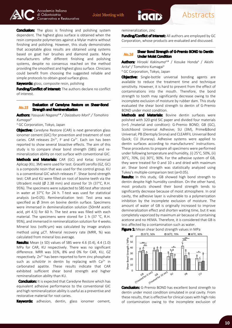
No.16 Shear Bond Strength of G-Premio BOND to Dentin Under Moist Condition
Authors: Hiroaki Kakinumaa* / Kosuke Hondaa / Akishi Aritaa / Tomohiro Kumagaia a GC Corporation, Tokyo, Japan Objective: Single-bottle universal bonding agents are available to reduce the treatment time and technique sensitivity. However, it is hard to prevent from the effect of contaminations into the mouth. Therefore, the bond strength to tooth may significantly decrease owing to the incomplete exclusion of moisture by rubber dam. This study evaluated the shear bond strength to dentin of G-Premio BOND under moist condition. Methods and Materials: Bovine dentin surfaces were polished with 320-grid SiC paper and divided four materials (n=5 /material and condtion): G-Premio BOND; GB (GC), Scotchbond Universal Adhesive; SU (3M), Prime&Bond Universal; PB (Dentsply Sirona) and CLEARFIL Universal Bond Quick; CU (Kuraray). Adhesive systems were applied to dentin surfaces according to manufacturers’ instructions. These procedures to prepare all specimens were performed under following temperature and humidity, (i) 25°C, 50%, (ii) 30°C, 70%, (iii) 30°C, 90%. For the adhesive system of GB, they were treated for 0 and 10 s and dried with maximum air. Shear bond strength was statistically analyzed using Tukey’s multiple-comparison test (α=0.05). Results: In this study, GB showed high bond strength to dentin despite high humidity condition. On the other hand, most products showed their bond strength tends to significantly decrease because of moist atmosphere. In oral cavity, the adhesive layer is vulnerable to a polymerization inhibition by the incomplete exclusion of moisture. The amount of water of GB is originally increased to improve demineralization effect and shorten waiting time, but it was completely vaporized by maximum air because of containing acetone and no HEMA. Therefore, it is considered that GB is less affected by a contamination such as water. Figure 1: Mean shear bond strength values in MPa
Conclusion: G-Premio BOND has excellent bond strength to dentin under moist condition simulated in oral cavity. From these results, that is effective for clinical cases with high risks of contamination owing to the incomplete exclusion of
0
10
20
30
40
50
60
Shea
r Bon
d St
reng
th /
MPa
25℃, 50% 30℃, 70% 30℃, 90%
** ***
****
AB
A
C
B
A AA,B B
C
A
A
B B B*
10
Abstracts
moisture because of the reduction of application time and completely dry with maximum air. Keywords: adhesives, bonding agent, dentin, shear bond strength Funding/Conflict of interest: All authors are employed by GC Corporation, whose products are evaluated and discussed.
No.17 Repair of Indirect Composites Regarding Surface Pretreatments and Universal Adhesives
Authors: Hande Sar Sancaklia* / Mürşide Gültekinb a Istanbul University, Istanbul, Turkey b Private Practice, Kocaeli, Turkey Objective: To evaluate the bond strength determining the repair capacity of resin composite CAD/CAM blocks with contemporary universal bonding agents following various surface pretreatment techniques. Methods and Materials: CAD/CAM resin composite blocks (Lava Ultimate, 3M) were prepared in cubes (5x5x5 mm) and randomly divided into four groups according to the surface pretreatments applied: tribochemical silica coating; phosphoric acid surface etching only; diamond bur surface roughening, and diamond bur surface roughening + phosphoric acid surface etching. Furthermore, each pretreated group was subgrouped according to the adhesives used with or without silane application: Adper Single Bond (etch & rinse adhesive, 3M), Scotchbond Universal Bonding (universal adhesive with MDP, 3M) and Futurabond U (universal adhesive, Voco) into totally 24 experimental groups. All specimens were pretreated and bonded according to the above-mentioned group's requirements and repaired with a microhybrid composite resin (Filtek Z250, 3M). The samples were aged (1,000 thermal cycles from 5-55°C) and cut into microbars with the dimension of 1×1×8 mm. Microbars were then tested using a micro tensile device. Surface characteristics of the composite following each pretreatment were evaluated by AFM. The statistical analyses of MTBS data was performed by one-way ANOVA and Bonferroni post-hoc tests. Results: Among the four different surface pretreatments applied on the Lava Ultimate surface, diamond bur surface roughening revealed the highest microtensile bond strength whereas the solely phosphoric acid surface roughening resulted in lowest bond strength, regardless of the adhesives used (p<0.01). Concerning the effect of adhesives on the repair potential following all surface pretreatments performed, the significantly lowest microtensile bond strength was found when Adper Single Bond was applied (p<0.01). Universal adhesives were found to give higher results for all the groups, however, resulting in higher bond strength values when used with silane (p<0.01). AFM evaluations revealed the highest surface porosities within bur-roughened surfaces with or without acid etching supporting the micro tensile data obtained. Conclusion: Mechanical surface pretreatments positively influence the repair capacity of resin composite CAD/CAM materials; however, this advantage is enhanced when followed by use of a universal adhesive. Regarding the silane
content of the universal adhesives, Singlebond Universal stands as an active adhesive eliminating an adjoint use of silane primer when indicated as a repair procedure material. Keywords: CAD/CAM resin composite, microtensile bond strength, silane, surface pretreatment, universal adhesive Funding/Conflict of interest: The present work was supported by the Research Fund of Istanbul University Project No: 55196.
No.18 Modeling Liquids Effect on Translucency and Chromatic Stability of Composites In-Vitro
Authors: Giovanni Salvatia* / Adrianaa / Alessandra Bandela / Gabriella Scavellaa / Alessandro Vichib / Enrico Felice Gherlonea / Giuseppe Cantatorea / Gaetano Paolonea a Università Vita-Salute San Raffaele, Milano, Italy bUniversity of Portsmouth Dental Academy, Portsmouth, United Kingdom Objective: To Evaluate the effect of the presence of adhesives or modeling resins between composite layers on translucency and long-term color stability of restorative materials. Methods and Materials: Resin adhesives (RA) and modeling resins (MR) have been applied between resin composite (RC) increments. Composite Wetting Agent (Ultradent), Estenia C & B Modeling Resin (Kuraray Noritake Dental), GC Modeling Liquid (GC) were used as MR. Prime & Bond XP (Dentsply Sirona), Clearfil SE Bond 2 (Kuraray Noritake Dental) bond liquid, Prime & Bond Active (Dentsply Sirona), were used as RA. Mosaic Universal Composite, (Ultradent) was used as RC. Composite disks (ø 9 mm, thickness 2 mm) were prepared layering four increments of resin composite. On every increment (0.5 mm), RA or MR was applied before light curing. Specimen were divided in 7 groups (n=10) 3 RA, 3 MR and one control group (no RA or MR between increments). Polished and unpolished subgroups were also created. Measurements of color stability and translucency (after immediate and 1, 28 days of coffee storage and after repolishing) were performed using a spectrophotometer (Vita Easyshade, VITA). An average specimen reading was computed from three continuous measurements. Data were analyzed using ANOVA and Tukey test (α=0.05) and ΔE=3.3 was defined as the threshold of clinical acceptability. Results: Groups with RA or MR showed significant influences color at baseline, after staining (t=28) and repolishing (p<0.01). Translucency at baseline was influenced by RA or MR (p<0.05). Influence of RA and MR on translucency was also reported after staining and repolishing. Conclusion: The use of modeling liquids influences color at baseline, after staining (t=28) and repolishing (p<0.01). Translucency at t=0 was influenced by RA and MR (p<0.05). Influence on translucency was reported after staining and repolishing Keywords: chromatic, composite, dentin, enamel, polishing, translucency Funding/Conflict of interest: The authors declare no conflict of interest.
11
Abstracts
No.19 Roughness, Translucency, and Gloss Analyses of Dental Ceramics After Polishing
Authors: Carlo Monacoa / Antonio Arenaa* / Lorenzo Schedaa / Lorenzo Breschia a University of Bologna, Bologna, Italy Objective: To evaluate the effect of prophylactic polishing pastes (PPPs) on 2/3D roughness, translucency and gloss of different ceramic materials. Methods and Materials: A total of 120 flat samples (thickness: 2 mm) obtained from CAD-CAM blocks of leucite glass ceramic (Empress CAD, Ivoclar Vivadent), lithium disilicate glass ceramic (e.max CAD, Ivoclar Vivadent) and zirconium oxide (Zenostar, Ivoclar Vivadent) were glazed and sintered. Next, 40 samples of each material were divided into four groups, which were polished (one group each) with Cleanic Fine (CF, Kerr), Nupro Fine (NF, Dentsply Sirona) and Proxyt Fine (PF, Ivoclar Vivadent), whereas the control group was untreated (n=10). Samples were polished for 2 min with a prophy cup mounted on a handpiece applying a constant load of 400 g at 2,000 rpm. Surface roughness was measured using a mechanical profilometer and a 3D optical profilometer. One sample per group was randomly selected for observation by SEM at 200x magnification. The translucency parameter (TP) and gloss value were calculated using a spectrophotometer and a glossmeter. Pearson correlation analyses were performed to evaluate the relationships between 2D roughness and gloss and between 2D roughness and translucency. Differences in means were compared using Two-way ANOVA and Tukey's test. The level of significance was set at α=0.05. Results: 2D analysis showed for Empress an increase of roughness using CF and NF (p<0.05). 2D roughness of Empress was lower than that of e.max (p<0.05). TP values of Empress and Zenostar were decreased using NF (p<0.05). Zenostar showed a translucency lower than e.max and Empress (p<0.05). PPPs had a minimal effect on gloss (p> 0.05). The gloss of Empress was higher than that of Zenostar and e.max (p<0.05). A correlation between gloss and surface roughness was found (p<0.0001). Conclusion: Polishing procedures should be performed with caution to avoid damaging the restoration surface. Keywords: polishing, roughness, gloss, translucency, ceramic Funding/Conflict of interest: The authors declare no conflict of interest.
No. 20 STUDENT
Interfacial Evaluation of Dentin-Cement Interface in Fiber-Post Supported Restorations
Authors: Andrea Querroa* / Allegra Combab / Gianpaolo Serinoc / Guido Audeninoc / Mario Alovisia / Giorgia Carpegnaa / Andrea Baldia / Nicola Scottia
a University of Turin, Turin, Italy b University of Bologna, Bologna, Italy c Polytechnic University of Turin, Turin, Italy Objective: The aim of this in vitro study was to evaluate the effect of curing time on mechanical properties of fiber-post
luting cement. The null hypothesis was that curing time does not affect hardness and young modulus. Methods and Materials: 24 premolars were endodontically treated and a class II cavity with 1.5 mm residual wall thickness and a 8 mm deep post space was prepared. Fiber posts were luted trough a standardized procedure: etching for 20 s; rinse for 20 s; rinse with ethanol for 30 s; universal adhesive system application (UBQ, Kuraray); fiber post insertion (Rabilda, Voco) after luting cement (DC Core, Kuraray) placement in the post space. Samples were divided in 3 groups according to the curing time (n=8 each): G1) no light; G2) 20 s; G3) 120 s. Light curing was performed with LED lamp (Celalux 2, Voco) at 1,000 mW/s. with the tip placed in contact with the fiber post. The occlusal cavity was restored with a nano filled resin composite (Filtek Supreme XTE, 3M). After 7 days, half of each group samples were submitted to cyclic fatigue test with a chewing simulator for 500,000 cycles at 50 N load, 2 mm excursion, under water (CS 4.4, SD Mechatronik). Samples were sectioned in 1mm thick slices perpendicularly to the fiber post long axis. Samples were tested with a Nanoindenter XP, equipped with a diamond Berkovich indenter and characterized by a theoretical force resolution of 50 nN and a theoretical displacement resolution lower than 0.01 nm. The loading-displacement (P-h) curves were analyzed by using the Oliver-Pharr method in way to obtain Young Modulus and Hardness. Obtained data were analyzed with ANOVA test (α<0.05). Results: Young modulus and hardness were not correlated to the curing time (p=0.0623). Cyclic fatigue significantly reduced the luting cement’s mechanical properties (p=0.0001) except when 120 s curing time was performed. Conclusion: The null hypothesis was partially rejected since curing time significantly affect luting cement mechanical behavior. Keywords: fiber-post, luting cement, nanohardness Funding/Conflict of interest: The authors declare no conflict of interest.
No. 21 FIB: Focused Ion-Beam Milling. An Alternative to the Micro-Indenter Test for the Evaluation of Dental Aging and Wear of Restorative Materials
Authors: Guido Pasquantonioa / Manuele Mancinia / Roberta Condòa / Loredana Cerronia / Nicol Bianchia / Andrea Notargiacomob / Luca Maioloc* a University of Rome Tor Vergata, Rome, Italy b Institute for Photonics and Nanotechnologies, Rome, Italy c Institute for Microelectronics and Microsystems, Rome, Italy Introduction: Aging of the human body is a process that also affects the oral cavity and teeth and occurs through wear and tear. Physiological dental aging occurs with a tissue loss of about 0.029 mm per year; therefore, if the wear is physiological, there will be a loss of tissue of about 1.5-2 mm over a period of 60 years. To date, to evaluate the wear
12
Abstracts
coefficient and the hardness of dental tissues and restorative materials, we have worked with the micro-indenter based on the scale of Vickers or Knoop. Objective: The aim of this study was to introduce an innovative protocol based on the Ga+ focused ion beam (FIB) milling of dental material, as an alternative method for the evaluation of the hardness not only of the restoration materials, but also of the hard substrates of teeth. Methods and Materials: FIB milling was performed at 30 kV on each material using different values of ion dose thus producing cavities of different depth. The milling depth difference was then evaluated in order to discard effects due to a surface layer which may likely have properties different from the bulk material. This “corrected” milling depth parameter, obtained in the same experimental conditions both on dental materials and on teeth, when correlated to hardness and wear, could trigger the choice of the most suitable restoration material during a prosthetic and conservative rehabilitation. Results: Conclusion: Wear of teeth and dental materials must be further studied to allow the selection of appropriate restorative material in clinical practice. Authors aimed to determine and validate a new evaluation scale using this new method in dentistry and then giving rules on the choice of the best material to use, thus facilitating rehabilitation in patients with physiological and pathological dental aging. To date, no references are known for the evaluation of the hardness and wear coefficient of any material with this innovative technique. Keywords: dental aging, dentin, enamel, FIB, wear Funding/Conflict of interest: The authors declare no conflict of interest.
No. 22
STUDENT CAD-CAM Materials Wear in Bruxism Patients: an In-Vitro Study
Authors: Danilo Cavallaroa* / Andrea Baldia / Edoardo Alberto Verganoa / Allegra Combab / Giorgia Carpegnaa / Mario Alovisia / Damiano Pasqualinia / Nicola Scotti a
a University of Turin, Turin, Italy b University of Bologna, Bologna, Italy Objective: The aim of this in vitro study was to evaluate the occlusal wear of CAD-CAM materials in a simulated condition of bruxism. The null hypothesis is that wear rate is not affected by the material employed for dental restorations. Methods and Materials: 160 extracted sound molars were selected. A standardized adhesive overlay preparation was performed in all specimens, except in the control group. An intraoral scanner (Cerec Omnicam) was employed to scan preparations. Specimens were then divided in 6 groups (n=16) according to the material employed for the overlay fabrication: G1) sound teeth; G2) Cerasmart (GC); G3) E-Max CAD (Ivoclar); G4) Katana Zirconia (Kuraray); G5) Grandio Block (VOCO); G6) Venus Pearls (Kultzer). Overlays of 2mm thickness were luted following a standardized adhesive procedure (3-step etch-and-rinse, Optibond FL, Kerr) and
light cured with a LED lamp for 2 min (D-Light Pro, GC). After 24 h, specimens were scanned (True-Depth, 3M) and then submitted to cyclic fatigue test: 500,000 cycles, 80 N, semicircular movement, 8 Hz. (CS4.4, SD Mechatronik) Each study group underwent a chewing simulation run using as an antagonist specimen belonging to each other group. After test, specimens were scanned again. Obtained STL files were superimposed with Geomagic Software to calculate volume loss after fatigue, expressed in mm3. Data were statistically analyzed with ANOVA test and post-hoc Tukey test (α=0.05). Results: ANOVA test showed a significant difference between groups. Post-hoc Tukey test showed that ceramic materials (G3 and G4) induced an increased volume loss than other tested materials to enamel (p=0.00001). The lowest volume loss was observed when chewing was done with same materials occluding. Enamel wear rate was significantly affected by the materials employed. Thus, the initial null hypothesis was rejected. Conclusion: Based on the obtained results, the choice of materials for overlay restorations in bruxism patient is strongly related to the opposing arch. Ceramic materials induced heavy wear on natural enamel. Keywords: bruxism, CAD-CAM, wear Funding/Conflict of interest: The authors declare no conflict of interest.
No.23
JUNIOR Wear and Marginal Gap of Direct Composites on Endodontically Treated Teeth
Authors: Andrea Baldia* / Nicola Scottia / Allegra Combab / Riccardo Michelotto Tempestaa / Edoardo Alberto Verganoa / Camilla Fogliaa / Mario Alovisia / Damiano Pasqualinia / Elio Beruttia
a University of Turin, Turin, Italy b University of Bologna, Bologna, Italy Objective: The aim of this in vitro study was to evaluate the effect of different direct restoration techniques on endodontically treated anterior teeth, with or without fiber posts, analyzing interfacial adaptation, wear and fracture resistance. Methods and Materials: 36 extracted single-rooted anterior teeth were selected. Endodontic treatment was carried out in all samples. After 24 h of storage in water at 37°C, samples were divided in 3 groups according to the cavity design: 1) endodontic access; 2) endodontic access + 1 Class III cavity; 3) endodontic access + 2 Class III cavities. Samples were then divided in 3 subgroups (n=4) according to the restoration technique: SB1) direct composite restoration (DCR); SB2) DCR supported by a fiber post (Rebilda Post, Voco); SB3) DCR supported by vertical fibers (Rebilda Post GT, Voco). All specimens were scanned with X-ray computed micro-tomography (micro-CT SKYSCAN, BRUKER), with following parameters: 100 kV, 100 µA, Al-Cu filter, 10 µm pixel size, rotation step 0.1°, 6 h total scan duration. Then, specimens of each group were subjected to mechanical fatigue test in a dual-axis masticatory simulator (CS4.4, SD Mechatronik). A force of 50 N was applied using a ceramic steatite ball with a
13
Abstracts
diameter of 4 mm as an antagonist for 100,000 cycles, with frequency of 1 Hz, downward speed 16 mm/s, 2mm sliding movement starting from palatial cingulum towards the incisal edge, with initial angle of 45°. After fatigue, micro-CT scanning was performed again to evaluate the interface behavior and wear resistance. Micro-CT images, before and after cycling load, were analyzed with Mimics (segmentation) and Geomagic Software (alignment and analysis) to evaluate composite wear and interfaces gap progression before and after mechanical load. Finally, a static fracture test with universal machine (Instron) was performed to measure the fracture resistance of the samples after fatigue tests (4 mm-diameter metal cone at constant speed of 0.5 mm/min and an angle of 30°). Statistical analysis was performed with two-way ANOVA test to evaluate the effect of cavity configuration and restoration of wear, interfacial gap and fracture resistance Results: Gap progression and volume are significantly related to the build-up technique (p<0.001) as well as to the cavity configuration (p=0.032). The 2-way ANOVA showed that both the variable cavity (p=0.0020) and the variable material (p=0.0013) significantly influence the fracture resistance. No other significant interactions were reported by ANOVA test (p=0.5130). Conclusion: Based on the results obtained, endodontically treated anterior teeth should be restored with composite restorations supported by fiber structures, especially in the case of loss of both marginal ridges. Further studies are needed to better understand the influence of fiber post on interfacial adaptation, wear and fracture resistance over time. Keywords: anterior, gap, microCT, wear Funding/Conflict of interest: The authors declare no conflict of interest.
No.24
JUNIOR Marginal Fit of Lithium Disilicate Overlays with Two Preparation Designs
Authors: Daniele Angeramea / Matteo De Biasia / Massimiliano Lenhardta* / Fernando Zaroneb / Roberto Sorrentinob
a University of Trieste, Trieste, Italy b University Federico II, Naples, Italy Objective: To assess the influence of the preparation design on the marginal fit of CAD/CAM lithium disilicate overlays. Methods and Materials: Twenty maxillary molars received 1-mm cusp reduction and were randomly allocated to two groups: 90° rounded shoulder margin (n=10) and marginal chamfer (n=10). After the preparation process, immediate dentin sealing was performed. IPS e.max CAD (Ivoclar Vivadent) restorations were obtained with the Cerec 3 CAD/CAM system (Dentsply Sirona). The intaglio surface of the occlusal veneers was conditioned with hydrofluoric acid (Porcelain etch, Ultradent Products) and silane (Monobond Plus, Ivoclar Vivadent), while the tooth surface underwent silicatization (Cojet System, 3M), enamel etching (Fill Etch, DentalWorld) and self-etch adhesive system application (Clearfil SE Bond 2, Kuraray). The occlusal veneers were luted
with Variolink II cement (Ivoclar Vivadent). The specimens underwent thermomechanical aging with a chewing simulator (CS-4, SD-Mechatronik; 1,250,000 cycles, 1 Hz, 5-55°C). The marginal fit was quantitatively assessed on resin replicas of the specimens obtained before and after the thermomechanical aging at the scanning electron microscope. The marginal fit at different experimental time points was compared within the same group with a paired-sample t-test, while the two groups were compared at the same time point with an independent-sample t-test (α=0.05). Results: After aging, the overlays with a 90° rounded shoulder margin group showed a mean marginal gap of 111.83±49.66 µm, while the fit in the marginal chamfer group was 119.57±54.30 µm. The two preparation designs showed comparable marginal fit at both experimental time points (before aging, p=0.438; after aging, p=0.650). In both groups, thermomechanical aging did not cause a significant worsening of the marginal fit (90° shoulder, p=0.599; chamfer, p=0.715). Conclusion: CAD/CAM lithium disilicate overlays prepared with a marginal chamfer can offer comparable marginal fit to that of a standard 90° rounded shoulder preparation. Keywords: CAD/CAM, lithium silicate, marginal fit, overlay Funding/Conflict of interest: The authors declare no conflict of interest.
No.25 JUNIOR
Interfacial Gaps and Fracture Resistance of Indirect CAD-CAM Restorations of Endodontically Treated Teeth
Authors: Edoardo Alberto Verganoa* / Andrea Baldia / Greta Zoppettoa / Riccardo Michelotto Tempestaa / Mario Alovisia / Damiano Pasqualinia / Allegra Combab / Nicola Scottia
a University of Turin, Turin, Italy b University of Bologna, Bologna, Italy Objective: The aim of this in vitro study was to evaluate the effect of three different CAD-CAM processed materials on the interfacial gap, wear and fracture resistance of endodontically treated molars. Methods and Materials: 48 maxillary molars were selected and endodontically treated. On each specimen a standardized MOD cavity was prepared. Specimens were then divided in two groups (n=24 each) according to the build-up technique employed: G1) build-up with a bulk fill composite material (Admira Fusion X-Tra, Voco); G2: fiber post supported build-up. Then, a standardized overlay preparation, 2 mm thick, exposing enamel margins was performed. Specimens were scanned with Cerec Omnicam (Denstply Sirona) and the indirect restoration was milled with Cerec MXCL. Each group was divided in 3 subgroups (n=8 each) according to the CAD-CAM material employed: SG1) Grandio-Bloks (Voco); SG2) Cerasmart (GC); SG3) Celtra Duo (Dentsply). Each overlay, once completed, was luted following a standardized procedure. All specimens were scanned with micro-CT at high-resolution scans (voltage 100kV, current 80A, source-to-object distance 80 mm, source-to-detector distance 220
14
Abstracts
mm, pixel binning 292, exposure time/projection 3 s). Then, specimens of each group were subjected to mechanical fatigue test in a dual-axis chewing (CS-4.4, SD Mechatronik). A force of 5 kg was applied using a ceramic steatite ball for 500,000 cycles. After fatigue, micro-CT scanning was performed to evaluate the interface gap. Replicas were obtained for external gap evaluation with SEM. Micro-CT images, before and after cycling load, were analyzed with Geomagic Software and Mimics to evaluate interfaces gap progression before and after mechanical load. Statistical analysis was performed with two-way ANOVA test. Results: Interfacial gap was not significantly influenced by the build-up technique (p=0.061). SG2 and SG3 showed a lower gap (p=0.001) and a higher fracture resistance rate (p=0.0001). No differences were found between SEM and 3D gap evaluation Conclusion: Interfacial gap and fracture resistance could be influenced by the CAD-CAM material employed to restore endodontically treated molars. Keywords: 3D interfacial gap, adhesion, adhesive overlay, CAD-CAM restoration, fracture resistance, micro-CT evaluation Funding/Conflict of interest: The authors declare no conflict of interest.
No. 26 STUDENT
Interfacial Gap and Fracture Resistance of Ceramic Overlays in Endodontically Treated Teeth
Authors: Edoardo Italiaa* / Giorgio Ferreroa / Andrea Baldia / Edoardo Alberto Verganoa / Allegra Combab / Mario Alovisia / Damiano Pasqualinia / Nicola Scottia
a University of Turin, Turin, Italy b University of Bologna, Bologna, Italy Objective: To evaluate 3D interfacial behavior, occlusal wear and fracture resistance of endodontically treated molars restored with an adhesive overlay with 2 different CAD-CAM materials after chewing simulation. Methods and Materials: 24 molars were selected and endodontically treated. Then a standardized MOD cavity was prepared and then a build-up was performed (Clearfil Majesty ES-2, Kuraray) after adhesive application (Clearfil SE Bond 2, Kuraray). A standardized overlay preparation was made. Samples were then divided in two groups, according to material restorations: G1: Zirconia-reinforced lithium silicate (Celtra DUO, Dentsply) G2: Cubic zirconia (Katana, Kuraray) Preparations were scanned with Cerec Omnicam and restorations were milled with Cerec MXCL (Sirona). Restoration was then luted (Panavia V5, Kuraray) following the manufacturer instructions. After 7 days, the following tests were performed:1. Micro-CT scan (Skyscan, Bruker). 2. Optical scan (Sinergiascan, Nobimetal), 3. Fatigue and fracture resistance were performed with a chewing simulator (CS4.4, SD-Mechatronik) and after wards the
restorations were subjected to a static load up to fracture using a universal testing machine (Instron; Canton, MA, USA) with a 6 mm diameter steel sphere crosshead welded to a tapered shaft and applied to the specimens at a constant speed of 0.5 mm/min and an angle of 30° to the long axis of the tooth. Load was applied perpendicular to the triangular crest of the palatal cusp. Samples were loaded until fracture; the maximum breaking loads were recorded in Newton (N). Each sample was scanned, radiographically and optically, before and after chewing. Data were imported into Mimics after smoothing and region growing; only external gap was considered in the analysis. Obtained STL optimal quality masks were imported into Geomagic software for noise removal and volume calculation. Interfacial gap progression and occlusal wear data, expressed in mm3, and fracture resistance, expressed in N, were collected and statistically analyzed with two-way ANOVA test (α=0.05). Results: Two-way ANOVA test showed that interfacial gap was not influenced by the material employed (p = 0.0657). Concerning the wear rate, lithium silicate showed a much larger volume reduction than cubic zirconia. Considering fracture resistance of overlays there is no difference between the two materials (p=0.0578). Conclusion: The obtained results showed that the restoration of endodontically treated molars with bonded overlays could be equally performed with either lithium silicate or cubic zirconia. Further studies are necessary to confirm present data. Keywords: bruxism, CAD-CAM, overlays Funding/Conflict of interest: The authors declare no conflict of interest.
No. 27
STUDENT Fatigue Resistance of Monolithic Ceramic Crowns: In-Vitro Comparison with Teeth
Authors: Paolo Baldissaraa / Francesca Gradaraa* / Maria Rosaria Gattoa / Leonardo Cioccaa
a University of Bologna, Bologna, Italy Objective: To compare the tooth fatigue resistance to lithium-disilicate, UTML (Ultra-Translucent-Multi-Layered zirconia), and Y-TZP (Yttrium-stabilized tetragonal zirconia) monolithic crowns, tested using a new simplified fatigue testing machine (Ball-mill) and a system of digital-type wear analysis. Methods and Materials: Twenty extracted human molars were used. In addition, 60 monolithic crowns have been produced, subdivided into 3 groups (n = 20) with different materials (lithium-disilicate 1.5 mm - IPS e-max CAD LT, IvoclarVivadent; UTML 1.5 mm - Katana STML, Kuraray Noritake and Y-TZP 1.0 mm – Katana, Kuraray Noritake). Teeth were subjected to three 10-min cycles of Ball-mill and to one 30-min cycle. For monolithic crowns, twelve 60-min cycles were performed (since in a preliminary pilot study no significant volumetric losses were observed before 60 min). At T0 and after each cycle an analysis was carried out using a stereo microscope (Wild M3C, Heerbrugg) to evaluate the wear morphology, an intraoral scanner (CS 3500,
15
Abstracts
Carestream Dental) and a software (Rhinoceros, McNeel) to calculate the volumetric loss of the samples caused by the wear. Results: The lithium-disilicate and Y-TZP crowns showed a 100% survival rate and high wear resistance; UTML had 95% survival. Lithium-disilicate and UTML showed many parting-cleavage fractures (>90%). The wear generated on teeth at T60 was statistically greater (p<0,05) than the one of crowns, Y-TZP in particular (p < 0,0001); all couples of compared materials significantly differ (p=0.0008) except for lithium-disilicate and UTML crowns. From comparison with the anthropometric classification of Miles it was estimated that 60 min of Ball-mill testing correspond approximately to an average time of clinical wear of 18 years. Conclusion: The Y-TZP crowns showed the best result in terms of wear resistance. The lithium-disilicate presented excellent resistance, but with uniform 1.5 mm thickness all over the crown (marginal and occlusal area). The Ball-mill testing machine proved effective results in reproducing the clinical conditions: its in-vitro outcomes evidenced the same mechanical characteristics of the 3 ceramics demonstrated by other fatigue test machines. The investigation methodology was effective when using digital technologies to compare materials and calculate the volumetric dental loss over time. Keywords: enamel, fatigue cycle, fatigue cycle, tooth wear, monolithic crown, tooth wear Funding/Conflict of interest: The authors declare no conflict of interest.
No. 28 STUDENT
Efficacy of Adhesive Crowns to Restore Endodontically Treated Molars: A Micro-CT Study
Authors: Giorgio Ferreroa* / Edoardoa / Andrea Baldia / Edoardo Alberto Verganoa / Allegra Combab / Mario Alovisia / Damiano Pasqualinia / Nicola Scottia
a University of Turin, Turin, Italy b University of Bologna, Bologna, Italy Objective: To evaluate 3D interfacial behavior, occlusal wear and fracture resistance of endodontically treated molars restored with a full-crown restoration made with 2 different CAD-CAM materials after chewing simulation. Methods and Materials: 24 sound molars were selected and endodontically treated, shaped with PathFile and ProTaper Next (Dentsply Sirona) and filled with a warm condensation technique. Then a standardized MOD cavity was prepared and a fiber post (Panavia Post, Kuraray) supported build-up was performed. A standardized preparation for complete crown with chamfer margin was done. Samples were then divided in two groups, according to material restorations: G1: Zirconia-reinforced lithium silicate (Celtra DUO, Dentsply); G2: Cubic zirconia (Katana, Kuraray). Preparations were scanned with Cerec Omnicam and restorations were milled with Cerec MXCL. Restoration was then luted (Panavia V5, Kuraray) following the manufacturer instructions. After 7 days, the following tests were performed:
• Micro-CT scan (Skyscan, Bruker). • Optical scan (Sinergiascan, Nobimetal) • Fatigue and fracture resistance were performed with a chewing simulator (CS4.4, SD-Mechatronik) and after a static fracture using a universal testing machine (Instron) with a 6 mm diameter steel sphere crosshead welded to a tapered shaft and applied to the specimens at a constant speed of 0.5 mm/min and an angle of 30° to the long axis of the tooth. Load was applied perpendicular to the triangular crest of the palatal cusp. Samples were loaded until fracture; the maximum breaking loads were recorded in Newton (N). Each sample was scanned, radiographically and optically, before and after chewing. Data were imported into Mimics after smoothing and region growing; only external gap was considered in the analysis. Obtained STL optimal quality masks were imported into Geomagic software for noise removal and volume calculation. Interfacial gap progression and occlusal wear data, expressed in mm3, and fracture resistance, expressed in N, were collected and statistically analyzed with two-way ANOVA test (α<0.05). Results: G1 showed mean gap of 0.43mm3, while G2 had 0.39 mm3. Two-way ANOVA test showed that interfacial gap was not influenced by the material employed (p = 0.0674). Concerning the wear rate, lithium silicate showed a much larger volume reduction in mm3 than cubic zirconia (p=0.00001). Fracture resistance (G1= 1351,52; G2= 1136,43) was significantly influenced by the material employed (p=0.0001). Conclusion: The obtained results showed that cubic zirconia seems to better perform in the restoration of endodontically treated molars. Further studies are necessary to confirm present data. Keywords: CAD-CAM, crowns, micro-CT Funding/Conflict of interest: The authors declare no conflict of interest.
No. 29 Fracture Resistance of CAD/CAM-Crowns After Margin-relocation Using Two Different Composite Resins
Authors: Giulio Marchesia */ Roberto Spreaficob / Gianluca Turcoa / Sebastiano Fantona / Milena Cadenaro Ma / Lorenzo Breschic a University of Trieste, Trieste Italy b Private practice, Varese, Italy c University of Bologna, Bologna, Italy Objective: Aim of this study was to evaluate the effect of cervical marginal relocation (CMR) with two different composite resins on the fracture loads of CAD/CAM monolithic lithium disilicate molar crowns. The null-hypothesis tested was that the fracture loads of crowns would not be influenced by the CMR with two different composite resins. Methods and Materials: Standard crown preparations were created in 20 human molars. The margins were located in enamel, except for mesial proximal box, where the cervical margin was 2.0 mm below the cement-enamel junction. The
16
Abstracts
CMR technique was performed by applying 2mm thick layers of conventional or flowable composite (Filtek Supreme XTE, 3M or Filtek Supreme Flow XTE, 3M) into the mesial box bonded with Optibond FL (Kerr). 20 standardized crowns were prepared with the CEREC CAD/CAM system (Dentsply Sirona) using CAD/CAM disilicate blocks (IPS e.max, Ivoclar Vivadent). Restorations were then luted with a dual-adhesive cement (RelyX Ultimate, 3M). All specimens were loaded until fracture after thermo-mechanical loading (240,000 mechanical cycles x 50N and 7800 thermo-cycles between 5° and 55C° using a chewing simulator (CS-4.4, SD Mechatronik). Force values were statistically analyzed by two-way ANOVA and Tukey’s post-hoc test (α=0.05). Results: The null hypothesis was accepted since no statistically significant differences were found among the groups (p>0.05). Mean fracture load values (N) were for group CMR 1 (1628 ± 296 N) and for Group 2 CMR 2 (1644±336 N). Conclusion: The CMR after thermo-mechanical loading using conventional or flowable composite had no effect on fracture load of CAD/CAM molar crowns. Keywords: relocation, resin composite, CAD/CAM crown, resin cements, lithium disilicate Funding/Conflict of interest: The authors declare no conflict of interest.
No. 30 STUDENT
Fracture Resistance of Endodontically Treated Premolars Restored with Different Posts
Authors: Irene Ballaa* / Luca Bartolettia / Allegra Combaa / Tatjana Maravica / Nicola Scottib / Andrea Baldib / Annalisa Mazzonia / Lorenzo Breschia
a University of Bologna, Bologna, Italy b University of Torino, Torino, Italy Objective: Fiber posts are widely employed to restore endodontically treated teeth with limited dental sound substrate. The aim of this in vitro study was to test the static fracture resistance and failure mode of endodontically treated premolars restored with different post systems. Methods and Materials: 15 monoradicular premolars, extracted for periodontal or orthodontic reasons, were endodontically treated and randomly divided in 3 group according to the restorative procedure performed: G1) direct composite restoration; G2) ParaPost Taper Lux (Coltene) and a CAD/CAM composite crown (BRILLIANT Crios, Coltene); G3) Rebilda PostGT (Voco) and a CAD/CAM composite crown. Restorations were aged for 30 days in artificial saliva then tested with a universal loading machine (maximum strength 2,000 N). A compressive force was applied at 30 degrees to the long axis of the teeth until fracture, using a 6mm diameter sphere. Different modalities of fracture were evaluated by three different operators and were classified as restorable/unrestorable depending on the fracture level in relation to the CEJ. Data were analyzed using
a one-way ANOVA test with the significance level set at α=0.05. Results: No significant differences in fracture resistance were detected among the three groups. The samples restored with Rebilda PostGT showed the highest percentage of restorable fractures compared to the other groups (80%). Conclusion: Based on the obtained results the use of an endodontic fiber post did not improve the fracture resistance of endodontically treated monoradicular premolars. However, the presence of the post is able to improve the number of restorable fractures. Further studies are needed to better understand the results of the present in vitro study. Keywords: adhesion, CAD/CAM, composite, fiber post, fracture resistance Funding/Conflict of interest: The authors declare no conflict of interest.
No. 31 STUDENT
Fracture Resistance of Endodontically Treated Premolars Restored with Different Techniques
Authors: Luca Bartolettia* / Irene Ballaa / Allegra Combaa / Tatjana Maravica / Andrea Baldib / Nicola Scottib / Annalisa Mazzonia / Lorenzo Breschia a University of Bologna, Bologna, Italy b University of Torino, Torino, Italy Objective: The aim of this study was to test the static fracture resistance of composite endocrowns versus a conventional restoration with a post, core and crown and a direct composite restoration. Methods and Materials: 15 single-rooted canal premolars were endodontically treated and randomly divided in 3 groups (N=5 teeth per group) according to the restoration protocol: G1: teeth restored with a CAD/CAM composite endocrown (BRILLIANT Crios, Coltene); G2: teeth restored placing a fiber post (ParaPost, Coltene), composite core, and then a composite CAD/CAM crown (BRILLIANT Crios); G3: teeth restored with a direct composite restoration (control, synergy D6, Coltene). Specimens were then digitally scanned and indirect restorations were produced using CEREC (Dentsply Sirona). Fracture resistance was tested with an Instron Universal machine after a 30-day storage in artificial saliva at 37°C. Samples were embedded in resin, positioned in the machine at a 30° angle and loaded until fracture with a 6 mm diameter sphere at the speed of 1 mm/min, and fracture loads were recorded. Fracture patterns were analyzed at a stereomicroscope and fractures occurring above the cement-enamel junction (CEJ) were considered restorable. Data were analyzed using a one-way ANOVA test with the significance level set at α=0.05. Results: No statistically significant differences in fracture resistance between the tested groups were found (p>0.05). However, there were differences between the groups regarding the fracture modes. Endocrowns showed the most unfavorable mode of fracture compared to the other tested
17
Abstracts
groups. In fact, all the fractures in the teeth restored with an endocrown occurred below the CEJ. Table 1. Mean fracture resistances in Newton (±standard deviation) and failure modes.
Mean fracture resistance ± SD
Non-restorable fractures
Restorable fractures
Direct
restoration 747.74±163.98 3 2
Post and
crown
restoration
866.89±126.31 4 1
Endocrown 867.31±108.13 5 0
Conclusion: Based on the results of the present study, we can speculate that endocrown should not be the first choice in the restoration of endodontically treated premolars. However, due to their ergonomic advantages and lower price, they could be considered when conventional restorations are not possible. Keywords: CAD/CAM, composite, endocrown, fiber post, fracture resistance Funding/Conflict of interest: The authors declare no conflict of interest.
No.32
JUNIOR Fracture Resistance of Teeth Restored with Lithium-Silicate and Composite Endocrowns
Authors: Daniele Angerame a / Matteo De Biasia* / Giulio Marchesia / Andrea Frassettoa / Lorenzo Bevilacquaa
a University of Trieste, Trieste, Italy Objective To assess the influence of the restorative material and margin relocation on the resistance to load of CAD/CAM endocrowns luted on maxillary molars. Methods and Materials: Sample size was calculated before the experimentation (α=0.05; β=0.20; δ=500.0; σ=300.0). Thirty-six sound maxillary molars of comparable size received a standardized endodontic treatment and were randomly allocated to four experimental groups of nine teeth each: Lava Ultimate endocrown (3M) without margin relocation; Lava Ultimate endocrown with margin relocation; Celtra DUO (Dentsply Sirona) without margin relocation; Celtra DUO with margin relocation. The margin relocation procedure entailed the preparation of a mesial box of fixed dimensions and subsequent margin relocation with a flowable composite (AP+ flow, Sweden & Martina) placed after self-etch adhesive procedures (Clearfil SE Bond 2, Kuraray). The endocrowns were obtained with the Cerec 3 CAD/CAM system (Dentsply Sirona) and luted with RelyX cement (3M). The restored teeth were subjected to thermomechanical aging (1,250,000 cycles, 1Hz, 5-55°C) with a chewing machine (CS-4, Willytech) and were axially loaded to fracture with an universal testing machine (Quasar, Galdabini) equipped with a round-head stylus. A qualitative assessment of fracture types was performed. The groups
were compared in term of mean maximum load to fracture with a two-way ANOVA and Duncan post-hoc test (α=0.05). Results In all the tested groups, the mean maximum load values were greater than the threshold of the masticatory forces. The greatest resistance values were registered in the Lava Ultimate endocrown without margin relocation (1870.6±279.9 N, p<0.05), while the lowest in the Celtra DUO with margin relocation (1289.5±303.4 N, p<0.05). Irrespective of the tested material, margin relocation did not significantly reduce the resistance values (Lava Ultimate, p=0.495; Celtra DUO, p=0.929). The most prevalent type of restoration failure was non-restorable fracture. Conclusion Margin relocation in a proximal box did not affect the resistance to fracture of teeth restored with CAD/CAM endocrowns. Despite bot the tested materials exhibited satisfactory results, lithium silicate seemed to be capable of slightly inferior resistance to load compared to the tested composite material. Keywords: CAD/CAM, composite resin, endocrown, lithium-silicate, load Funding/Conflict of interest: The authors declare no conflict of interest.
No.33 Root Canal Obturation Quality Evaluation Using Different Techniques: SEM Study
Authors: Marine Mamaladzea* / Nikoloz Jalabadzeb / Vadachkoria Otarc / Tea Tchumburidzed a Tbilisi State Medical University, Tbilisi, Georgia b Institute of Physical Materials, Science and Materials Technologies, Tbilisi, Georgia c Dental Clinic ZIP, Munich, Germany d Dental Clinic, Training and Research Center – UniDent, Tbilisi, Georgia Objective: The evaluation of the completeness of root canal obturation with Gutta Percha cones based on revealing of Gutta-Percha non-filled areas (GPNFA) that was performed via SEM examination of cross-section samples of roots at the coronal and apical thirds. Methods and Materials: Fifty-six extracted human incisors were collected. Indications for extraction included periodontitis and trauma. After properly cleaning, teeth were stored in 5% sodium hypochlorite. For the investigation the crowns were separated from the roots and remaining roots were divided randomly into three groups (n= 18,18,20). The root canals of the teeth were prepared with Step-Back technique. In group I cold lateral condensation technique of GP was done, in groups II and III, warm method of obturation was performed. For group II Gutta-Master was selected and for group III – Gutta Fusion cones were used. Two days later, teeth cross sections were obtained in two areas: in the coronal part 2 mm downwards from root isthmus and in the apical part 2 mm upwards from the apex. Total number of investigated segments was 112 (36, 36, 40). Separation of roots was performed by double sided diamond disc. Next step was photographing of samples, SEM investigation and scanning of slides for appearance GPNFA.
18
Abstracts
The results have been statistically interpreted with ANOVA and student t-test. Results: Our study revealed different quality of root canals obturation. The most non-filled areas, both, in coronal and apical thirds, was revealed in group I (14,19±1,03). In groups II and III the results were less 10,53±1,14 (II), 8,71±1,07 (III). The study has revealed that in apical part of root canals GPNFA was more frequent than in coronal third. Conclusion: Usage of cold lateral condensation technique was not able to fill root canals properly. Usage of warm method obturation techniques improves the quality of root filling; In all groups, the coronal part of the RC was filled up more than the apical one. Keywords: gutta-fusion, sealer, gutta-master, non-filled root canals Funding/Conflict of interest: The authors declare no conflict of interest.
No.34 Direct Composite Restorations of Anterior Teeth: a Retrospective Clinical Study
Authors: Giulia Massanoa* / Allegra Combaa / Garombo Ea, Andrea Baldia / Mario Alovisia / Damiano Pasqualinia / Multari Sa, Nicola Scottia a University of Turin, Turin, Italy Objective: The aim of this in vivo study is to evaluate the longevity of Class III/IV restorations performed with different nanofilled resin composites. The tested null hypothesis is that nanofilled composites restorations show good clinical performance in class III and IV cavities. Methods and Materials: 53 patients, who came to the Department of Cariology and Operative Dentistry of the University of Turin to treat caries and fractures on anterior teeth were enrolled. Patients were randomly divided in seven groups according to the composite employed (Amaris; Adonis; Asteria; Clearfil ES2; Filtek Supreme; Inspiro; Ceram-X). They were treated by the same experienced operator following a standardized clinical procedure: rubber dam positioning after shade selection, cavity detersion and finishing, application of a 3-step etch-and-rinse adhesive system (Optibond FL, Kerr), composite layering following natural layering technique, composite finishing and polishing. After 7 days each patient was recalled for an eventual shade correction and final radiograph evaluation. Patients were then scheduled for follow-up visits. Composite restorations evaluation was performed by 2 blinded calibrated operators, following USPHS modified criteria. Obtained data were analyzed with Chi Square test and Kaplan-Meier regression analysis; significance was set at α<0.05. Results: 93 restorations were evaluated at follow-up visits (mean follow-up: 32.3 months). The statistical analysis showed that there was not a significant difference between the tested composites (p>0,05). Among all the evaluated parameters, at 48 months a significant shift to worse scores for surface roughness, marginal adaptation, marginal discoloration and secondary caries was recorded. At 60 months wear and fracture of restorations were significantly affected parameters, regarding color match, no statistically
significant differences were found until 96 months. The annual failure rate was calculated and set at 2.4%. Conclusion: Based on the present study results, the null hypothesis tested was accepted since nano-filled composites showed a good clinical longevity in anterior direct restorations. Keywords: None Funding/Conflict of interest: The authors declare no conflict of interest.
No.35
CLINICAN Epidemiological Study on the Prevalence of Non-Carious Cervical Lesions
Authors: Salvatore Emanuele Teresia* / Jacopo Lenzia a University of Bologna, Bologna, Italy Objective: The aim of this study was to evaluate the prevalence of single, multiple and total non-carious cervical lesions (NCCL). Methods and Materials: An archive research was conducted on patients seen at the department of periodontology, University of Bologna, between 01/01/2001 and 31/12/2017. All patients with gingival recessions (GR) with not-periodontal etiology were included. The variables analyzed were sex, age, smoking, tooth, quadrant, and sextant affected by NCCL. Statistical analyses were performed using Pearson's χ² test and Student's t-test. Results: 3026 patients were examined, of which 685 (22.6%) had GR with not-periodontal etiology and 179 (5.9%) had NCCL. The mean age of patients with GR was 37.2±12.9 years, while the mean age of patients with NCCL was of 41.3±11.4 years. 21.7% of males and 28.0% of females with GR had NCCL. 15.6% of patients with GR had single NCCL, while 10.5% of patients with GR and 40.2% of patients with NCCL had multiple NCCL (>3 teeth). Female sex, unlike age and smoking, was associated with multiple NCCL (84.7% of NCCL) (Table 1), however under the age of 50 males were more affected. The teeth with GR were 4779, of which 756 (15.8%) had an NCCL. The most affected teeth by NCCL were the first and second premolars and canines (27.9%, 21.7%, 20.6%); the first molars and central and lateral incisors were mildly affected (10.8%, 9.9%, 8.2%); the least affected teeth were the second molars (0.8%) (Table 2). The NCCL, similar to the GR, were more prevalent in the left quadrants (54%) but, unlike the GR, were more prevalent in the posterior (61.3%) and upper sextants (58.1%). Conclusion: In this epidemiological study conducted on an Italian population, the distribution of NCCL was strongly unequal based on the analyzed parameters. Keywords: dentin, epidemiological study, non-carious cervical lesions, prevalence Funding/Conflict of interest: The authors declare no conflict of interest.
19
Abstracts
No.36 Clinical Experience of Indirect CAD/CAM Overlays Placed by Dental Students
Authors: Alvise Camurri Pilonia* / Roberto Di Lenardaa / Giulio Marchesia a University of Trieste, Trieste, Italy Objective: Bonded indirect CAD/CAM restorations are increasingly gaining popularity for the restoration of large defects in posterior teeth. In addition, different composite resin blocks and polymer-infiltrated ceramic network (PICN) have been developed. The aim of this study is to present a treatment approach for indirect restoration of posterior teeth using the CEREC chair-side system (Omnicam, CEREC MC X; Sirona Dentsply). Clinical cases are presented by graduated students to illustrate the technique step by step. Case Report: In this follow-up case series 10 dental students placed 22 (Lava Ultimate, 3M) composite resin inlays in posterior teeth (16 adults) and 11 (Enamic, VITA) hybrid ceramic onlays or overlays in posterior teeth (10 adult). After defect-oriented preparations, monolithic composite of Lava Ultimate and Enamic were fabricated with a CAD/CAM system (Omnicam, CEREC MC X). Clinical evaluation was performed at baseline and up to 1 year. Results: Every patient was asked to evaluate the method considering time saving and aesthetic satisfaction. No failed cases, fractures or debondings were observed. The authors rated the adequacy of proximal contact and occlusal relationships as acceptable to good for all restorations. Conclusion: Minimally invasive CAD/CAM restorations showed a favorable clinical performance over an observation period of 12 months. The patients seem to appreciate the chair side opportunity. This technique is particularly indicated in posterior teeth for graduate students. However, clinical long-term data are desirable. Keywords: clinical study, CAD/CAM, inlays, lithium, longevity Funding/Conflict of interest: The authors declare no conflict of interest.
No.37 Full Mouth Adhesive Rehabilitation in a Severe Case of Erosion
Authors: Giacomo Dallaria* / Carlo Monacoa / Ivan Scalzoa / Riccardo Maria Rosatia a University of Bologna, Bologna, Italy Objective: The aim of this clinical report was to re-establish function and aesthetics of a 52-year-old patient’s smile by using a minimally invasive and totally adhesive approach. Case Report: A 52-year-old male patient was referred to our prosthodontic unit, at University of Bologna, for a severe loss of dental structure due to acid erosion given by excessive consumption of lemon juice for over 10 years. His clinical examination consisted of generalized dental erosion involving occlusal, buccal and palatal surfaces in the upper and lower arches. He was classified as grade 5 according to Ace’s classification. No symptoms were
present at the moment of the evaluation and no evidence of TMJ disorders were found. During the first appointment upper and lower jaw impressions and a face-bow record were taken. After a 2-week period treatment with Deprogrammer, RC was detected and, according to the DSD project, a diagnostic wax-up was fabricated. A mock-up was built for aesthetical evaluation and left in function for 2 months. After the treatment plan was agreed on and no symptoms were exacerbated, every tooth was minimally prepared through the mockup and the dentin sealed where necessary. The impressions were taken with poly-vinyl-siloxane according to the putty wash technique. Prescription to the laboratory consisted of four tabletops (#17, 27, 37, 47), two overlays (#16, 26), eight veneerlays (#15, 14, 25, 24, 35, 34, 45, 44), six V-shape veneers (#13, 12, 11, 21, 22, 23), five laminate veneers (#33, 32, 31, 42, 43) and one full contour veneer to align a central lower incisor lingually (#41). All the restorations were fabricated in pressed and stained lithium disilicate (Ivoclar Vivadent). Every single restoration was cemented by using an adhesive protocol, strictly under rubber dam isolation, using a flowable composite cement (Variolink Esthetic DC, Ivoclar Vivadent). At the end of the treatment an acrylic splint was fabricated for maintenance. Results: With 1 year follow up we were able to preserve the teeth’s vitality and tooth structure through the application of modern restorations. The interesting fact about this case is the presence of most of the partial indirect restorations classified so far by the literature. Conclusion: Evolution in adhesive techniques and materials made it ethically mandatory to always look for minimally invasive treatment plans. With Digital support in data recording we will be helped more and more in treating such patients. Keywords: adhesion, dentin, enamel, erosion Funding/Conflict of interest: The authors declare no conflict of interest.
No.38
CLINICIAN Wear from Acid Erosion: Case Report of a Minimally Invasive Rehabilitation
Authors: Andrea Toffolia* / Francescoa / Beatrice Spaggiaria / Federico Rivaraa / Beatrice Mozzonia / Ludovica Parisia / Guido Maria Macalusoa / Edoardo Manfredia a University of Parma, Parma, Italy Objective: The aim of this case report is to show an effective and minimally invasive way to treat a dentition which suffered the effects of severe acid erosion. Case Report: The patient was a 51-year-old man who came complaining about the appearance of his teeth. The patient reported he had been suffering from gastroesophageal reflux disease for 7 years and that he did not underwent any dental therapy except for 3 direct restoration performed 15 years before. Clinical and radiographical examinations showed a severely worn dentition presenting the typical pattern of acid erosion and a concrete loss in vertical dimension of occlusion (VDO). All teeth responded positively
20
Abstracts
to the dental pulp vitality test, so it was decided to perform minimally invasive full mouth restorations with lithium disilicate veneers to restore form and function of the dentition. First, the increase of VDO was determined and then the Digital Smile Design (DSD) protocol was used to create a preview of the new shape and dimension of the teeth and to produce a wax-up of the restored dentition and then two silicon indexes to create the mock-up. Subsequently, the mock-up on the worn teeth was printed and phonetic tests were performed. Since the patient was satisfied with the esthetic and function of the new dentition, we proceeded with our therapy plan. We performed a mock-up driven preparation technique which resulted in minimally invasive preparations and took VPS impressions for lithium disilicate veneers that were cemented in different appointments starting from the posterior areas and carefully following the adhesive cementation protocol. Results: The rehabilitation successfully restored form and function of the dentition. At the six months recall the patient was satisfied and the veneers presented a perfect integration with the surrounding soft tissues. Conclusion: In case of severe acid erosion, the presented protocol represents a viable, predictable and minimally invasive therapeutic option. Keywords: adhesion, dental veneers, dentin, enamel, gastroesophageal reflux Acknowledgement/Funding/Conflict of interest: The authors would like to thank Mr. Franco Pozzi (Lab Pozzi & C., Parma) and Mr. Sergio Marturano. The authors declare no conflict of interest.
No.39 Full-Mouth Adhesive Rehabilitation in a Bulimic Patient: a Case Report
Authors: Alberto Murri Dello Diagoa* / Arnaldo Petraccaa / Luca Giannettia a University of Modena and Reggio Emilia, Modena, Italy Objective: The aim of this article is to illustrate the treatment of a 25-year-old patient with a bulimic history with a non-invasive adhesive technique, showing how a treatment with no-tooth preparation allows us to reach excellent results from a biologic and functional point of view. Case Report: After an aesthetic and functional evaluation, the models were articulated in maximum intercuspation position, and the increase of vertical dimension occlusion was first decided arbitrarily on the articulator. Due to the less conspicuous destruction, the occlusal plane and the incisal edges of the future restorations can be determined by analyzing the initial casts, without the need for a maxillary vestibular mock up. Then by using a deprogrammer a new reference position is identified in a repeatable and unforced way and a posterior occlusal wax up was made in the articulator. The posterior teeth were first restored with provisional composite onlays, directly performed in the patient’s mouth, thanks to translucent silicon keys. The rehabilitation was completed, with the establishment of a functional anterior guidance, in the mandibular anterior sextant with direct composite restorations, and in the
maxillary anterior sextant with six composite occlusal-palatal onlays. The case was finalized with the delivery of lithium-disilicate buccal-occlusal veneers on the premolars and first molar of each quadrant and direct restoration of the second molars. Results: The purely additive strategy utilized in this case allows us to achieve a very good and stable functional and aesthetic result. After 2 years, all teeth maintained pulp vitality and no hypersensitivity was developed. Conclusion: The restorative therapy of patients affected by severe dental erosion should be based on a minimal invasive approach to guarantee the biological success. The treatment described is strategically planned in a way that allows rehabilitating patients quadrant-wise simplifying the therapy. Keywords: additive dentistry, adhesion, bulimia, palatal onlay Funding/Conflict of interest: The authors declare no conflict of interest.
No.40
CLINICIAN Autologous Veneers
Authors: Andrea Gerardia* a Private Practice, Creazzo, Italy Objective: The choice of recovering a severely compromised tooth, instead of proceeding with its substitution through more invasive methods, such as bridges or dental implants, is an extremely topical subject. The aim of this clinical case presentation was to seek new limits in the direct restoration of structurally compromised elements, following the concept of maximum preservation and recovery of biological tissue, thanks to the proven effectiveness of modern adhesive techniques. Case Report: A clinical case is presented concerning the treatment of the right maxillary central incisor with a coronal fracture extended to the cervical third of the crown. The treatment plan included the endodontic retreatment of the tooth and the coronal reconstruction through a composite build-up together with the luting of a veneer obtained from the patient's autologous enamel. After a three-step etch-and-rinse adhesive (Optibond FL, Kerr) was applied, a light-curing composite abutment was built-up (UD1 Enamel Plus, Micerium). The fractured crown was carefully hollowed on the inside with a bur, preserving only the enamel portion. This was relined with composite, after a separating agent was applied, on the newly rebuilt abutment in order to obtain a personalized veneer. A proper adhesive multistep procedure was adopted on both the tooth and the veneer: sandblasting with 50 μm alumina oxide, etching with H3PO4 acid (15 s on dentin and 30 s on enamel and composite), primer and bonding application according to the manufacturer’s instructions. As a luting material, the same composite as for the build-up was used after a pre-heating (8 min/55°C). After light curing, finishing and polishing steps were performed. All procedures were carried out under rubber dam isolation. Results: One-year follow-up showed optimal biological and esthetic integration of the restored tooth; however, its
21
Abstracts
biomechanical resistance still remains a concern and requires longer term follow-up. Conclusion: Accuracy of execution together with the respect of protocols and indications are fundamental characteristics for the success of the treatment. This is particularly true when the restorative strategies adopted are based on adhesion. Keywords: adhesion, adhesive dentistry, veneer, dentin, enamel Funding/Conflict of interest: The authors declare no conflict of interest.
No.41 Conservative Management of a Complicated Crown Fracture
Authors: Yoo-Jin Haa* / Jiwon Jeonga / Donghyun huanga / Su-Jung Parka a Wonkwang University, Iksan, Korea Objective: Complicated crown fractures of anterior teeth is a common traumatic dental injury. These types of fractures require professional preparation and an immediate approach. Fragment reattachment appears to be the most conservative approach and allows the maintenance of natural dental structures. However, in cases of absence of the fragment, direct resin restoration is an esthetic, functional, and economical alternative. This case report describes cases of complicated crown fractures of the maxillary left central incisor (tooth 21, FDI), treated with direct resin restoration and the maxillary left lateral incisor (tooth 22, FDI), treated with fragment reattachment. Case Report: A 17-year-old male reported with crown fractures of his maxillary left central incisor (tooth 21, FDI) and lateral incisor (tooth 22, FDI) due to an accident during a baseball game. Only the fragment of the lateral incisor was present. Complicated crown fractures of tooth 21 and 22 were diagnosed under clinical and radiographic examination. It was decided to treat by partial pulpotomy. The pulp was amputated to a depth of 2~3mm. After irrigation and drying of wound surface, resin-modified calcium silicate was applied and light-cured. And then, tooth 21 was provisionally restored with a flowable resin composite. On the other hand, tooth fragment reattachment was performed on tooth 22 using dentin adhesive and flowable resin composite. A week later, the patient reported no pain and discomfort and the teeth was sensible. After removal of provisional restoration, tooth 21 was restored with dentin adhesive and resin, composite using silicone matrix technique. A silicone matrix was used to guide palatal side of restoration. Results/Conclusion: At the follow-up 7 months later, both maxillary left incisors showed satisfying aesthetics and function. And the pulp vitality of them was maintained. Keywords: crown fracture, reattachment, direct resin composite restoration Funding/Conflict of interest: The authors declare no conflict of interest.
No.42 Management of a Crown Fractured Tooth by Tooth Reattachment
Authors: Jiwon Jeong a* / Yoo-Jin Haa / Yoorina Choia / Su-Jung Parka a Wonkwang University, Iksan, Korea Objective: Crown fractures of the anterior teeth are common in cases of trauma. This case reports describes two cases of the management of a complicated and uncomplicated crown fractured tooth by fragment reattachment. Tooth reattachment is a simple and reasonable method, which provides better esthetic results and maintains its original contouring, form, and color. Case Report: Case I: A 11-year old male fell and sustained a crown fracture in his maxillary left central incisor (tooth 21, FDI). We diagnosed the tooth was uncomplicated crown fracture with normal pulp, normal apical tissue under clinical and radiographic examination. The pulp was not exposed, but remaining dentin thickness seemed to be less than 0.5mm. Therefore, we decided to reattach the tooth fragment after partial pulpotomy. Fragment reattachment was performed with dentin adhesive and composite resin. Case II: A 39-year old male fell and sustained a crown fracture in his maxillary right central incisor (tooth 11, FDI). The pulp was exposed, and it was not indicated for vital pulp therapy because pulp was inflamed. I diagnosed as complicated crown fracture with irreversible pulpitis, normal apical tissue, and planned to root canal treatment. Tooth fragment reattachment was performed after pulpectomy. Reattachment was performed with dentin adhesive and composite resin. After 3 weeks, the root canal treatment was completed, tooth was restored with fiber post and resin core. Results: Case I: At the follow-up appointment12 months later, the patient complained about hypersensitivity in his maxillary left central incisor (tooth 21, FDI). He responded sensitively to ice test and percussion. Therefore, root canal treatment was performed, and the tooth was restored with fiber post and a resin core. At the follow-up 15 months after trauma, the tooth showed good esthetics and function was well maintained. Case II: At the follow-up 8 months later, the tooth showed good esthetics and function, and was well maintained. Keywords: adhesion, adhesive dentistry, reattachment, tooth fragment, trauma Funding/Conflict of interest: The authors declare no conflict of interest.
No.43 Upper Molar Partial Fracture Treated with Indirect Technique: 2-yrs Follow-Up
Authors: Emanuele Bergantina* / Annalisa Mazzonia / Lorenzo Breschia a University of Bologna, Bologna, Italy Objective: Fractures are one of the major conditions in human teeth that cause pain. However, when a tooth fracture is incomplete, diagnosis can be difficult since signs
22
Abstracts
and symptoms are often confusing and are not easily recognized by clinicians. The aim of the present report was the treatment of a case of incomplete fracture of an upper molar without pulpal involvement treated with an indirect technique. Case Report: A 40-year old male patient with absent medical history of general diseases showed absence of periodontal or endodontic problems at clinical and radiographic examinations. An amalgam restoration on tooth 26 showed marginal deficiencies and leakage, but no symptoms were reported by the patient. After dental dam isolation, the old restoration and carious tissue was removed. During the cavity preparation an incomplete vertical fracture of the enamel and dentin without pulp involvement was detected. The preparation was polished, and the surfaces were cleansed with air flow (glycine powder). The dental substrate was hybridized with a self-etch adhesive system (Clearfil SE Bond, Kuraray Noritake) then a build-up was performed. After finishing of the margins, silicon impressions were obtained. Casts were poured and a composite overlay was prepared. The indirect restoration was cemented during a second appointment and a radiographic control was performed to check the presence of material excess in the interproximal area. Follow-up were planned to control function and symptomatology of the tooth. Results: After 2 years of function, the overlay showed a good aesthetic result, no symptoms, maintenance of the vitality, no periodontal problems and the complete satisfaction of the patient. The x-ray examination presented no periodontal and endodontic disease. Conclusion: The cuspal coverage with a composite overlay can be considered a valid, economic and minimally invasive alternative to total crown or disilicate overlay in the treatment of a symptomatic vertical incomplete fracture. Keywords: adhesion, composite overlay, fracture Funding/Conflict of interest: The authors declare no conflict of interest.
No.44 Reattachment of Tooth Fragment on a Crown-root Fractured Maxillary Incisor
Authors: Dong-Hyun Huanga* / Ji won Jeonga / Yoo-Jin Haa / Su-Jung Parka a Wonkwang University, Iksan, Korea Objective: Tooth fragment reattachment has been regarded as an important technique in restoration of fractured anterior teeth, which would be the most conservative and natural approach by using the original tooth fragment. This case report describes a case of traumatic crown-root fracture of a maxillary incisor, with successful adhesive reattachment of the tooth fragment accompanied by a use of fiber reinforced composite post. Case report: A 61-year-old male who fell down the stairs and sustained a crown-root fracture in maxillary central incisor (tooth 21 FDI) reported from the emergency room. Based on the clinical and radiographic findings, a diagnosis of the complicated horizontal crown-root fracture was achieved.
Pulp extirpation and canal shaping was done, and provisional filling was placed on the fracture site. Once the fragment was properly cleaned from any oral tissue, it was stored in distilled water to be used at a later stage. After two weeks, the outer enamel surface of tooth was roughened with a diamond bur. The tooth fragment was tried in and adjusted until fully seated. Etching was performed, then rinsed. Adhesive were applied and light cured for 20 s. The fragment was attached using a flowable composite resin, and excess composite was wiped. Light curing was performed for 40 s. Occlusal contacts were checked, adjusted and polished. After three weeks, root canal treatment was completed. The fiber post was cemented with self-adhesive resin cement and the coronal portion was restored with a resin core composite. The restoration was finished and polished. Result: At the follow-up 6 months later, esthetics and function were satisfactory. Conclusion: The reattachment of original tooth fragment in a fractured tooth may be the most conservative and desirable treatment for anterior teeth, if possible. Adhesive technique using a fiber post can reinforce the restored segment and improve retention, durability, and survival. Keywords: adhesion, post and core technique, reattachment Funding/Conflict of interest: The authors declare no conflict of interest.
No.45 The Biological Active Intrasulcular Restoration (BAIR) Technique
Authors: Luca Giachettia* a University of Florence, Florence, Italy Objective: to change the emergence profile by direct restorations. The proposed method allows the management of the portion of tooth covered by soft tissues, through the use of a circular metal matrix. Case Report: This technique is based on the use of a common circular metal matrix (Automatrix Band Dentsply/Caulk) that allows us to isolate the operative site, at the same time, to move the soft tissues and provide a clear access to the intrasulcular portion of the tooth. The matrix is positioned around the tooth, tilted, pushed in the cervical direction keeping it adjacent to the tooth, and slid down to fit into the gingival sulcus. Once in the sulcus, the metal matrix is gently pushed to move the soft tissues. The space created by the matrix allows for the easy application of adhesive and composite thus facilitating the rebuilding of a new "artificial CEJ". The first composite layer is particularly important: it must be applied while the edge of the matrix is in close contact with the tooth, in order to obtain a change of the emergence angle without causing overhangs. The new emergence profile will guide the soft tissues to adapt into the desired position. Results: The subgingival portion of the restoration is perfectly smooth because it is polymerized in contact with the metal matrix, in the absence of oxygen. Therefore, it does not require any finishing and polishing. It is possible to obtain a lengthening of the clinical crown without any
23
Abstracts
surgical intervention, very often without even the need for anesthesia. BAIR technique proves useful in the closure of pronounced diastemas, in the transformation of malformed, small or peg-shaped teeth, in the balancing of the gingival contour. In the treated cases the soft tissues did not show signs of suffering, both in the immediate post-operative and follow-up, indeed the gingival situation even improved. No patient has ever complained or reported any discomfort or bleeding. It is possible to change a smile in a single appointment, in a non-invasive way, at low economic and biological costs, even in young patients. Conclusion: The BAIR Technique, based on the use of metal matrices that allow at the same time to isolate the operative site and displace the soft tissues in a non-traumatic way, makes possible the rebuilding of a new intrasulcular “artificial CEJ" and the changing of the natural emergence angle. The new emergence profile will drive the gingiva to adapt to the desired position. Using this rapid, non-invasive and reversible therapy it is possible to solve aesthetic and functional problems, not only at the teeth level but also at the gingival one. Keywords: adhesion, composite resin, emergence profile, esthetic dentistry, interdental papilla Funding/Conflict of interest: The authors declare no conflict of interest.
No.46 Combined Endo-Restorative Treatment of a Traumatized Central Incisor: a Five-Year Follow-Up
Authors: Mario Alovisia / Riccardo Michelotto Tempestaa* / Damiano Pasqualini a / Nicola Scotti a a University of Turin, Turin, Italy Objective: Epidemiological data showed that 16-30% of 7-21 years old patients have experienced dental traumas. Identify the real dimension and the margins of the fracture and which dental and/or periodontal tissues are involved is mandatory for planning the right treatment, which could improve the long-term prognosis of the element. Indirect restorative techniques are the main choice for the rehabilitation of anterior teeth, however, due the development of new composite resins and bonding techniques, direct restorations could achieve good esthetic and functional results with a significant savings of dental tissues. This approach is far more in agreement with modern less invasive restorative concepts, especially in young patients. The aim of the study was to describe functional and aesthetic parameters of a direct restorative treatment with a five years follow-up. Case Report: The patient, male of eleven years old, was referred to the Endodontic Department of the University of Turin – Dental School for a traumatic episode on upper right central incisor. Clinical and radiographic examination showed the presence of complicated crown-root fracture and incomplete root development with periapical radiolucency and inadequate endodontic treatment with overfilling.
Orthograde retreatment was executed obtaining an apical seal with 3 mm layer of white MTA (ProRoot MTA; Tulsa Dental). Afterwards the excess material and the expected fibrous tissues was removed with apical microsurgery, performed with microscopic vision (OPMI Pro Ergo, Carl Zeiss). Since the flap was raised, the coronal fracture margin was exposed and marginal relocation was carried out, using flowable composite. After two weeks a direct restoration was executed using a wax up to plan and manage the treatment. A silicon index was created to serve as a guide for the direct composites. Additive restorations were then performed following a standardized clinical procedure: rubber dam isolation, etch-and-rinse adhesive system application and direct additive restorations performed using Clearfil-Majesty ES2 (Kuraray) with an incremental layering technique with the help of silicon guides. Dentin and enamel shades were then layered to complete the direct restorations. Finally, teeth were finished and polished with diamond bur and soft flex discs. Results: A five-years follow up showed a complete healing of the periapical lesion and a correct preservation of function and aesthetic parameters. Conclusion: Nano-filled composite resins and the use of new adhesive systems positively influence the longevity of the direct composite restorations, which become a reliable approach with the maximum spare of sound tooth structure in the era of the modern minimally invasive dentistry. Keywords: surgical micro-endodontics, dental trauma, permanent tooth, prognosis, direct composite restoration, marginal relocation Funding/Conflict of interest: The authors declare no conflict of interest.
No.47 Multiple Diastema Closure and Malposition Camouflage Using Direct Composite Restorations
Authors: Maria Rosaria Ferraroa* a Private practice, Florence, Italy Introduction: A 19-year-old boy asked for help to improve his aesthetic appearance. The clinical examination showed the presence of multiple diastema in the anterior areas of both arches and teeth malposition. Furthermore, tooth 71 persisted, with the missing respective permanent tooth. Objective: The patient refused orthodontic or prosthetic treatment, so I proposed additive direct composite restorations: a non-invasive, fast and economic way to solve his problem. Case Report: After professional oral hygiene, appropriate instructions, and proper functional analysis, I took videos, photos, impressions, bite registrations and face bow. Then, I mounted the casts in an articulator, and I did the wax-up. A silicon key was used to transfer the mock-up into the mouth to evaluate aesthetics, phonetics and all the other functional aspects. After shade selection and upper teeth isolation with rubber dam teeth surfaces were cleaned and etched for 30 seconds (without any preparation). After Prime&Bond Active application -rubbing motion for 20 s- and LED curing for 20 s
24
Abstracts
at 1 mm of distance, the silicon key was used as palatal index. Sectional matrices were helpful to build interproximal walls, then buccal surfaces were restored. A single increment of body-composite (Ceram.x Universal A2) was used. Contouring was realized after drawing transition lines with a pencil: this helped me to obtain a good appearance in terms of width-height ratio of the teeth. I used a diamond bur in order to copy the natural buccal texture, then I completed the polishing. On the lower teeth the procedures were the same. The tooth 71 was taken out of occlusal and eccentric movements, because of its slight mobility. Special attention was given to soft-tissue friendly setting and bio-integration of the restorations. Results/Conclusion: This case shows how proper protocols and modern materials allow us to simplify the procedures with good aesthetic results. Keywords: adhesion, diastema, direct restorations, enamel Funding/Conflict of interest: The authors declare no conflict of interest.
No.48 Resolution of a Dental Malposition by Indirect Restorations in Feldspathic Glass Ceramics
Authors: Massimo Francesco Manfredoniaa* / Valentina Arditoa / Giuseppe Mondaa a Private practice, Foggia, Italy Objective: A 33-year old patient came in October 2012 to our clinic with the request to "rehabilitate her smile"; the objective examination shows the presence of the dark maxillary right lateral incisor and a malposition of the left and right maxillary central incisors; the radiograph shows the presence of an apical lesion on the right maxillary lateral with a previous endodontic treatment. Case Report: The treatment plan included subsequent internal whitening and then a lithium disilicate crown for the right maxillary lateral and three veneers made from feldspathic glass ceramic both maxillary central incisors and the left lateral incisor. First of all, the retreatment of the right lateral incisor took place: the canal was shaped and after proper cleansing it was obturated to the correct working length. Therefore, after having interposed a Glass-Ionomer Cement seal in the middle third of the channel, internal bleaching with a sodium perborate and physiological solution-based product was performed instead of hydrogen peroxide for a lower aggressiveness. For the correct centering of the two central incisors a diagnostic wax-up was used; we carefully analyzed the spaces and established the left central incisor as the dominant tooth from the point of view of the spaces. This allowed to obtain the correct preparation of the right central incisor. Traditional feldspathic glass ceramic (CC-creation-Willi Geller) was preferred due to its high aesthetic potential and its etchability for increased retention. Priority was given to cementing the three veneers according to the traditional adhesive principles: treatment with hydrofluoric acid (Ultradent Porcelain Etch 9%), silanization (Monobond Ivoclar-Vivadent), and cementation with heated microhybrid composite (Enamel plus HFO).
Once the correct integration of the color of the feldspathic ceramic was been carried out, the crown for the right lateral incisor was manufactured in lithium disilicate reinforced glass ceramic (IPS e.max press and IPS e.max Ceram, Ivoclar Vivadent). The crown has also been treated by etching and silanization. Results/Conclusion: The six-year control shows the excellent aesthetic and periodontal integration that took place. Keywords: adhesion, enamel Funding/Conflict of interest: The authors declare no conflict of interest.
No.49 Ultra-Thin Disilicate Restorations Stabilizing Occlusion in a Unilateral Posterior Open-Bite
Authors: Stefano Gelli a* / Renzo Revelantb a Complutense University of Madrid, Madrid, Spain b Odontotecnico, Magnano in Reviera, Italy Objective: Development of new and better performing materials and the enhancement of adhesive protocols allow clinicians to restore patient’s teeth with very thin restorations under normal occlusal forces. The objective of this clinical report is to show the possibility to stabilize occlusion with a minimal-invasive treatment. Case Report: A very young patient presented with a unilateral posterior open-bite (first and fourth quadrants) with the lack of occlusal contacts of the two right premolars and the mesial portion of the first right molars. He refused orthodontic treatment. Silicon study impressions were taken together with a wax bite registration. Evaluating the model and measuring the interocclusal space at the open bite point a height of more than 0.5 mm was detected. The measured space was enough not to perform ulterior tooth preparation. Looking at the occlusal plane, it was decided to compensate the lack of contacts adding restorations on the two right mandibular premolars and on the mesial portion of the first right mandibular molar. An in-enamel preparation was performed smoothing all the sharp angles and developing a light chamfer all around the occlusal face margins of the involved premolars. A definitive silicon impression was taken. The technician produced three disilicate monolithic restorations (press system). An adhesive cementation protocol was developed choosing a photocurable cement. Discussion: The choice of the press technique was due to the limit of the CAD-CAM systems in milling disilicate restorations under 0.7 mm. Lithium disilicate was selected instead of zirconia because of its adhesive characteristics and instead of composite because of the higher wear resistance. In order to ensure higher fracture resistance no layering was developed in a region of low aesthetics. Conclusion: A minimal-invasive restorative approach is a therapeutic option to compensate occlusal disfunctions permitting us to preserve tooth structure, avoiding the use of anesthesia and obtaining predictable results. Keywords: adhesion, minimal invasive Funding/Conflict of interest: The authors declare no conflict of interest.
25
Abstracts
No.50
CLINICIAN A Case-Report of Smile Improvement with Direct Technique: Two-year Follow-Up
Authors: Allegra Combaa* / Annalisa Mazzonia / Nicola Scottib / Lorenzo Breschia a University of Bologna, Bologna, Italy b University of Torino, Torino, Italy Objective: In modern society, where aesthetics is predominant in different aspects of everyday life, smile has acquired a fundamental role. Indirect restorative technique is a well-established procedure in the literature, however sometimes it could be considered invasive and expensive for the patient. The aim of the present case report is to show a direct alternative approach for smile enhancement in adult patients. Methods and Materials: A 40-year old female patient asking for the correction and the improvement of the smile with cost containment and no orthodontic treatment was examined and two impressions of the initial situation were taken. The upper anterior region presented old composite restorations and asymmetric diastemas between the teeth. A wax-up of the final results was performed and the mock-up was approved by the patient. A silicon index was created to serve as a guide for the direct composites. After dental dam isolation, the old restorations and carious tissue were removed and a 3-step etch-and-rinse adhesive (OptiBond FL, Kerr) was applied on the prepared surfaces. Then, Class V cavities were filled with composite (Asteria, Tokuyama) and the diastemas were closed with the help of the silicon index. After curing each layer of resin composite for 20 s, finishing procedures were performed and a radiographic control was done to check the presence of material excess in the interproximal area. Results: After 2 years of function the case showed a good aesthetic result and the complete satisfaction of the patient. The x-ray examination presented no interferences between the composite and the surrounding periodontal tissue with perfect integration of the composite restorations. Conclusion: The direct restorative approach can be considered a valid alternative to indirect restorations for the improvement of the smile with a reduced biological and economic cost, even in adult patients. Keywords: adhesion, dentin, diastema closure, direct composites, enamel Funding/Conflict of interest: The authors declare no conflict of interest.
No.51 A Combined Minimal Invasive Treatment of Dental Fluorosis discolorations
Authors: Giovanni Sammarcoa* a Private Practice, Trento, Italy Objective: To describe an effective combination of minimally invasive procedures to deal with a severe form of dental fluorosis. Case Report: A woman (aged 45) complained about the appearance of her upper frontal teeth. The patient had no
contraindications for dental treatment. The analysis of features of the white discolorations (WDs) and the medical history evaluation resulted in the diagnosis of dental fluorosis at stage 3/4 of TSIF classification. To reduce the high chromaticity appearance and camouflage WDs by increasing the brightness of the teeth, a 10-day home bleaching treatment by means of 6% hydrogen peroxide in pre-charged trays was prescribed. In the next session (15 days after the last bleaching application), in order to remove a surface layer of enamel, a 5 steps microabrasion (MiA) session was carried out. MiA was performed by means of abrasive, water-soluble paste containing 6.6% hydrochloric acid and silicon carbide microparticles rubbed on enamel with a special cup attached to a gear-reduction contra-angle (100 to 500 rpm), with slight pressure for 60 sec (single step). To deal with the remaining WDs, thanks to the fact that the surface of the enamel was already properly abraded and etched, a Resin Infiltration (RI) procedure was performed in the same session, avoiding any form of further enamel etching. This last stage of treatment camouflaged the remaining white areas due to the optical characteristics of the infiltrant resin (TEG-DMA). Results: At the end of the treatment and subsequent follow-up appointments, the frontal teeth had lost almost all WDs and show a pleasant appearance. Transilluminated photographs also confirmed the removal of most of the discolorations. Conclusion: The combination of minimally invasive treatments such as bleaching, enamel micro-abrasion and resin infiltration is able, in some cases, to achieve a satisfactory esthetic result without significant removal of the dental tissue. Keywords: bleaching, fluorosis, microabrasion, resin infiltration, white spot Funding/Conflict of interest: The authors declare no conflict of interest.
No.52 The White Spot Treatment Using an Infiltration Resin. A Spectrophotometric Evaluation of Esthetic Improvement Durability
Authors: Francesco Puleio a / Fabiana Nicitaa / Giancarlo Brunoa* / Fabrizio Lo Giudicea / Cristina Lo Giudicea / Frank Liparia a Messina University, Messina, Italy Introduction: The conservative treatment of white spot lesions (WSLs) is essential to prevent the enamel demineralization and the cavity formation. An ultraconservative approach considers the lesion infiltration with low-viscosity light-curing resins with refractive index similar to the enamel. The resin penetration into the thickness of the lesion inhibits the demineralization progression and masks the opaque white aspect of the WSLs. Objective: The objective of this clinical study is to evaluate the esthetic improvements and the time stability of this conservative method.
26
Abstracts
Methods and Materials: Eleven non cavitated, unrestored idiopathic or post-orthodontic WSLs were treated by resin infiltration technique (Icon, DMG). The observations have been done before and after treatment (T0 ,T1) and after 6 (T2) and 12 months (T2). Color and lightness of both the WSLs and the sound adjacent enamel (SAE) were assessed in three different points, with a spectrophotometer (SpectroShade), using the CIE L*a*b*system. A photographic control was also performed. In our study only the L*value was used since this parameter can be related to the demineralization degree and consents to evaluate the camouflage action. T-test was used to compare the measurements. Results: The mean L-value decreased significantly from T0
(80.42±4.15) to T1 (73.28±2.26) in all the treated lesions (p<0.01). After therapy every tooth analyzed showed a similar L-value in both WSLs and the SAE. The statistical analysis revealed no significant difference (p>0.05). The WSLs color modifications recorded between T1 and T2
(72.83±2.34) or T2 and T3 (71.97±2.56) showed no significant differences (p >0.05) Conclusion: The results show an immediate camouflage effect on the dichromatism with a significant decrease of the L-value. Similar values are recorded between treated WSLs and surrounding sound enamel. The results stability is highlighted by the lack of cavities and the absence of significant lightness differences recorded in different observation times. Keywords: demineralization, resin infiltration, spectrophotometer, white spot Funding/Conflict of interest: The authors declare no conflict of interest.
No.53 Minimally Invasive Esthetic Treatment with Microabrasion and Resin Infiltration Technique
Authors: Seungjae Do a* / Youngmi Moon a / Minseock Seoa a Wonkwang University, Daejon, Korea Objective: The present clinical report is to describe the treatment of a patient with localized tooth stains treated with the enamel microabrasion technique using resin infiltration as a complementary treatment to aesthetic improvement. Case Report: A 18-year old boy came to the Department of conservative Dentistry to solve his aesthetic problem ‘The white spot lesions on my upper maxillary teeth’. Severe white spot lesions with enamel loss and moderate white spot lesions were observed. Decision was made to perform microabrasion with Opalustre (Ultradent), resin infiltration technique with Icon (DMG) on #15-#25, and resin restoration on #11, 12, 13, 14. Result: The association between a larger amount of hydrochloric acid gel and resin infiltration resulted in deeper penetration depths in enamel, improving aesthetics. Conclusion: The deep penetration of the resin infiltration led to the plugging of porosities within the enamel, which might
be the factor that improved the color due to the similar light refraction index as shown in the present study. Keywords: microabrasion, resin infiltration, white spot lesion Funding/Conflict of interest: The authors declare no conflict of interest.
No.54 Complete Digital Workflow: from Interim Denture to Implant-Supported Fixed Prostheses
Authors: Nicola Ragazzinia* / Carlo Monacoa / Leonardo Cioccaa a University of Bologna, Bologna, Italy Objective: To describe the complete digital workflow of full-arch rehabilitation, from the preoperative complete denture (PCD) to the delivery of the implant-supported fixed definitive prosthesis. Methods and Materials: A digital impression of the existing complete denture was used to transfer all esthetic and functional parameters to the full-arch fixed prosthesis and to project the surgical template for implants. The solid triangulation language (STL) file of the PCD is superimposed to the STL file of the digital impression of the scan abutments on implants, and the occlusion in the virtual articulator is determined using the digital record of the provisional prosthesis. The digital artificial teeth are designed and the provisionals are manufactured. To finalize the project, after three months, the provisionals are scanned again to register every occlusal functional abrasion and digitally replicated to produce the final restoration. The cobalt/chrome framework, the pink resin and the teeth are singularly projected and manufactured to be bonded together and clinically evaluated. Results: The clinical outcomes of this digital workflow are: the transfer of occlusal, esthetic, and functional data from the provisionals to the definitive rehabilitation; the creation of a prosthetically guided surgical template for implants; and the reduction of the cost and time of the prosthetic procedure, using the CAD-CAM technology. Conclusion: A complete digital workflow using CAD-CAM technologies represents a viable alternative for edentulous patients to plan implant-supported full-arch rehabilitation. Keywords: digital, full-arch, implants, prosthetic dentistry Funding/Conflict of interest: The authors declare no conflict of interest.
No.55 Digital Options for the Rehabilitation of the Atrophic Edentulous Maxilla
Authors: Leonardo Cioccaa* / Gerardo Pellegrinoa / Paolo Baldissaraa / Giuseppe Corinaldesia a University of Bologna, Bologna, Italy Objective: The aim of this study was to describe fully digital workflows for the Prosthetically Guided Reconstructive Surgery (PGRS) of edentulous patients affected by atrophic maxilla. Case Report: The preoperative provisional removable full-denture was used as starting point to determine aesthetics
27
Abstracts
parameters, occlusion of the final prosthetic rehabilitation as well as the respective maxillary implant positions using conventional software for the diagnosis of available bone and for the design of the full-arch prosthesis. Four patients were involved in this protocol. The regenerative option (2 patients; bone augmentation using customized titanium mesh), or the long implant option (2 patients; navigated zygomatic implants) were both digitally projected depending on the patient needs. After implants healing, two sets of digital impressions were acquired: set #1 was the digital impression of implant positions; set #2 was the digitalization of the provisional full denture together with the impression of the rear side. The two sets were digitally combined into a single file. The metal framework of the final prosthesis was designed and manufactured using digital hybrid manufacturing technology and clinically evaluated with Sheffield test and radiographs; at the same time a resin prototype was produced for clinical try-in of aesthetic and functional parameters. Results: Aesthetic parameters and the occlusion of the provisional removable full-denture were digitally transferred to the provisional and definitive full-arch fixed prosthesis. The digital models were 3D-printed in occlusion to finalize in three steps the full-arch prosthesis. Conclusion: The workflows of PGRS allowed realizing the full process of the prosthetic and surgical rehabilitation of edentulous patients with atrophic maxilla using a complete digital CAD-CAM technology. Keywords: CAD-CAM, full-arch prosthesis, maxillary atrophy, Prosthetic Dentistry Funding/Conflict of interest: The authors declare no conflict of interest.
Poster Competition JudgesProf. Uwe Blunck, Charité - Universitätsmedizin Berlin, Berlin, GermanyDr. Franco Brenna, Private Practice, Como, ItalyProf. Marcelo Giannini, University of Campinas, Piracicaba, SP, BrazilProf. Massimo Gagliani, University of Milan, ItalyProf. Francesco Mangani, University of Rome “ Tor Vergata”, Rome, ItalyDr. Paolo Maestripieri, Private Practice, Spoleto, ItalyDr. Massimo Nuvina, Private Practice, Turin, ItalyProf. Mutlu Özcan, University of Zürich, Zürich, SwitzerlandProf. Jin-Ho Phark, University of Southern California, Los Angeles, CA, USAProf. Jean-François Roulet, University of Florida, Gainesville, FL, USAProf. Francesca Vailati, University of Geneva, Geneva, Switzerland
3
3
28
Poster Session and Competition ChairProf. Jin-Ho Phark, University of Southern California, Los Angeles, CA, USA
Abstract ReviewersProf. Jin-Ho Phark, University of Southern California, Los Angeles, CA, USAProf. Jean-François Roulet, University of Florida, Gainesville, FL, USA
Presenters of the prizes• “Presidential IAAD Scientist Award” presented by IAAD President - Prof. Lorenzo Breschi, University of Bologna, Bologna, Italy• “Fusayama IAAD Scientist Award“ presented by IAAD President-elect - Prof. Marcelo Giannini, University of Campinas, Piracicaba, SP, Brazil• “IAAD Clinician Award“ presented by IAAD Immediate Past President - Prof. Markus B. Blatz, University of Pennsylvania, Philadelphia, PA, USA