Embed Size (px)
Citation preview
Posterior White Matter Disease Distribution
as a Predictor of Cerebral Amyloid Angiopathy
Sekh Thanprasertsuk, M.D. Faculty of Medicine, Chulalongkorn University, Thailand
Stroke Research Center, Massachusetts General Hospital, Boston, USA
Posterior White Matter Disease Distribution
as a Predictor of Cerebral Amyloid Angiopathy

Cerebral Amyloid Angiopathy
Cerebrovascular disease
Large Vessel
Disease
SVD
CAA
SVD – small vessel disease
CAA – cerebral amyloid angiopathy
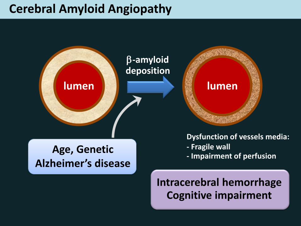
Cerebral Amyloid Angiopathy
lumen lumen
-amyloid deposition
Dysfunction of vessels media: - Fragile wall - Impairment of perfusion
Age, Genetic Alzheimer’s disease
Intracerebral hemorrhage Cognitive impairment
Cerebral Amyloid Angiopathy
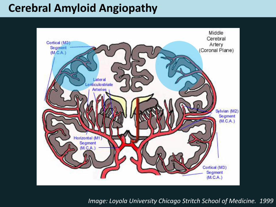
• Cortical and leptomeningeal arteries (not deep arteries)
• Posterior (occipital) predilection
Cerebral Amyloid Angiopathy
Image: Loyola University Chicago Stritch School of Medicine. 1999
Cerebral Amyloid Angiopathy
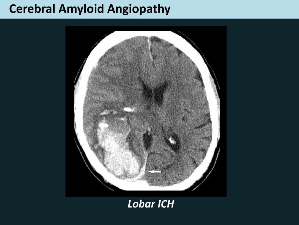
Lobar ICH
Cerebral Amyloid Angiopathy
MRI markers of CAA:
- Cerebral microbleeds (strictly Lobar pattern)
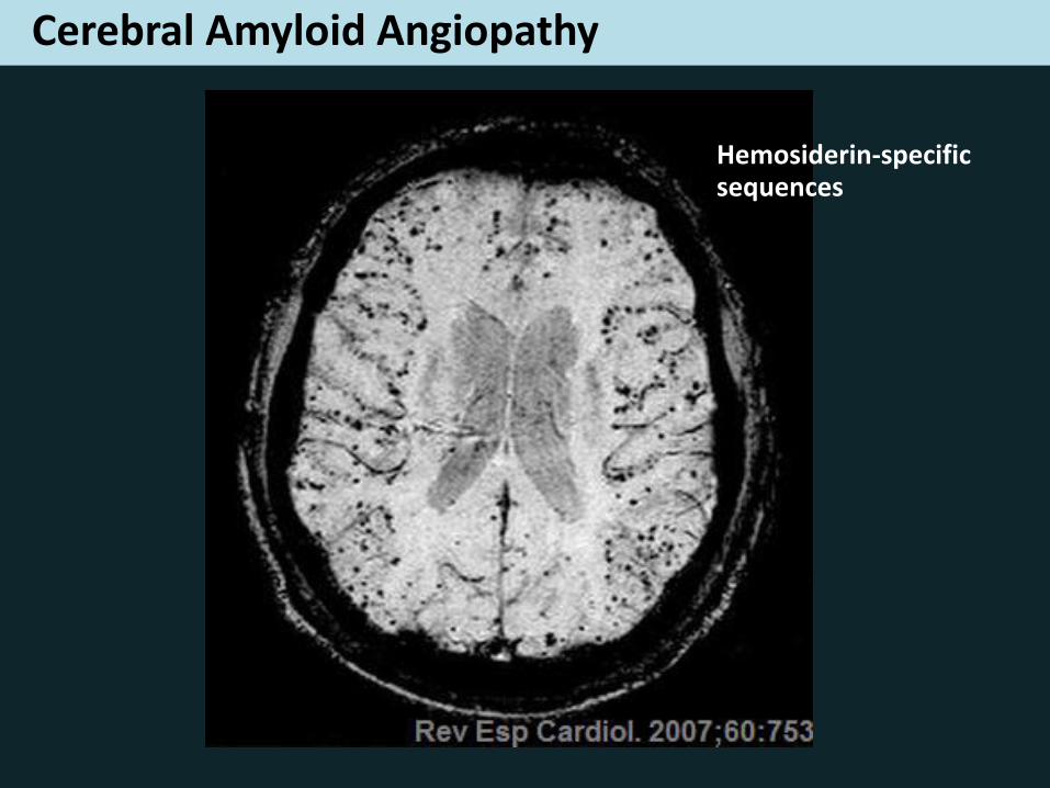
Cerebral Amyloid Angiopathy
Hemosiderin-specific sequences
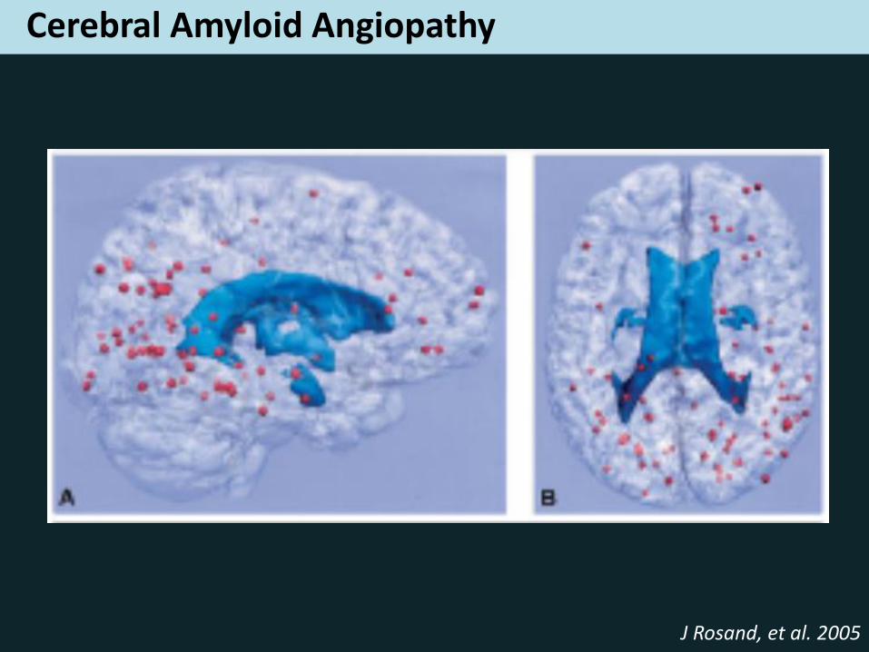
Cerebral Amyloid Angiopathy
J Rosand, et al. 2005
Cerebral Amyloid Angiopathy
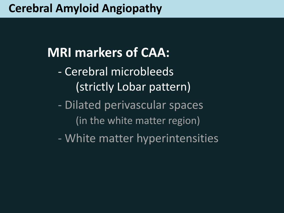
MRI markers of CAA:
- Cerebral microbleeds (strictly Lobar pattern)
- Dilated perivascular spaces (in the white matter region)
- White matter hyperintensities

Posterior White Matter Disease Distribution
as a Predictor of Cerebral Amyloid Angiopathy
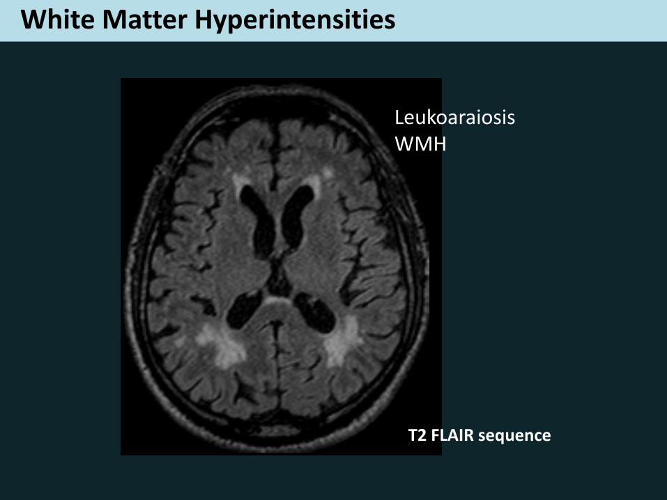
White Matter Hyperintensities
Leukoaraiosis WMH
T2 FLAIR sequence
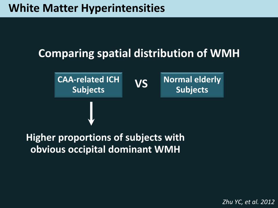
White Matter Hyperintensities
Comparing spatial distribution of WMH
Zhu YC, et al. 2012
CAA-related ICH Subjects
Normal elderly Subjects
VS
Higher proportions of subjects with obvious occipital dominant WMH
Objectives
To evaluate and compare the anteroposterior (AP) distribution of WMH
between CAA and non-CAA patients, in the absence of ICH
Hypotheses
In the absence of ICH:
- A posterior distribution of WMH should associate with a strictly lobar pattern of MB and a high burden of WM-DPVS
- The posterior WMH distribution should be a predictor of CAA
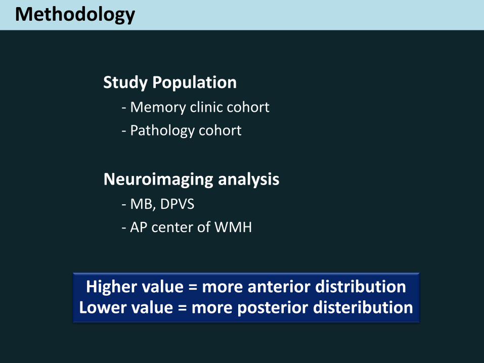
Methodology
Study Population
- Memory clinic cohort
- Pathology cohort
Neuroimaging analysis
- MB, DPVS
- AP center of WMH
Exclusion criteria
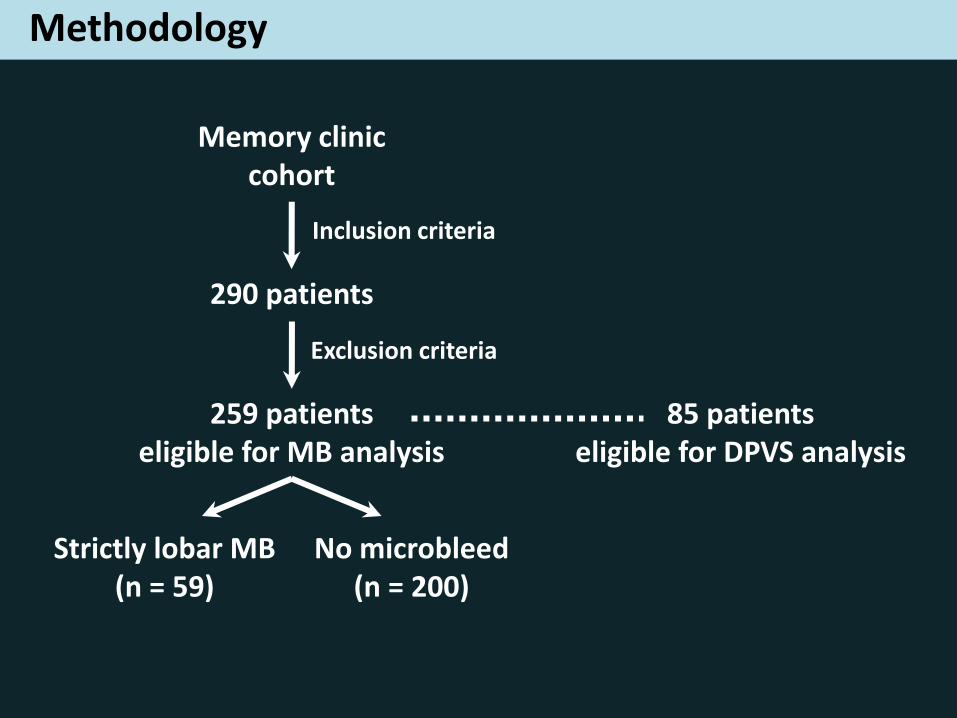
Methodology
Memory clinic cohort
Inclusion criteria
290 patients
259 patients eligible for MB analysis
Strictly lobar MB (n = 59)
No microbleed (n = 200)
85 patients eligible for DPVS analysis
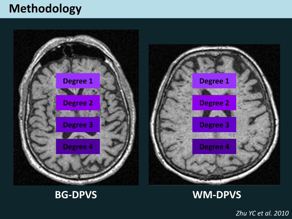
Methodology
BG-DPVS WM-DPVS
Degree 1
Degree 2
Degree 3
Degree 4
Degree 1
Degree 2
Degree 3
Degree 4
Zhu YC et al. 2010
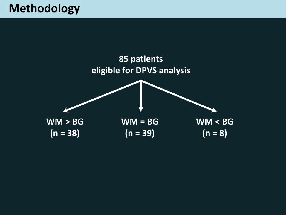
Methodology
85 patients eligible for DPVS analysis
WM > BG (n = 38)
WM = BG (n = 39)
WM < BG (n = 8)
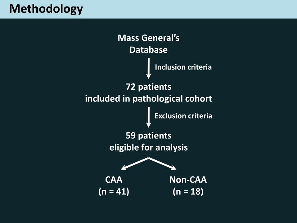
Methodology
Mass General’s Database
Inclusion criteria
72 patients included in pathological cohort
59 patients eligible for analysis
Exclusion criteria
CAA (n = 41)
Non-CAA (n = 18)
Methodology
Study Population
- Memory clinic cohort
- Pathology cohort
Neuroimaging analysis
- MB, DPVS
- AP center of WMH
Higher value = more anterior distribution Lower value = more posterior disteribution
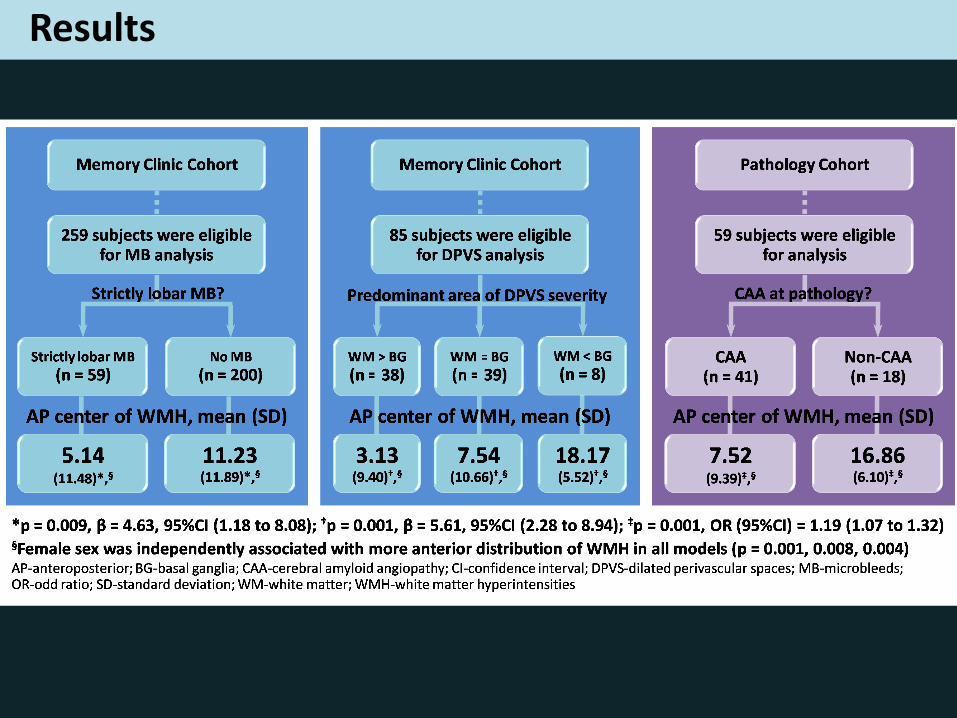
Results
Interpretations
• In patients without ICH, strictly lobar MB and high degree of WM-DPVS was found to be associated with posterior WMH distribution
• Posterior WMH distribution may be a potential predictor of CAA at pathology
• Female patients tended to have more anterior distribution of WMH.
Conclusions
Assessment of AP distribution of WMH
may have additional diagnostic importance
in patients with suspected CAA, prior to ICH

Thank you!
Namnao National Park, Thailand