Embed Size (px)
Citation preview
MICHAEL A. CLARKMICHAEL B. WORRELLJOHN E. PLESS
Introduction
The processes that reduce a human body to a skeleton through the postmortem destructionof soft tissues are complex. As with other biologic phenomena, there will be much variationfrom person to person and it is impossible to assign absolute times for any of the processes.The following discussion of decomposition is limited to bodies which have not beenembalmed, since this process retards and, in some cases, completely prevents decomposition.
Determination of Death and Early Postmortem Changes
In a hospital setting, determination of death usually involves the use of such instruments asthe electrocardiograph and the electroencephalograph. The more traditional and time-hon-ored methods of determination of cessation of vital function are simple and straightforward.The absence of a heartbeat over the left side of the chest or lack of an arterial pulse in theneck or the wrists means that the patient is dead. Listening for a heartbeat with an ear to thechest or with a stethoscope is the next level of sophistication. Cessation of breathing afterdeath can be observed by noting the lack of chest movement as well as by listening to theairway with or without a stethoscope.
Changes which occur within the first 2 hours after death are referred to as early post-mortem changes. These alterations are caused by lack of effective cardiac pumping of oxy-genated blood resulting in a loss of the usual skin color. This “pallor” is first noticed in verylight-skinned people as early as 15 to 30 minutes after death, but it may go unnoticed in dark-skinned persons. At the same time, the skeletal muscles of the body, including the sphincters,relax. It is during this period that fecal soiling may occur. In addition, if a body is movedduring the period of muscular relaxation, regurgitation of gastric contents or “purging” mayoccur.
External and internal alterations of the eye occur during the early postmortem period.Externally, a dark band of dried corneal epithelium may be visualized across the front of theeyes since relaxation of the orbicularis oculi muscles of the eyelids exposes the corneas to airdrying. This phenomenon is sometimes referred to as “tache noir sclerotique.” By using anophthalmoscope, sludging and intravascular coagulation of blood in the retinal vessels canbe seen. This is sometimes called sausaging or boxcarring.
Following death, the blood gradually becomes acidic due to the accumulation of carbondioxide and other chemicals released as the result of tissue breakdown (Cotran et al. 1994).This causes the activation of the intrinsic coagulation mechanism resulting in blood clots inboth arteries and veins throughout the body. This generalized coagulation of the blood usually
9
©1997 CRC Press LLC
occurs at about the same time as the development of rigor mortis or stiffening of the muscles.As decomposition continues, the blood pH further decreases, and enzymes called intrinsicfibrinolysins are activated. These enzymes cause reliquification of the coagulated blood. Thisgenerally occurs at the same time as rigor mortis begins to dissipate.
Late Postmortem Changes
Rigor mortis, algor mortis, and livor mortis are referred to as late postmortem changes becausethey are first observed beginning at 2 to 4 hours after death. These processes are independentof one another, but usually occur simultaneously.
Rigor mortis, or the postmortem stiffening of the muscles, is a reversible chemical changeof the muscles. It begins in all skeletal muscles shortly after death, but is first noticeable inthe facial muscles as tightening of the jaw at 2 to 3 hours postmortem. After 24 hours, theentire body will be rigid to the extent that it may be capable of supporting its own weight(Spitz 1993:27). As chemicals in the muscles are consumed, they relax by about 48 hours.Since rigor mortis is a chemical process, it is accelerated by heat. This means that if anindividual dies following exertion or with a fever, rigidity will develop faster than in anindividual whose body temperature is normal at the time of death. Rigor mortis does notcause muscular contraction and, Hollywood movies to the contrary, dead bodies do not situp, grasp objects, or walk about due to rigor.
Algor mortis is the normal cooling of a body which takes place as the body equilibrateswith the environment after death. The normal metabolic processes of the body which maintaina core temperature of 98.6˚F during life cease at death and the body temperature will tend toapproach the ambient temperature. In most circumstances this means that bodies cool afterdeath at an approximate rate of 1.5°F per hour. If death occurs in a very warm environment,body temperature will rise after death. Elevated body temperature during life or hyperthermiawill obviously modify postmortem changes in body temperature.
Livor mortis (also called livor or lividity) refers to the gravitational pooling of blood independent parts which occurs after death. In other words, the blood pools on the down sideof the body because it is no longer being circulated by the heart. Livor can be first recognizedas soon as 15 minutes after death by trained observers, but it is ordinarily first evident atabout 2 hours postmortem. The normal color of livor mortis changes from red to purple asoxygen gradually dissociates from the hemoglobin of the red blood cells. This produces apigment in the red cells called deoxyhemoglobin which is purple. When it is initially seen,Iivor mortis is nonfixed. This means that when a blunt object, such as the back of a finger, isdrawn across an area of lividity, the pressure will force blood from the engorged capillariesresulting in an area of blanching which quickly refills. As the body cools (i.e., algor mortis),the fat in the dermis which surrounds the capillaries solidifies and pinches them such thatpressure will no longer force blood away from the area of lividity. This phenomenon is calledfixation of lividity and it occurs at 4 to 6 hours after death or after rigor mortis is easilydetected (Figure 1). It should be noted that in patients who are profoundly anemic or havelost large quantities of blood, lividity may not easily be seen.
Lividity typically varies from red to purple and becomes darker as the postmorteminterval increases. Deviations from these normal colors can be of profound importance.Specifically, cherry red lividity is diagnostic of carbon monoxide poisoning until provenotherwise. Bodies that have been exposed to very cold temperatures soon after death willappear pink because the cold inhibits dissociation of oxygen from the hemoglobin. The leastcommon cause of red lividity is cyanide poisoning, where the cyanide inhibits dissociation ofoxygen by blocking the cytochrome oxydase enzymes. All of these changes look similar. Totell the difference, laboratory testing of the blood must be done and these results should be
©1997 CRC Press LLC
compared with information from the scene of death. Finally, the death investigation andlaboratory data must be the subject of interpretation by a forensic pathologist who thencompares that information with the findings at autopsy.
Tissue Changes
Now that early and late postmortem changes have been considered, we will begin a discussionof the changes which occur in the tissues eventually leading to skeletonization of the remains.The entire process is referred to as decomposition, and this process is further subdivided intoautolysis and putrefaction.
Autolysis is a process whereby hydrolytic enzymes that are present in cytoplasmic granulesin all cells, called lysosomes, are released into the cytoplasm. Autolysis is thought to betriggered by the decrease in intracellular pH which occurs as a result of decreased oxygenlevels which occur after death (Cormack 1987). The hydrolytic enzymes in the lysosomesdigest carbohydrates and proteins, while fats are affected to a lesser degree. Because the cellmembranes are also disrupted during autolysis, these molecules are released and are utilizedas nutrients by microorganisms (see below). Following death of the organism, homeostasisis no longer operative and all cells undergo autolysis beginning shortly after death. The timeat which autolysis begins in different cell types and organs is quite variable. As a general rule,autolysis begins much sooner after death in those cell types which contain large numbers oflysosomes (e.g., pancreas) than in those which contain few hydrolytic enzymes (e.g., muscle).The process of autolysis is temperature dependent, and refrigeration of a body soon afterdeath will retard the enzymatic self digestion of cells. Autolysis will be accelerated by ante-mortem fever, exertion, or a high ambient temperature.
The changes produced by autolysis initially can be seen only by use of a microscope, butas the process progresses the features can be seen with the naked eye. These changes will firstbe observed at about 48 hours after death. Externally, a phenomenon called skin slippage willbe seen. In skin slippage, the postmortem release of hydrolytic enzymes by cells at thedermal–epidermal junction of the skin results in a loosening of the epidermis from the
Figure 1 This elderly lady with a history of heart disease, was last seen alive 24 hours prior tobeing found sitting upright in her bathroom on the toilet. Note fixed lividity with blanching in areasof pressure produced by the weight of the body.
©1997 CRC Press LLC
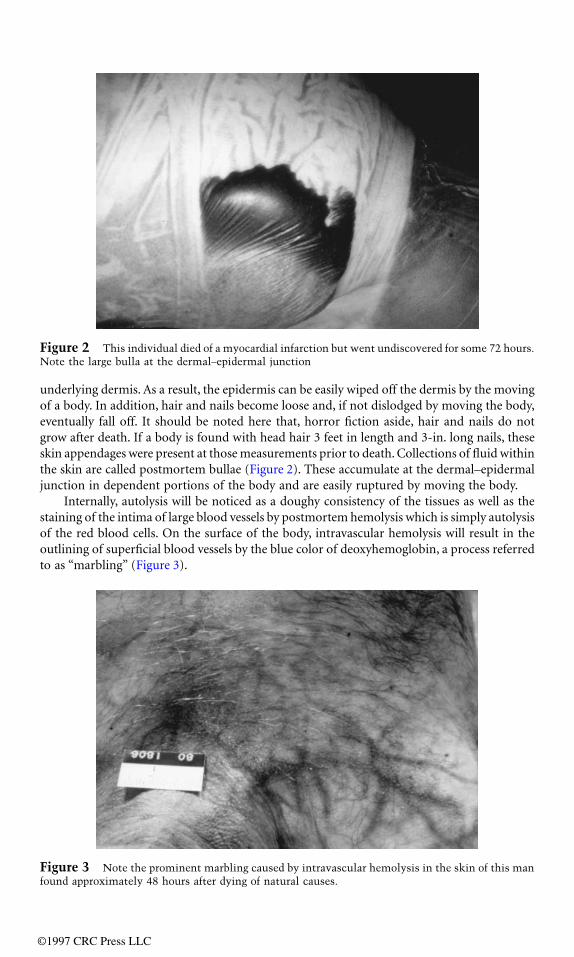
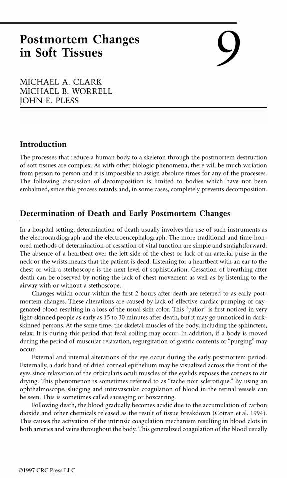
underlying dermis. As a result, the epidermis can be easily wiped off the dermis by the movingof a body. In addition, hair and nails become loose and, if not dislodged by moving the body,eventually fall off. It should be noted here that, horror fiction aside, hair and nails do notgrow after death. If a body is found with head hair 3 feet in length and 3-in. long nails, theseskin appendages were present at those measurements prior to death. Collections of fluid withinthe skin are called postmortem bullae (Figure 2). These accumulate at the dermal–epidermaljunction in dependent portions of the body and are easily ruptured by moving the body.
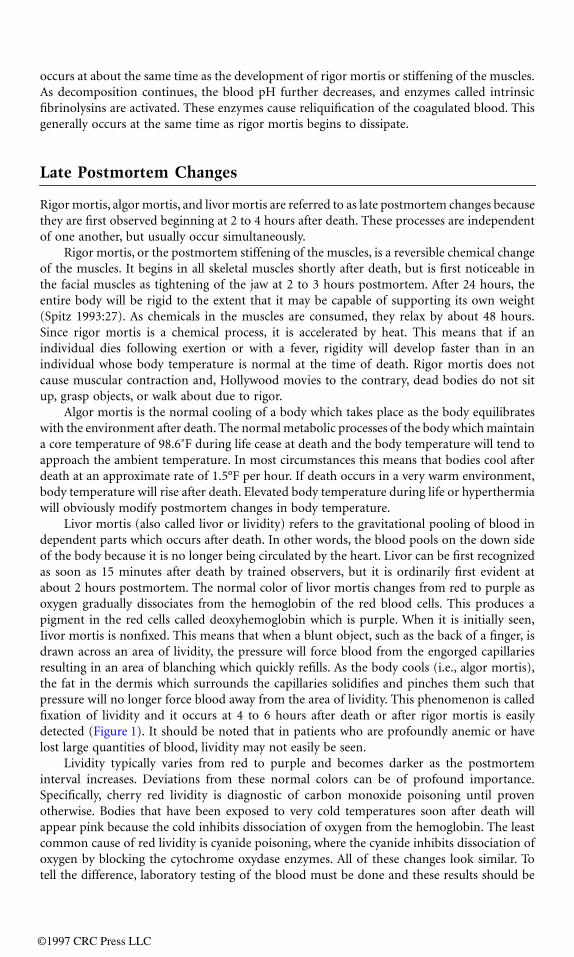
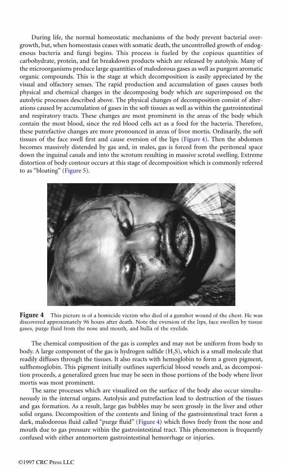
Internally, autolysis will be noticed as a doughy consistency of the tissues as well as thestaining of the intima of large blood vessels by postmortem hemolysis which is simply autolysisof the red blood cells. On the surface of the body, intravascular hemolysis will result in theoutlining of superficial blood vessels by the blue color of deoxyhemoglobin, a process referredto as “marbling” (Figure 3).
Figure 2 This individual died of a myocardial infarction but went undiscovered for some 72 hours.Note the large bulla at the dermal–epidermal junction
Figure 3 Note the prominent marbling caused by intravascular hemolysis in the skin of this manfound approximately 48 hours after dying of natural causes.
©1997 CRC Press LLC
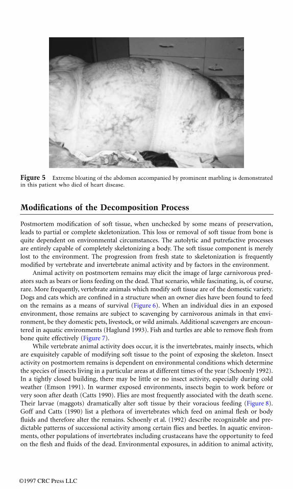
During life, the normal homeostatic mechanisms of the body prevent bacterial over-growth, but, when homeostasis ceases with somatic death, the uncontrolled growth of endog-enous bacteria and fungi begins. This process is fueled by the copious quantities ofcarbohydrate, protein, and fat breakdown products which are released by autolysis. Many ofthe microorganisms produce large quantities of malodorous gases as well as pungent aromaticorganic compounds. This is the stage at which decomposition is easily appreciated by thevisual and olfactory senses. The rapid production and accumulation of gases causes bothphysical and chemical changes in the decomposing body which are superimposed on theautolytic processes described above. The physical changes of decomposition consist of alter-ations caused by accumulation of gases in the soft tissues as well as within the gastrointestinaland respiratory tracts. These changes are most prominent in the areas of the body whichcontain the most blood, since the red blood cells act as a food for the bacteria. Therefore,these putrefactive changes are more pronounced in areas of livor mortis. Ordinarily, the softtissues of the face swell first and cause eversion of the lips (Figure 4). Then the abdomenbecomes massively distended by gas and, in males, gas is forced from the peritoneal spacedown the inguinal canals and into the scrotum resulting in massive scrotal swelling. Extremedistortion of body contour occurs at this stage of decomposition which is commonly referredto as “bloating” (Figure 5).
The chemical composition of the gas is complex and may not be uniform from body tobody. A large component of the gas is hydrogen sulfide (H2S), which is a small molecule thatreadily diffuses through the tissues. It also reacts with hemoglobin to form a green pigment,sulfhemoglobin. This pigment initially outlines superficial blood vessels and, as decomposi-tion proceeds, a generalized green hue may be seen in those portions of the body where livormortis was most prominent.
The same processes which are visualized on the surface of the body also occur simulta-neously in the internal organs. Autolysis and putrefaction lead to destruction of the tissuesand gas formation. As a result, large gas bubbles may be seen grossly in the liver and othersolid organs. Decomposition of the contents and lining of the gastrointestinal tract form adark, malodorous fluid called “purge fluid” (Figure 4) which flows freely from the nose andmouth due to gas pressure within the gastrointestinal tract. This phenomenon is frequentlyconfused with either antemortem gastrointestinal hemorrhage or injuries.
Figure 4 This picture is of a homicide victim who died of a gunshot wound of the chest. He wasdiscovered approximately 96 hours after death. Note the eversion of the lips, face swollen by tissuegases, purge fluid from the nose and mouth, and bulla of the eyelids.
©1997 CRC Press LLC
Modifications of the Decomposition Process
Postmortem modification of soft tissue, when unchecked by some means of preservation,leads to partial or complete skeletonization. This loss or removal of soft tissue from bone isquite dependent on environmental circumstances. The autolytic and putrefactive processesare entirely capable of completely skeletonizing a body. The soft tissue component is merelylost to the environment. The progression from fresh state to skeletonization is frequentlymodified by vertebrate and invertebrate animal activity and by factors in the environment.
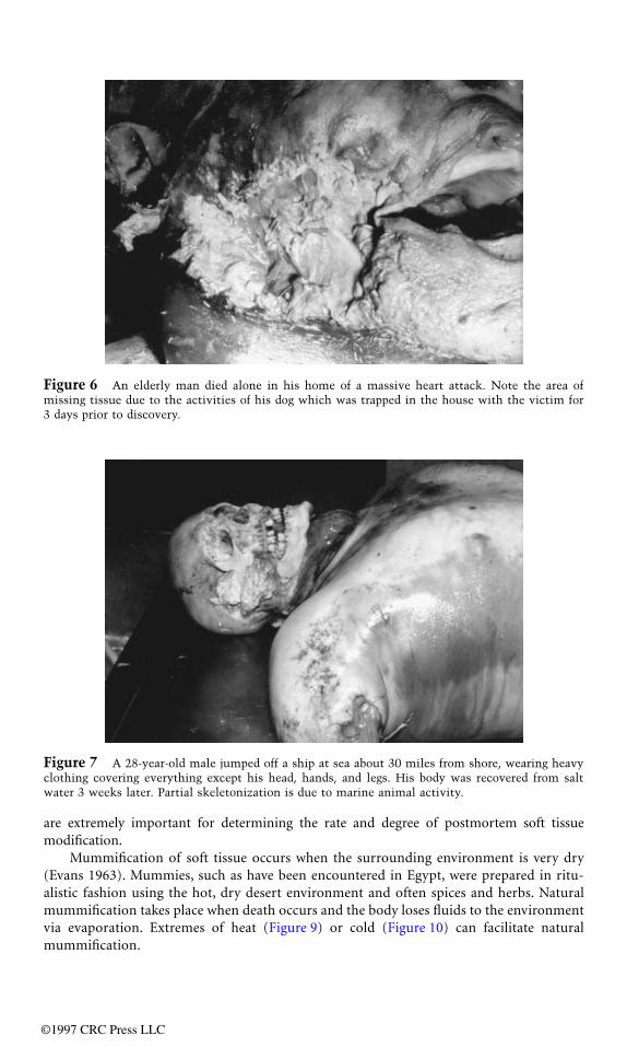
Animal activity on postmortem remains may elicit the image of large carnivorous pred-ators such as bears or lions feeding on the dead. That scenario, while fascinating, is, of course,rare. More frequently, vertebrate animals which modify soft tissue are of the domestic variety.Dogs and cats which are confined in a structure when an owner dies have been found to feedon the remains as a means of survival (Figure 6). When an individual dies in an exposedenvironment, those remains are subject to scavenging by carnivorous animals in that envi-ronment, be they domestic pets, livestock, or wild animals. Additional scavengers are encoun-tered in aquatic environments (Haglund 1993). Fish and turtles are able to remove flesh frombone quite effectively (Figure 7).
While vertebrate animal activity does occur, it is the invertebrates, mainly insects, whichare exquisitely capable of modifying soft tissue to the point of exposing the skeleton. Insectactivity on postmortem remains is dependent on environmental conditions which determinethe species of insects living in a particular areas at different times of the year (Schoenly 1992).In a tightly closed building, there may be little or no insect activity, especially during coldweather (Emson 1991). In warmer exposed environments, insects begin to work before orvery soon after death (Catts 1990). Flies are most frequently associated with the death scene.Their larvae (maggots) dramatically alter soft tissue by their voracious feeding (Figure 8).Goff and Catts (1990) list a plethora of invertebrates which feed on animal flesh or bodyfluids and therefore alter the remains. Schoenly et al. (1992) describe recognizable and pre-dictable patterns of successional activity among certain flies and beetles. In aquatic environ-ments, other populations of invertebrates including crustaceans have the opportunity to feedon the flesh and fluids of the dead. Environmental exposures, in addition to animal activity,
Figure 5 Extreme bloating of the abdomen accompanied by prominent marbling is demonstratedin this patient who died of heart disease.
©1997 CRC Press LLC
are extremely important for determining the rate and degree of postmortem soft tissuemodification.
Mummification of soft tissue occurs when the surrounding environment is very dry(Evans 1963). Mummies, such as have been encountered in Egypt, were prepared in ritu-alistic fashion using the hot, dry desert environment and often spices and herbs. Naturalmummification takes place when death occurs and the body loses fluids to the environmentvia evaporation. Extremes of heat (Figure 9) or cold (Figure 10) can facilitate naturalmummification.
Figure 6 An elderly man died alone in his home of a massive heart attack. Note the area ofmissing tissue due to the activities of his dog which was trapped in the house with the victim for3 days prior to discovery.
Figure 7 A 28-year-old male jumped off a ship at sea about 30 miles from shore, wearing heavyclothing covering everything except his head, hands, and legs. His body was recovered from saltwater 3 weeks later. Partial skeletonization is due to marine animal activity.
©1997 CRC Press LLC
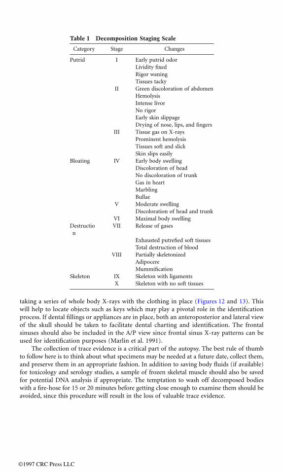
Adipocere is a malodorous, cheesy, compound of fatty acids also referred to as grave wax(Evans 1963). Adipocere formation occurs most commonly when tissues are submerged incool water where the oxygen content is very low (Figure 11). Over time, adipocere has beenseen to become hard and brittle even in aquatic environments (Haglund 1993). Adipocereforms in both embalmed and unembalmed bodies via the hydrolysis and hydrogenation offats to fatty acids. This process requires the presence of water either from an exogenous sourceor from the body itself. Areas covered by clothing appear to produce conditions which favoradipocere formation (Mellen et al. 1993). Dehydration and mummification may accompanyadipocere formation in bodies where little or no exogenous water is available. As fats are
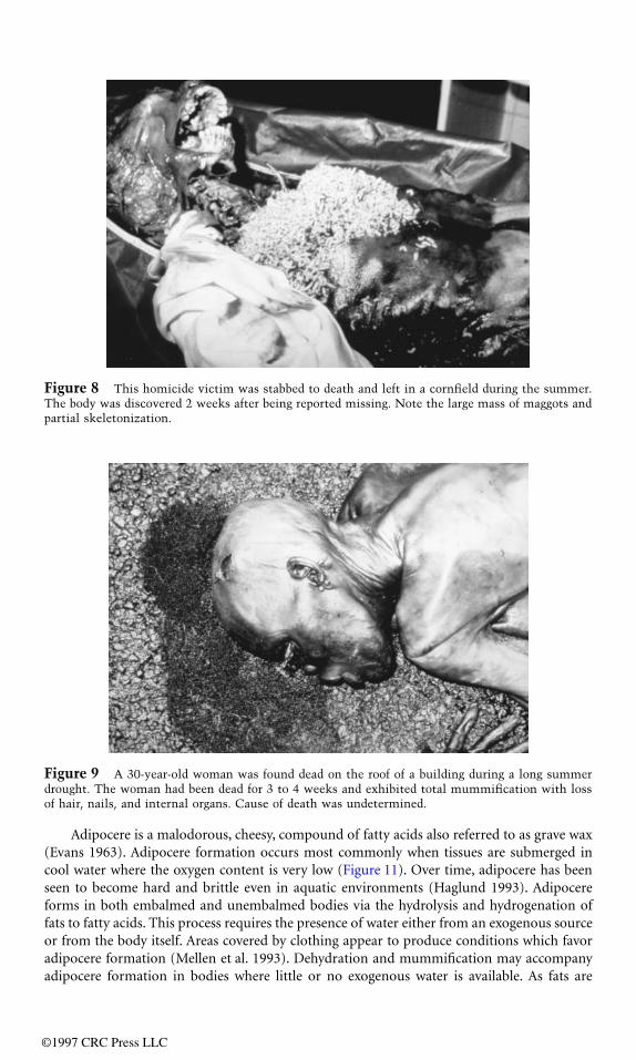
Figure 8 This homicide victim was stabbed to death and left in a cornfield during the summer.The body was discovered 2 weeks after being reported missing. Note the large mass of maggots andpartial skeletonization.
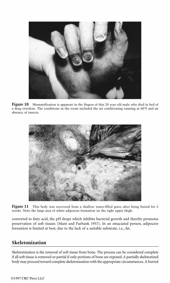
Figure 9 A 30-year-old woman was found dead on the roof of a building during a long summerdrought. The woman had been dead for 3 to 4 weeks and exhibited total mummification with lossof hair, nails, and internal organs. Cause of death was undetermined.
©1997 CRC Press LLC
converted to fatty acid, the pH drops which inhibits bacterial growth and thereby promotespreservation of soft tissues (Mant and Furbank 1957). In an emaciated person, adipocereformation is limited at best, due to the lack of a suitable substrate, i.e., fat.
Skeletonization
Skeletonization is the removal of soft tissue from bone. The process can be considered completeif all soft tissue is removed or partial if only portions of bone are exposed. A partially skeletonizedbody may proceed toward complete skeletonization with the appropriate circumstances. A buried
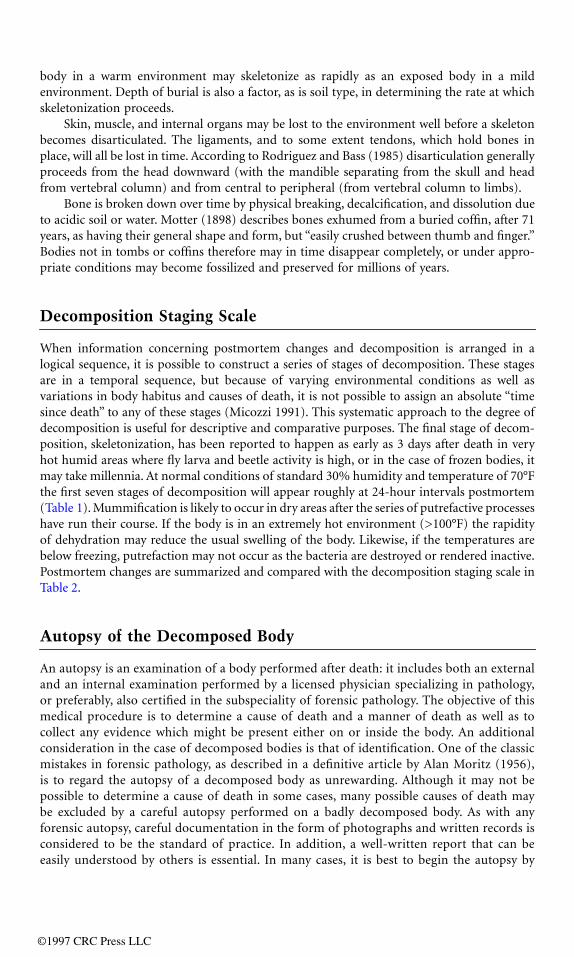
Figure 10 Mummification is apparent in the fingers of this 29 year old male who died in bed ofa drug overdose. The conditions in the room included the air conditioning running at 60°F and anabsence of insects.
Figure 11 This body was recovered from a shallow water-filled grave after being buried for 3weeks. Note the large area of white adipocere formation on the right upper thigh.
©1997 CRC Press LLC
body in a warm environment may skeletonize as rapidly as an exposed body in a mildenvironment. Depth of burial is also a factor, as is soil type, in determining the rate at whichskeletonization proceeds.
Skin, muscle, and internal organs may be lost to the environment well before a skeletonbecomes disarticulated. The ligaments, and to some extent tendons, which hold bones inplace, will all be lost in time. According to Rodriguez and Bass (1985) disarticulation generallyproceeds from the head downward (with the mandible separating from the skull and headfrom vertebral column) and from central to peripheral (from vertebral column to limbs).
Bone is broken down over time by physical breaking, decalcification, and dissolution dueto acidic soil or water. Motter (1898) describes bones exhumed from a buried coffin, after 71years, as having their general shape and form, but “easily crushed between thumb and finger.”Bodies not in tombs or coffins therefore may in time disappear completely, or under appro-priate conditions may become fossilized and preserved for millions of years.
Decomposition Staging Scale
When information concerning postmortem changes and decomposition is arranged in alogical sequence, it is possible to construct a series of stages of decomposition. These stagesare in a temporal sequence, but because of varying environmental conditions as well asvariations in body habitus and causes of death, it is not possible to assign an absolute “timesince death” to any of these stages (Micozzi 1991). This systematic approach to the degree ofdecomposition is useful for descriptive and comparative purposes. The final stage of decom-position, skeletonization, has been reported to happen as early as 3 days after death in veryhot humid areas where fly larva and beetle activity is high, or in the case of frozen bodies, itmay take millennia. At normal conditions of standard 30% humidity and temperature of 70°Fthe first seven stages of decomposition will appear roughly at 24-hour intervals postmortem(Table 1). Mummification is likely to occur in dry areas after the series of putrefactive processeshave run their course. If the body is in an extremely hot environment (>100°F) the rapidityof dehydration may reduce the usual swelling of the body. Likewise, if the temperatures arebelow freezing, putrefaction may not occur as the bacteria are destroyed or rendered inactive.Postmortem changes are summarized and compared with the decomposition staging scale inTable 2.
Autopsy of the Decomposed Body
An autopsy is an examination of a body performed after death: it includes both an externaland an internal examination performed by a licensed physician specializing in pathology,or preferably, also certified in the subspeciality of forensic pathology. The objective of thismedical procedure is to determine a cause of death and a manner of death as well as tocollect any evidence which might be present either on or inside the body. An additionalconsideration in the case of decomposed bodies is that of identification. One of the classicmistakes in forensic pathology, as described in a definitive article by Alan Moritz (1956),is to regard the autopsy of a decomposed body as unrewarding. Although it may not bepossible to determine a cause of death in some cases, many possible causes of death maybe excluded by a careful autopsy performed on a badly decomposed body. As with anyforensic autopsy, careful documentation in the form of photographs and written records isconsidered to be the standard of practice. In addition, a well-written report that can beeasily understood by others is essential. In many cases, it is best to begin the autopsy by
©1997 CRC Press LLC
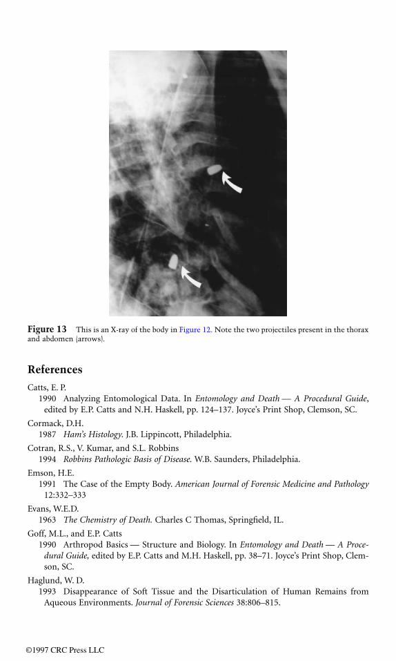
taking a series of whole body X-rays with the clothing in place (Figures 12 and 13). Thiswill help to locate objects such as keys which may play a pivotal role in the identificationprocess. If dental fillings or appliances are in place, both an anteroposterior and lateral viewof the skull should be taken to facilitate dental charting and identification. The frontalsinuses should also be included in the A/P view since frontal sinus X-ray patterns can beused for identification purposes (Marlin et al. 1991).
The collection of trace evidence is a critical part of the autopsy. The best rule of thumbto follow here is to think about what specimens may be needed at a future date, collect them,and preserve them in an appropriate fashion. In addition to saving body fluids (if available)for toxicology and serology studies, a sample of frozen skeletal muscle should also be savedfor potential DNA analysis if appropriate. The temptation to wash off decomposed bodieswith a fire-hose for 15 or 20 minutes before getting close enough to examine them should beavoided, since this procedure will result in the loss of valuable trace evidence.
Table 1 Decomposition Staging Scale
Category Stage Changes
Putrid I Early putrid odorLividity fixedRigor waningTissues tacky
II Green discoloration of abdomenHemolysisIntense livorNo rigorEarly skin slippageDrying of nose, lips, and fingers
III Tissue gas on X-raysProminent hemolysisTissues soft and slickSkin slips easily
Bloating IV Early body swellingDiscoloration of headNo discoloration of trunkGas in heartMarblingBullae
V Moderate swellingDiscoloration of head and trunk
VI Maximal body swellingDestruction
VII Release of gases
Exhausted putrefied soft tissuesTotal destruction of blood
VIII Partially skeletonizedAdipocereMummification
Skeleton IX Skeleton with ligamentsX Skeleton with no soft tissues
©1997 CRC Press LLC

Table 2 Postmortem Changes
Time after Death Postmortem Changes Modifiers Category Stage
0 minutes Circulation and breathing stop Temperature See Table 1Pallor Humidity Early changesEarly lividity Outdoor locationMuscular relaxation Indoor locationSphincters may relax Submerged in water
2 hours Vascular changes in eyeRigor mortis begins Late changesAlgor mortis beginsLividity easily seen
4–5 hours Coagulation of bloodFixation of lividity
24 hours Drying of cornea Putrid IRe-liquefication of blood Tissue changes II
48 hours Rigor dissappears IIIIntravascular hemolysis
72 hours Loss of hair and nails96 hours Skin slippage and bulla formation Insect activity Bloated IV
Bacterial overgrowth Animal activity VDays-months Green discoloration VI
Bloating Destruction VIIRelease of gasses Mummification VIIIRelease of liquified internal organs Adipocere formation Skeleton IXGradual loss of soft tissues XPartial skeletonizationComplete skeletonization
Figure 12 This body was recovered from a central Indiana cornfield in July. The victim had beendead for approximately 3 days. The clothing was in place prior to autopsy. Note the heavy maggotinfestation.
©1997 CRC Press LLC
References
Catts, E. P.1990 Analyzing Entomological Data. In Entomology and Death — A Procedural Guide,
edited by E.P. Catts and N.H. Haskell, pp. 124–137. Joyce’s Print Shop, Clemson, SC.
Cormack, D.H.1987 Ham’s Histology. J.B. Lippincott, Philadelphia.
Cotran, R.S., V. Kumar, and S.L. Robbins1994 Robbins Pathologic Basis of Disease. W.B. Saunders, Philadelphia.
Emson, H.E.1991 The Case of the Empty Body. American Journal of Forensic Medicine and Pathology
12:332–333
Evans, W.E.D.1963 The Chemistry of Death. Charles C Thomas, Springfield, IL.
Goff, M.L., and E.P. Catts1990 Arthropod Basics — Structure and Biology. In Entomology and Death — A Proce-
dural Guide, edited by E.P. Catts and M.H. Haskell, pp. 38–71. Joyce’s Print Shop, Clem-son, SC.
Haglund, W. D.1993 Disappearance of Soft Tissue and the Disarticulation of Human Remains from
Aqueous Environments. Journal of Forensic Sciences 38:806–815.
Figure 13 This is an X-ray of the body in Figure 12. Note the two projectiles present in the thoraxand abdomen (arrows).
©1997 CRC Press LLC
Mant, A.K., and R. Furbank1957 Adipocere — A Review. Journal of Forensic Medicine 4:18–35.
Marlin, D.C., M.A. Clark, and S.M. Standish1991 Identification of Human Remains by Comparison of Frontal Sinus Radiographs: A
Series of Four Cases. Journal of Forensic Sciences 36:1765–1772.
Mellen, P.F.M., M.A. Lowry, and M.S. Micozzi1993 Experimental Observations on Adipocere Formation. Journal of Forensic Sciences
38:91–93.
Micozzi, M.S.1991 Postmortem Change in Human and Animal Remains. Charles C Thomas, Springfield,
IL.
Moritz, A.R.1956 Classical Mistakes in Forensic Pathology. American Journal of Clinical Pathology
26:1383–1397.
Motter, M.G.1898 A Contribution to the Study of the Fauna of the Grave. A Study of One Hundred
and Fifty Disinterments, with Some Additional Experimental Observations. Journal ofthe New York Entomological Society 6:201–230.
Rodriguez, W.C., and W.M. Bass1985 Decomposition of Buried Bodies and Methods That May Aid in Their Location.
Journal of Forensic Sciences 30:836–852.
Schoenly, K.1992 A Statistical Analysis of Successional Patterns in Carrion-Arthropod Assemblages:
Implications for Forensic Entomology and Determination of the Postmortem Interval.Journal of Forensic Sciences 37:1489–1513.
Schoenly, K., M.L. Goff, and M. Early1992 A BASIC Algorithm for Calculating the Postmortem Interval from Arthropod Suc-
cessional Data. Journal of Forensic Sciences 37:808–823.
Spitz, W.U.1993 Spitz and Fisher’s Medicolegal lnvestigation of Death. 3rd ed. Charles C Thomas,
Springfield, IL.
©1997 CRC Press LLC